Epizootic Hemorrhagic Disease Virus: Comparison
Please note this is a comparison between Version 1 by Luis Jimenez-Cabello and Version 2 by Lindsay Dong.

Epizootic Hemorrhagic Disease (EHD) of ruminants is a viral pathology that has significant welfare, social, and economic implications. The causative agent, epizootic hemorrhagic disease virus (EHDV), belongs to the Orbivirus genus and leads to significant regional disease outbreaks among livestock and wildlife in North America, Asia, Africa, and Oceania, causing significant morbidity and mortality. Here, we comprehensively review the current knowledge on EHDV.

  • EHDV
  • epidemiology
  • vaccines
  • white-tailed deer
  • cattle

1. Introduction

The appearance and spread of outbreaks caused by arboviruses that affect both wild and domestic ruminants carry a high risk of generating important direct and indirect economic losses. Therefore, control of diseases caused by arboviruses is essential to ensure the welfare of livestock, as well as to prevent possible detrimental effects on local, regional, and national economies. In this regard, implementation of vaccination campaigns against this type of pathogen has successful, with a great impact on animal health. However, a variety of anthropogenic factors, climate change, and growing global trade increase the risk of appearance of viral diseases, some of them transmitted by arthropod insects, in non-endemic territories.
Epizootic hemorrhagic disease (EHD) is an arthropod-transmitted viral illness of wild and domestic species of the suborder Ruminantia, included in the list of notifiable diseases of the World Organization for Animal Health (WOAH) since 2008. The causative agent of EHD is epizootic hemorrhagic disease virus (EHDV). This virus belongs to the genus Orbivirus within the family Sedoreoviridae, similar to bluetongue virus (BTV) and African horse sickness virus (AHSV). EHDV and BTV share a variety of domestic and wild ruminant hosts, including sheep, white-tailed deer (WTD), and cattle, although susceptibility to clinical disease associated with these viral infections varies greatly among host species, individuals, and viral serotype. For BTV, more than 29 serotypes have been described, while just 7 serotypes of EHDV are currently described. 

2. EHDV, the Etiological Agent of EHD

Similar to members of the Orbivirus genus, EHDV non-enveloped virions present a structure characterized by an icosahedral capsid, which is divided into three consecutive protein layers: the inner and intermediate layers (core) and an outer capsid [1] (Figure 1). The genome is located inside the core particle and comprises ten linear double-strand RNA segments that encode for seven structural (VP1–VP7) and at least four non-structural proteins (NS1, NS2, NS3/NS3A, NS4, and probably the putative NS5 as for BTV [2]) [1][3][4][1,3,4]. As for the prototype BTV, the outer protein layer is made of 60 trimers of VP2, the most exposed virion protein, and 120 trimers of VP5 [5]. The inner capsid is made of the subcore, formed by VP3, and the intermediate layer, constituted by VP7, along with three minor structural proteins with enzymatic activities: VP1 (RNA-dependent RNA polymerase), VP4 (capping enzyme), and VP6 (helicase) [4][6][7][8][9][4,6,7,8,9].
Figure 1. Diagrammatic representation of the viral particle of EHDV (~80 nm in diameter). Three concentric layers constituted by VP2 and VP5 form the outer capsid. The intermediate layer and the subcore are composed by VP7 and VP3. The inner capsid contains the RNA polymerase complex, composed by structural proteins VP1, VP4, and VP6. At least four additional proteins (NS1, NS2, NS3/NS3A, and NS4) are expressed during the replicative cycle.
Outer capsid proteins VP2 and VP5, encoded by segments 2 and 6, respectively, are the most variable proteins among EHDV serotypes, especially VP2, probably due to great selective pressure [10]. Similar to BTV, VP2 and VP5 accomplish key roles during the early stages of infection, being involved in virus attachment and virus entry into host cells [11]. VP2 is also mainly responsible for the induction of virus neutralizing antibodies (nAbs), thereby defining virus serotype based on cross-neutralization assays and supported by extensive phylogenetic studies [10]. Rapid, sensitive, and specific molecular typing assays have been developed by identification of segment 2 nucleotide regions unique to each EHDV serotype [12][13][12,13].
VP1 (segment 1), VP4 (segment 4), and VP6 (segment 9), involved in genome replication, are also highly conserved proteins, showing at least 85% of amino acid sequence identities when eastern and western strains were considered separately [14]. Identification of conserved nucleotide regions in segment 9 allowed development of a highly sensitive EHDV pan-reactive TaqMan real-time RT-PCR assay for diagnosis and genome detection in tissues [12].
VP7 (encoded by segment 7) shows a highly conserved amino acid sequence (more than 90% sequence identity) among EHDV serotypes [14][15][14,15]. VP7 is the immunodominant serogroup-specific protein and is used in serogroup specific enzyme-linked immunosorbent assays (ELISAs) for disease diagnosis [16]. Recently, researchers have shown the usefulness of purified and unpurified baculovirus-expressed VP7 for development of competitive enzyme-linked immunosorbent assays [17][18][17,18].
Non-structural proteins, found in infected cells but not in virus particles [19], play supportive roles in crucial viral processes such as genome packaging, intracellular transport, capsid assembly, virus release, and control of the immune response. NS1, the most expressed protein during orbivirus replication in infected cells and positive regulator of viral protein synthesis, forms tubules in the cytoplasm that are involved in viral transport within infected cells and have a role in cellular pathogenicity [20][21][22][20,21,22]. NS1 has been described as an almost identical protein among EHDV serotypes (more than 90% sequence homology) [23].
NS2 (segment 8) acts as ATPase, having a role in RNA packaging and translation [24][26]. This phosphoprotein is the major component of viral inclusion bodies (VIB), acting as virus assembly sites [25][26][27,28]. In terms of amino acid identity, NS2 varies more than NS1. The sequence identity seen in NS2 among all strains varies around 79.7% [23]. TAuthe scholaors also reported the conservation of a domain at the N-terminus of NS2, between amino acids 75–83. This domain was shown to be important for binding single-stranded RNA and formation of VIBs during EHDV infection [24][26]. Differences at the sequence of this domain between western and eastern strains could explain differential pathogenicity in cattle [23]. NS3 (segment 10) is important in virus release from infected cells, being expressed at greater levels. NS3, which possess transmembrane domains [27][29], facilitates virus egress via budding, rather than lysis, in Culicoides cells [28][30]. NS3 and NS3A are closely related proteins, both translated from sequential ORFs of segment 10 [29][31]. The sequence identity mean of NS3 is 89.7% at the amino acid level between eastern and western viruses [23]. For NS4 (encoded by segment 9), which is a virulence factor in the case of BTV [30][31][32,33], no sequence homology analysis between EHDV serotypes has been conducted so far.

3. Changes in EHDV Epidemiology

To date, seven serotypes of EHDV have been identified, named as 1–2 and 4–8, designated based on extensive phylogenetic studies, sequencing data, and cross-neutralization assays [10]. Genetic analyses demonstrated that previously identified serotype 3 [32][34] (Nigerian strain Ib Ar 22619) was serotype 1 [10]. EHDV has been isolated in North and South America, Africa, Asia, the Middle East, and Oceania (Figure 2a). To date, it is endemic in parts of North America, Australia, and certain countries of Asia and Africa [33][35]. EHDV was first detected in the USA in 1955 when WTD were severely affected showing high mortality [34][36]. Among the seven serotypes proposed, EHDV-1, 2, and 6 have been reported to be present in North America, where WTD is the most severely affected host and the scale of individual outbreaks increased with time [35][36][37][37,38,39]. In Australia, six out of seven serotypes (EHDV-1, -2, -5, -6, -7, and -8) have been detected over the years [33][38][35,40]. Globally, the presence of EHDV has been noted in Japan (serotypes 2 and 7, and serotypes 5 and 6 recently isolated from Culicoides insect vectors), China (serotypes 1, 5, 6, 7, 8), Morocco (serotype 6), Algeria (serotype 6), Libya (serotype 6), Turkey (serotype 6), Tunisia (serotypes 6 and 8), Egypt (serotype 1), Oman (serotype 2 and 6), Sudan (serotype 5 and 6), Nigeria (serotypes 1 and 4), the island of Mayotte (serotype 6), French Guiana (serotypes 6 and 7), Ecuador (serotype 1), Trinidad (serotype 6) and Israel (serotypes 1, 6 and 7) [39][40][41][42][43][44][45][46][47][48][49][50][51][52][53][54][55][56][57][41,42,43,44,45,46,47,48,49,50,51,52,53,54,55,56,57,58,59]. Genome detection or serological evidences also indicate the presence of EHDV (unknown serotypes) in Indonesia, Taiwan, Zimbabwe, Kenya, Kazakhstan, and Brazil [58][59][60][61][62][60,61,62,63,64]. Nucleotide sequencing and neutralization tests suggest novel strains of EHDV identified in South Africa and China as new putative serotypes [63][64][65,66].

Outbreaks during 2020 in Turkey and North Africa were originated by serotype 6, while serotype 8 was the causative virus of several outbreaks in cattle in Tunisia during 2021 [48][50]. This was the first evidence of EHDV-8 circulation since 1982, when this serotype was isolated in Australia. In Europe, there was no evidence of the presence of EHDV. However, it emerged on the continent for the first time in October 2022. After BTV-like clinical signs were noticed in some animals, EHDV was identified as the causative agent of cattle disease outbreaks in Sicily and southwestern Sardinia (Figure 2b). Subsequently, it was confirmed that these outbreaks were caused by EHDV-8, an identical strain to the one circulating in Tunisia between 2021 and 2022, pointing to North Africa as the direct origin [65][80]. Consecutively, EHDV outbreaks were detected in southern Spain, with EHDV serotype 8 confirmed and notified to the WOAH as the causative agent (Figure 2b).

Figure 2. Epidemiology of EHDV across the world (a) and in Europe (b). (a) Colored areas represent the countries were different serotypes of EHDV have caused outbreaks over the years or serological evidence exists. The corresponding serotypes of each country are indicated. Where outbreaks are located within a specific region, the whole country is indicated as infected. (b) Representation of the Italian and Spanish affected regions.
Epidemiologic changes of arboviral diseases are also related to genetic evolution of their causative viruses. Recent works have established a distinction between EHDV strains based on their eastern or western origin, which could possibly be influenced by both regional and genetic factors [14][23][14,23]. Genetic variation present among EHDV strains derives from mechanisms of recombination, gene duplication, and point mutation [10][23][10,23]. In this sense, an African ancestry of US and Australian strains has been suggested by the identification of a recombination event in segment 8 of EHDV. Gene reassortment is another major force that characterizes EHDV evolutive strength to maintain viral fitness. In the USA, a novel EHDV-6 reassortant strain (Indiana strain) was identified as the causative virus of an outbreak in WTD during September and October 2006. At first, researchers identified a reassortment between the North American topotype of EHDV-2 and an exotic strain of EHDV-6 [66][70]. Thereafter, this exotic strain was confirmed to have an Australian origin [67][81].

4. Disease and Pathology

EHD was firstly described in WTD in New Jersey in 1955 [34][36]. WTD (Odocoileus virginianus) are especially susceptible to severe disease. In North America, where EHDV is a major cause of mortality in WTD [68][102], periodic outbreaks result in significant mortality, but susceptibility to disease has been shown to vary between subspecies [69][103]. Clinical disease due to EHDV has been also reported in mule (black-tailed) deer, bighorn, yak, elk, brocket deer, and pronghorn antelope [61][68][70][71][72][73][63,102,104,105,106,107]. While sheep are highly susceptible to BTV with significant mortality, these are often resistant to EHDV-induced disease. However, it is important to consider animals with subclinical infections as reservoirs of infection that can amplify virus circulation, although the role of sheep in EHDV epidemiology seems to be negligible [74][108]. Other species that have also been seropositive are fallow deer, wapiti, bison, goat, red deer, and roe deer [75][109].
After blood feeding from an infected animal, EHDV first replicates in the midgut epithelium of the biting midge and disseminates through the hemolymph to secondary infection sites including the salivary glands [76][110], where viral particles can undergo changes related to infectivity [77][111]. When an infected Culicoides bites a susceptible ruminant host, the virus primarily infects dendritic cells and macrophages. These subsequently migrate carrying the virus to regional lymph nodes, draining the entry site. Here, the primary replication occurs, and, then, the virus is disseminated to many organs, particularly the spleen and lungs (although the virus has also been detected in the heart, cerebrum, cerebellum, and testes). Mononuclear phagocytes and endothelium cells are mainly infected [78][112]. EHDV replicates in the vascular epithelium, benefiting from autophagy and inducing cell death by apoptosis [78][79][80][112,113,114], leading to hemorrhage and thrombosis. In addition to direct viral damage caused by replication in the endothelium, replication in macrophages and endothelial cells leads to the release of pro-inflammatory cytokines such as interleukin 1 (IL-1) and interleukin 6 (IL-6) [81][115], which could enhance viral pathogenesis. Thus, inflammatory mediators may contribute to the severity of clinical disease, as well as the induction of vasoactive mediators, such as nitric oxide [82][83][116,117].
During the initial steps of infection, a type I IFN response determines whether the disease makes progress in the infected host. In experimental infection of deer with EHDV, peak viraemia coincided with peak IFN type I levels and both then rapidly declined [84][118]. Importantly, host genetics related to the innate immune response probably play a role in disease outcome [85][119]. Leukopenia and lymphopenia are common features of EHD. Clinical outcomes vary depending on the different forms of disease. The sub-acute form is characterized by development of ulcers in the oral cavity and the gastrointestinal tract [68][102]. The acute forms portrays a hemorrhagic disease that includes hyperaemia of the conjunctiva and the oral mucosa, pulmonary edema, pleural effusion, and multifocal haemorrhages in a variety of organs affected by vascular damage and coagulopathy [68][102]. The peracute disease causes fulminant death, probably due to the development of pulmonary vascular injury with subsequent pulmonary oedema, probably assocciated with the cytokine storm [82][116]. Prolonged infections have also been observed in experimentally infected WTD and cattle, which can be explained by association of EHDV viral particles with invaginations in the erythrocyte membrane [86][120].

5. Experimental Animal Models of EHDV

5.1. White-Tailed Deer (WTD) and Other Cervid Species

As the global incidence of EHD is constantly increasing, the study of pathogenesis, transmission, and diagnosis as well as the evaluation of vaccine candidates in natural wildlife hosts is a key issue. In this sense, cervids have been used for EHDV study in natural hosts (Table 1). Different species of cervids, including red, fallow, roe, and muntjac deer, were experimentally infected with the New Jersey strain of EHDV-1, which is highly virulent in WTD. In contrast, these animals did not display any signs of disease, although productive infection could be detected [87][123]. Among the species of cervids susceptible to EHDV infection, WTD stand out as the most affected host of EHDV. Experimental infection of WTD with EHDV followed the first outbreak detected in the USA. WTD inoculated with EHDV-1 displayed severe illness in most cases, resembling the clinical disease observed in nature and displaying gross and histopathological lesions [88][89][124,125]. A high mortality among infected WTD was recorded [89][125]. In general, experimental infection of WTD with EHDV induces acute disease leading to high mortality rates, independent from the age or sex of the infected animals. WTD infected with the reassortant North American EHDV-6 resembled infection and clinical disease caused by the US strains of EHDV-1 and EHDV-2. Researchers also characterized virological parameters of EHDV infection, detecting viraemia from the third day post-inoculation until more than two weeks later (viraemia was detectable for more than seven weeks) in surviving animals, even in presence of high nAb titers. Viral RNA was detected in tissue samples of organs that presented macro- and microscopic lesions [90][126]. A field isolate of EHDV-7 that caused intense and widespread epizootic in domestic cattle in Israel was also assessed to determine whether WTD was susceptible to infection. This virus strain led to a clinical disease identical to that observed in experimental infections with North American isolates, and virological parameters were similar to those of EHDV-6 infected WTD [91][127]. The fact that “exotic” strains of EHDV can productively infect WTD inducing fatal clinical disease highlights the marked susceptibility of WTD to EHDV infection. Nonetheless, it is important to note that not all subspecies of WTD share the same susceptibility grade, with some of them, such as the subspecies Odocoileus virginianus texanus (which inhabits regions where EDHV has been endemic for a long period), showing innate resistance to EHDV [92][128]. Differential expression of pro-inflammatory cytokines could explain this resistance [81][115]. Host genetic factors can also influence susceptibility to EHDV infection [85][119]. This illustrates the complexity of understanding the pathogenesis and virulence of EHDV even in WTD.

5.2. Cattle and Other Farm Animals

WTD and other wildlife experimental models involve limitations (reviewed in [93][138]) that are not shared by suitable traditional livestock experimental animals widely used in infectious disease research. As stated above, during recent EHDV outbreaks in North America, the Mediterranean Basin, and Reunion Island, an apparent increase in pathogenicity of EHDV in cattle raised concerns. Therefore, cattle can be considered as a more accessible and cheaper alternative to WTD for studying viral pathology and evaluating vaccine efficacy. In later studies, cattle, sheep, pigs, and goats were inoculated with the New Jersey strain of EHDV-1, which was virulent in deer [87][123]. Despite technical limitations concomitant with that time, researchers were able to observe viraemia in inoculated sheep and cattle while none of the goats or pigs were viraemic. Interestingly, the virus was recovered from the vulva of a recently lambed viraemic sheep. Clinical disease was not observed in any animal [87][123]. Similarly, other researchers observed that inoculation of cattle with the EHDV-1 New Jersey strain did not induce clinical disease, but the virus could be isolated from day 9 to day 23 post-inoculation and viraemia was detected by gel-based reverse transcriptase-PCR between days 3 and 28 post-infection. Similar results were observed for cattle infected with the Alberta strain of EHDV-2 [94][139]. In another work, subsequent inoculation of cattle with two US isolates led to transient viraemia as well as the induction of a neutralizing immune response in absence of clinical disease [95][140].

5.3. Mouse Models

Availability of appropriate laboratory animal models is a major concern when studying disease pathogenesis and developing efficient and safe therapies against viral diseases. Mouse models are a reliable tool that reproduce or, at least, partially mimic the disease pathogenesis in a variety of cases [96][148]. For vaccine development, utilization of valid mouse models endorses the basis of every traditional vaccine development procedure as it implies several advantages, such as reduction of costs and time, facility to handle and accessibility of a huge number of optimal reagents. When considering vaccine evaluation against veterinary diseases, mouse models offer a more accessible and affordable animal housing compared to natural hosts. Immunocompromised mouse models, like mice deficient in the type I IFN (IFN-α/β) receptor (IFNAR(−/−)), have been extensively used for vaccine efficacy assessment. The IFNAR(−/−) knock-out receptor mouse model has been employed to study viral infection, disease, pathogenesis and vaccine testing against a plethora of viral diseases [96][148]. This laboratory animal model has been exploited for extensive study of BTV and AHSV [97][98][99][149,150,151]. Infection of IFNAR(−/−) mice with BTV and AHSV reproduces the pathology observed in natural hosts of these viruses.

6. Classic and Novel Vaccine Approaches against EHDV

Vaccination entails the most effective countermeasure to successfully contain several human and veterinary viral diseases. In the case of EHDV, vaccines based on conventional approaches have been developed and their commercialization has been circumscribed to regions where the virus has circulated causing a significant economic impact. In Japan, two vaccines against EHDV-2 are commercially available: a monovalent live attenuated vaccine and an inactivated bivalent vaccine (against EHDV-2 and bovine ephemeral fever, caused by bovine ephemeral fever virus). LAVs (live attenuated vaccines) against BTV are used in the United States, Turkey, the Republic of South Africa, India, and Israel, among others [100][161]. However, BTV LAVs, which show highly immunogenicity, are often associated with several drawbacks relating toanimal welfare and transmission to insect vectors (reviewed elsewhere [100][161]). For these reasons, and after being used to control several outbreaks of BTV over the years, immunization with BTV LAVs was reduced and, eventually, completely substituted by inactivated vaccines in the European Union (EU) [101][162]. Therefore, LAVs are not a recommended choice to consider in EHDV vaccination campaigns in the EU, as the possibility of virus spillover to unaffected regions through uptake and spread by midges or in-contact transmission is clear. Inactivated vaccines against BTV are produced and licensed in Europe, and, although some pitfalls exist (reviewed in [101][162]), this approach has demonstrated more than enough efficacy to control this disease [101][162].  Next-generation vaccines against EHDV must overcome inherent disadvantages of classical approaches that occur with BTV and AHSV. First, they must allow differentiation between naturally infected and vaccinated animals (DIVA), which has fundamental implications in the economic field. Second, these newly generated vaccines should induce protection against multiple EHDV serotypes, whose expansion to non-endemic latitudes is highly probable. To date, the unique vaccine candidate that has been evaluated is based on recombinant VP2 protein of EHDV-2 [102][164]. This DIVA subunit vaccine has shown promising results in terms of immunogenicity and protection in the primarily EHDV affected host, WTD, preventing it from EHDV clinical disease, infection, and viraemia. Prime-boost immunization with rVP2 of serotype 2 induced high titers (ranging from 1:240 to 1:320) of homologous nAbs in immunized WTD. After viral challenge with virulent EHDV-2, immunized animals did not display EHD-related signs of disease and showed steady rectal temperatures and peripheral lymphocyte counts. No viraemia nor RNA were detected in EHDV-target organs, and immunized WTD showed an absence of gross and histopathological lesions. Although not yet evaluated, its efficacy for avoiding transmission to the insect vector seems plausible as no RNA was detected in blood. This EHDV-2 rVP2-based vaccine is currently under field trial in the USA. Importantly, cattle immunized following the same immunization strategy also developed a potent humoral response. Not only that, but the authors also achieved the expression and purification of the rVP2 of EHDV-6, which induced high titers of homologous nAbs in cattle. In this sense, bi- or multivalent vaccines could be formulated as proposed in the study [102][164]. Different vaccine platforms widely used for novel BTV and AHSV vaccines [103][104][152,165] should be applied for generation of novel EHDV vaccines, e.g., subunit vaccine and viral vector-based vaccines.

7. Conclusions

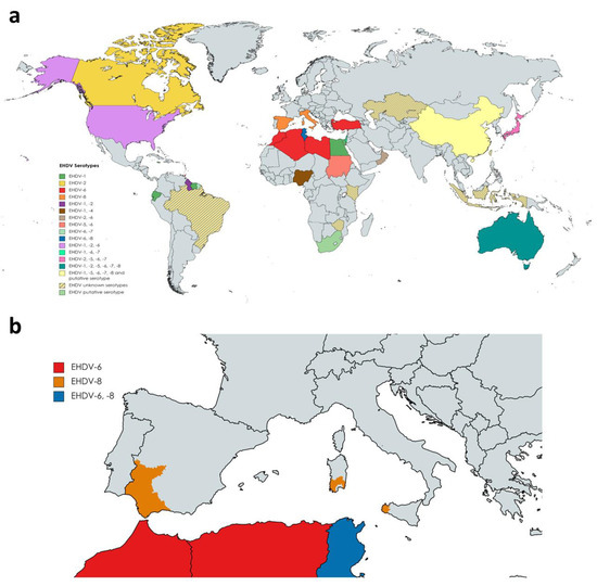
EHDV, an important arthropod-transmitted RNA virus that infects different wild and domestic ruminants, has experienced a northern spread into novel areas in the last 20 years. Global warming may result in expansion of vector species to previously vector-free regions, and in altered vector competence of some midge species. These factors and others related with human activities are likely to increase the risk of EHDV outbreaks in new territories. Worryingly, the virus has recently been detected for the first time in the European Union in October 2022. White-tailed deer are especially susceptible to severe illness caused by EHDV infection; nevertheless, disease can also occur in bovines. The increased virulence of certain EHDV strains observed in cattle and the expansion of competent vectors involved in EHDV transmission make necessary further investigation regarding the development of new diagnostic techniques, safe DIVA vaccines, and the evaluation of laboratory animal models that will facilitate the study of the protective capacity of new vaccine candidates against EHDV.
ScholarVision Creations