

 +1 credit
| Version | Summary | Created by | Modification | Content Size | Created at | Operation |
|---|---|---|---|---|---|---|
| 1 | Vitalii Ivchuk | -- | 1734 | 2026-11-02 00:04:10 | | | |
| 2 | Catherine Yang | Meta information modification | 1734 | 2026-11-02 02:49:17 | | |
Video Upload Options
Ferroptosis: Core Mechanisms and Metabolic Regulation
EditThis entry summarizes the molecular basis of ferroptosis, its metabolic and signalling regulation, its roles in major disease groups, and the principal therapeutic strategies targeting this cell-death pathway. Ferroptosis is an iron-dependent form of regulated cell death driven by uncontrolled peroxidation of polyunsaturated fatty acid-containing phospholipids. It is morphologically and biochemically distinct from apoptosis, necroptosis, autophagy, and pyroptosis and is characterized by iron accumulation, glutathione depletion, glutathione peroxidase 4 (GPX4) inactivation, and lipid peroxide accumulation. The core execution machinery involves labile iron, PUFA-phospholipid remodelling by ACSL4 and LPCAT3, lipoxygenase- and oxidoreductase-mediated lipid peroxidation, and Fenton chemistry. Cellular defence is organized around the system Xc-/GSH/GPX4 axis and GPX4-independent systems, including FSP1-CoQ10-NADPH, GCH1-BH4-DHFR, DHODH-CoQH2, and MBOAT1/2-mediated phospholipid remodelling. Ferroptosis is regulated by p53, NRF2, MAPK-AMPK signalling, hypoxia, mitochondrial metabolism, and immune cells such as macrophages. It contributes to cancer biology, cardiovascular and metabolic diseases, liver injury, neurological disorders, and inflammatory conditions. Therapeutic strategies include ferroptosis inducers, iron chelators, radical-trapping antioxidants, natural products, and nanomedicine, but biomarker validation and clinical translation remain unresolved.
1. Introduction
Ferroptosis was defined in 2012 as a non-apoptotic, iron-dependent form of regulated cell death that is driven by lethal lipid peroxidation and is suppressible by iron chelators and lipophilic antioxidants [1][2][3]. Unlike apoptosis, which depends on caspase activation and apoptotic body formation, ferroptosis is characterized morphologically by mitochondrial shrinkage, increased mitochondrial membrane density, reduced or absent cristae, and rupture of the outer mitochondrial membrane, while the nucleus remains relatively intact [1][4]. Biochemically, ferroptosis reflects a collapse of redox homeostasis: intracellular iron accumulates, glutathione (GSH) is depleted, GPX4 activity decreases, and lipid peroxides accumulate [1][2][3]. The pathway is now recognized as a nexus linking iron metabolism, lipid metabolism, amino acid metabolism, mitochondrial function, and immunity [1][3][5][6][7].
The importance of ferroptosis extends beyond a single cell-death programme. It acts as a tumour-suppressive mechanism in some contexts, contributes to therapy resistance in others, and mediates tissue injury in ischaemia-reperfusion, neurodegeneration, metabolic disorders, and inflammation [2][4][8][9][10][11][12][13]. Consequently, pharmacological modulation of ferroptosis has become a major translational objective. However, the field still lacks robust in vivo biomarkers and reliable therapeutic windows, and the relationship between ferroptosis and other regulated cell-death modalities remains incompletely defined [1][3][14][15].
2. Core Molecular Mechanisms
2.1. Iron Metabolism and the Labile Iron Pool
Iron is essential for oxygen transport, electron transfer, DNA synthesis, and enzyme function, but excess redox-active iron is toxic. Circulating ferric iron binds transferrin and enters cells through transferrin receptor 1 (TFR1). After endocytosis, Fe(III) is reduced to Fe(II) by STEAP3 and released into the cytoplasm through DMT1/SLC11A2, forming a labile iron pool (LIP) [2][3][7]. Iron is stored in ferritin, composed of FTH1 and FTL, and exported by ferroportin (FPN). Ferritin can be degraded by NCOA4-mediated ferritinophagy, releasing free iron and sensitizing cells to ferroptosis [2][3][16].
The LIP is central to ferroptosis because Fe(II) catalyses Fenton chemistry, generating hydroxyl radicals that attack PUFA-containing phospholipids. Iron also serves as a cofactor for lipoxygenases and other oxygenases. Therefore, increased TFR1 expression, reduced ferritin storage, enhanced ferritinophagy, or decreased FPN-mediated export can promote ferroptosis [2][3][16]. Conversely, iron chelators and increased iron storage suppress ferroptosis [1][2][4]. Iron metabolism is thus both a trigger and a therapeutic target.
2.2. Lipid Peroxidation and PUFA-Phospholipid Remodelling
The execution of ferroptosis depends on oxidation of polyunsaturated fatty acid-containing phospholipids (PUFA-PLs). Free PUFAs, particularly arachidonic acid and adrenic acid, are activated by ACSL4 to PUFA-CoAs and esterified into phosphatidylethanolamines by LPCAT3 [1][2][8][11]. These PUFA-PE species are susceptible to peroxidation through enzymatic and non-enzymatic mechanisms. Lipoxygenases (LOXs) and cytochrome P450 oxidoreductase (POR) contribute to enzymatic lipid peroxidation, whereas Fenton chemistry drives non-enzymatic autoxidation [1][2][17].
The balance between PUFA-PLs and monounsaturated fatty acid-containing phospholipids is critical. ACSL4 promotes ferroptosis sensitivity, whereas ACSL3 and SCD1 promote synthesis of monounsaturated fatty acids that displace oxidizable PUFAs and suppress ferroptosis [1][2][6]. MBOAT1/2 can also remodel phospholipids to incorporate MUFAs and protect cells independently of GPX4 [1][14]. Thus, ferroptosis vulnerability is determined not simply by iron load but by the lipid composition of membranes.
2.3. GPX4-Dependent and GPX4-Independent Antioxidant Systems
The system Xc-/GSH/GPX4 axis is the canonical ferroptosis defence. System Xc-, composed of SLC7A11 and SLC3A2, imports cystine and exports glutamate; intracellular cystine is reduced to cysteine, which is used for GSH synthesis. GPX4 uses GSH to reduce lipid hydroperoxides to lipid alcohols, thereby terminating lipid peroxidation [1][2][3][11]. Inhibition of system Xc-, depletion of GSH, or direct inactivation of GPX4 induces ferroptosis [1][8][14].
GPX4-independent systems provide parallel protection. FSP1 reduces CoQ10 to ubiquinol using NAD(P)H, and ubiquinol traps lipid peroxyl radicals at the plasma membrane [1][2][3][14]. The GCH1-BH4-DHFR axis produces tetrahydrobiopterin, a radical-trapping antioxidant, and DHFR regenerates BH4 [1][2][3]. DHODH in the mitochondrial inner membrane reduces CoQ10 to CoQH2 and suppresses mitochondrial lipid peroxidation [1][2][6][14]. These systems are not simply redundant; their subcellular localization and metabolic dependencies differ, and simultaneous impairment can overwhelm cellular antioxidant capacity [1][3].
3. Metabolic and Signalling Regulation
3.1. Mitochondria and Energy Stress
Mitochondria are both sources and targets of ferroptotic damage. Mitochondrial iron uptake, respiratory chain activity, and ROS production influence ferroptosis sensitivity [6][18]. Mitochondrial GPX4 and DHODH provide compartment-specific defence, while excessive mitochondrial lipid peroxidation can trigger ferroptosis [1][6]. Energy stress activates AMPK, which phosphorylates acetyl-CoA carboxylase and reduces PUFA biosynthesis, thereby inhibiting ferroptosis in some contexts [6][19]. Conversely, mitochondrial glutaminolysis and TCA-cycle metabolites can promote ferroptosis under cysteine deprivation.[6] The mitochondrial contribution is therefore context-dependent.
3.2. p53, NRF2, MAPK-AMPK, and Hypoxia
p53 has a dual role. It can promote ferroptosis by repressing SLC7A11 and limiting cystine uptake, but it can also suppress ferroptosis by blocking DPP4 activity or inducing p21.[1][8]. NRF2 is a major negative regulator that transcriptionally activates antioxidant genes, including SLC7A11, GPX4, FTL, FTH1, and enzymes of GSH synthesis [1][2][14]. MAPK pathways regulate iron metabolism, lipid metabolism, and amino acid metabolism, and MAPK-AMPK crosstalk influences autophagy-dependent and p53-dependent ferroptosis.[19]. Hypoxia modulates ferroptosis through HIF-dependent changes in iron uptake, lipid storage, and SLC7A11 expression [13][18].
3.3. Macrophages, Inflammation, and Immunity
Macrophages are closely linked to ferroptosis through iron recycling, cytokine secretion, and ROS production [5]. Iron overload promotes M1 polarization and inflammatory cytokine release, while M1 and M2 macrophages differ in iron storage and ferroptosis sensitivity [5]. Ferroptotic cells release damage-associated molecular patterns, including HMGB1 and oxidized phospholipids, which recruit and activate macrophages [5]. In turn, macrophages can either promote ferroptosis through IL-6, TNF-α, and IL-1β or protect cancer cells by secreting factors that modulate iron and lipid metabolism [5]. This bidirectional interaction places ferroptosis within the broader immune landscape.
4. Disease Relevance
4.1. Cancer
Ferroptosis is implicated in tumour suppression, metastasis, and therapy resistance [8][9][17][20]. Mesenchymal and dedifferentiated cancer cells, which are often resistant to apoptosis and conventional therapy, can be highly sensitive to ferroptosis [1][8][14]. Ferroptosis inducers can reverse resistance to chemotherapy, targeted therapy, and immunotherapy [9][17][20]. However, ferroptosis can also promote tumorigenesis through inflammation and immunosuppression, and cancer cells may evade ferroptosis by upregulating SLC7A11, GPX4, FSP1, or MUFA synthesis [2][8][14][15]. The therapeutic goal is therefore selective induction in malignant cells without systemic toxicity.
4.2. Cardiovascular and Metabolic Diseases
In cardiovascular disease, ferroptosis contributes to ischaemia-reperfusion injury, doxorubicin cardiomyopathy, heart failure, atherosclerosis, and diabetic cardiovascular complications [10][21][22][23]. In diabetes, hyperglycaemia, iron overload, advanced glycation end products, and lipid accumulation increase cardiac susceptibility to ferroptosis.[10] Iron chelation, GPX4 activation, and inhibition of lipid peroxidation have shown protective effects in preclinical models [10][21][22]. In metabolic dysfunction-associated fatty liver disease, ferroptosis contributes to hepatocyte injury, inflammation, and fibrosis [4][11]. These findings support ferroptosis as a mechanistic link between metabolic stress and organ damage.
4.3. Liver and Gastrointestinal Diseases
The liver is highly susceptible to ferroptosis because of its central role in iron storage and metabolism. Ferroptosis contributes to acute liver injury, non-alcoholic steatohepatitis, fibrosis, cirrhosis, and hepatocellular carcinoma [4][11]. In ulcerative colitis, ferroptosis mediates intestinal epithelial cell death and mucosal barrier dysfunction, and inhibition of ferroptosis attenuates experimental colitis [24]. These observations link ferroptosis to chronic inflammatory and metabolic liver-gut axes.
4.4. Neurological Diseases
Ferroptosis has been implicated in intracerebral haemorrhage, ischaemic stroke, traumatic brain injury, Alzheimer’s disease, Parkinson’s disease, and Huntington’s disease[12][13][16][18]. After intracerebral haemorrhage, haemoglobin degradation releases iron, which catalyses lipid peroxidation and neuronal death [12]. Ferritinophagy and NCOA4-mediated iron release contribute to cerebral ischaemia-reperfusion injury [16]. Hypoxia and HIF signalling further modulate neuronal ferroptosis sensitivity [13][18]. The shared feature is a combination of iron overload, oxidative stress, and impaired antioxidant defence.
5. Therapeutic Implications
Ferroptosis inducers include class I compounds that inhibit system Xc- (erastin, sulfasalazine, sorafenib), class II compounds that inhibit GPX4 (RSL3, ML162), class III compounds that deplete GPX4 and CoQ10 (FIN56), and class IV compounds that promote iron loading [1][8][11]. Ferroptosis inhibitors include iron chelators (deferoxamine, deferiprone, deferasirox), radical-trapping antioxidants (ferrostatin-1, liproxstatin-1, vitamin E), ACSL4 inhibitors, and LOX inhibitors [1][2][4][11]. Natural products and nanoparticles are being explored to improve selectivity, bioavailability, and tumour targeting [1][23][25]. NADPH availability is a central determinant of ferroptosis defence, and strategies that restore NADPH or GSH supply may protect normal tissues [3].
Translation remains challenging. There is no validated in vivo ferroptosis biomarker, and most evidence derives from preclinical models [1][3][14]. The therapeutic window for ferroptosis induction in cancer and ferroptosis inhibition in degenerative diseases must be defined. Cell-type-specific delivery, combination with immunotherapy or radiotherapy, and careful assessment of long-term safety are required before clinical implementation (Figure 1) [3][8][15][17][20].
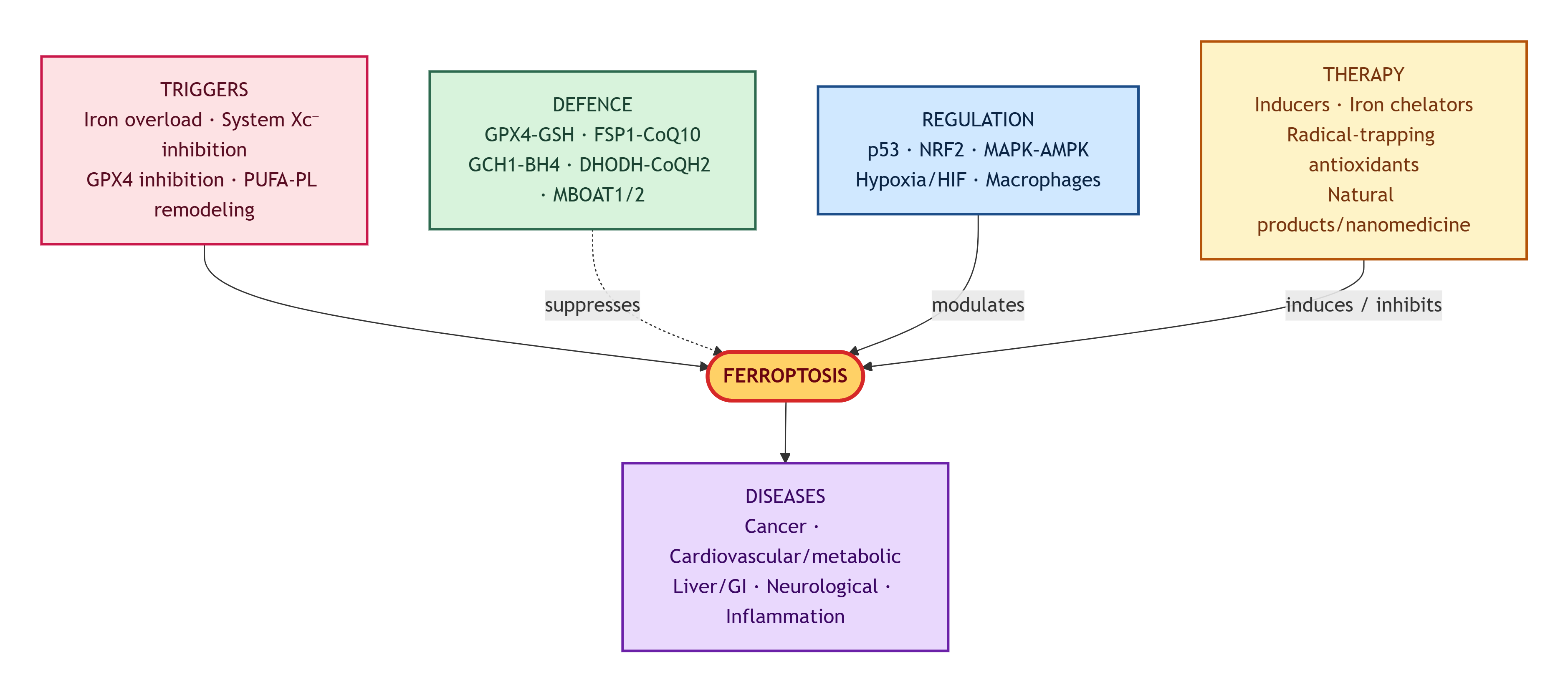
Figure 1. Integrated overview of ferroptosis: triggers, core execution mechanisms, antioxidant defence, regulatory pathways, disease relevance, and therapeutic modulation. Ferroptosis is triggered by iron overload, inhibition of system Xc-, GPX4 inactivation, and PUFA-phospholipid remodelling. These events promote Fenton chemistry, ROS accumulation, GSH depletion, GPX4 inactivation, and lipid peroxidation, ultimately leading to ferroptotic cell death. Antioxidant defence systems, including the system Xc-/GSH/GPX4 axis, FSP1-CoQ10-NADPH, GCH1-BH4-DHFR, DHODH-CoQH2, and MBOAT1/2-mediated MUFA remodelling, suppress ferroptosis. Regulatory pathways such as p53, NRF2, MAPK-AMPK, hypoxia/HIF, and macrophage-derived signals modulate ferroptosis sensitivity. Ferroptosis is implicated in cancer, cardiovascular and metabolic diseases, liver and gastrointestinal diseases, neurological disorders, and inflammation. Therapeutic strategies include ferroptosis inducers, iron chelators, radical-trapping antioxidants, and natural-product-based modulators or nanomedicine-based modulators.
6. Conclusions
Ferroptosis is a metabolically driven cell-death programme centred on iron-dependent phospholipid peroxidation. Its core mechanisms involve labile iron, PUFA-PL remodelling, Fenton chemistry, and insufficient GPX4-dependent and GPX4-independent antioxidant defence. The pathway is regulated by p53, NRF2, MAPK-AMPK signalling, mitochondrial metabolism, hypoxia, and immune cells. Ferroptosis contributes to cancer, cardiovascular and metabolic diseases, liver injury, neurological disorders, and inflammation. Therapeutic modulation of ferroptosis is promising but remains preclinical in many areas, requiring validated biomarkers, selective delivery systems, and rigorous clinical trials.
References
- Shumin Sun; Jie Shen; Jianwei Jiang; Fudi Wang; Junxia Min; Targeting ferroptosis opens new avenues for the development of novel therapeutics. Signal Transduct. Target. Ther. 2023, 8, 1-30. [CrossRef]
- Qin Ru; Yusheng Li; Lin Chen; Yuxiang Wu; Junxia Min; Fudi Wang; Iron homeostasis and ferroptosis in human diseases: mechanisms and therapeutic prospects. Signal Transduct. Target. Ther. 2024, 9, 271. [CrossRef]
- Francesca Alves; Darius Lane; Triet Phu Minh Nguyen; Ashley I. Bush; Scott Ayton; In defence of ferroptosis. Signal Transduct. Target. Ther. 2025, 10, 1-29. [CrossRef]
- Wenli Hu; Kehong Liang; Hong Zhu; Chong Zhao; Hongbo Hu; Shutao Yin; Ferroptosis and Its Role in Chronic Diseases. Cells 2022, 11, 2040. [CrossRef]
- Yan Yang; Yu Wang; Lin Guo; Wen Gao; Ting-Li Tang; Miao Yan; Interaction between macrophages and ferroptosis. Cell Death Dis. 2022, 13, 1-10. [CrossRef]
- Yu’e Liu; Shiping Lu; Lei-Lei Wu; Liang Yang; Lixue Yang; Jinghan Wang; The diversified role of mitochondria in ferroptosis in cancer. Cell Death Dis. 2023, 14, 1-12. [CrossRef]
- Ling Xu; Yu’E Liu; Xi Chen; Hua Zhong; Yi Wang; Ferroptosis in life: To be or not to be. Biomed. Pharmacother. 2023, 159, 114241. [CrossRef]
- Qian Zhou; Yu Meng; Daishi Li; Lei Yao; Jiayuan Le; Yihuang Liu; Yuming Sun; Furong Zeng; Xiang Chen; Guangtong Deng; Ferroptosis in cancer: from molecular mechanisms to therapeutic strategies. Signal Transduct. Target. Ther. 2024, 9, 1-30. [CrossRef]
- Chen Zhang; Xinyin Liu; Shidai Jin; Yi Chen; Renhua Guo; Ferroptosis in cancer therapy: a novel approach to reversing drug resistance. Mol. Cancer 2022, 21, 1-12. [CrossRef]
- Ziyi Wang; Chao Wu; Dong Yin; Kefei Dou; Ferroptosis: mechanism and role in diabetes-related cardiovascular diseases. Cardiovasc. Diabetol. 2025, 24, 1-16. [CrossRef]
- Junyi Chen; Xiaopeng Li; Chaodong Ge; Junxia Min; Fudi Wang; The multifaceted role of ferroptosis in liver disease. Cell Death Differ. 2022, 29, 467-480. [CrossRef]
- Yuanyuan Sun; Qian Li; Hongxiu Guo; Quanwei He; Ferroptosis and Iron Metabolism after Intracerebral Hemorrhage. Cells 2022, 12, 90. [CrossRef]
- Inês Costa; Daniel José Barbosa; Sofia Benfeito; Vera Silva; Daniel Chavarria; Fernanda Borges; Fernando Remião; Renata Silva; Molecular mechanisms of ferroptosis and their involvement in brain diseases. Pharmacol. Ther. 2023, 244, 108373. [CrossRef]
- Fangquan Chen; Rui Kang; Daolin Tang; Jiao Liu; Ferroptosis: principles and significance in health and disease. J. Hematol. Oncol. 2024, 17, 1-41. [CrossRef]
- Vitalii Ivchuk. Ferroptosis: Molecular Mechanisms of Regulation and Therapeutic Potential in the Pathogenesis of Multifactorial Diseases; MDPI AG: Basel, Switzerland, 2026. [CrossRef]
- Xiaoyue Liu; Canming Xie; Yao Wang; Jing Xiang; Litong Chen; Jia Yuan; Chutao Chen; Haomei Tian; Ferritinophagy and Ferroptosis in Cerebral Ischemia Reperfusion Injury. Neurochem. Res. 2024, 49, 1965-1979. [CrossRef]
- Guang Lei; Chao Mao; Yuelong Yan; Li Zhuang; Boyi Gan; Ferroptosis, radiotherapy, and combination therapeutic strategies. Protein Cell 2021, 12, 836-857. [CrossRef]
- Xin Gao; Wei Hu; Dianlun Qian; Xiangfeng Bai; Huilin He; Lin Li; Shibo Sun; The Mechanisms of Ferroptosis Under Hypoxia. Cell. Mol. Neurobiol. 2023, 43, 3329-3341. [CrossRef]
- Xinyue Wang; Xiao Tan; Jinping Zhang; Jiaping Wu; Hongjuan Shi; The emerging roles of MAPK-AMPK in ferroptosis regulatory network. Cell Commun. Signal. 2023, 21, 1-17. [CrossRef]
- Lei Zhao; Xiaoxue Zhou; Feng Xie; Lei Zhang; Haiyan Yan; Jun Huang; Chong Zhang; Fangfang Zhou; Jun Chen; Long Zhang; Ferroptosis in cancer and cancer immunotherapy. Cancer Commun. 2022, 42, 88-116. [CrossRef]
- Yang Zhang; Laiyun Xin; Mi Xiang; Chang Shang; Yuling Wang; Yan Wang; Xiangning Cui; Yingdong Lu; The molecular mechanisms of ferroptosis and its role in cardiovascular disease. Biomed. Pharmacother. 2022, 145, 112423. [CrossRef]
- Guoqing Liu; Xiaoyong Xie; Wang Liao; Siyuan Chen; Rumao Zhong; Jiahui Qin; Peichun He; Jian Xie; Ferroptosis in cardiovascular disease. Biomed. Pharmacother. 2023, 170, 116057. [CrossRef]
- Xiaojiao Yi; Qi Wang; Mengjie Zhang; Qi Shu; Junfeng Zhu; Ferroptosis: A novel therapeutic target of natural products against doxorubicin-induced cardiotoxicity. Biomed. Pharmacother. 2024, 178, 117217. [CrossRef]
- Dan Long; Chenhan Mao; Yingtao Huang; Yin Xu; Ying Zhu; Ferroptosis in ulcerative colitis: Potential mechanisms and promising therapeutic targets. Biomed. Pharmacother. 2024, 175, 116722. [CrossRef]
- Shuangyuan Hu; Maoyaun Zhao; Wei Li; Pengfei Wei; Qingsong Liu; Shuanglan Chen; Jinhao Zeng; Xiao Ma; Jianyuan Tang; Preclinical evidence for quercetin against inflammatory bowel disease: a meta-analysis and systematic review. Inflammopharmacology 2022, 30, 2035-2050. [CrossRef]