Submitted Successfully!
To reward your contribution, here is a gift for you: A free trial for our video production service.
Thank you for your contribution! You can also upload a video entry or images related to this topic.
Version Summary Created by Modification Content Size Created at Operation
1 -- 3144 2024-03-20 14:52:57 |
2 Reference format revised. Meta information modification 3144 2024-03-22 09:16:44 |

Video Upload Options

Do you have a full video?

Confirm

Are you sure to Delete?
Cite
If you have any further questions, please contact Encyclopedia Editorial Office.
Arebro, J.; Lee, C.; Bennewith, K.L.; Garnis, C. Cancer-Associated Fibroblast Heterogeneity in Malignancy. Encyclopedia. Available online: https://encyclopedia.pub/entry/56438 (accessed on 28 April 2024).
Arebro J, Lee C, Bennewith KL, Garnis C. Cancer-Associated Fibroblast Heterogeneity in Malignancy. Encyclopedia. Available at: https://encyclopedia.pub/entry/56438. Accessed April 28, 2024.
Arebro, Julia, Che-Min Lee, Kevin L. Bennewith, Cathie Garnis. "Cancer-Associated Fibroblast Heterogeneity in Malignancy" Encyclopedia, https://encyclopedia.pub/entry/56438 (accessed April 28, 2024).
Arebro, J., Lee, C., Bennewith, K.L., & Garnis, C. (2024, March 20). Cancer-Associated Fibroblast Heterogeneity in Malignancy. In Encyclopedia. https://encyclopedia.pub/entry/56438
Arebro, Julia, et al. "Cancer-Associated Fibroblast Heterogeneity in Malignancy." Encyclopedia. Web. 20 March, 2024.
Cancer-Associated Fibroblast Heterogeneity in Malignancy
Edit

Oral squamous cell carcinoma (OSCC) remains an understudied and significant global cancer killer and dismal survival rates have not changed in decades. A better understanding of the molecular basis of OSCC progression and metastasis is needed to develop new approaches for treating this disease. The supportive network surrounding cancer tumor cells known as the tumor microenvironment (TME) has gained increasing interest lately since it performs essential protumorigenic functions. Cancer-associated fibroblasts (CAFs) are one of the main cell types in the TME and are known to play a key role in influencing the biological behavior of tumors.

cancer-associated fibroblast CAF subtype oral squamous cell carcinoma tumor microenvironment

1. Introduction

Head and neck cancer consists of several subtypes, with oral squamous cell carcinoma (OSCC) being the most common. The 5-year survival rate for all OSCC patients is approximately 50%, and this measure decreases to approximately 30% when looking only at patients diagnosed with more advanced stages of the disease [1][2]. Frequent late-stage diagnoses and high rates of recurrence are the key drivers of this poor prognosis. Interventions such as immunotherapy and targeted therapies have enhanced cancer treatment efficacy and improved cancer patient survival in general, building on successes from standard care involving surgery, radiation, and chemotherapy. However, gains in survival have been modest. There remains a clear need to develop new approaches for treating this disease. A better understanding of the molecular basis of OSCC progression and metastasis is needed to provide new treatment/disease management options.
Recent years have seen an expanded research focus on the tumor microenvironment (TME) based on the emerging evidence that it plays a crucial role in tumor progression and metastasis. Cancer-associated fibroblasts (CAFs) represent one of the main cell constituents in the TME, and these are known to contribute to stromal desmoplasia, which is linked to worse outcomes for many tumor types including head and neck cancer [3][4]. Expanded research into CAFs has indicated a key role for these fibroblasts in tumor progression and metastasis, making them attractive factors for targeted therapy and immunotherapy research [5]. However, effective targeting of CAFs requires a robust understanding of the heterogeneity of CAF cell populations, as well as the biological and clinical significance of various CAF subpopulations.

2. Defining Fibroblasts

Fibroblasts exist in almost all tissue types and are the main cell type in stroma that produces the structural framework of human tissue through creation and maintenance of the extracellular matrix (ECM). Fibroblasts are derived mainly from primitive mesenchymal cells, although alternative origins have been reported (this is described in a subsequent section). There is no single marker used to identify fibroblast cells. In practice, fibroblasts taken from tumors/ tissues are usually first defined through negative selection: they do not express epithelial (EpCAM), endothelial (CD31), leukocyte (CD45), or erythrocyte (CD235a) markers. Next, positive expression of mesenchymal markers such as Vimentin (VIM), alpha-smooth muscle actin (αSMA), and others is used to indicate that a given cell is a fibroblast. In culture, fibroblasts can also be characterized by their spindle-like appearance.
Fibroblast functions include tissue homeostasis, wound healing response through secretion of extracellular matrix proteins to help heal the area, and/or through secretion of cytokines to recruit immune cells and initiate an inflammatory response. Normal fibroblasts can also serve as progenitors for specialized mesenchymal cell types, such as bone-forming osteoblasts and lipid-filled adipocytes [6]. Further, fibroblasts activated in a TME are called CAFs and these are known to display a vast array of functional abilities affecting tumor progression.

3. Fibroblast Activation and CAF Markers

3.1. Fibroblast Plasticity

CAFs may originate from normal tissue fibroblasts, as reported by others [7], with lineage tracing studies in genetically engineered mouse models supporting this concept [8]. These lineage tracing mouse models utilize a specific fibroblast gene promoter attached to a reporter molecule (e.g., mCherry or GFP). This reporter is expressed once the cell begins to express the fibroblast gene used to promote the reporter in the mouse (e.g., a Col5a1 promoter). The cell continues to express the reporter once activated, and expression of the marker is passed down to all daughter cells. This approach has allowed tracking of a chosen cell type as it morphs into a CAF in a tumor setting. An example of a lineage tracing model from Garcia et al. showed that mesenchymal cells driven by Hoxb6 and Gli1 expression become pancreatic fibroblasts but only Gli1 positive mesenchymal cells become CAFs in pancreatic ductal adenocarcinoma (PDAC) [8].
However, lineage tracing studies have also challenged the belief that most CAFs originate from normal fibroblasts in the tissue—Helms et al. have shown that pancreatic stellate cells (PSCs, the resident fibroblast-like cell of the pancreas) that were lineage traced with Fabp4 only contributed to a fraction of the CAFs in PDAC, contrary to popular belief that most CAFs in PDAC come from PSCs [9]. This work demonstrated that there is heterogeneity in the origin of cells that become CAFs in PDAC and potentially in other tumor types as well. Other than lineage tracing, single-cell RNA-sequencing (scRNA-seq) in tandem with either flow-cytometry sorting or mass spectrometry have allowed researchers to identify specific CAF subsets by surface markers that have distinct gene signatures denoting different functions.

3.2. Fibroblast Activation

Fibroblasts can be activated into CAFs through several mechanisms. Among growth factors, the TGFβ family ligands are well-established as activating signals [10]. Lysophosphatidic acid, a lipid mediator, can also drive expression of the myofibroblast marker αSMA while also increasing the activity of the contractile cytoskeleton [11]. Fibroblast activation through Notch signaling [12][13] has been linked to poor prognosis of ductal breast carcinoma [12][13]. Several inflammatory cytokines drive CAF subtype differentiation, with IL-6 activating STAT signaling [14] and IL-1 acting through NF-κB [15]. Stromatogenesis as a result of connective tissue reaction to cancer has been shown to promote desmoplastic fibroblast differentiation in three-dimensional cultures [16], and YAP-mediated enhancement of matrix stiffening helps to maintain CAF phenotypes in breast cancer [17].
Physiological and genomic stress can also drive fibroblast activation info CAFs, with factors like reactive oxygen species [18] and double-stranded DNA breaks [19] serving as examples of this phenomenon. There is increasing evidence that cancer cell-derived extracellular vesicles (EVs) can also drive CAF activation [20][21][22][23], though EV content differs and can include miRNAs, TGFβ, and cells surface proteins, as well as other proteins, mRNAs, and lncRNAs [21][24][25]. It should be noted, however, that many CAF subtypes and functions exist on a spectrum, rather than distinctive subtypes—and that these subtypes can have overlapping functions, a reality that amplifies the complexity inherent in defining CAFs subtypes.

3.3. Fibroblast Markers

CAFs lack a single, specific biomarker in various tumor types. This can be a significant challenge in the context of CAF research. Besides widely mentioned mesenchymal biomarkers such as VIM, αSMA, fibroblast activation protein (FAP), podoplanin (PDPN), integrin beta-1 (ITGB1, also known as CD29), FSP-1 (S100A4), and platelet-derived growth factor alfa (PDGFR-α), PDGFR-β [20][26], the role of matrix metalloproteinases (MMPs), through which CAFs can facilitate tumor growth, invasion, and metastasis has also been evaluated [27]. It is now commonplace for researchers to try to identify useful CAF markers through either flow sorting followed by RNA-seq and/or scRNA-seq to discover new potential CAF markers that are then validated in vivo. The LRRC15 protein represents one example of this—and may also serve as a putative target for precision medicine. High expression of LRRC15 has been reported in CAFs, but not in normal fibroblasts, for multiple tumor types [28].

4. CAF Subtypes in Various Cancer Types

Flow cytometry and scRNA-seq studies have begun to define the phenotypic and functional heterogeneity of CAFs for individual cancer types, revealing a variety of CAF subtypes (which may also be referred to as “subsets” or “subpopulations”). A cell subtype is defined by specific markers, unique functional properties, and has a secretome pattern that differs from other subtypes. Turley et al. have created a resource for searching CAF and fibroblast markers called “FibroXplorer” [29]. It includes data for both human and mouse CAFs/fibroblasts. This is a scRNA-seq object that was created from pooling data from 17 tissues, 50 datasets, and 11 tumor types. Although this may not help with subtyping CAFs in a specific model or a patient sample, it is a great resource for looking at specific CAF markers in the context of various disease states or tissues of origin. Work by the group behind the FibroXplorer resource has also been used to define two universal fibroblast transcriptional sites of origin using mouse tissues, characterize Pi16+ and Col15a1+ expressing fibroblasts, and describe use of dermatopontin (Dpt) to trace fibroblasts [30]. While almost all healthy tissue fibroblasts expressed Dpt, especially in Pi16+ and Col15a1+ expressing fibroblasts, Dpt expression has been reported to be lost as a fibroblast becomes specialized either into a CAF or a specific fibroblast niche.

4.1. Breast Cancer

Costa et al., Givel et al., and Bonneau et al. have demonstrated the presence of four CAF subtypes in breast cancer when samples were characterized using multicolor flow cytometry [26][31][32]. The CAF-S1 subset, typically positive for FAP and CD29 and lacking CAV1, contributes to immunosuppression through attracting CD4+ T cells, increasing CD4+CD25+ T lymphocyte survival, and promoting their differentiation into FOXP3+ cells [26]. This CAF-S1 subset was preferentially detected in aggressive breast cancer [26], in cases of relapsing luminal breast cancer [31], and in mesenchymal high-grade serous ovarian cancer associated with poor prognosis [32]. On the other hand, the presence of the FAP low CAF-S4 subset in metastatic axillary lymph nodes of breast cancer correlated with the later development of distant metastasis, a finding that suggests that evaluating CAF subtypes may be useful as a prognostic marker for recurrence or metastasis. Compared to the CAF-S1 subset, the CAF-S4 subset expressed lower levels of PDGFRβ but the highest level of CD29. CAF-S2 and CAF-S3 were found not only in tumors but also in healthy tissue, suggesting that they are normal-like resident fibroblasts [31].
Using scRNA-seq, Sebastian et al. detected myofibroblast-like CAFs (myCAFs), inflammatory CAFs (iCAFs), and antigen-presenting CAFs (apCAFs) in breast cancer [33]. Interestingly, these subsets with similar transcriptomes could also be detected in pancreatic cancer, based on analyses of publicity available RNA-seq data [34]. The myCAF and iCAF subtypes were also identified by Wu et al., showing strong enrichment of iCAF signature genes to be associated with cytotoxic T lymphocyte (also known as CD4+ T cells) dysfunction [35]. In line with this, patients with a low iCAF dysfunction signature level had a significant survival benefit associated with high cytotoxic T lymphocyte levels [35]. MyCAFs, on the other hand, had elevated capabilities for collagen deposition, which has been connected to cancer invasion and disease progress [35]. Using immunohistochemistry (IHC), myCAFs were found to be in close proximity to the invasive tumor interface while iCAFs were in an area distal to this interface but with high presence of tumor infiltrating lymphocytes [35].

4.2. Pancreatic and Gastric Cancer

Pancreatic ductal adenocarcinoma (PDAC) has been the primary subtype of focus of CAF research in pancreatic malignancy, with several authors having delineated CAFs into myCAFs and iCAFs [34][36][37][38]. These two subpopulations of CAFs have been identified using immunofluorescence (IF), IHC, and microscopy [37]. Using scRNA-seq, 962 fibroblast cells could be analyzed in vivo and two distinct subclusters could be formed with unique gene signatures [34]. The subcluster 1 had enriched expression of Il-6 and Il-8 and chemokines such as Cxcl1, Cxcl2, Ccl2, and Cxcl12, and was identified as iCAFs. The IL-6 expressing iCAFs were located far away from neoplastic cells in the desmoplastic stroma according to IHC [37]. The αSMA positive subcluster 2 was identified as myCAFs and spatially distributed in direct proximity to neoplastic cells forming a periglandular ring surrounding the clusters of cancer cells [34][37]. Using Gene Set Enrichment Analysis (GSEA), enriched pathways could be identified enclosing an upregulation of inflammatory pathways such as IFNγ response, TNF/NF-κB, IL2/STAT5, IL6/JAK/STAT3, and the complement pathway in iCAFs, while myCAFs displayed an upregulation in pathways such as smooth muscle contraction, focal adhesion, ECM organization and collagen formation. Biffi et al. further revealed IL1 to be critical for generation of iCAFs [36]. When analyzing PDAC tumors in Kras+/LSL-G12D; Trp53+/LSL-R172H; Pdx1-Cre (KPC) mice, a new subpopulation—namely, apCAF—were described [34]. These MHC II-expressing and Cd74-expressing apCAFs could activate CD4+ T cells in an antigen-specific fashion based on use of an ovalbumin-specific TCR transgenic OTII mouse model. An apCAF subtype has also been suggested by Zhang et al. in HNSCC [39].
Another group also divided PDAC CAFs into myCAFs and iCAFs, with IL1 inducing an iCAF phenotype similar to the one reported by Biffi et al. [40]. However, this other group also revealed that hypoxia promoted the iCAF-like state. The TME is characterized by varied extent of hypoxia and associated with poor prognosis of patients with PDAC. This finding links the direct functional impact of hypoxia on CAFs in the TME. A third group also delineated CAFs into myCAFs and iCAFs [41]. They showed in an orthotopic model of diffuse-type gastric cancer (DGC) that EVs from highly metastatic DGC cells transferred various miRNAs and induced chemokine expression (CXCL1 and CXCL8) in fibroblasts.

4.3. Hepatocellular Carcinoma and Cholangiocarcinoma

Ying et al. reviewed studies summarizing CAF subpopulations and their markers in hepatocellular carcinoma (HCC) and cholangiocarcinoma (CCA), highlighting the problematic lack of specific CAF markers [42][43]. Six distinct fibroblast subsets were detected using scRNA-seq on human intrahepatic cholangiocarcinoma (ICC) by Zhang et al.: vascular CAFs (vCAF), matrix CAFs (mCAF), inflammatory CAFs (iCAF), antigen-presenting CAFs (apCAF), EMT-like CAFs (eCAF), and lipofibroblasts. The authors determined that the vCAF subgroup was the most prevalent in the tumors. These vCAFs were shown to secrete IL-6, which induced significant epigenetic alterations in tumor cells and furthered malignancy [44].

5. CAF Subtypes in OSCC

Given the emerging complexity and varied reports about CAF subtypes, it is clear that resolving the role of these cells in cancer processes will remain challenging for the near future. It is tempting to suggest that CAF subtypes described by one group are the same as subtypes described by another group with the only difference being the name. F
While several research groups have detected and categorized CAF subtypes in breast, ovarian, pancreatic, and hepatocellular cancer, the work to describe CAF subtypes in OSCC has just begun. Figure 1 illustrates CAF subtypes identified in several cancer types including OSCC. Galbo et al. attempted to find common ground between CAF subgroupings by analyzing results from head and neck squamous cell carcinoma (HNSCC, most frequently presented as OSCC), melanoma, and lung cancer (LC) [45].
Figure 1. CAF subtypes detected in several cancer forms including OSCC and their possible functions and pathways driving tumor progression. Abbreviations: myofibroblastic CAF (myCAF), inflammatory CAF (iCAF), antigen-presenting CAF (apCAF).

6. The Prognostic Role of CAF Subtypes in OSCC

Looking upon CAFs as a heterogeneous population of cells with different marker proteins, transcriptomes, and secretomes, it follows that different CAFs may have different functional properties—and that they may be associated with tumor progression and clinical features including prognosis. It is suggested that CAFs in HNSCC (including OSCC) are mainly comprised of myofibroblasts, playing a central role in tumor progression through Akt3 expression [46]. Akt3 depletion resulted in impaired CAF immunosuppressive activity, potentially counteracting tumor progression, and intratumor infiltration of Akt3 positive CAFs correlated to unfavorable prognosis among HNSCC patients. Yang et al. defined CAFs by t-SNE plots into myofibroblasts (mCAFs) (highly expressing Acta2, Tagln, Mmp11, Myl9, Postn, Tpm1, and Tpm2), and iCAFs (highly expressing chemokines) [47]. The authors could see a correlation between mCAF/iCAF ratio and tumor stage in OSCC, with a higher proportion of mCAFs detected in more advanced stages of disease, suggesting that this is directly related to OSCC progression. However, in regard to patient survival, high expression of mCAF or iCAF genes predicted poor overall survival in OSCC, while high iCAF-related gene expression alone predicted poor overall survival after five years.

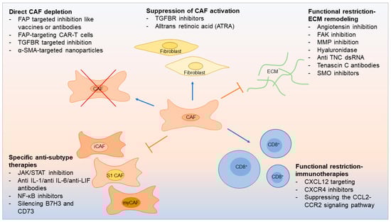
7. Therapeutic Opportunities Linked to CAF Subtypes

In PDAC, up to 90% of the tumor volume consists of stroma [40]. In HNSCC, CAFs can account for up to 80% of the tumor mass [48]. The tumor–stroma ratio has been shown to have prognostic importance [49], and evaluating this ratio can also garner insights into clinical disease [50]. Tumor stroma may block delivery of therapy to cancer cells, support cancer growth, and promote therapy resistance [25]. On the other hand, the tumor stroma may also contain potential targets for potential therapy. Recently, the number of preclinical experiments targeting CAFs to restore the anticancer immune response has increased dramatically. Further, strategies for CAF-based immunotherapy have emerged: direct CAF depletion including a novel therapeutic that uses FAP-targeting chimeric antigen receptor (CAR)-T cells [5] resulting in reduced CAF activation [51] and functional suppression of CAF-induced ECM remodeling [52][53][54] (Figure 2).
Figure 2. CAF-related therapeutics.
Multiple rationales exist for targeting CAFs in anti-cancer treatment, although the heterogeneity of CAF populations—and the reality that CAFs are always solely tumor promoting—complicate this approach. Supporting this, recent studies aiming to target stromal cells in general have not always been successful [55][56][57]. A new therapeutic approach would be to embrace the detection of CAF heterogeneity and to understand the importance of subtypes in terms of treatment opportunities. 
In terms of targeting specific markers on CAFs in general, FAP is one of the most widely studied. FAP has been used as a target in preclinical therapeutic strategies with vaccines, antibodies, and CAR-T cells [58][59][60][61]. In fact, a promising new FAP-targeting CAR-T cell has been shown to loosen the desmoplasia that restricts T cell invasion into tumors, which enables other tumor-targeting CAR-T cells to be used to finish the job and shrink the tumor [5].
Numerous previous studies have linked the interaction between CAFs and tumor cells in tumorigenesis. Additionally, infiltrating immune cells like macrophages, dendritic cells, lymphocytes, and neutrophils are another key component in the TME. Cancer immunotherapy has been one of the biggest medical breakthroughs in recent years. Several reports reveal improved patient survival with those who have increased CD3+/CD8+ T cell tumor infiltration [26][62][63]. The importance of lymphocytes as a positive prognostic factor in the TME has also been studied in oral cancer [64]. The interplay between tumor cells and immune cells throughout tumor progression involves multiple steps with both stimulating and inhibitory factors. The interaction between CAFs, immune cells, and tumor cells in the TME and their role in tumor progression is an ongoing topic of study. It is, however, known that CAFs regulate and enable the pro-tumor activity of TME immune cells both directly and indirectly via the ECM and secretion of cytokines such as IL-6 [15][54][65][66][67].
Most research into iCAFs describes them as protumorigenic and prognostically unfavorable, which aligns with the role of iCAFs and IL-6 secretion. Since cancer immunology has largely been focused on T cells and CD3+/CD8+ T cell response, it is crucial to determine CAF subtype functions in oral cancer, especially linked to CD8+ and CD4+ T cells as conducted by Costa et al. in S1 CAFs in breast and ovarian cancer [26][32]. For instance, CXCL12 secreted by FAP+ CAFs decreases tumor infiltration of CD8+ T cells [58]. This makes CXCL12 or its associated signaling pathway a potential therapeutic target. Similar analyses of CAF subtypes in the context of disease will reveal additional therapeutic targets with the potential to impact patient outcomes.

References

  1. Suresh, G.M.; Koppad, R.; Prakash, B.; Sabitha, K.; Dhara, P. Prognostic indicators of oral squamous cell carcinoma. Ann. Maxillofac. Surg. 2019, 9, 364–370.
  2. American Cancer Society. Cancer Facts & Figures 2023; American Cancer Society: Atlanta, GA, USA, 2023.
  3. Zainab, H.; Sultana, A.; Shaimaa. Stromal desmoplasia as a possible prognostic indicator in different grades of oral squamous cell carcinoma. J. Oral Maxillofac. Pathol. 2019, 23, 338–343.
  4. Graizel, D.; Zlotogorski-Hurvitz, A.; Tsesis, I.; Rosen, E.; Kedem, R.; Vered, M. Oral cancer-associated fibroblasts predict poor survival: Systematic review and meta-analysis. Oral Dis. 2020, 26, 733–744.
  5. Xiao, Z.; Todd, L.; Huang, L.; Noguera-Ortega, E.; Lu, Z.; Huang, L.; Kopp, M.; Li, Y.; Pattada, N.; Zhong, W.; et al. Desmoplastic stroma restricts T cell extravasation and mediates immune exclusion and immunosuppression in solid tumors. Nat. Commun. 2023, 14, 5110.
  6. Plikus, M.V.; Wang, X.; Sinha, S.; Forte, E.; Thompson, S.M.; Herzog, E.L.; Driskell, R.R.; Rosenthal, N.; Biernaskie, J.; Horsley, V. Fibroblasts: Origins, definitions, and functions in health and disease. Cell 2021, 184, 3852–3872.
  7. Kalluri, R. The biology and function of fibroblasts in cancer. Nat. Rev. Cancer 2016, 16, 582–598.
  8. Garcia, P.E.; Adoumie, M.; Kim, E.C.; Zhang, Y.; Scales, M.K.; El-Tawil, Y.S.; Shaikh, A.Z.; Wen, H.-J.; Bednar, F.; Allen, B.L.; et al. Differential Contribution of Pancreatic Fibroblast Subsets to the Pancreatic Cancer Stroma. Cell. Mol. Gastroenterol. Hepatol. 2020, 10, 581–599.
  9. Helms, E.J.; Berry, M.W.; Chaw, R.C.; DuFort, C.C.; Sun, D.; Onate, M.K.; Oon, C.; Bhattacharyya, S.; Sanford-Crane, H.; Horton, W.; et al. Mesenchymal Lineage Heterogeneity Underlies Nonredundant Functions of Pancreatic Cancer–Associated Fibroblasts. Cancer Discov. 2022, 12, 484–501.
  10. De Wever, O.; Nguyen, Q.-D.; Van Hoorde, L.; Bracke, M.; Bruyneel, E.; Gespach, C.; Mareel, M. Tenascin-C and SF/HGF produced by myofibroblasts in vitro provide convergent proinvasive signals to human colon cancer cells through RhoA and Rac. FASEB J. 2004, 18, 1016–1018.
  11. Radhakrishnan, R.; Ha, J.H.; Jayaraman, M.; Liu, J.; Moxley, K.M.; Isidoro, C.; Sood, A.K.; Song, Y.S.; Dhanasekaran, D.N. Ovarian cancer cell-derived lysophosphatidic acid induces glycolytic shift and cancer-associated fibroblast-phenotype in normal and peritumoral fibroblasts. Cancer Lett. 2019, 442, 464–474.
  12. Su, Q.; Zhang, B.; Zhang, L.; Dang, T.; Rowley, D.; Ittmann, M.; Xin, L. Jagged1 upregulation in prostate epithelial cells promotes formation of reactive stroma in the Pten null mouse model for prostate cancer. Oncogene 2017, 36, 618–627.
  13. Strell, C.; Paulsson, J.; Jin, S.-B.; Tobin, N.P.; Mezheyeuski, A.; Roswall, P.; Mutgan, C.; Mitsios, N.; Johansson, H.; Wickberg, S.M.; et al. Impact of Epithelial–Stromal Interactions on Peritumoral Fibroblasts in Ductal Carcinoma in Situ. J. Natl. Cancer Inst. 2019, 111, 983–995.
  14. Sanz-Moreno, V.; Gaggioli, C.; Yeo, M.; Albrengues, J.; Wallberg, F.; Viros, A.; Hooper, S.; Mitter, R.; Féral, C.C.; Cook, M.; et al. ROCK and JAK1 Signaling Cooperate to Control Actomyosin Contractility in Tumor Cells and Stroma. Cancer Cell 2011, 20, 229–245.
  15. Erez, N.; Truitt, M.; Olson, P.; Hanahan, D. Cancer-Associated Fibroblasts Are Activated in Incipient Neoplasia to Orchestrate Tumor-Promoting Inflammation in an NF-κB-Dependent Manner. Cancer Cell 2010, 17, 135–147.
  16. Amatangelo, M.D.; Bassi, D.E.; Klein-Szanto, A.J.; Cukierman, E. Stroma-Derived Three-Dimensional Matrices Are Necessary and Sufficient to Promote Desmoplastic Differentiation of Normal Fibroblasts. Am. J. Pathol. 2005, 167, 475–488.
  17. Calvo, F.; Ege, N.; Grande-Garcia, A.; Hooper, S.; Jenkins, R.P.; Chaudhry, S.I.; Harrington, K.; Williamson, P.; Moeendarbary, E.; Charras, G.; et al. Mechanotransduction and YAP-dependent matrix remodelling is required for the generation and maintenance of cancer-associated fibroblasts. Nat. Cell Biol. 2013, 15, 637–646.
  18. Weinberg, F.; Ramnath, N.; Nagrath, D. Reactive Oxygen Species in the Tumor Microenvironment: An Overview. Cancers 2019, 11, 1191.
  19. Fordyce, C.; Fessenden, T.; Pickering, C.; Jung, J.; Singla, V.; Berman, H.; Tlsty, T. DNA Damage Drives an Activin A–Dependent Induction of Cyclooxygenase-2 in Premalignant Cells and Lesions. Cancer Prev. Res. 2010, 3, 190–201.
  20. Arebro, J.; Towle, R.; Lee, C.-M.; Bennewith, K.L.; Garnis, C. Extracellular vesicles promote activation of pro-inflammatory cancer-associated fibroblasts in oral cancer. Front. Cell Dev. Biol. 2023, 11, 1240159.
  21. Dai, G.; Yao, X.; Zhang, Y.; Gu, J.; Geng, Y.; Xue, F.; Zhang, J. Colorectal cancer cell–derived exosomes containing miR-10b regulate fibroblast cells via the PI3K/Akt pathway. Bull. Cancer 2018, 105, 336–349.
  22. Goulet, C.R.; Bernard, G.; Tremblay, S.; Chabaud, S.; Bolduc, S.; Pouliot, F. Exosomes Induce Fibroblast Differentiation into Cancer-Associated Fibroblasts through TGFβ Signaling. Mol. Cancer Res. 2018, 16, 1196–1204.
  23. Ping, Q.; Yan, R.; Cheng, X.; Wang, W.; Zhong, Y.; Hou, Z.; Shi, Y.; Wang, C.; Li, R. Cancer-associated fibroblasts: Overview, progress, challenges, and directions. Cancer Gene Ther. 2021, 28, 984–999.
  24. Zhang, X.; Zhang, Y.; Qiu, X.; Cai, J.; Yang, Z.; Song, F. Extracellular Vesicles Derived from Lung Cancer Cells Induce Transformation of Normal Fibroblasts into Lung Cancer-Associated Fibroblasts and Promote Metastasis of Lung Cancer by Delivering lncRNA HOTAIR. Stem Cells Int. 2022, 2022, 3805013.
  25. Shoucair, I.; Mello, F.W.; Jabalee, J.; Maleki, S.; Garnis, C. The Role of Cancer-Associated Fibroblasts and Extracellular Vesicles in Tumorigenesis. Int. J. Mol. Sci. 2020, 21, 6837.
  26. Costa, A.; Kieffer, Y.; Scholer-Dahirel, A.; Pelon, F.; Bourachot, B.; Cardon, M.; Sirven, P.; Magagna, I.; Fuhrmann, L.; Bernard, C.; et al. Fibroblast heterogeneity and immunosuppressive environment in human breast cancer. Cancer Cell 2018, 33, 463–479.e10.
  27. Öhlund, D.; Elyada, E.; Tuveson, D. Fibroblast heterogeneity in the cancer wound. J. Exp. Med. 2014, 211, 1503–1523.
  28. Purcell, J.W.; Tanlimco, S.G.; Hickson, J.A.; Fox, M.; Sho, M.; Durkin, L.; Uziel, T.; Powers, R.; Foster, K.; McGonigal, T.; et al. LRRC15 Is a Novel Mesenchymal Protein and Stromal Target for Antibody–Drug Conjugates. Cancer Res. 2018, 78, 4059–4072.
  29. Available online: https://fibroxplorer.com/ (accessed on 1 November 2023).
  30. Buechler, M.B.; Pradhan, R.N.; Krishnamurty, A.T.; Cox, C.; Calviello, A.K.; Wang, A.W.; Yang, Y.A.; Tam, L.; Caothien, R.; Roose-Girma, M.; et al. Cross-tissue organization of the fibroblast lineage. Nature 2021, 593, 575–579.
  31. Bonneau, C.; Eliès, A.; Kieffer, Y.; Bourachot, B.; Ladoire, S.; Pelon, F.; Hequet, D.; Guinebretière, J.-M.; Blanchet, C.; Vincent-Salomon, A.; et al. A subset of activated fibroblasts is associated with distant relapse in early luminal breast cancer. Breast Cancer Res. 2020, 22, 76.
  32. Givel, A.-M.; Kieffer, Y.; Scholer-Dahirel, A.; Sirven, P.; Cardon, M.; Pelon, F.; Magagna, I.; Gentric, G.; Costa, A.; Bonneau, C.; et al. miR200-regulated CXCL12β promotes fibroblast heterogeneity and immunosuppression in ovarian cancers. Nat. Commun. 2018, 9, 1056.
  33. Sebastian, A.; Hum, N.R.; Martin, K.A.; Gilmore, S.F.; Peran, I.; Byers, S.W.; Wheeler, E.K.; Coleman, M.A.; Loots, G.G. Single-Cell Transcriptomic Analysis of Tumor-Derived Fibroblasts and Normal Tissue-Resident Fibroblasts Reveals Fibroblast Heterogeneity in Breast Cancer. Cancers 2020, 12, 130.
  34. Elyada, E.; Bolisetty, M.; Laise, P.; Flynn, W.F.; Courtois, E.T.; Burkhart, R.A.; Teinor, J.A.; Belleau, P.; Biffi, G.; Lucito, M.S.; et al. Cross-Species Single-Cell Analysis of Pancreatic Ductal Adenocarcinoma Reveals Antigen-Presenting Cancer-Associated Fibroblasts. Cancer Discov. 2019, 9, 1102–1123.
  35. Wu, S.Z.; Roden, D.L.; Wang, C.; Holliday, H.; Harvey, K.; Cazet, A.S.; Murphy, K.J.; Pereira, B.; Al-Eryani, G.; Bartonicek, N.; et al. Stromal cell diversity associated with immune evasion in human triple-negative breast cancer. EMBO J. 2020, 39, e104063.
  36. Biffi, G.; Oni, T.E.; Spielman, B.; Hao, Y.; Elyada, E.; Park, Y.; Preall, J.; Tuveson, D.A. IL1-Induced JAK/STAT Signaling Is Antagonized by TGFβ to Shape CAF Heterogeneity in Pancreatic Ductal Adenocarcinoma. Cancer Discov. 2019, 9, 282–301.
  37. Ohlund, D.; Handly-Santana, A.; Biffi, G.; Elyada, E.; Almeida, A.S.; Ponz-Sarvise, M.; Corbo, V.; Oni, T.E.; Hearn, S.A.; Lee, E.J.; et al. Distinct populations of inflammatory fibroblasts and myofibroblasts in pancreatic cancer. J. Exp. Med. 2017, 214, 579–596.
  38. Bernard, V.; Semaan, A.; Huang, J.; Lucas, F.A.S.; Mulu, F.C.; Stephens, B.M.; Guerrero, P.A.; Huang, Y.; Zhao, J.; Kamyabi, N.; et al. Single-Cell Transcriptomics of Pancreatic Cancer Precursors Demonstrates Epithelial and Microenvironmental Heterogeneity as an Early Event in Neoplastic Progression. Clin. Cancer Res. 2019, 25, 2194–2205.
  39. Zhang, Q.; Wang, Y.; Xia, C.; Ding, L.; Pu, Y.; Hu, X.; Cai, H.; Hu, Q. Integrated analysis of single-cell RNA-seq and bulk RNA-seq reveals distinct cancer-associated fibroblasts in head and neck squamous cell carcinoma. Ann. Transl. Med. 2021, 9, 1017.
  40. Schwörer, S.; Cimino, F.V.; Ros, M.; Tsanov, K.M.; Ng, C.; Lowe, S.W.; Carmona-Fontaine, C.; Thompson, C.B. Hypoxia Potentiates the Inflammatory Fibroblast Phenotype Promoted by Pancreatic Cancer Cell–Derived Cytokines. Cancer Res 2023, 83, 1596–1610.
  41. Naito, Y.; Yamamoto, Y.; Sakamoto, N.; Shimomura, I.; Kogure, A.; Kumazaki, M.; Yokoi, A.; Yashiro, M.; Kiyono, T.; Yanagihara, K.; et al. Cancer extracellular vesicles contribute to stromal heterogeneity by inducing chemokines in cancer-associated fibroblasts. Oncogene 2019, 38, 5566–5579.
  42. Chiavarina, B.; Ronca, R.; Otaka, Y.; Sutton, R.B.; Rezzola, S.; Yokobori, T.; Chiodelli, P.; Souche, R.; Pourquier, D.; Maraver, A.; et al. Fibroblast-derived prolargin is a tumor suppressor in hepatocellular carcinoma. Oncogene 2022, 41, 1410–1420.
  43. Ying, F.; Chan, M.S.M.; Lee, T.K.W. Cancer-Associated Fibroblasts in Hepatocellular Carcinoma and Cholangiocarcinoma. Cell. Mol. Gastroenterol. Hepatol. 2023, 15, 985–999.
  44. Zhang, M.; Yang, H.; Wan, L.; Wang, Z.; Wang, H.; Ge, C.; Liu, Y.; Hao, Y.; Zhang, D.; Shi, G.; et al. Single-cell transcriptomic architecture and intercellular crosstalk of human intrahepatic cholangiocarcinoma. J. Hepatol. 2020, 73, 1118–1130.
  45. Galbo, P.M., Jr.; Zang, X.; Zheng, D. Molecular Features of Cancer-associated Fibroblast Subtypes and their Implication on Cancer Pathogenesis, Prognosis, and Immunotherapy Resistance. Clin. Cancer Res. 2021, 27, 2636–2647.
  46. Takahashi, H.; Rokudai, S.; Kawabata-Iwakawa, R.; Sakakura, K.; Oyama, T.; Nishiyama, M.; Chikamatsu, K. AKT3 Is a Novel Regulator of Cancer-Associated Fibroblasts in Head and Neck Squamous Cell Carcinoma. Cancers 2021, 13, 1233.
  47. Yang, W.; Zhang, S.; Li, T.; Zhou, Z.; Pan, J. Single-cell analysis reveals that cancer-associated fibroblasts stimulate oral squamous cell carcinoma invasion via the TGF-β/Smad pathway. Acta Biochim. Biophys. Sin. 2022, 55, 262–273.
  48. Bienkowska, K.J.; Hanley, C.J.; Thomas, G.J. Cancer-Associated Fibroblasts in Oral Cancer: A Current Perspective on Function and Potential for Therapeutic Targeting. Front. Oral Health 2021, 2, 686337.
  49. Almangush, A.; Heikkinen, I.; Bakhti, N.; Mäkinen, L.K.; Kauppila, J.H.; Pukkila, M.; Hagström, J.; Laranne, J.; Soini, Y.; Kowalski, L.P.; et al. Prognostic impact of tumour–stroma ratio in early-stage oral tongue cancers. Histopathology 2018, 72, 1128–1135.
  50. Hendry, S.M.; Salgado, R.; Gevaert, T.; Russell, P.A.M.; John, T.; Thapa, B.M.; Christie, M.M.; van de Vijver, K.; Estrada, M.; Gonzalez-Ericsson, P.I.; et al. Assessing Tumor-Infiltrating Lymphocytes in Solid Tumors: A Practical Review for Pathologists and Proposal for a Standardized Method from the International Immuno-Oncology Biomarkers Working Group: Part 2: TILs in Melanoma, Gastrointestinal Tract Carcinomas, Non–Small Cell Lung Carcinoma and Mesothelioma, Endometrial and Ovarian Carcinomas, Squamous Cell Carcinoma of the Head and Neck, Genitourinary Carcinomas, and Primary Brain Tumors. Adv. Anat. Pathol. 2017, 24, 311–335.
  51. Froeling, F.E.; Feig, C.; Chelala, C.; Dobson, R.; Mein, C.E.; Tuveson, D.; Clevers, H.; Hart, I.R.; Kocher, H.M. Retinoic Acid–Induced Pancreatic Stellate Cell Quiescence Reduces Paracrine Wnt–β-Catenin Signaling to Slow Tumor Progression. Gastroenterology 2011, 141, 1486–1497.e14.
  52. Li, Z.-L.; Zhang, H.-L.; Huang, Y.; Huang, J.-H.; Sun, P.; Zhou, N.-N.; Chen, Y.-H.; Mai, J.; Wang, Y.; Yu, Y.; et al. Autophagy deficiency promotes triple-negative breast cancer resistance to T cell-mediated cytotoxicity by blocking tenascin-C degradation. Nat. Commun. 2020, 11, 3806.
  53. Cazet, A.S.; Hui, M.N.; Elsworth, B.L.; Wu, S.Z.; Roden, D.; Chan, C.-L.; Skhinas, J.N.; Collot, R.; Yang, J.; Harvey, K.; et al. Targeting stromal remodeling and cancer stem cell plasticity overcomes chemoresistance in triple negative breast cancer. Nat. Commun. 2018, 9, 2897.
  54. Mao, X.; Xu, J.; Wang, W.; Liang, C.; Hua, J.; Liu, J.; Zhang, B.; Meng, Q.; Yu, X.; Shi, S. Crosstalk between cancer-associated fibroblasts and immune cells in the tumor microenvironment: New findings and future perspectives. Mol. Cancer 2021, 20, 131.
  55. Özdemir, B.C.; Pentcheva-Hoang, T.; Carstens, J.L.; Zheng, X.; Wu, C.-C.; Simpson, T.R.; Laklai, H.; Sugimoto, H.; Kahlert, C.; Novitskiy, S.V.; et al. Depletion of Carcinoma-Associated Fibroblasts and Fibrosis Induces Immunosuppression and Accelerates Pancreas Cancer with Reduced Survival. Cancer Cell 2015, 28, 831–833.
  56. Rhim, A.D.; Oberstein, P.E.; Thomas, D.H.; Mirek, E.T.; Palermo, C.F.; Sastra, S.A.; Dekleva, E.N.; Saunders, T.; Becerra, C.P.; Tattersall, I.W.; et al. Stromal elements act to restrain, rather than support, pancreatic ductal adenocarcinoma. Cancer Cell 2014, 25, 735–747.
  57. Lee, J.J.; Perera, R.M.; Wang, H.; Wu, D.-C.; Liu, X.S.; Han, S.; Fitamant, J.; Jones, P.D.; Ghanta, K.S.; Kawano, S.; et al. Stromal response to Hedgehog signaling restrains pancreatic cancer progression. Proc. Natl. Acad. Sci. USA 2014, 111, E3091–E3100.
  58. Feig, C.; Jones, J.O.; Kraman, M.; Wells, R.J.; Deonarine, A.; Chan, D.S.; Connell, C.M.; Roberts, E.W.; Zhao, Q.; Caballero, O.L.; et al. Targeting CXCL12 from FAP-expressing carcinoma-associated fibroblasts synergizes with anti-PD-L1 immunotherapy in pancreatic cancer. Proc. Natl. Acad. Sci. USA 2013, 110, 20212–20217.
  59. Kraman, M.; Bambrough, P.J.; Arnold, J.N.; Roberts, E.W.; Magiera, L.; Jones, J.O.; Gopinathan, A.; Tuveson, D.A.; Fearon, D.T. Suppression of Antitumor Immunity by Stromal Cells Expressing Fibroblast Activation Protein–α. Science 2010, 330, 827–830.
  60. Lo, A.; Li, C.-P.; Buza, E.L.; Blomberg, R.; Govindaraju, P.; Avery, D.; Monslow, J.; Hsiao, M.; Puré, E. Fibroblast activation protein augments progression and metastasis of pancreatic ductal adenocarcinoma. JCI Insight 2017, 2, e92232.
  61. Peltier, A.; Seban, R.-D.; Buvat, I.; Bidard, F.-C.; Mechta-Grigoriou, F. Fibroblast heterogeneity in solid tumors: From single cell analysis to whole-body imaging. Semin. Cancer Biol. 2022, 86, 262–272.
  62. Fridman, W.H.; Pagès, F.; Sautès-Fridman, C.; Galon, J. The immune contexture in human tumours: Impact on clinical outcome. Nat. Rev. Cancer 2012, 12, 298–306.
  63. Jimenez, D.G.; Sobti, A.; Askmyr, D.; Sakellariou, C.; Santos, S.C.; Swoboda, S.; Forslund, O.; Greiff, L.; Lindstedt, M. Tonsillar Cancer with High CD8+ T-Cell Infiltration Features Increased Levels of Dendritic Cells and Transcriptional Regulation Associated with an Inflamed Tumor Microenvironment. Cancers 2021, 13, 5341.
  64. Almangush, A.; Bello, I.O.; Heikkinen, I.; Hagström, J.; Haglund, C.; Kowalski, L.P.; Nieminen, P.; Coletta, R.D.; Mäkitie, A.A.; Salo, T.; et al. Stromal categorization in early oral tongue cancer. Virchows Arch. 2021, 478, 925–932.
  65. Harper, J.; Sainson, R.C. Regulation of the anti-tumour immune response by cancer-associated fibroblasts. Semin. Cancer Biol. 2014, 25, 69–77.
  66. Liu, T.; Han, C.; Wang, S.; Fang, P.; Ma, Z.; Xu, L.; Yin, R. Cancer-associated fibroblasts: An emerging target of anti-cancer immunotherapy. J. Hematol. Oncol. 2019, 12, 86.
  67. Karakasheva, T.A.; Lin, E.W.; Tang, Q.; Qiao, E.; Waldron, T.J.; Soni, M.; Klein-Szanto, A.J.; Sahu, V.; Basu, D.; Ohashi, S.; et al. IL-6 Mediates Cross-Talk between Tumor Cells and Activated Fibroblasts in the Tumor Microenvironment. Cancer Res. 2018, 78, 4957–4970.
More
Information
Subjects: Oncology
Contributors MDPI registered users' name will be linked to their SciProfiles pages. To register with us, please refer to https://encyclopedia.pub/register : , , ,
View Times: 53
Revisions: 2 times (View History)
Update Date: 22 Mar 2024
1000/1000