Submitted Successfully!
To reward your contribution, here is a gift for you: A free trial for our video production service.
Thank you for your contribution! You can also upload a video entry or images related to this topic.
Version Summary Created by Modification Content Size Created at Operation
1 -- 3226 2024-03-18 09:14:01 |
2 Reference format revised. Meta information modification 3226 2024-03-19 02:42:02 |

Video Upload Options

Do you have a full video?

Confirm

Are you sure to Delete?
Cite
If you have any further questions, please contact Encyclopedia Editorial Office.
Xu, W.; Chen, H.; Li, Y.; Liu, S.; Wang, K.; Liu, J. DNA-Based Artificial Transmembrane Channels for Biomedical Applications. Encyclopedia. Available online: https://encyclopedia.pub/entry/56372 (accessed on 28 April 2024).
Xu W, Chen H, Li Y, Liu S, Wang K, Liu J. DNA-Based Artificial Transmembrane Channels for Biomedical Applications. Encyclopedia. Available at: https://encyclopedia.pub/entry/56372. Accessed April 28, 2024.
Xu, Wanyu, Hui Chen, Yang Li, Shuangna Liu, Kemin Wang, Jianbo Liu. "DNA-Based Artificial Transmembrane Channels for Biomedical Applications" Encyclopedia, https://encyclopedia.pub/entry/56372 (accessed April 28, 2024).
Xu, W., Chen, H., Li, Y., Liu, S., Wang, K., & Liu, J. (2024, March 18). DNA-Based Artificial Transmembrane Channels for Biomedical Applications. In Encyclopedia. https://encyclopedia.pub/entry/56372
Xu, Wanyu, et al. "DNA-Based Artificial Transmembrane Channels for Biomedical Applications." Encyclopedia. Web. 18 March, 2024.
DNA-Based Artificial Transmembrane Channels for Biomedical Applications
Edit

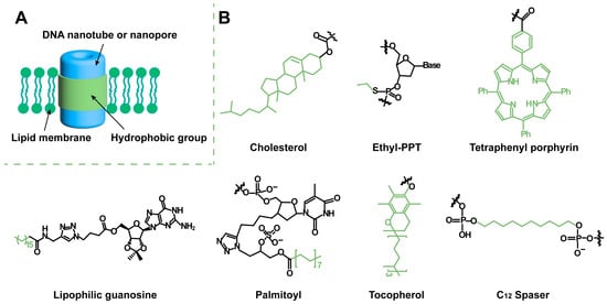
Biomolecular channels on the cell membrane are essential for transporting substances across the membrane to maintain cell physiological activity. Artificial transmembrane channels used to mimic biological membrane channels can regulate intra/extracellular ionic and molecular homeostasis, and they elucidate cellular structures and functionalities. Due to their program design, facile preparation, and high biocompatibility, DNA nanostructures have been widely used as scaffolds for the design of artificial transmembrane channels and exploited for ionic and molecular transport and biomedical applications. DNA-based artificial channels can be designed from two structural modules: DNA nanotubes/nanopores as transport modules for mass transportation and hydrophobic segments as anchor modules for membrane immobilization. 

artificial transmembrane channel DNA nanostructure biosensing biomedical application

1. Introduction

Transmembrane channels are integral membrane proteins with channels or pores that allow particular ions or small molecules to cross a lipid bilayer [1][2]. These channels are critical for regulating ion homeostasis, transporting molecules, maintaining normal cell physiological functions, and performing related life activities [3]. However, their formation requires genetic coding in living cells under strict spatiotemporal control, which is difficult to reproduce in vitro [4]. To date, various materials, such as biological macromolecules, synthetic organic compounds, and inorganic substances, have been successfully used for the design of artificial nanochannels with diverse structures and functions [5]. The design of synthetic biomolecules that mimic the structures and functions of natural transmembrane channels has garnered substantial interest among molecular biology researchers as models for studying fundamental information, creating alternative drugs, and developing advanced biosensors.
Deoxyribonucleic acid (DNA) is an irreplaceable building material for the design of artificial channels [6], since DNA is an easily accessible biomacromolecule with high biocompatibility and programmable self-assembly ability, and it can be safely used in natural biological processes [7]. In addition, each strand used to construct DNA channels can be independently functionalized through the precise design and modification of various biochemical molecules [8]. With excellent shapes, structures, and functions, DNA-based artificial channels have been widely used for the transmembrane transport of ions or molecules, the transduction of intercellular signals, and the regulation of cell physiological activities [9]. Various methods have been reported for the design of DNA-based transmembrane channels [10].

2. Hydrophobic Modification for Artificial Transmembrane Channels

The purposes of designing DNA-based artificial channels include the transmembrane transport of substances for signal exchange, the adjustment of the concentrations of substances, and the regulation of cell behaviors. The design method, structure (pore size and length), and interaction force between the channel and phospholipid membrane can directly affect the material transport efficiency [11]. DNA-based artificial transmembrane channels transport ions or biomolecules between phospholipid membranes, which mainly rely on the arrangement and assembly of DNA strands to form nanotubes or nanopores with holes [12]. Generally, DNA-based artificial transmembrane channels are composed of two structural modules: DNA nanotubes/nanopores as transport modules for mass transportation; and hydrophobic segments as anchor modules for membrane immobilization (Figure 1A). For example, phospholipids consist of a hydrophilic head and two hydrophobic tails. Hydrophobic fragments can be inserted into the internal hydrophobic site, allowing DNA nanostructures to be well anchored to phospholipid membranes.
Figure 1. Structure model of artificial transmembrane channels and different hydrophobic modifications. (A) Schematic diagram of transmembrane channels formed by hydrophobic-modified DNA nanotubes or nanopores. (B) Various hydrophobic modifications: cholesterol, ethyl-PPT, tetraphenyl porphyrin, lipophilic guanosine, palmitoyl, tocopherol, and C12 spacer.
A core challenge of DNA nanotubes or nanopores embedded in phospholipid membranes regarding the formation of transmembrane channels is to overcome the adverse interactions between the hydrophilic, negatively charged DNA nanostructures and the hydrophobic membrane environment [6]. Many strategies have been developed to insert DNA nanostructures into the hydrophobic centers of the lipid bilayer. Keyser et al. have proposed several steps for constructing efficient membrane channels, especially for overcoming the high energy barrier of DNA across the bilayer hydrophobic core: (1) The connector between the hydrophobic anchor and the DNA core is shortened to achieve improved control over the anchor position. (2) When large structures are introduced, there should be a large spacing between the hydrophobic anchors to inhibit their simultaneous insertion into the bilayer without inducing crossing [13]. (3) The distance between the end of the structure and the membrane transdomain, as determined by anchor position, is reduced, limiting adverse interactions between the charged material and the hydrophobic core of the membrane [14].
Hydrophobic modification of DNA nanotubes or nanopores is a key step when designing transmembrane channels. Due to the excellent properties resulting from its membrane insertion, cholesterol is the preferred hydrophobic group for DNA modification. Cholesterol is most frequently used because of its strong hydrophobicity and because it can be modified at the DNA end or middle section, which is more conducive to the design of DNA artificial transmembrane channel structures. The amount and location of cholesterol are adjusted according to the designed channel shape and function. The number of cholesterols on artificial channels has been reported to be in the range of 2–60 [15][16][17][18][19][20][21][22]. DNA channels with small pore sizes and simple structures have little resistance to inserting into the membrane, and the corresponding modification method is relatively simple. Small-aperture artificial channels usually require only a few cholesterols at intervals along the sidewalls of the channel. However, as the pore size increases, it becomes increasingly difficult for DNA channels to be inserted into the lipid membrane. 
Ethyl-phosphorothioate (ethyl-PPT) [23], tetraphenyl porphyrin (TPP) [24], lipophilic guanosine [25], palmitoyl [20], tocopherol [26], and C12 spacer [27] are widely used for DNA hydrophobic modification (Figure 1B). The attachment of the ethyl group to the mercaptan group removes the negative charge of the typical phosphate anion. Ethyl iodide reacts with mercaptan groups through nucleophilic substitution to produce ethyl-protected PPT, which can be used for the hydrophobic modification of DNA channels [23]. To simplify the design of channels and minimize chemical intervention, other chemical labels with improved hydrophobicity have been proposed. It is expected that macroporous channels can be anchored to the membrane with a few chemical labels. TPP meets the requirements of hydrophobicity and can be easily coupled with DNA. Therefore, DNA channels modified with two porphyrin labels are constructed [24]. Acetylene-TPP is rigidly linked to the DNA strand by Sonogashira coupling to deoxyuridine. The T-pore-based artificial channels that were reported in 2016 are hydrophobically modified with tocopherol as a substitute for cholesterol [26].

3. Design of DNA Nanostructure for Artificial Transmembrane Channels

3.1. DNA Wireframe-Based Transmembrane Channels

DNA wireframe nanotubes are promising self-assembled nanostructures that have produced a range of nanotubes or nanopores with different cavity sizes and tube lengths [28][29][30][31][32][33]. A typical modular assembly process for designing a wireframe structure from short synthetic strands is as follows: First, DNA structures acting as modular building blocks with specific shapes, such as triangles, squares, pentagons, and hexagons, are designed as needed. DNA nanotube rungs are then formed by longitudinal assembly through connecting strands.
DNA wireframe nanotubes can act as artificial transmembrane channels after adding hydrophobic anchors for membrane insertion. Sleiman et al. [15] designed cuboidal DNA channels and found that changing the pattern of the cholesterol unit on the cuboidal DNA significantly alters its interaction pattern with the lipid membrane. Modification of cholesterol on a single face of the cube results in the peripheral anchoring of the DNA structure, while modification of cholesterol on two opposite faces of the cube enables the DNA pipeline structure to cross the phospholipid membrane. The DNA channels embedded in the membrane function as nanochannels for the transmembrane transport of dyes. Furthermore, researchers have designed a switch for the channel to avoid channel aggregation. This switch is achieved by adjusting the length of the cholesterol-DNA conjugate and the interval between the cubic binding segment of the strand and the cholesterol unit. The cholesterol units are initially hidden inside the cube and then exposed by a conformational switch for membrane insertion. Cholesterol-DNA cubes have become the first open-walled DNA channels that can be used as tools for sensing, drug delivery, and cellular behavior regulation applications.

3.2. DNA Helix Bundle-Based Transmembrane Channels

Small-pore DNA-based artificial channels can be designed based on helix bundle (HB)-based nanotube assembly through the concatenation of multiple DNA strands [16]. DNA helix bundles are prepared by cross-joining scaffolds and short strands in the middle or at the ends of the materials, and the helix bundles follow the structural layout of polygon arrays [23]. The six DNA helix bundle (6-HB) nanotubes form approximately 2 nm-wide pores at the center, which are hydrophobically modified to maintain structural stability in the lipid bilayer and to support a constant transmembrane current. The reported conductivities of these 6-HB DNA transmembrane channels range from approximately 0.3 nS to 1.6 nS [23][24]. Through molecular dynamics (MD) simulations, the following conclusions are found. (1) The chemical modification on the surface of the channel has an extremely large impact on the transport of water and ions across the membrane, and the type, number, and position of the hydrophobic group modification can directly affect the formation of transmembrane channels. (2) DNA nanochannels can be used to transfer charged solute pairs to antistatic gradients through electroosmosis. (3) The porous channel wall allows the transverse leakage of ions and water. (4) The central lumen of the DNA channel is cylindrical and filled with water and ions; the volumes of water at the opening regions of both ends fluctuate in time and exhibit mechanosensitive gating, creating a force sensor [34][35][36][37].

3.3. DNA Tile-Based Transmembrane Channels

DNA tiles are short intersecting DNA strands that contribute to structural control. They have cohesive ends that can be programmed to self-assemble to form various DNA nanostructures by clinging together [38][39]. DNA tiles have been widely used to prepare DNA nanotubes. The steps of their self-assembly mainly focus on the release of DNA input molecules to trigger the growth of nanostructures, which is a nonautonomous and irreversible reaction [40]. With the maturation of construction technology, several methods have been designed to synthesize DNA tile nanotubes with adjustable, reversible, or controllable termination characteristics [41][42][43]. By using DNA origami structures as seeds to construct channels, micron-long nanotubes can be obtained through the polymeric growth of DNA tiles. The hydrophobic unit on the seed can directly insert the nanotube into the membrane to form a transmembrane channel [44]. An additional DNA origami channel cap can be used to terminate the aggregation of tiles. Conductivity measurements reveal that the conductance values of seeds and nanochannels are lower than their uncapped counterparts when the channel caps are attached. The results show that ions move from one end of the channel to the other and that there is partial leakage through the channel wall. However, the observation experiments of fluorescent dyes crossing lipid membranes confirm that molecular transport can occur through DNA nanochannels and that it is mainly end-to-end rather than across the channel wall [45].

3.4. DNA Origami-Based Transmembrane Channels

DNA origami technology can be used for designing DNA-based channels; the shapes and sizes of the channels can be adjusted precisely, systematically, and abundantly [11]. DNA origami, proposed by Rothemund in 2006, is a relatively new method for DNA assembly. Based on the principle of complementary base pairing, by utilizing the structural characteristics of DNA molecules, the long DNA strands folded in specific regions are fixed by short strands to construct the expected structure [46]. Due to its simple experimental conditions and high assembly efficiency, DNA origami has become a popular technology for constructing artificial DNA nanochannels. Artificial DNA nanochannels have been comprehensively designed concerning their pore size, length, and morphology characteristics.
Artificial DNA nanochannels can be exploited for size-dependent, selective transmembrane transport. The pore diameter is critical for the selective delivery of substances of different sizes. Transmembrane channels with large pore sizes are required for the transport of macromolecular substances. To this end, long DNA strands that are arranged longitudinally are initially used as basic units to construct channels, and the inner diameters of these channels are <9 nm [17][18][19][20]. A method of lateral assembly of DNA strands to form channels has been proposed; the same amount of DNA strands can be used to obtain artificial DNA nanochannels with a relatively large pore size (~35 nm) [21][22].
The DNA nanochannels synthesized by Kjems et al. [20] have flanks to adjust the cholesterol exposure on demand. The DNA channels are composed of a double-layer irregular hexagonal cylindrical DNA structure with 46 hydrophobic spots (17 on the walls and 29 on the flanks). Three programmable DNA lobes are on the three nonadjacent sides of the channel and connected approximately 12 nm from the bottom of the channel by a single-stranded DNA hinge. In the closed state, each flank is near the channel wall due to two stable strands that are complementary to the bottom single-strand portion of the channel. The flanking closure can protect the hydrophobic moiety from the aqueous environment and limit hydrophobicity-driven channel aggregation. When a key strand fully complementary to the single-stranded DNA is present around the channel, the flanks are opened and the cholesterol is exposed, thereby driving channel insertion into the membrane. 

3.5. Other DNA-Based Transmembrane Channels

G-quadruplex, a DNA duplex with unique ion transport properties, is utilized to design transmembrane channels. Depending on the hydrogen bond between the nitrogen and oxygen atoms of guanosine and the π-π stacking between the bases, guanine (G)-rich DNA single strands can be deformed or aggregated to form a G-quadruplex structure with a central hole [47]. Moreover, the stability of the G-quadruplex depends heavily on certain cations, such as K+, Na+, NH4+, and Ca2+ [48]. Within the central channel, each ion is completely dehydrated and interacts with the guanine carbonyl O6 atom around the pore. This particular feature is reminiscent of the selectivity filters in K+ ion channel proteins first noted by Feigon et al. Because of this structural similarity, the G-quadruplex is a candidate for the design of artificial ion channels for selective transmembrane transport of K+ [49][50][51]. Dash et al. [25] have used telomere DNA to form a G-quadruplex and additionally modified lipophilic guanosine to construct an artificial potassium ion transmembrane transport carrier.

4. Artificial Transmembrane Channels for Biosensing and Biomedical Applications

4.1. DNA-Based Transmembrane Channels for Biosensors

4.1.1. Single-Molecule Nanochannel Sensors

Transmembrane DNA channels have been proposed for use as single-molecule nanochannel sensors. In biomimetic sensing experiments, the translocation of analyte molecules leads to current changes in membrane pores and the duration and depth, which are related to the charges and sizes of the analytes. For example, DNA channels designed by Simmel et al. [17] have been used for single-molecule sensing. A stable baseline current is detected on the lipid membrane containing the artificial DNA channels. The additions of the hairpin molecules at the beginning and ~30 min later show transient current blocking, in which the applied voltage can capture, decompress, and translocate the hairpin structure.

4.1.2. Ligand-Gated Artificial Transmembrane Channels

The DNA-based artificial transmembrane channels are mostly hollow tubes with openings. Voltage gating is observed in almost all DNA nanochannels, but channels with additional gates are highly flexible and controllable [52]. Therefore, the design of artificial channels with ligand-gated opening or closing properties has become the focus of research. The reported artificial DNA nanochannels with gates can specifically recognize key strands. Howorka et al. [53] built an artificial channel with a gate that can be opened with a key strand. The lock of the channel is tightly bound to the entrance by hybridizing with two docking sites to form a spiral bundle across the channel opening. The docking site is formed by the extension of two duplex support rods in opposite positions. The key can be hybridized with the lock strand to remove it, leaving the channel open. After being modified by cholesterol, such channels can be used to regulate the flow of small organic molecules (including many important drug compounds), which have broad application prospects in biomedicine.
To exploit the potential of DNA-based artificial channels as real-time smart sensing devices, Kjems et al. [20] have designed a bolt on the inside of the channel. PEG is used as a plug that connects the toehold sequence with 8 nucleotides to partially block the gateway and allows small molecules (ATTO 655) to pass through. The unplugged strand is combined with the toehold-mediated strand to remove PEG so that the macromolecular material (dTMR-40k) can pass through the artificial DNA nanochannel. Another reversible gated protein transport membrane channel is constructed based on a horizontal routing DNA origami design strategy with a large pore size of 20.4 nm [54]. The passageway opening is designed with a square cover, one side of which is attached to the cap by a flexure hinge. The other side of the cap carries two single strands that can be hybridized with the two single strands on the cap to form a complete double lock. Two key strands are added to the system to open the lock and lid. The lid is switched back to its closed state with a single-strand reverse key pair. This channel allows the precisely timed transport of folded proteins across the membrane. 

4.1.3. Environmental Stimuli-Responsive Artificial Transmembrane Channels

Environmental stimuli-responsive artificial channels have been designed to be sensitive to temperature [55], light [56][57][58], and ions [59]. The temperature-responsive DNA channel constructed by Howorka et al. [55] has two main parts: a transmembrane barrel-shaped nanotube and a reversibly sealed lid at the top. Biphasic segments 1–4 are designed between the channel and the lid, with a designed melting temperature of approximately 40 °C for segments 2–4 and 62.8 °C for segment 1. The lid is hybridized to the two elongated rings of the channel at room temperature to block the mass influx. Temperatures higher than 40 °C selectively separate the lid from loop segments 2–4 to allow the cap to open. By adjusting the temperature, the lid of this channel can achieve reversible on/off functionality. Azobenzene is a reversible cis-trans photoisomerization switchable compound. The conversion of azobenzene from the trans isomer to the cis isomer can be triggered by light irradiation with a wavelength λ < 400 nm, and the reverse effect can be achieved by illumination at λ > 400 nm [56]. The cis-trans isomerization of azobenzene can adjust the on/off state of the channel. Howorka et al. proposed a 6-HB-based DNA channel, and cis-azobenzene corresponded to the closed state [57].

4.2. DNA-Based Transmembrane Channels for Biomedical Applications

4.2.1. Cell Mimics for Transmembrane Transport

Artificial DNA nanochannels can serve as synthetic cell membrane components to mimic transmembrane transport. Currently, the transport selectivity of DNA transmembrane channels is largely determined by their pore size. The molecules or ions smaller than their pore size are easily mass-transportable. Meanwhile, the negatively charged DNA ion channel has a poor transport capacity for negatively charged ions. Artificial DNA transmembrane channels with large pores have been proposed for the transmembrane transport of drugs, immune proteins, and so on. In 2016, Howorka et al. [60] were inspired by organelles to create synthetic hybrid nanocontainers composed of polymersomes and DNA nanochannels. Nanocontainers exhibit size-dependent permeability. These containers enable the transport of the enzyme substrate across the membrane while retaining the relatively large enzyme inside the container. These nanodevices can be used to simulate the site where biocatalytic reactions occur. The 6-HB DNA nanochannels modified with three cholesterol molecules on this container mimic the protein channels in biofilms and enable specific substance transport.

4.2.2. Transmembrane Channels for Cell Death

Artificial transmembrane channels can selectively control ion transport across biological membranes, and artificial channels can disrupt cellular homeostasis of cell death. Howorka et al. [61] designed a DNA channel with a highly hydrophobic 2-nm band composed of ethyl phosphorothioate (EP) at one end, which can penetrate the cell membrane and cause cell cytotoxicity. Tan et al. [62] have found that phosphorothioate (PPT)-modified DNA nanochannels can be spontaneously inserted into the cell membrane, and they can transport ions and antitumor drugs to neurons and cancer cells, respectively. It has been proposed that their potency can be improved by specifically binding target cancer cells. Loading chemical toxins, such as doxorubicin, with DNA insertion enhances chemical toxicity.

References

  1. Shen, H.; Wang, Y.; Wang, J.; Li, Z.; Yuan, Q. Emerging Biomimetic Applications of DNA Nanotechnology. ACS Appl. Mater. Interfaces 2019, 11, 13859–13873.
  2. Shen, Q.; Xiong, Q.; Zhou, K.; Feng, Q.; Liu, L.; Tian, T.; Wu, C.; Xiong, Y.; Melia, T.J.; Lusk, C.P.; et al. Functionalized DNA-Origami-Protein Nanopores Generate Large Transmembrane Channels with Programmable Size-Selectivity. J. Am. Chem. Soc. 2022, 145, 1292–1300.
  3. Jiang, X.; Wang, L.; Liu, S.; Li, F.; Liu, J. Bioinspired artificial nanochannels: Construction and application. Mater. Chem. Front. 2021, 5, 1610–1631.
  4. Pugh, G.C.; Burns, J.R.; Howorka, S. Comparing proteins and nucleic acids for next-generation biomolecular engineering. Nat. Rev. Chem. 2018, 2, 113–130.
  5. Luo, Y.; Zhu, C.; Zhang, T.; Yan, T.; Liu, J. Self-assembled Supramolecular Artificial Transmembrane Ion Channels: Recent Progress and Application. Chem. Res. Chin. Univ. 2023, 39, 3–12.
  6. Langecker, M.; Arnaut, V.; List, J.; Simmel, F.C. DNA nanostructures interacting with lipid bilayer membranes. Acc. Chem. Res. 2014, 47, 1807–1815.
  7. Seeman, N.C.; Sleiman, H.F. DNA nanotechnology. Nat. Rev. Mater. 2017, 3, 17068.
  8. Ramezani, H.; Dietz, H. Building machines with DNA molecules. Nat. Rev. Genet. 2020, 21, 5–26.
  9. Suzuki, Y.; Endo, M.; Sugiyama, H. Mimicking Membrane-Related Biological Events by DNA Origami Nanotechnology. ACS Nano 2015, 9, 3418–3420.
  10. Wang, D.; Zhang, Y.; Liu, D. DNA nanochannels. F1000Res 2017, 6, 503.
  11. Niranjan, D.N.; Thiyagarajan, D.; Bhatia, D. DNA Origami in the Quest for Membrane Piercing. Chem. Asian. J. 2022, 17, e202200591.
  12. Chen, J.; Seeman, N.C. Synthesis from DNA of a molecule with the connectivity of a cube. Nature 1991, 350, 631–633.
  13. Ohmann, A.; Li, C.Y.; Maffeo, C.; Nahas, K.A.; Baumann, K.N.; Gopfrich, K.; Yoo, J.; Keyser, U.F.; Aksimentiev, A. A synthetic enzyme built from DNA flips 107 lipids per second in biological membranes. Nat. Commun. 2018, 9, 2426.
  14. Jones, S.F.; Joshi, H.; Terry, S.J.; Burns, J.R.; Aksimentiev, A.; Eggert, U.S.; Howorka, S. Hydrophobic Interactions between DNA Duplexes and Synthetic and Biological Membranes. J. Am. Chem. Soc. 2021, 143, 8305–8313.
  15. Chidchob, P.; Offenbartl-Stiegert, D.; McCarthy, D.; Luo, X.; Li, J.; Howorka, S.; Sleiman, H.F. Spatial Presentation of Cholesterol Units on a DNA Cube as a Determinant of Membrane Protein-Mimicking Functions. J. Am. Chem. Soc. 2018, 141, 1100–1108.
  16. Burns, J.R.; Howorka, S. Defined Bilayer Interactions of DNA Nanopores Revealed with a Nuclease-Based Nanoprobe Strategy. ACS Nano 2018, 12, 3263–3271.
  17. Langecker, M.; Arnaut, V.; Martin, T.G.; List, J.; Renner, S.; Mayer, M.; Dietz, H.; Simmel, F.C. Synthetic lipid membrane channels formed by designed DNA nanostructures. Science 2012, 338, 932–936.
  18. Gopfrich, K.; Li, C.Y.; Ricci, M.; Bhamidimarri, S.P.; Yoo, J.; Gyenes, B.; Ohmann, A.; Winterhalter, M.; Aksimentiev, A.; Keyser, U.F. Large-Conductance Transmembrane Porin Made from DNA Origami. ACS Nano 2016, 10, 8207–8214.
  19. Diederichs, T.; Pugh, G.; Dorey, A.; Xing, Y.; Burns, J.R.; Nguyen, Q.H.; Tornow, M.; Tampe, R.; Howorka, S. Synthetic protein-conductive membrane nanopores built with DNA. Nat. Commun. 2019, 10, 5018.
  20. Thomsen, R.P.; Malle, M.G.; Okholm, A.H.; Krishnan, S.; Bohr, S.S.; Sorensen, R.S.; Ries, O.; Vogel, S.; Simmel, F.C.; Hatzakis, N.S.; et al. A large size-selective DNA nanopore with sensing applications. Nat. Commun. 2019, 10, 5655.
  21. Fragasso, A.; De Franceschi, N.; Stommer, P.; van der Sluis, E.O.; Dietz, H.; Dekker, C. Reconstitution of Ultrawide DNA Origami Pores in Liposomes for Transmembrane Transport of Macromolecules. ACS Nano 2021, 15, 12768–12779.
  22. Xing, Y.; Dorey, A.; Jayasinghe, L.; Howorka, S. Highly shape- and size-tunable membrane nanopores made with DNA. Nat. Nanotechnol. 2022, 17, 708–713.
  23. Burns, J.R.; Stulz, E.; Howorka, S. Self-assembled DNA nanopores that span lipid bilayers. Nano Lett. 2013, 13, 2351–2356.
  24. Burns, J.R.; Gopfrich, K.; Wood, J.W.; Thacker, V.V.; Stulz, E.; Keyser, U.F.; Howorka, S. Lipid-bilayer-spanning DNA nanopores with a bifunctional porphyrin anchor. Angew. Chem. Int. Ed. Engl. 2013, 52, 12069–12072.
  25. Debnath, M.; Chakraborty, S.; Kumar, Y.P.; Chaudhuri, R.; Jana, B.; Dash, J. Ionophore constructed from non-covalent assembly of a G-quadruplex and liponucleoside transports K+-ion across biological membranes. Nat. Commun. 2020, 11, 469.
  26. Krishnan, S.; Ziegler, D.; Dietz, H.; Simmel, F.C. Molecular transport through large-diameter DNA nanopores. Nat. Commun. 2016, 7, 12787.
  27. Li, C.; Chen, H.; Zhou, L.; Shi, H.; He, X.; Yang, X.; Wang, K.; Liu, J. Single-stranded DNA designed lipophilic G-quadruplexes as transmembrane channels for switchable potassium transport. Chem. Commun. 2019, 55, 12004–12007.
  28. Aldaye, F.A.; Lo, P.K.; Karam, P.; McLaughlin, C.K.; Cosa, G.; Sleiman, H.F. Modular construction of DNA nanotubes of tunable geometry and single- or double-stranded character. Nat. Nanotechnol. 2009, 4, 349–352.
  29. Lo, P.K.; Karam, P.; Aldaye, F.A.; McLaughlin, C.K.; Hamblin, G.D.; Cosa, G.; Sleiman, H.F. Loading and selective release of cargo in DNA nanotubes with longitudinal variation. Nat. Chem. 2010, 2, 319–328.
  30. Hamblin, G.D.; Carneiro, K.M.M.; Fakhoury, J.F.; Bujold, K.E.; Sleiman, H.F. Rolling Circle Amplification-Templated DNA Nanotubes Show Increased Stability and Cell Penetration Ability. J. Am. Chem. Soc. 2012, 134, 2888–2891.
  31. Hariri, A.A.; Hamblin, G.D.; Gidi, Y.; Sleiman, H.F.; Cosa, G. Stepwise growth of surface-grafted DNA nanotubes visualized at the single-molecule level. Nat. Chem. 2015, 7, 295–300.
  32. Rahbani, J.F.; Vengut-Climent, E.; Chidchob, P.; Gidi, Y.; Trinh, T.; Cosa, G.; Sleiman, H.F. DNA Nanotubes with Hydrophobic Environments: Toward New Platforms for Guest Encapsulation and Cellular Delivery. Adv. Health Mater. 2018, 7, 1701049.
  33. Saliba, D.; Luo, X.; Rizzuto, F.J.; Sleiman, H.F. Programming rigidity into size-defined wireframe DNA nanotubes. Nanoscale 2023, 15, 5403–5413.
  34. Yoo, J.; Aksimentiev, A. Molecular Dynamics of Membrane-Spanning DNA Channels: Conductance Mechanism, Electro-Osmotic Transport, and Mechanical Gating. J. Phys. Chem. Lett. 2015, 6, 4680–4687.
  35. Maingi, V.; Lelimousin, M.; Howorka, S.; Sansom, M.S.P. Gating-like Motions and Wall Porosity in a DNA Nanopore Scaffold Revealed by Molecular Simulations. ACS Nano 2015, 9, 11209–11217.
  36. Maingi, V.; Burns, J.R.; Uusitalo, J.J.; Howorka, S.; Marrink, S.J.; Sansom, M.S. Stability and dynamics of membrane-spanning DNA nanopores. Nat. Commun. 2017, 8, 14784.
  37. Birkholz, O.; Burns, J.R.; Richter, C.P.; Psathaki, O.E.; Howorka, S.; Piehler, J. Multi-functional DNA nanostructures that puncture and remodel lipid membranes into hybrid materials. Nat. Commun. 2018, 9, 1521.
  38. Mathieu, F.; Liao, S.; Kopatsch, J.; Wang, T.; Mao, C.; Seeman, N.C. Six-Helix Bundles Designed from DNA. Nano Lett. 2005, 5, 661–665.
  39. Endo, M.; Seeman, N.C.; Majima, T. DNA Tube Structures Controlled by a Four-Way-Branched DNA Connector. Angew. Chem. Int. Ed. Engl. 2005, 44, 6074–6077.
  40. Mohammed, A.M.; Šulc, P.; Zenk, J.; Schulman, R. Self-assembling DNA nanotubes to connect molecular landmarks. Nat. Nanotechnol. 2016, 12, 312–316.
  41. Green, L.N.; Subramanian, H.K.K.; Mardanlou, V.; Kim, J.; Hariadi, R.F.; Franco, E. Autonomous dynamic control of DNA nanostructure self-assembly. Nat. Chem. 2019, 11, 510–520.
  42. Agarwal, S.; Franco, E. Enzyme-Driven Assembly and Disassembly of Hybrid DNA-RNA Nanotubes. J. Am. Chem. Soc. 2019, 141, 7831–7841.
  43. Jia, S.; Phua, S.C.; Nihongaki, Y.; Li, Y.; Pacella, M.; Li, Y.; Mohammed, A.M.; Sun, S.; Inoue, T.; Schulman, R. Growth and site-specific organization of micron-scale biomolecular devices on living mammalian cells. Nat. Commun. 2021, 12, 5729.
  44. Dhanasekar, N.N.; Li, Y.; Schulman, R. The ion permeability of DNA nanotube channels. BioRxiv 2022, 1–33.
  45. Li, Y.; Maffeo, C.; Schulman, R. Leakless end-to-end transport of small molecules through micron-length DNA nanochannels. Sci. Adv. 2022, 8, eabq4834.
  46. Rothemund, P.W. Folding DNA to create nanoscale shapes and patterns. Nature 2006, 440, 297–302.
  47. Forman, S.L.; Fettinger, J.C.; Pieraccini, S.; Gottarelli, G.; Davis, J.T. Toward Artificial Ion Channels: A Lipophilic G-Quadruplex. J. Am. Chem. Soc. 2000, 122, 4060–4067.
  48. Lee, M.P.H.; Parkinson, G.N.; Hazel, P.; Neidle, S. Observation of the Coexistence of Sodium and Calcium Ions in a DNA G-Quadruplex Ion Channel. J. Am. Chem. Soc. 2007, 129, 10106–10107.
  49. Akhshi, P.; Mosey, N.J.; Wu, G. Free-Energy Landscapes of Ion Movement through a G-Quadruplex DNA Channel. Angew. Chem. 2012, 124, 2904–2908.
  50. Akhshi, P.; Wu, G. Umbrella sampling molecular dynamics simulations reveal concerted ion movement through G-quadruplex DNA channels. Phys. Chem. Chem. Phys. 2017, 19, 11017–11025.
  51. Balasubramanian, S.; Senapati, S. Dynamics and Barrier of Movements of Sodium and Potassium Ions Across the Oxytricha nova G-Quadruplex Core. J. Phys. Chem. B 2020, 124, 11055–11066.
  52. Seifert, A.; Gopfrich, K.; Keyser, U.F.; Howorka, S. Bilayer-Spanning DNA Nanopores with Voltage-Switching between Open and Closed State. ACS Nano 2015, 9, 1117–1126.
  53. Burns, J.R.; Seifert, A.; Fertig, N.; Howorka, S. A biomimetic DNA-based channel for the ligand-controlled transport of charged molecular cargo across a biological membrane. Nat. Nanotechnol. 2016, 11, 152–156.
  54. Dey, S.; Dorey, A.; Abraham, L.; Xing, Y.; Zhang, I.; Zhang, F.; Howorka, S.; Yan, H. A reversibly gated protein-transporting membrane channel made of DNA. Nat. Commun. 2022, 13, 2271.
  55. Arnott, P.M.; Howorka, S. A Temperature-Gated Nanovalve Self-Assembled from DNA to Control Molecular Transport across Membranes. ACS Nano 2019, 13, 3334–3340.
  56. Li, P.; Xie, G.; Kong, X.Y.; Zhang, Z.; Xiao, K.; Wen, L.; Jiang, L. Light-Controlled Ion Transport through Biomimetic DNA-Based Channels. Angew. Chem. Int. Ed. Engl. 2016, 55, 15637–15641.
  57. Offenbartl-Stiegert, D.; Rottensteiner, A.; Dorey, A.; Howorka, S. A Light-Triggered Synthetic Nanopore for Controlling Molecular Transport Across Biological Membranes. Angew. Chem. Int. Ed. Engl. 2022, 61, e202210886.
  58. Li, C.; Chen, H.; Yang, X.; Wang, K.; Liu, J. An ion transport switch based on light-responsive conformation-dependent G-quadruplex transmembrane channels. Chem. Commun. 2021, 57, 8214–8217.
  59. Li, C.; Chen, H.; Chen, Q.; Shi, H.; Yang, X.; Wang, K.; Liu, J. Lipophilic G-Quadruplex Isomers as Biomimetic Ion Channels for Conformation-Dependent Selective Transmembrane Transport. Anal. Chem. 2020, 92, 10169–10176.
  60. Messager, L.; Burns, J.R.; Kim, J.; Cecchin, D.; Hindley, J.; Pyne, A.L.; Gaitzsch, J.; Battaglia, G.; Howorka, S. Biomimetic Hybrid Nanocontainers with Selective Permeability. Angew. Chem. Int. Ed. Engl. 2016, 55, 11106–11109.
  61. Burns, J.R.; Al-Juffali, N.; Janes, S.M.; Howorka, S. Membrane-spanning DNA nanopores with cytotoxic effect. Angew. Chem. Int. Ed. Engl. 2014, 53, 12466–12470.
  62. Lv, C.; Gu, X.; Li, H.; Zhao, Y.; Yang, D.; Yu, W.; Han, D.; Li, J.; Tan, W. Molecular Transport through a Biomimetic DNA Channel on Live Cell Membranes. ACS Nano 2020, 14, 14616–14626.
More
Information
Contributors MDPI registered users' name will be linked to their SciProfiles pages. To register with us, please refer to https://encyclopedia.pub/register : , , , , ,
View Times: 45
Revisions: 2 times (View History)
Update Date: 19 Mar 2024
1000/1000