Submitted Successfully!
To reward your contribution, here is a gift for you: A free trial for our video production service.
Thank you for your contribution! You can also upload a video entry or images related to this topic.
Version Summary Created by Modification Content Size Created at Operation
1 -- 1782 2024-03-13 13:38:55 |
2 format change Meta information modification 1782 2024-03-14 03:24:48 |

Video Upload Options

Do you have a full video?

Confirm

Are you sure to Delete?
Cite
If you have any further questions, please contact Encyclopedia Editorial Office.
Brown, R.B. Phosphate Toxicity Associated with Tumorigenesis and Obesity. Encyclopedia. Available online: https://encyclopedia.pub/entry/56211 (accessed on 07 May 2024).
Brown RB. Phosphate Toxicity Associated with Tumorigenesis and Obesity. Encyclopedia. Available at: https://encyclopedia.pub/entry/56211. Accessed May 07, 2024.
Brown, Ronald B.. "Phosphate Toxicity Associated with Tumorigenesis and Obesity" Encyclopedia, https://encyclopedia.pub/entry/56211 (accessed May 07, 2024).
Brown, R.B. (2024, March 13). Phosphate Toxicity Associated with Tumorigenesis and Obesity. In Encyclopedia. https://encyclopedia.pub/entry/56211
Brown, Ronald B.. "Phosphate Toxicity Associated with Tumorigenesis and Obesity." Encyclopedia. Web. 13 March, 2024.
Phosphate Toxicity Associated with Tumorigenesis and Obesity
Edit

Phosphate toxicity, the accumulation of excess phosphate in the body from dysregulated phosphate metabolism, is associated with tumorigenesis. High levels of hormones that regulate phosphate metabolism, such as parathyroid hormone and fibroblast growth factor 23, are also associated with obesity, providing a potential link between obesity and phosphate toxicity. Increased dietary intake of inorganic phosphate is linked to excessive consumption of foods processed with phosphate additives, and consumption of ultra-processed foods is associated with an increase in the incidence of obesity.

obesity cancer tumorigenesis dysregulated phosphate phosphate toxicity

1. Introduction

The World Health Organization (WHO) defined obesity as “abnormal or excessive fat accumulation that may impair health,” caused by “an energy imbalance between calories consumed and calories expended” [1]. Noting that “overweight and obesity, as well as their related noncommunicable diseases, are largely preventable,“ WHO recommended reducing obesity and overweight through supportive environments and communities that provide easier access to regular physical activity, with more affordable and greater availability of healthy food choices.
Next to smoking, obesity is the second leading preventable risk factor for cancer, but increasing rates of obesity and overweight are estimated to overtake smoking as the leading preventable cancer risk factor within the next two decades [2]. Yet, much of the population remains unaware of the association of obesity with cancer [3]. Evidence is sufficiently strong to link obesity with at least 13 sites of cancer: esophagus, stomach, colorectum, liver, gallbladder, pancreas, breast, uterus, ovary, kidney, meninges, thyroid, and white blood cells [4]. Paradoxically, a higher bodyweight has also been associated with reduced cancer risk—an obesity paradox that some researchers suggest may be related to selection bias from reliance on body mass index (BMI), which does not measure fat mass and fat-free mass [5].
Hypotheses of the pathophysiological mechanisms linking obesity and cancer commonly investigate hormonal and enzyme dysregulation of glucose and lipid metabolism [6]. For example, obesity in humans is associated with higher levels of the hepatic protein kinase Cβ (PKCβ), which negatively regulates glycogenesis [7]. Dysregulated PKCβ is common in many cancers and may be implicated in cancer development. Additionally, dysregulated inositol synthesis in Drosophila melanogaster fed a high-sucrose diet is a potential model for similar inositol abnormalities in human cancer and obesity [8]. However, few research studies have investigated dysregulated endocrine metabolism of dietary phosphate as a potential mediating factor in the association of obesity with cancer.

2. Phosphate Metabolism

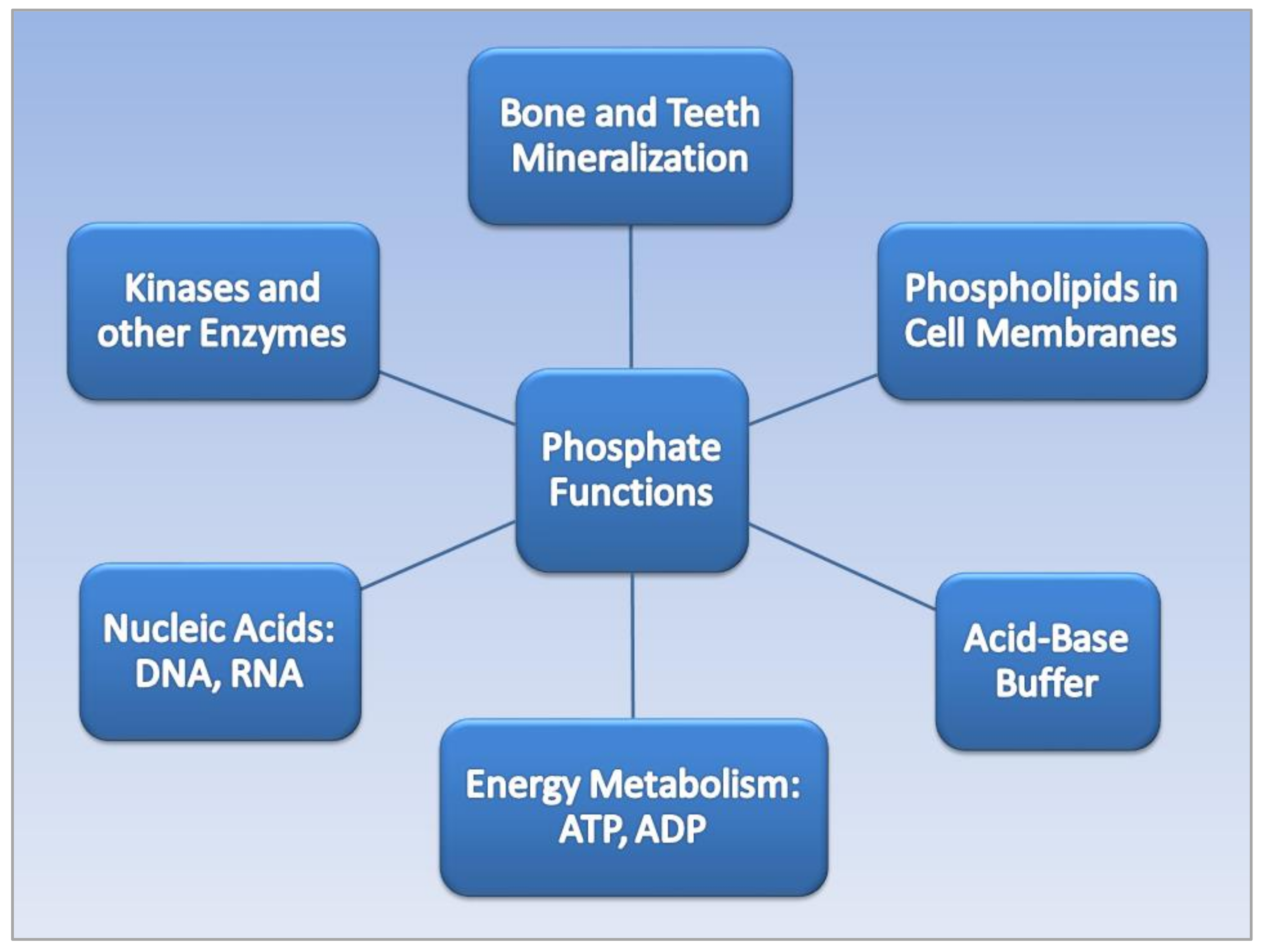
2.1. Phosphate Functions

The dietary mineral phosphorus, often found in chemical combination with oxygen as phosphate (PO4), is an essential micronutrient with a dietary reference intake (DRI) of 700 mg/day for adults [9]. Phosphate performs a wide variety of functions in the human body [10], as shown in Figure 1. Bones and teeth contain 85% of total body phosphate. Inorganic phosphate (Pi) forms hydroxyapatite with calcium, which mineralizes the extracellular matrix of bone. The lipid bilayers of cell membranes contain phosphate, and phosphate is a component in nucleic acids, deoxyribonucleic acid (DNA), and ribonucleic acid (RNA). Energy is derived from the metabolism of adenosine triphosphate (ATP) and adenosine diphosphate (ADP). Phosphate also acts as a urinary buffer, and many enzymatic reactions involve inorganic phosphate.
Figure 1. Phosphate functions in the human body.

2.2. Phosphate Regulation and Dysregulation

Serum Pi is regulated by a sensitive network of endocrine hormones that form a bone–kidney–parathyroid–intestine axis, as shown in Table 1. Ref. [11]. Pi absorption in the intestines occurs mainly through type II sodium-dependent phosphate cotransporters, and intestinal absorption is regulated by bioactive vitamin D3 released by the kidneys, 1,25(OH)2D3, or calcitriol. The kidneys regulate serum Pi through reabsorption largely in the renal proximal tubule. Fibroblast growth factor 23 (FGF23) released from osteocytes in the bones, together with parathyroid hormone (PTH) from the parathyroid glands, reduce high serum Pi levels by inhibiting kidney reabsorption of Pi and increasing Pi urinary excretion [12]. PTH also increases the resorption of calcium from bone to maintain serum calcium levels. Excess calcium phosphate formed in the blood serum from dysregulated phosphate metabolism can lead to ectopic calcification deposited throughout the soft tissue [13].
Table 1. Endocrine Regulation of Serum Pi through the Bone–Kidney–Parathyroid–Intestine Axis.

3. Phosphate Toxicity and Tumorigenesis

Phosphate toxicity, the accumulation of excess phosphate in the body from dysregulated phosphate metabolism, is associated with tumorigenesis [14]. Excessive cell growth during tumor promotion and progression is stimulated by the uptake of excess phosphate into cellular ribosomal RNA, and suppressed uptake of phosphorus within nuclear RNA was shown to delay carcinogenesis in precancerous tissue [15]. High levels of phosphate within the tumor microenvironment have been found to stimulate cell signaling in tumorigenesis [16] and tumor neovascularization [17]. Moreover, high phosphate levels in patients with hyperphosphatemia are associated with chromosome instability and increased parathyroid cell proliferation—further investigations of a direct effect of phosphate on DNA damage are needed [18]. Additionally, high extracellular levels of Pi have been linked to cancer progression through metastasis [19].
Cancer cells absorb and sequester large amounts of inorganic phosphate from the tumor extracellular microenvironment through sodium-phosphate cotransporters, which were overexpressed in cancer cells of the breast, lung, ovaries, and thyroid gland [20][21]. Levels of inorganic phosphate were up to twice as high in lung and colon tumor cells compared to normal cells [22], and high dietary phosphate levels were found to increase skin cancer growth in an animal model [23]. As well, lung tumorigenesis was stimulated in mice fed high levels of dietary inorganic phosphate [16]. Cancer in humans was also associated with higher serum phosphate levels in adults, with the exception of reproductive cancers in females, which may be due to phosphate shifting into the reproductive tissue for rapid growth [24][25].
More recently, H+-dependent Pi transporters in breast cancer cells were found to transport high concentrations of Pi at five-times the rate of sodium-phosphate cotransporters [26]. Patients with high levels of FGF23, which regulates Pi metabolism, had reduced overall survival compared to patients with low levels [27], and FGF23 levels were significantly higher in patients with urothelial carcinoma of the bladder and upper urinary tract compared to normal controls [28]. Additionally, compared to men with benign prostate hyperplasia, men with prostate cancer had higher levels of PTH, which also regulates Pi metabolism [29]. High serum PTH levels are associated with all-cause mortality in U.S. adults [30], and patients with primary hyperparathyroidism had significantly higher risks of diagnosis with cancers of the breast, skin, and kidney [31].

4. Phosphate Toxicity and Obesity

The same hormones that regulate phosphate metabolism are also associated with obesity, providing a transitive link between obesity and phosphate toxicity. For example, high serum FGF23 was associated with increased fat mass in a cohort of elderly white people with normal renal function [32]. Participants in the study who had the highest levels of FGF23 also had a significantly higher risk for overweight compared to individuals with the lowest FGF23 levels. A 10% increase in FGF23 in the cohort was associated with a 3% increase in body weight and BMI and a 2% increase in waist circumference (WC) and waist–hip ratio (WHR). Additionally, a 10% increase in FGF23 in males was associated with a 4% increase in total body fat mass and trunk fat mass.
Hyperparathyroidism is commonly found in high-grade obesity, and higher serum levels of PTH were found in 72% of bariatric patients with severe obesity who underwent laparoscopic sleeve gastrectomy (LSG) [33]. Patients’ PTH levels dropped along with BMI following surgery, likely related to a lower caloric density and volume of food intake following LSG [34]. These findings imply that diet is an etiological factor in hyperparathyroidism related to obesity, which provides an additional transitive link between obesity and phosphate toxicity from excessive dietary phosphate intake. Higher concentrations of serum PTH were also positively associated with obesity in females with metabolic abnormalities, while levels of 25(OH)D3, which are converted to calcitriol to increase phosphate absorption in the intestines, were inversely associated with obesity in males with metabolic abnormalities [35].
Obesity is often associated with inadequate intake of micronutrients such as calcium, magnesium, copper, iron, and zinc, but a recent analysis of the US National and Health Nutrition Examination Survey (NHANES) 2007–2014 found that phosphorus dietary intake in adults, along with sodium, was positively associated with BMI [36]. Of relevance, type II sodium-phosphate cotransporters (Na/Pi-2b) and type III inorganic phosphate transporters (Pit-1 and Pit2) are found in the salivary glands, and obese and overweight children were found to have significantly higher saliva levels of phosphate than normal-weight children, despite having normal serum phosphate levels [37]. A plausible mechanism accounting for higher saliva phosphate in obese and overweight children could be related to increased salivary gland cotransport of phosphate from higher levels of ingested dietary phosphate—more investigations are needed in this area.
Hypophosphatemia has also been linked to high BMI, but rather than resulting from low dietary phosphate intake or phosphate losses in urine, low serum phosphate is most frequently caused by a transcellular shift of internal phosphate [38]. Because dietary carbohydrates require phosphorylation of glucose compounds during glycolysis, excessive intake of carbohydrates releases increased levels of insulin which may cause phosphate to shift from extracellular blood serum to liver and skeletal muscle cells during metabolism of a heavy load of sugars [39]. Moreover, abdominal obesity is a part of metabolic syndrome [40], and high serum levels of catecholamines in metabolic syndrome also increase metabolic glycolysis of sugars, which further contributes to a phosphate transcellular shift that lowers serum phosphate levels.
A paradoxical relationship also exists in the association of obesity with a lower risk of breast cancer in premenopausal women compared to postmenopausal women [41]. The mid-luteal phase of the menstrual cycle is when the uterus undergoes growth [42], and women self-reported consuming more animal-based foods during this phase [43]. Animal-based foods contain high levels of protein and phosphorus, which are needed to support the monthly growth of the uterus. The need for more phosphorus during the menstrual cycle could mitigate the risk of phosphate toxicity associated with heavy dietary phosphate intake in obese premenopausal women, which would lower the risk of breast cancer compared to obese postmenopausal women.
Before closing this section on phosphate toxicity and obesity, it should be mentioned that low levels of vitamin D are consistently linked with obesity and with cancer, but researchers found that “the mediating role of vitamin D in the biological pathways linking obesity and cancer is low” [44]. On the other hand, high serum Pi can signal endocrine-controlled reductions in the renal biosynthesis of calcitriol (bioactive vitamin D), thus lowering dietary phosphate intestinal absorption. In other words, low vitamin D appears to be a consequence rather than a cause of dysregulated Pi metabolism in obesity and cancer.

References

  1. who.int. Obesity and Overweight. Available online: https://www.who.int/news-room/fact-sheets/detail/obesity-and-overweight (accessed on 28 November 2021).
  2. Cancerresearchuk.Org. When could overweight and obesity overtake smoking as the biggest casue of cancer in the UK? Available online: https://www.cancerresearchuk.org/sites/default/files/obesity_tobacco_cross_over_report_final.pdf (accessed on 19 December 2021).
  3. Renehan, A.G.; Lloyd, K.; Renehan, I. Awareness of the link between obesity and cancer in UK school curricula. Lancet 2019, 393, 1591–1592.
  4. Lauby-Secretan, B.; Scoccianti, C.; Loomis, D.; Grosse, Y.; Bianchini, F.; Straif, K. Body Fatness and Cancer-Viewpoint of the IARC Working Group. N. Engl. J. Med. 2016, 375, 794–798.
  5. Lee, D.H.; Giovannucci, E.L. The Obesity Paradox in Cancer: Epidemiologic Insights and Perspectives. Curr. Nutr. Rep. 2019, 8, 175–181.
  6. Stone, T.W.; McPherson, M.; Gail Darlington, L. Obesity and Cancer: Existing and New Hypotheses for a Causal Connection. EBioMedicine 2018, 30, 14–28.
  7. Shu, Y.; Hassan, F.; Ostrowski, M.C.; Mehta, K.D. Role of hepatic PKCβ in nutritional regulation of hepatic glycogen synthesis. JCI Insight 2021, 6, e149023.
  8. Rivera, M.J.; Contreras, A.; Nguyen, L.T.; Eldon, E.D.; Klig, L.S. Regulated inositol synthesis is critical for balanced metabolism and development in Drosophila melanogaster. Biol. Open 2021, 10, bio058833.
  9. IOM. Dietary Reference Intakes for Calcium, Phosphorus, Magnesium, Vitamin D, and Fluoride; Institute of Medicine Standing Committee on the Scientific Evaluation of Dietary Reference Intakes; National Academy of Sciences: Washington, DC, USA, 1997.
  10. Qadeer, H.A.; Bashir, K. Physiology, Phosphate; StatPearls Publishing: Treasure Island, FL, USA, 2021.
  11. Brown, R.B. Stress, inflammation, depression, and dementia associated with phosphate toxicity. Mol. Biol. Rep. 2020, 47, 9921–9929.
  12. Saki, F.; Kassaee, S.R.; Salehifar, A.; Omrani, G.H.R. Interaction between serum FGF-23 and PTH in renal phosphate excretion, a case-control study in hypoparathyroid patients. BMC Nephrol. 2020, 21, 176.
  13. Brown, R.B.; Razzaque, M.S. Dysregulation of phosphate metabolism and conditions associated with phosphate toxicity. BoneKEy Rep. 2015, 4, 705.
  14. Brown, R.B.; Razzaque, M.S. Phosphate toxicity and tumorigenesis. Biochim. Et Biophys. Acta (BBA)-Rev. Cancer 2018, 1869, 303–309.
  15. Ward, D.N.; Griffin, A.C. Phosphorus incorporation into nucleic acids and proteins of liver nuclei of normal and azo dye-fed rats. Cancer Res. 1955, 15, 456–461.
  16. Jin, H.; Xu, C.-X.; Lim, H.-T.; Park, S.-J.; Shin, J.-Y.; Chung, Y.-S.; Park, S.-C.; Chang, S.-H.; Youn, H.-J.; Lee, K.-H. High dietary inorganic phosphate increases lung tumorigenesis and alters Akt signaling. Am. J. Respir. Crit. Care Med. 2009, 179, 59–68.
  17. Lin, Y.; McKinnon, K.E.; Ha, S.W.; Beck, G.R., Jr. Inorganic phosphate induces cancer cell mediated angiogenesis dependent on forkhead box protein C2 (FOXC2) regulated osteopontin expression. Mol. Carcinog. 2015, 54, 926–934.
  18. Chudek, J.; Nagy, A.; Kokot, F.; Podwinski, A.; Wiecek, A.; Ritz, E.; Kovacs, G. Phosphatemia is related to chromosomal aberrations of parathyroid glands in patients with hyperparathyroidism. J. Nephrol. 2007, 20, 164–172.
  19. Bobko, A.A.; Eubank, T.D.; Driesschaert, B.; Dhimitruka, I.; Evans, J.; Mohammad, R.; Tchekneva, E.E.; Dikov, M.M.; Khramtsov, V.V. Interstitial inorganic phosphate as a tumor microenvironment marker for tumor progression. Sci. Rep. 2017, 7, 41233.
  20. Levan, K.; Mehryar, M.; Mateoiu, C.; Albertsson, P.; Bäck, T.; Sundfeldt, K. Immunohistochemical evaluation of epithelial ovarian carcinomas identifies three different expression patterns of the MX35 antigen, NaPi2b. BMC Cancer 2017, 17, 303.
  21. D’Arcangelo, M.; Brustugun, O.; Xiao, Y.; Choi, Y.; Behrens, C.; Solis, L.; Wang, Y.; Firestein, R.; Boyle, T.; Lund-Iversen, M. 194P prevalence and prognostic significance of sodium-dependent phosphate transporter 2B (NAPI2B) protein expression in non-small cell lung cancer (NSCLC). Ann. Oncol. 2014, 25, iv66–iv67.
  22. Elser, J.J.; Kyle, M.M.; Smith, M.S.; Nagy, J.D. Biological stoichiometry in human cancer. PLoS ONE 2007, 2, e1028.
  23. Camalier, C.E.; Young, M.R.; Bobe, G.; Perella, C.M.; Colburn, N.H.; Beck, G.R. Elevated phosphate activates N-ras and promotes cell transformation and skin tumorigenesis. Cancer Prev. Res. 2010, 3, 359–370.
  24. Papaloucas, C.; Papaloucas, M.; Kouloulias, V.; Neanidis, K.; Pistevou-Gompaki, K.; Kouvaris, J.; Zygogianni, A.; Mystakidou, K.; Papaloucas, A. Measurement of blood phosphorus: A quick and inexpensive method for detection of the existence of cancer in the body. Too good to be true, or forgotten knowledge of the past? Med. Hypotheses 2014, 82, 24–25.
  25. Wulaningsih, W.; Michaelsson, K.; Garmo, H.; Hammar, N.; Jungner, I.; Walldius, G.; Holmberg, L.; Van Hemelrijck, M. Inorganic phosphate and the risk of cancer in the Swedish AMORIS study. BMC Cancer 2013, 13, 257.
  26. Lacerda-Abreu, M.A.; Russo-Abrahão, T.; Cosentino-Gomes, D.; Nascimento, M.T.C.; Carvalho-Kelly, L.F.; Gomes, T.; Rodrigues, M.F.; König, S.; Rumjanek, F.D.; Monteiro, R.Q.; et al. H(+)-dependent inorganic phosphate transporter in breast cancer cells: Possible functions in the tumor microenvironment. Biochim. Biophys. Acta Mol. Basis Dis. 2019, 1865, 2180–2188.
  27. Mansinho, A.; Ferreira, A.R.; Casimiro, S.; Alho, I.; Vendrell, I.; Costa, A.L.; Sousa, R.; Abreu, C.; Pulido, C.; Macedo, D.; et al. Levels of Circulating Fibroblast Growth Factor 23 (FGF23) and Prognosis in Cancer Patients with Bone Metastases. Int. J. Mol. Sci. 2019, 20, 695.
  28. Li, J.R.; Chiu, K.Y.; Ou, Y.C.; Wang, S.S.; Chen, C.S.; Yang, C.K.; Ho, H.C.; Cheng, C.L.; Yang, C.R.; Chen, C.C.; et al. Alteration in serum concentrations of FGF19, FGF21, and FGF23 in patients with urothelial carcinoma. Biofactors 2019, 45, 62–68.
  29. Kim, W.T.; Bang, W.J.; Seo, S.P.; Kang, H.W.; Byun, Y.J.; Piao, X.M.; Jeong, P.; Shin, K.S.; Choi, S.Y.; Lee, O.J.; et al. Parathyroid hormone is associated with prostate cancer. Prostate Int. 2020, 8, 116–120.
  30. Kato, H.; Ito, N.; Makita, N.; Nangaku, M.; Leung, A.M.; Inoue, K. Association of Serum Parathyroid Hormone Levels With All-Cause and Cause-Specific Mortality Among U.S. Adults. Endocr. Pract. 2022, 28, 70–76.
  31. Palmieri, S.; Roggero, L.; Cairoli, E.; Morelli, V.; Scillitani, A.; Chiodini, I.; Eller-Vainicher, C. Occurrence of malignant neoplasia in patients with primary hyperparathyroidism. Eur. J. Intern. Med. 2017, 43, 77–82.
  32. Mirza, M.A.; Alsiö, J.; Hammarstedt, A.; Erben, R.G.; Michaëlsson, K.; Tivesten, A.; Marsell, R.; Orwoll, E.; Karlsson, M.K.; Ljunggren, O.; et al. Circulating fibroblast growth factor-23 is associated with fat mass and dyslipidemia in two independent cohorts of elderly individuals. Arterioscler. Thromb. Vasc. Biol. 2011, 31, 219–227.
  33. Ministrini, S.; Ricci, M.A.; Daviddi, G.; Scavizzi, M.; De Vuono, S.; D’Abbondanza, M.; Paganelli, M.T.; Boni, M.; Roscini, A.R.; Scarponi, A.M.; et al. Determinants of High Parathyroid Hormone Levels in Patients With Severe Obesity and Their Relationship With the Cardiometabolic Risk Factors, Before and After a Laparoscopic Sleeve Gastrectomy Intervention. Obes. Surg. 2020, 30, 2225–2232.
  34. Coluzzi, I.; Raparelli, L.; Guarnacci, L.; Paone, E.; Del Genio, G.; le Roux, C.W.; Silecchia, G. Food Intake and Changes in Eating Behavior After Laparoscopic Sleeve Gastrectomy. Obes. Surg. 2016, 26, 2059–2067.
  35. Ha, J.; Jo, K.; Lim, D.J.; Lee, J.M.; Chang, S.A.; Kang, M.I.; Cha, B.Y.; Kim, M.H. Parathyroid hormone and vitamin D are associated with the risk of metabolic obesity in a middle-aged and older Korean population with preserved renal function: A cross-sectional study. PLoS ONE 2017, 12, e0175132.
  36. Jiang, S.; Ma, X.; Li, M.; Yan, S.; Zhao, H.; Pan, Y.; Wang, C.; Yao, Y.; Jin, L.; Li, B. Association between dietary mineral nutrient intake, body mass index, and waist circumference in U.S. adults using quantile regression analysis NHANES 2007-2014. PeerJ 2020, 8, e9127.
  37. Hartman, M.-L.; Groppo, F.; Ohnishi, M.; Goodson, J.M.; Hasturk, H.; Tavares, M.; Yaskell, T.; Floros, C.; Behbehani, K.; Razzaque, M.S. Can salivary phosphate levels be an early biomarker to monitor the evolvement of obesity. In Phosphate and Vitamin D in Chronic Kidney Disease; Karger Publishers: Basel, Switzerland, 2013; Volume 180, pp. 138–148.
  38. Håglin, L.; Törnkvist, B.; Bäckman, L. Obesity, smoking habits, and serum phosphate levels predicts mortality after life-style intervention. PLoS ONE 2020, 15, e0227692.
  39. Stoian, M.; Stoica, V. The role of disturbances of phosphate metabolism in metabolic syndrome. Maedica 2014, 9, 255–260.
  40. Engin, A. The Definition and Prevalence of Obesity and Metabolic Syndrome. Adv. Exp. Med. Biol. 2017, 960, 1–17.
  41. García-Estévez, L.; Cortés, J.; Pérez, S.; Calvo, I.; Gallegos, I.; Moreno-Bueno, G. Obesity and Breast Cancer: A Paradoxical and Controversial Relationship Influenced by Menopausal Status. Front. Oncol. 2021, 11, 705911.
  42. Piiroinen, O.; Kaihola, H.L. Uterine size measured by ultrasound during the menstrual cycle. Acta Obstet. Gynecol. Scand. 1975, 54, 247–250.
  43. Gorczyca, A.M.; Sjaarda, L.A.; Mitchell, E.M.; Perkins, N.J.; Schliep, K.C.; Wactawski-Wende, J.; Mumford, S.L. Changes in macronutrient, micronutrient, and food group intakes throughout the menstrual cycle in healthy, premenopausal women. Eur. J. Nutr. 2016, 55, 1181–1188.
  44. Shanmugalingam, T.; Crawley, D.; Bosco, C.; Melvin, J.; Rohrmann, S.; Chowdhury, S.; Holmberg, L.; Van Hemelrijck, M. Obesity and cancer: The role of vitamin D. BMC Cancer 2014, 14, 712.
More
Information
Subjects: Oncology
Contributor MDPI registered users' name will be linked to their SciProfiles pages. To register with us, please refer to https://encyclopedia.pub/register :
View Times: 54
Revisions: 2 times (View History)
Update Date: 14 Mar 2024
1000/1000