Submitted Successfully!
To reward your contribution, here is a gift for you: A free trial for our video production service.
Thank you for your contribution! You can also upload a video entry or images related to this topic.
Version Summary Created by Modification Content Size Created at Operation
1 -- 3548 2024-03-07 21:04:24 |
2 format change + 5 word(s) 3553 2024-03-08 03:05:10 |

Video Upload Options

Do you have a full video?

Confirm

Are you sure to Delete?
Cite
If you have any further questions, please contact Encyclopedia Editorial Office.
Scalia, P.; Marino, I.R.; Asero, S.; Pandini, G.; Grimberg, A.; El-Deiry, W.S.; Williams, S.J. Autocrine IGF-II-Associated Cancers. Encyclopedia. Available online: https://encyclopedia.pub/entry/55990 (accessed on 19 May 2024).
Scalia P, Marino IR, Asero S, Pandini G, Grimberg A, El-Deiry WS, et al. Autocrine IGF-II-Associated Cancers. Encyclopedia. Available at: https://encyclopedia.pub/entry/55990. Accessed May 19, 2024.
Scalia, Pierluigi, Ignazio R. Marino, Salvatore Asero, Giuseppe Pandini, Adda Grimberg, Wafik S. El-Deiry, Stephen J. Williams. "Autocrine IGF-II-Associated Cancers" Encyclopedia, https://encyclopedia.pub/entry/55990 (accessed May 19, 2024).
Scalia, P., Marino, I.R., Asero, S., Pandini, G., Grimberg, A., El-Deiry, W.S., & Williams, S.J. (2024, March 07). Autocrine IGF-II-Associated Cancers. In Encyclopedia. https://encyclopedia.pub/entry/55990
Scalia, Pierluigi, et al. "Autocrine IGF-II-Associated Cancers." Encyclopedia. Web. 07 March, 2024.
Autocrine IGF-II-Associated Cancers
Edit

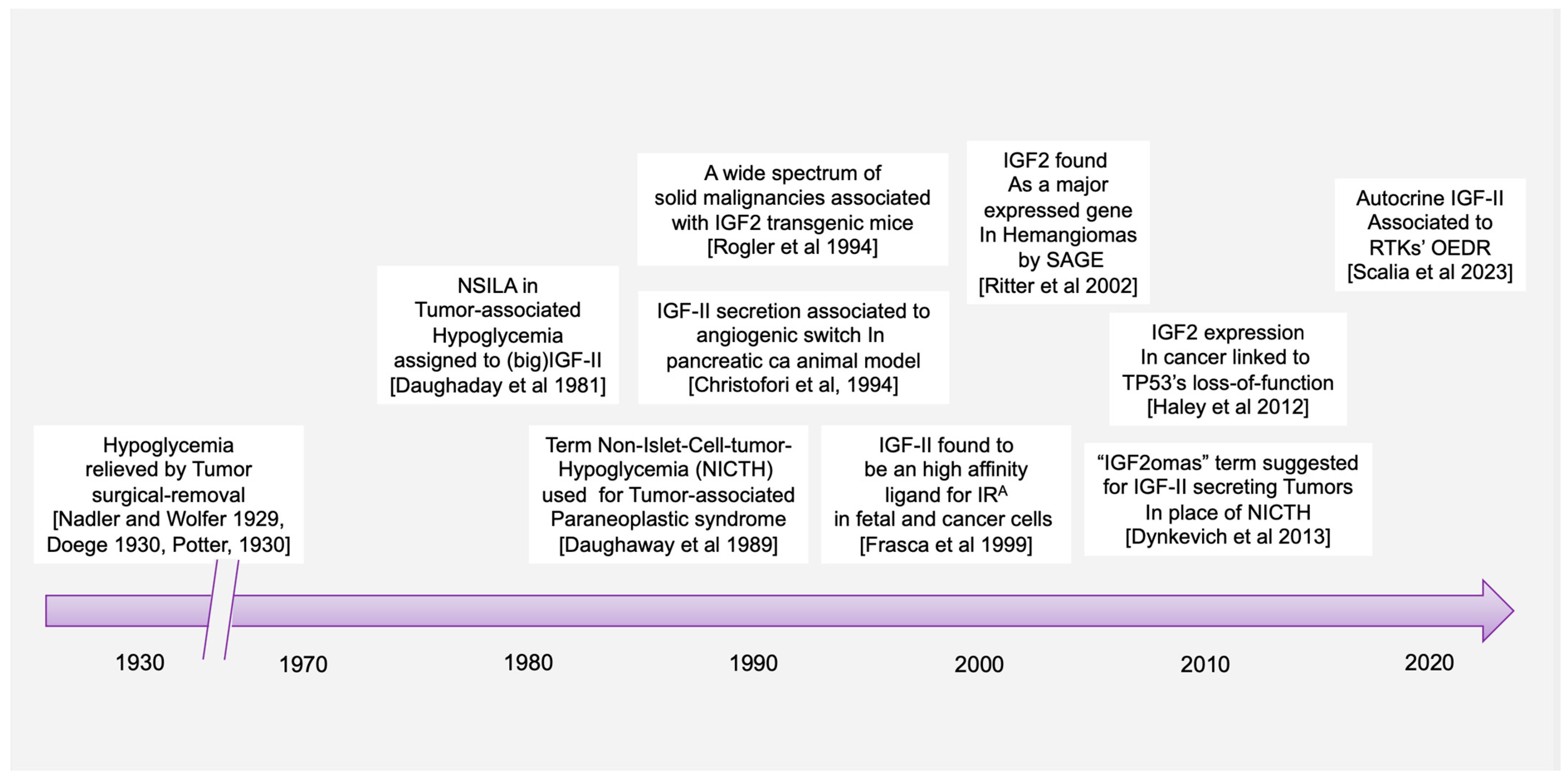
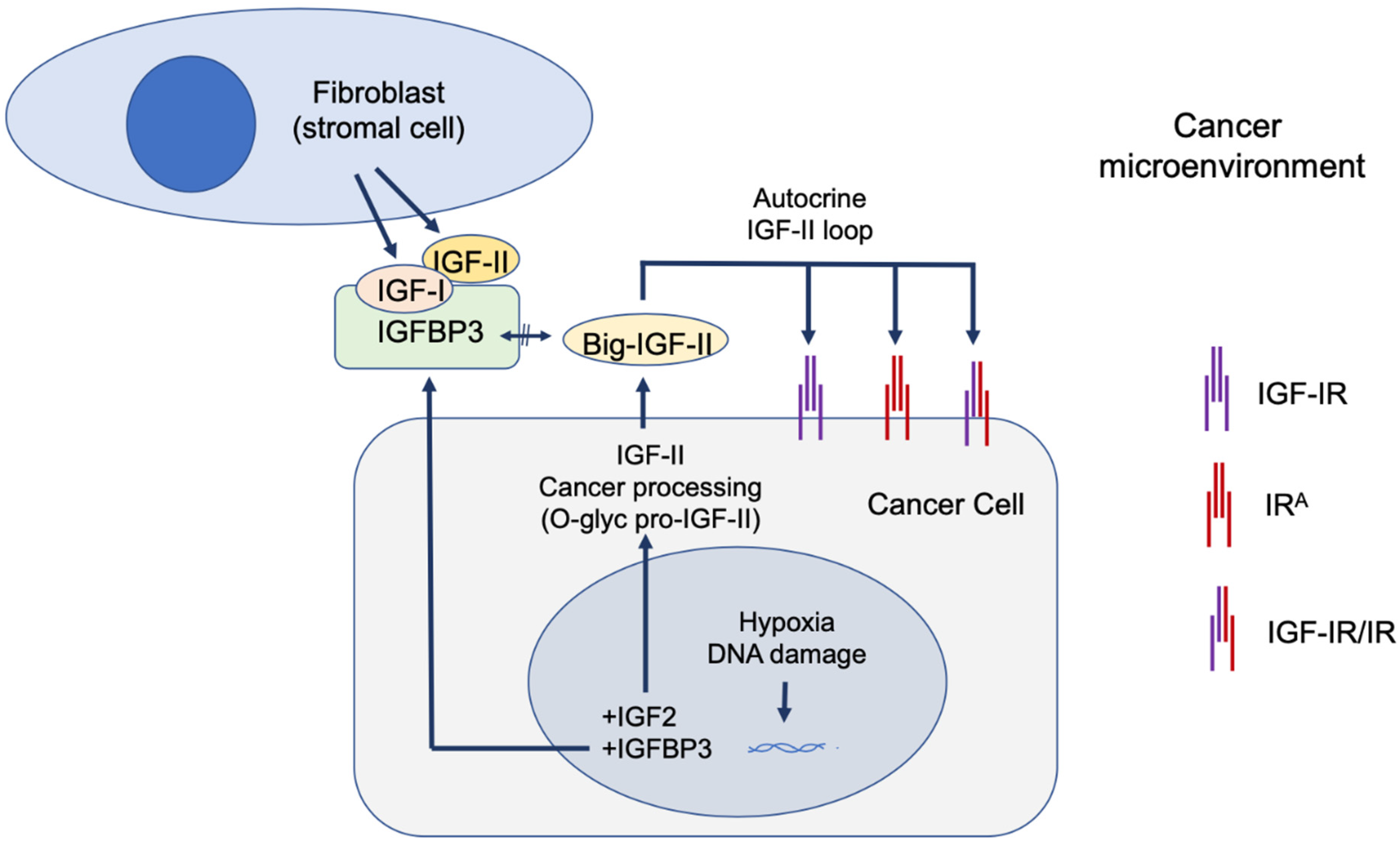
The paraneoplastic syndrome referred in the literature as non-islet-cell tumor hypoglycemia (NICTH) and extra-pancreatic tumor hypoglycemia (EPTH) was first reported almost a century ago, and the role of cancer-secreted IGF-II in causing this blood glucose-lowering condition has been widely established. The landscape emerging, based on molecular and cellular findings, supports a broader role for IGF-II in cancer biology beyond its involvement in the paraneoplastic syndrome. In particular, a few key findings are constantly observed during tumorigenesis, (a) a relative and absolute increase in fetal insulin receptor isoform (IRA) content, with (b) an increase in IGF-II high-molecular weight cancer-variants (big-IGF-II), and (c) a stage-progressive increase in the IGF-II autocrine signal in the cancer cell, mostly during the transition from benign to malignant growth. An increasing and still under-exploited combinatorial pattern of the IGF-II signal in cancer is shaping up in the literature with respect to its transducing receptorial system and effector intracellular network. Interestingly, while surgical and clinical reports have traditionally restricted IGF-II secretion to a small number of solid malignancies displaying paraneoplastic hypoglycemia, a retrospective literature analysis, along with publicly available expression data from patient-derived cancer cell lines conveyed in the present perspective, clearly suggests that IGF-II expression in cancer is a much more common event, especially in overt malignancy.

NICTH EPTH NSILA IGF1-IGF2 (gene) IGF-I-IGF-II (protein) IRA IGF-IR HRA/B IGF2oma IGF2ST

1. Introduction

The earliest reports of the paraneoplastic syndrome associating what has been later referred as non-suppressible insulin-like activity (NSILA) [1] to hypoglycemia in cancer goes back to reports from W.H. Nadler and J.A. Wolfer in 1929 [2] and Karl W. Doege [3] and R.P. Potter in 1930 [4]. In possible oversight of the earlier report, the term of Doege–Potter Syndrome was adopted to describe these surgically treated intrathoracic tumors associated with hypoglycemia. Later reports confirmed that paraneoplastic hypoglycemia could indeed be found in cancers from all other (extra-thoracic) body districts and not limited to those of fibrous (connective/soft tissue) origin (namely sarcomas), as already suggested by the first under-looked report in 1929, but almost equally associated with epithelial/parenchymal tissue-derived cancers (carcinomas) [5]. The first findings linking IGF-II to cancer paraneoplastic hypoglycemia were related in the work of Doughaday et al. [6][7]. The added value of his work is linked to the observation that cancer-secreted IGF-II differs from physiologically produced IGF-II and that such difference confers cancer-secreted (“Big”)IGF-II key biologic advantages underlying its now widely proven autocrine loop effects. Specifically, cancer-secreted IGF-II corresponds to the IGF-II pro-hormone retaining its E domain, allowing its O-Glycosylation [6][8][9]. This processing defect increases the life-span and bioavailability of the IGF-II variants, both in the tumor microenvironment and in the systemic circulation, by reducing binding to IGFBP-3 and the IGF-II scavenger protein SpI2-6 (deceivingly known as the IGF-II “receptor” but actually causing IGF-II sequestration and degradation) [10][11][12]. The timeline of key discoveries connecting IGF-II to paraneoplastic hypoglycemia and proving its unique biological features are conveyed in Figure 1.
Figure 1. Historical timeline for key discoveries conferring a central role to IGF-II in the Insulin/IGF receptorial system in cancer. References within figure are as follows: Nadler and Wolfer, 1929 [2]; Doege 1930 [3]; Potter 1930 [4]; Daughaday et al., 1981 [13]; Daughaday 1989 [6]; Rogler et al., 1994; [14]; Christifori et al., 1994 [15]; Frasca et al., 1999 [16]; Ritter et al., 2002 [17]; Haley et al., 2012 [18]; Dynkevich et al., 2013 [5]; Salia et al., 2023 [19].

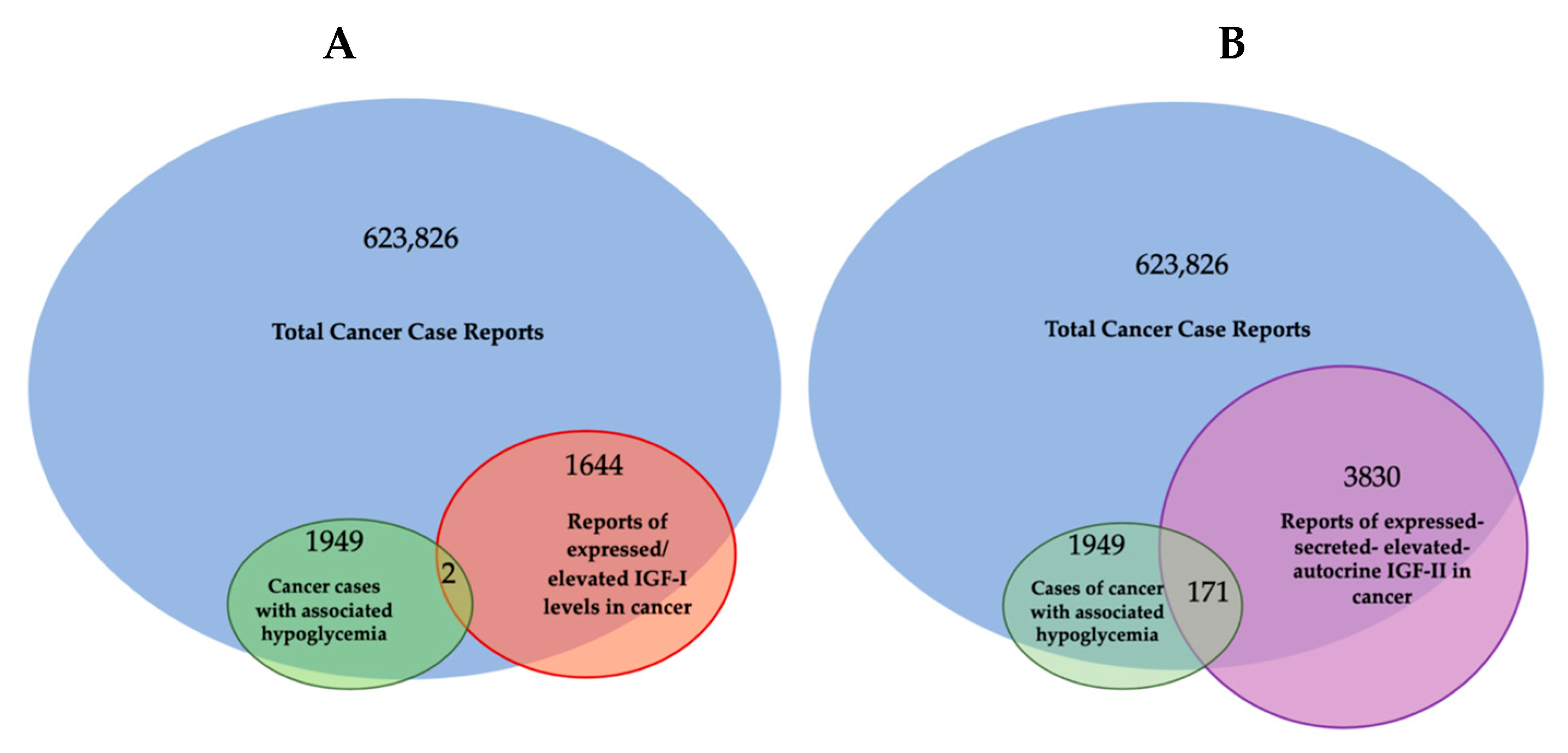
2. Cancer-Secreted IGF-II and Paraneoplastic Hypoglycemia: Is There Sufficient Evidence Supporting IGF-II as the Key IGF Ligand Involved in Solid Malignancy?

Despite the finding of IGF-II expression and secretion in cancer having long being established through the literature (Table 1), some authors have been supporting a comparable/interchangeable cancer-driving role for IGF-I, which mediates growth hormone effects during post-natal development in all vertebrates. This view, which implies a biological equivalence for IGF-I and IGF-II in cancer, cannot be supported any longer based on a number of available lines of evidence further discussed herein. Among these, the researchers are adding the literature and expression correlation analysis assigning cancer-secreted IGF-II a distinctive hallmark compared to IGF-I. This is summarized in Figure 1 and Table 1 and discussed herein.
Table 1. Literature analyses of cancer case reports involving IGF-I and IGF-II in relation to cancer-associated hypoglycemia.
* Mostly associated to stromal component secretion.
Overall, the number of cancers with hypoglycemic symptoms secreting IGF-II and associated with malignancies exceed the number and types of tumors (mainly pituitary in origin) linked to IGF-I expression/secretion. This is in apparent conflict with the epidemiology results displaying an association between IGF-I blood levels and solid cancer risk.
In this context, it is useful to trace back the lines of evidence which have led us to the view linking IGF-I and IGF-II to cancer in order to highlight eventual incongruences. A literature search again provides a quantity of actionable evidence to this regard. In particular, given the physiological roles of these growth factors on developmental growth such as those summarized based on genetic knock-down work in rodents [20][21][22][23][24], the researchers specifically minimized the inclusion of studies on IGFs genetics and physiology and focused the research on the work on IGFs in solid malignancies at the cellular, molecular, and clinical levels.
This parallel search shows that only a minor number of published works have looked at both IGF-I and IGF-II in the same studied cancer model (cellular, molecular, clinical, or epidemiologic). On the other hand, a larger number of highly referenced studies (e.g., trying to reconstitute signaling events) in vitro have made extensive use of exogenous stimulation of IGF-IR-expressing cellular models, often using supraphysiologic amounts of IGF-I (e.g., 100 nM and higher) without properly integrating or reconstituting the in vivo ligands and receptors landscape in their experimental design.
Overall, such a reductionistic in vitro approach, if it has, on one hand, advanced the understanding on the mechanistic aspects of this ligands/receptor system, has, on the other hand, been misleading in that the following aspects:
(a)
It does not take in consideration the actual in vivo IGFs ligands and receptors co-expression context, which, taken together, supports a specific and independent role for cancer-secreted IGF-II and its autocrine loops;
(b)
It does not succeed in explaining the failure of the individual pharmacological blockers of IGF-IR in clinical trials towards meeting the invoked therapeutic advantages suggested by the in vitro and epidemiologic studies;
(c)
It has kept excluding alternative hypotheses and proper controls in experimental design which have been suggested by additional evidence available since the late nineties and proving the existence of an IGF-II- Insulin fetal receptor isoform (IRA) axis in mammalian fetal and cancer cells [16], as well as the expression and biological impact of IGF-IR/IR isoform-specific hybrids [25] in the studied cancer models.
Arguably, even relatively recent studies published on reputable journals [26] keep restricting the study focus on the IGF-I/IGF-IR axis as a standalone system in cancer without including parallel analysis of the IGF-II/IRA ligand/RTK system in their experimental design [27], reiterating the persistence of an unsupported bias in the interpretation of the available experimental and observational data. The retroactive analysis of the published literature in regard to the IGFs’ involvement in cancer cases displaying NSILA-dependent hypoglycemia is conveyed in Table 1 and graphically summarized in Figure 1 above.
Table 1: Based on the available literature out of all cases of cancer-associated hypoglycemia (1949 cases since 1929), 171 cases (10.3%) were reported after the available immunometric test had been developed and could be clearly associated with high IGF-II secretion levels versus 38 cases also reporting increased levels of IGF-I (1.94%) along with IGF-II. IGF-II association with such paraneoplastic condition was underestimated due to the fact that the IGF-II testing had been made available only in the early 1970′s. * Compatible with cancer stromal component secretion as source of increased levels.

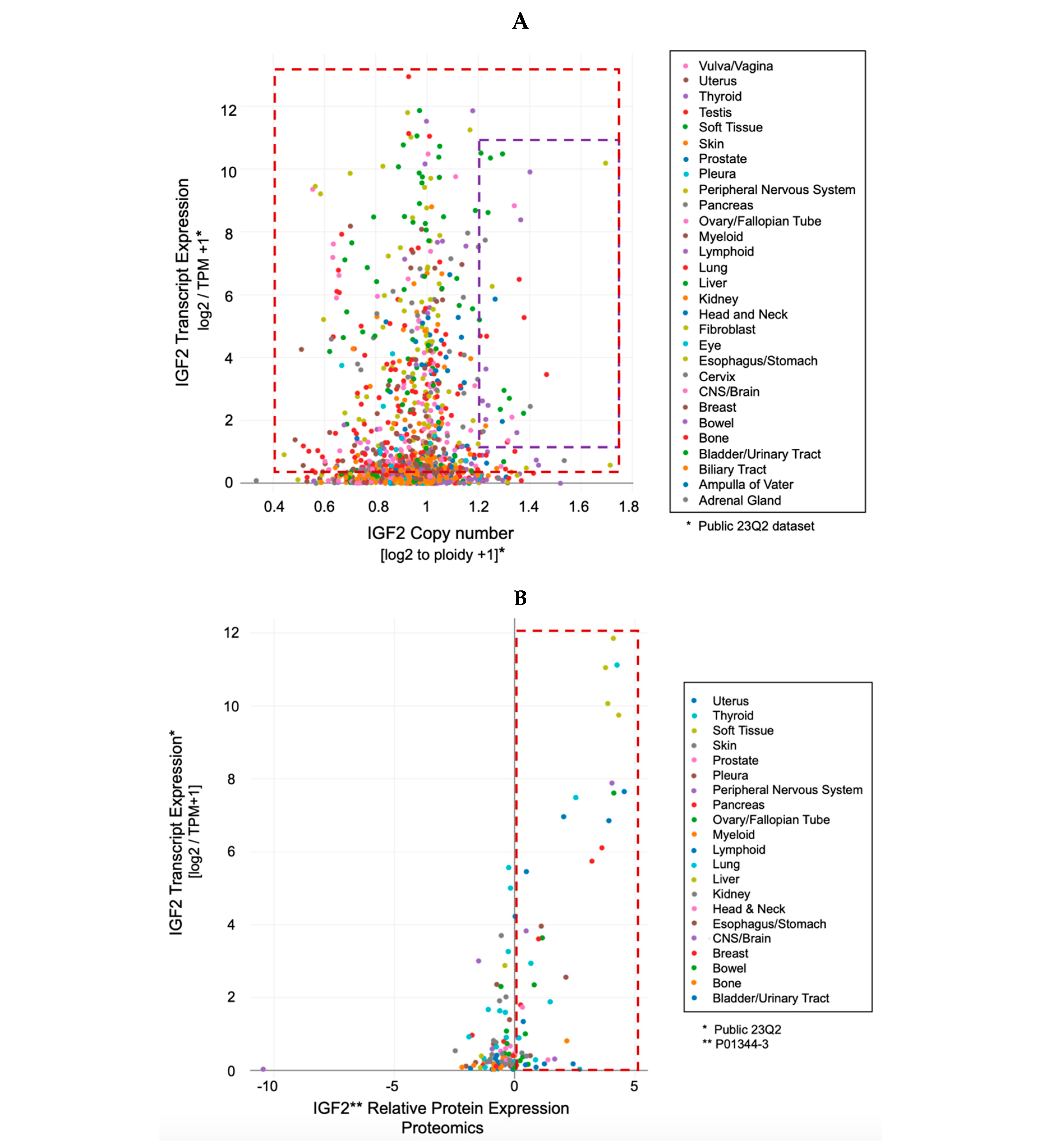
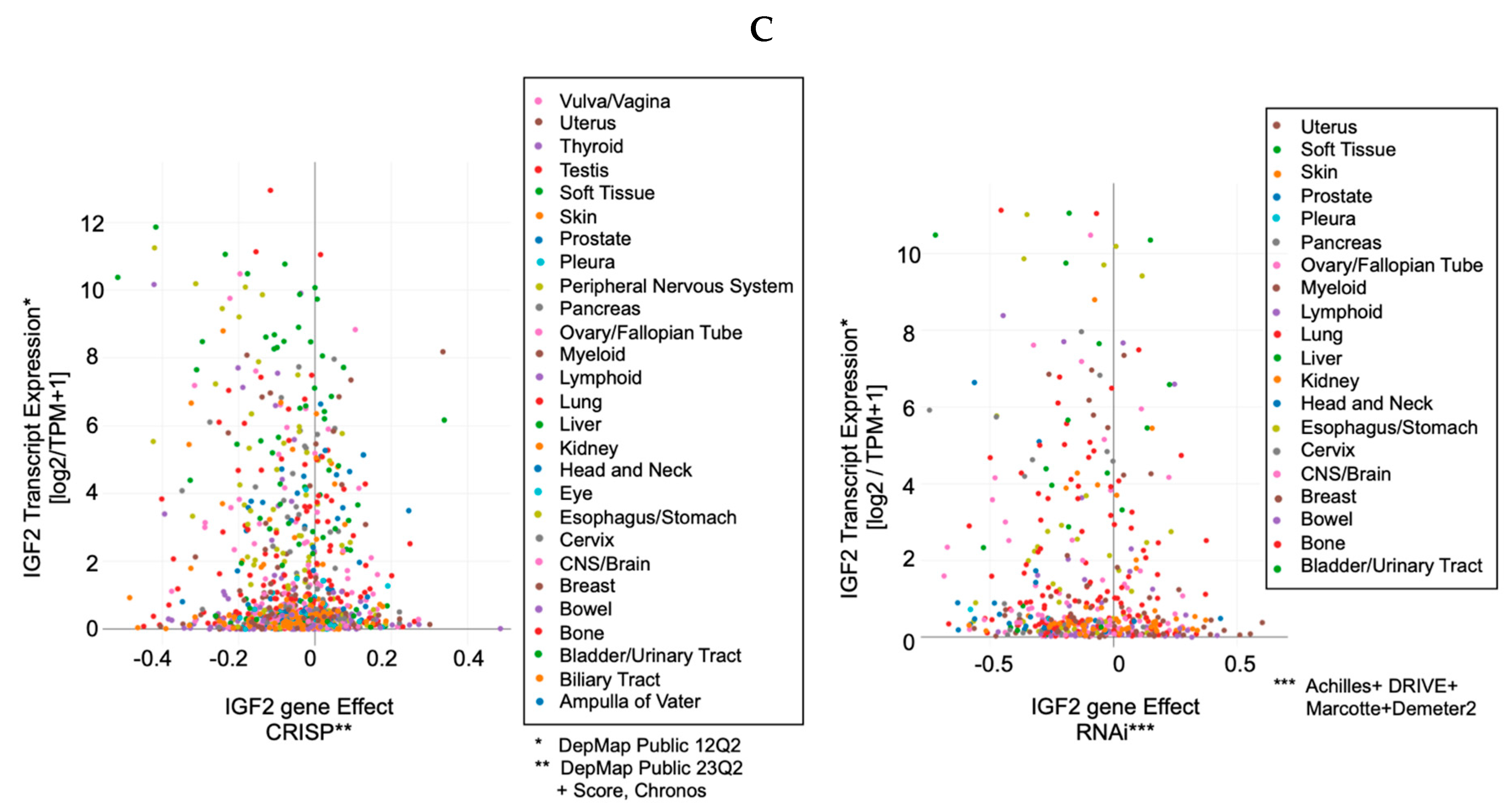
3. IGF-II Over-Expression Is a Common Event in Cancer Cell-Lines

While IGF2 expression in somatic cells is regulated via parental imprinting, its regulation in cancer cells is determined by a combination of both imprinting and transcriptional regulation mechanisms reviewed elsewhere [[28], ibidem]. Ultimately, independently of the underlying genetic, translational, and post-translational mechanisms involved, the phenotypic and functional effects of such increased expression is reflected in the secretion of high molecular IGF-II pro-hormone variants [9] and its autocrine signal, which has been associated with both paraneoplastic hypoglycemia and malignancy (summarized in Figure 2 and Table 1).
Figure 2. Cancer cases in the scientific literature exhibiting hypoglycemia associated with IGFs secretion. Venn diagram produced with online software available at https://www.meta-chart.com/venn#/display (accessed on 21 October 2023). (A) IGF-I-related cases. (B) IGF-II-related cases. The data analysis was the result of a PubMed literature search conveyed in Table 1. Note: the IGF reports in the Venn diagram followed the advent of IGF-I and IGF-II immunometric testing development (1972).
Indeed, the idea of IGF-II secretion as a rare associated event in cancer has been maintained in the scientific literature till present [29], somehow implying that IGF-II-secreting tumors could be mostly benign in nature and fully surgically treatable. This has motivated a group of authors to name such tumors as “IGF2omas” recalling the rare and surgically removable features of the early reports [5]. However, the cumulative evidence based on expression studies conducted at the histological and cellular level suggests a different scenario than that proposed by clinical reports of its rarer hypoglycemic-associated syndrome. In fact, based on the retrospective analysis of the published literature, which the researchers conveyed herein in Figure 1 and Table 1, it is clear that IGF-II secretion in tumors is a much more common event than generally implied by IGF studies focusing on mechanistic and reductionistic experimental design. Interestingly, IGF-II expression by cancer cells and bioptic tissues from solid malignancies exceeds, by several orders of magnitude, the number of cancers overtly displaying hypoglycemia. Although there is still not sufficient published evidence, increases in hypoxia and CO2 levels with resultant body acidification in cancer patients may also result as a highly associated event with IGF-II secretion in patients diagnosed with a solid tumor. The rational for this predicted association is based on the demonstrated IGF-II expression increase in response to HIF-1 stimuli reported in a variety of experimental cancer models [30]. To further characterize the expression levels and patterns of IGF-II in cancer, the researchers turned to the DepMap expression database, a publicly available tool managed by the Broad Institute [31] (available at https://depmap.org/portal/ accessed on 24 September 2023), and focused on a few key parameters conveyed in Figure 3. This analysis, relative to a number of well-characterized human-derived cancer cell lines, has provided the following results:
Figure 3. IGF2 (gene, transcript, and protein) expression pattern in patient-derived cancer cells. DepMap-originated expression levels conveyed in Figure are relative to the respective content in normal cells; (A) IGF2 transcript expression is a common event in cancer cells (as summarized in the red boxed area) and rarely associates with its gene duplication events (compare red with purple boxed areas); (B) IGF-II ligand expression (proteomics) is a common event in cancer cells, even at low transcription expression levels (see red boxed area spanning from the 0 value corresponding to expression levels comparable to normal cells, up to 12 times the IGF2 transcript expression in normal cells); (C) comparative effect of IGF2 gene editing (left) and/or transcript silencing (right) in cancer cell lines. Note the consistent distribution of human cancer cells among those responding to IGF2 gene block (by either CRISP or RNAi) irrespective of the folds of IGF2 transcript native over-expression.
  • The IGF-II transcript expression in cancer cells exceeds the expression of normal cells and tissues by a range of 0.1- to 12-fold (Figure 3A–C);
  • The IGF-II transcript (mRNA) expression is not commonly associated with gene duplication events (Figure 3A);
  • The IGF-II protein expression in human-derived cancer cells exceeds normal cells/tissues by 0.1- to 5-fold (Figure 3B);
  • IGF-II gene editing and or transcript silencing negatively affects ~60–65% of cancer cells (Figure 3C).
As for IGF2 expression and its correlation to solid cancer, despite the established association of IGF2 transcript and ligand (IGF-II) expression in a wider spectrum of solid tumors (summarized in Table 1), a few studies have specifically looked at the cause–effect between IGF-II overexpression and malignant switch. Two seminal studies addressing this point are discussed in the following. The first, authored by Rogler et al. and conducted in a IGF2 transgenic mice model [14], observed development of a broad spectrum of solid malignancies (3.25-fold higher than normal control animals), with resulting transgenic mice bearing an IGF2 transgene construct able to drive 20- to 30-times-higher plasma levels than control animals. Interestingly, the study shows that in these mice, hypoglycemia increased proportionally with the increase in the circulating IGF-II levels. In particular, in animals with up to 20 times the mean levels of circulating IGF-II, the measured glycemic levels were still in the normal range despite hypoglycemia being more frequent with aging. On the other hand, all IGF2 transgenic mice displaying more than 30 times the IGF-II levels compared to non-transgenic control animals did constantly display reduced blood glucose levels and symptoms of hypoglycemia. This particular finding implies that increased level of IGF2 transcript expression and consequent IGF-II ligand secretion might affect a larger number of solid malignancies before setting or even in the absence of underlying hypoglycemic symptoms. This is also consistent with the retrospective literature findings conveyed herein (Table 1 and Figure 1) supporting the idea that IGF2 transcript or IGF-II protein expression is a broader event in cancer compared to the established but rarer paraneoplastic hypoglycemic symptoms linked to IGF-II’s non-suppressible insulin-like activity (NSILA) [1], also referred as non-islet-cell tumor hypoglycemia (NICTH) [8] and extra-pancreatic tumor hypoglycemia (EPTH) [32].

4. The Role of IGF-II in Cancer Is Not Alternative to IGF-I

Traditionally, IGF-I and IGF-II have been considered almost to be interchangeable and/or redundant ligands triggering the oncogenic effects of the IGF-IR. Nonetheless, unlike IGF-II, IGF-I is not commonly found to be over-expressed or secreted by cancer cells and it has been found to be negligibly associated with NICTH (summarized in Figure 1 and Table 1). Indeed, there are a plethora of studies involving IGF-I in cancer. The current lines of evidence supporting its role can be conveyed in (a) epidemiologic studies associating relatively high levels of circulating IGF-I to increased incidence of prostate, breast, and other cancers [33][34][35][36], and (b) other studies in vitro with human tumor cells implicating IGF-I in growth, survival, migration, and metastatic behavior upon activation of the expressed IGF-IR [37][38][39], as well as resistance to chemotherapeutic and radiation therapies [40].
Physiologically, IGF-I levels in all mammalian species including humans are known to peak during the pubertal phase and slowly decrease throughout lifetime in response to GH, which shares a similar age-related trend [41]. This general concentration decreasing pattern is not different in that group of patients with increased cancer risk, despite such (relative increase in) circulating IGF-I amounts being significantly lower compared to the same subject during pubertal age. In other words, there is no dose–response correspondence between absolute IGF-I levels in blood and cancer risk given the very low prevalence of cancer in the pubertal population.
Noteworthy, in the epidemiologic studies associating higher levels of IGF-I to increased cancer risk, no specific attention has been given to the cellular source or cancer tissue component responsible for IGF-I production. Additionally, while epidemiology has suggested a link between high IGF-I blood levels and increased cancer risk, a cancer-protective role of low IGF-I dose exposures, such as in IGF-I treated subjects affected by Laron syndrome (a genetic form of IGF-1 deficiency), has been more difficult to demonstrate given the fact that these subjects have cancer risk comparable to those exposed to higher IGF-I doses [reviewed by Werner and Laron [42]]. Interestingly, updated FDA recommendations for rhIGF-I usage in IGF-I deficiency conditions warn about increased occurrence of neoplasia, especially when used at higher dosages, including some rare malignancies not typically observed in children. This is in line with the widely described effects of supra-physiological levels of IGF-I stimulation reported in vitro [20].
As a result, the type of broader evidence currently available to support IGF-I’s role in cancer, suggesting foreseeable advantages in IGF-I targetability compared to the single block of big-IGF-II in cancer, are highly debatable unless and until this is differently demonstrated using appropriate experimental design (namely with selective IGF-I and IGF-II ligands block and using positional biology multi-plex, or better, multi-omic methods to pinpoint the exact cellular source of protein expression in the cancer tissue context). This concept is even more actual on the base of the differential effects of these ligands in terms of malignant switch, as further discussed in the next chapter.
Consistent with the concept of a differential effect of IGFs in cancer, increased IGF-II bioavailability in the tumor microenvironment is also provided by reduced expression of its high affinity scavenger receptor SpI2-6 [12], formerly referred as IGF2 receptor, secondary to its loss of heterozygosity [43][44][45]. Indeed, SpI2-6/IGF2R tumor suppressor functions have been widely demonstrated to be linked to its ability to sequestrate and degrade IGF-II through direct cell internalization [10][46], while this does not apply to IGF-I, which displays negligible binding to SpI2-6/IGF2R at physiological concentrations [47][48]. On the other hand, while locally expressed IGFBPs do bind both IGF-I and IGF-II (7.5 KDa), big-IGF-II variants can escape such binding and exert biological advantages [49].
In this regard, IGF-I bioavailability in cancer can be further reduced via IGFBP-3 upregulation, which is triggered by (wild-type) TP53 activation induced via DNA damage and/or hypoxia [50]. Hypoxia also upregulates IGF2 transcription via HIF-1 [51]. This parallel increase in IGF2 transcription, coupled with defective cancer processing, generates the known high-molecular IGF-II pro-hormone variants [11], which are refractive to IGFBP-3 [49][52] (and SpI2-6/IGF2R) binding [49] but not to the IGF-II RTKs (IGF-IR and IRA) which are efficiently activated [11][49]. This contextual increase in big-IGF-II and IGFBP-3 in the extracellular microenvironment can ultimately decrease IGF-I bioavailability [53][54] and favor the big-IGF-II autocrine tumorigenic signal and effects. This scenario is likely to play a distinctive role at the transition between benign and malignant growth [15] when the urge for tridimensional growth in the absence of an established vascular network in the growing tissue triggers inner mass hypoxia towards favoring an angiogenic switch. Under these circumstances, based on the above bioavailability scenario, the big-IGF-II autocrine growth stimuli may prevail over the combined IGFs paracrine stimuli. These contextual mechanisms are graphically conveyed in Figure 4. Interestingly, EGFR overexpression also induces IGFBP-3 in cancer cell lines [55], supporting the idea that EGFR and the IGF-II autocrine signals might act synergistically in a variety of solid cancers. It is worth mentioning that such contextual circuitry fits with early-stage tumorigenic phases where TP53 function is maintained. As for those advanced cancers (more than 50%) with loss of function of TP53, this condition has been shown to further trigger IGF2 transcription [18] and further consolidate the ability of a cancer cell to maintain its malignant features. Although the genetic and epigenetic mechanisms underlying IGF2 expression in cancer have been reviewed elsewhere [[28], ibidem] and are not the subject of the present perspective, the researchers included this mechanism as an example of the role of TP53 in the regulation of IGFBP-3, which is directly involved in the high-affinity binding of mature IGF-I and IGF-II but not of cancer-secreted big-IGF-II.
Other factors have been shown to play a mandatory role in IGF-I and IGF-II biosynthesis, such as GRP94 [[56][57], reviewed in [58]]. The relevance of this chaperone protein towards sustaining paracrine and autocrine loops is also suggested by its increased expression in cancer [59][60]. Since GRP94 exerts its maturation-/secretion-promoting activity on IGFs by physically associating to its pro-hormones [56][57], it will be interesting to clarify its specific role towards the production/secretion of big-IGF-II variants given their ability to escape IGFBP proteins’ high-affinity binding. In terms of bioavailability at the microenvironmental level, it is reasonable to think that anytime IGF-I levels potentially escape sequestration/neutralization by extracellular IGFBPs in the cancer microenvironment (e.g., by increased local cleavage of IGFBPs) [61], its signal may provide a further advantage towards cancer cells’ viability and serum independence. Nonetheless, the exact biological impact of IGF-I towards the acquisition and maintenance of malignant features has not yet been demonstrated in vivo, unlike IGF-II [15]. Altogether, the published evidence discussed above further supports differential roles between IGF-I and IGF-II in cancer. Although it has been shown that the IGF-I signal seems to be provided mostly by the cancer-surrounding stromal component [62][63], or what the researchers call the cancer microenvironment, it will be important to evaluate the contribution of stromal IGFs in terms of function and potential synergistic effect with that provided by the big-IGF-II autocrine loop throughout the tumorigenic process. A feasible scenario of this dynamic landscape and individual contribution, in tight relationship with the underlying contextual receptorial system, is provided in Figure 4.
Figure 4. Role of IGFBP-3 in differential IGFs bioavailability in cancer microenvironment. Under hypoxic conditions, IGF2 and IGFBP-3 are upregulated at the transcriptional level and consequently over-expressed at the protein level. In cancer cells, the IGF2 transcript undergoes defective processing, leading to its high molecular variant (big-IGF-II) which is secreted, along with IGFBP3, in the cancer microenvironment. The refractory binding of big-IGF-II with IGFBP-3 favors the selective sequestration of IGF-I and IGF-II secreted by the cancer stromal component [62][64], while big-IGF-II is able to effectively stimulate autocrine parallel signals via the IGF-IR, the IRA, and the IGF-IR/IR hybrid variant.

References

  1. Rinderknecht, E.; Humbel, R.E. Polypeptides with nonsuppressible insulin-like and cell-growth promoting activities in human serum: Isolation, chemical characterization, and some biological properties of forms I and II. Proc. Natl. Acad. Sci. USA 1976, 73, 2365–2369.
  2. Nadler, W.H.; Wolfer, J.A. Hepatogenic hypoglycemia associated with primary liver cell carcinoma. Arch. Intern. Med. 1929, 44, 700–710.
  3. Doege, K.W. Fibro-Sarcoma of the Mediastinum. Ann. Surg. 1930, 92, 955–960.
  4. Potter, R.P. Intrathoracic Tumors. Radiology 1930, 14, 60–61.
  5. Dynkevich, Y.; Rother, K.I.; Whitford, I.; Qureshi, S.; Galiveeti, S.; Szulc, A.L.; Danoff, A.; Breen, T.L.; Kaviani, N.; Shanik, M.H.; et al. Tumors, IGF-2, and hypoglycemia: Insights from the clinic, the laboratory, and the historical archive. Endocr. Rev. 2013, 34, 798–826.
  6. Daughaday, W.H.; Kapadia, M. Significance of abnormal serum binding of insulin-like growth factor II in the development of hypoglycemia in patients with non-islet-cell tumors. Proc. Natl. Acad. Sci. USA 1989, 86, 6778–6782.
  7. Daughaday, W.H.; Trivedi, B.; Baxter, R.C. Serum “big insulin-like growth factor II” from patients with tumor hypoglycemia lacks normal E-domain O-linked glycosylation, a possible determinant of normal propeptide processing. Proc. Natl. Acad. Sci. USA 1993, 90, 5823–5827.
  8. van Doorn, J. Insulin-like growth factor-II and bioactive proteins containing a part of the E-domain of pro-insulin-like growth factor-II. Biofactors 2020, 46, 563–578.
  9. Duguay, S.J.; Jin, Y.; Stein, J.; Duguay, A.N.; Gardner, P.; Steiner, D.F. Post-translational processing of the insulin-like growth factor-2 precursor. Analysis of O-glycosylation and endoproteolysis. J. Biol. Chem. 1998, 273, 18443–18451.
  10. Oka, Y.; Rozek, L.M.; Czech, M.P. Direct demonstration of rapid insulin-like growth factor II Receptor internalization and recycling in rat adipocytes. Insulin stimulates 125I-insulin-like growth factor II degradation by modulating the IGF-II receptor recycling process. J. Biol. Chem. 1985, 260, 9435–9442.
  11. Greenall, S.A.; Bentley, J.D.; Pearce, L.A.; Scoble, J.A.; Sparrow, L.G.; Bartone, N.A.; Xiao, X.; Baxter, R.C.; Cosgrove, L.J.; Adams, T.E. Biochemical characterization of individual human glycosylated pro-insulin-like growth factor (IGF)-II and big-IGF-II isoforms associated with cancer. J. Biol. Chem. 2013, 288, 59–68.
  12. Scalia, P.; Williams, S.J.; Fujita-Yamaguchi, Y.; Giordano, A. Cell cycle control by the insulin-like growth factor signal: At the crossroad between cell growth and mitotic regulation. Cell Cycle 2023, 22, 1–37.
  13. Daughaday, W.H.; Trivedi, B.; Kapadia, M. Measurement of insulin-like growth factor II by a specific radioreceptor assay in serum of normal individuals, patients with abnormal growth hormone secretion, and patients with tumor-associated hypoglycemia. J. Clin. Endocrinol. Metab. 1981, 53, 289–294.
  14. Rogler, C.; Yang, D.; Rossetti, L.; Donohoe, J.; Alt, E.; Chang, C.; Rosenfeld, R.; Neely, K.; Hintz, R. Altered body composition and increased frequency of diverse malignancies in insulin-like growth factor-II transgenic mice. J. Biol. Chem. 1994, 269, 13779–13784.
  15. Christofori, G.; Naik, P.; Hanahan, D. A second signal supplied by insulin-like growth factor II in oncogene-induced tumorigenesis. Nature 1994, 369, 414–418.
  16. Frasca, F.; Pandini, G.; Scalia, P.; Sciacca, L.; Mineo, R.; Costantino, A.; Goldfine, I.D.; Belfiore, A.; Vigneri, R. Insulin receptor isoform, A.; a newly recognized, high-affinity insulin-like growth factor II receptor in fetal and cancer cells. Mol. Cell. Biol. 1999, 19, 3278–3288.
  17. Ritter, M.R.; Dorrell, M.I.; Edmonds, J.; Friedlander, S.F.; Friedlander, M. Insulin-like growth factor 2 and potential regulators of hemangioma growth and involution identified by large-scale expression analysis. Proc. Natl. Acad. Sci. USA 2002, 99, 7455–7460.
  18. Haley, V.L.; Barnes, D.J.; Sandovici, I.; Constancia, M.; Graham, C.F.; Pezzella, F.; Bühnemann, C.; Carter, E.J.; Hassan, A.B. Igf2 pathway dependency of the Trp53 developmental and tumour phenotypes. EMBO Mol. Med. 2012, 4, 705–718.
  19. Scalia, P.; Williams, S.J. Over-expression by degradation rescue of RTKs via cancer-secreted autocrine growth factors: A Phospho-degron-driven actionable layer of post-translational regulation? Front. Oncol. 2023, 13, 1278402.
  20. Sell, C.; Dumenil, G.; Deveaud, C.; Miura, M.; Coppola, D.; DeAngelis, T.; Rubin, R.; Efstratiadis, A.; Baserga, R. Effect of a null mutation of the insulin-like growth factor I receptor gene on growth and transformation of mouse embryo fibroblasts. Mol. Cell. Biol. 1994, 14, 3604–3612.
  21. DeChiara, T.M.; Efstratiadis, A.; Robertson, E.J. A growth-deficiency phenotype in heterozygous mice carrying an insulin-like growth factor II gene disrupted by targeting. Nature 1990, 345, 78–80.
  22. Baker, J.; Liu, J.P.; Robertson, E.J.; Efstratiadis, A. Role of insulin-like growth factors in embryonic and postnatal growth. Cell 1993, 75, 73–82.
  23. Louvi, A.; Accili, D.; Efstratiadis, A. Growth-promoting interaction of IGF-II with the insulin receptor during mouse embryonic development. Dev. Biol. 1997, 189, 33–48.
  24. Ludwig, T.; Eggenschwiler, J.; Fisher, P.; D’Ercole, A.J.; Davenport, M.L.; Efstratiadis, A. Mouse mutants lacking the type 2 IGF receptor (IGF2R) are rescued from perinatal lethality in Igf2 and Igf1r null backgrounds. Dev. Biol. 1996, 177, 517–535.
  25. Pandini, G.; Frasca, F.; Mineo, R.; Sciacca, L.; Vigneri, R.; Belfiore, A. Insulin/insulin-like growth factor I hybrid receptors have different biological characteristics depending on the insulin receptor isoform involved. J. Biol. Chem. 2002, 277, 39684–39695.
  26. Rieger, L.; O’Shea, S.; Godsmark, G.; Stanicka, J.; Kelly, G.; O’Connor, R. IGF-1 receptor activity in the Golgi of migratory cancer cells depends on adhesion-dependent phosphorylation of Tyr(1250) and Tyr(1251). Sci. Signal. 2020, 13, 633.
  27. Crudden, C.; Girnita, L. The tale of a tail: The secret behind IGF-1R’s oncogenic power. Sci. Signal. 2020, 13, 633.
  28. Scalia, P.; Williams, S.J.; Fujita-Yamaguchi, Y. Human IGF2 Gene Epigenetic and Transcriptional Regulation: At the Core of Developmental Growth and Tumorigenic Behavior. Biomedicines 2023, 11, 1655.
  29. Soumerai, T.E.; Cote, G.M.; Goiffon, R.J.; Yerevanian, A.I.; Sy, A.L. Case 20-2023: A 52-Year-Old Man with a Solitary Fibrous Tumor and Hypoglycemia. N. Engl. J. Med. 2023, 388, 2467–2477.
  30. Feldser, D.; Agani, F.; Iyer, N.V.; Pak, B.; Ferreira, G.; Semenza, G.L. Reciprocal positive regulation of hypoxia-inducible factor 1alpha and insulin-like growth factor 2. Cancer Res. 1999, 59, 3915–3918.
  31. Tsherniak, A.; Vazquez, F.; Montgomery, P.G.; Weir, B.A.; Kryukov, G.; Cowley, G.S.; Gill, S.; Harrington, W.F.; Pantel, S.; Krill-Burger, J.M.; et al. Defining a Cancer Dependency Map. Cell 2017, 170, 564–576.e16.
  32. Zapf, J.; Walter, H.; Froesch, E.R. Radioimmunological determination of insulinlike growth factors I and II in normal subjects and in patients with growth disorders and extrapancreatic tumor hypoglycemia. J. Clin. Investig. 1981, 68, 1321–1330.
  33. Renehan, A.G.; Zwahlen, M.; Minder, C.; O’Dwyer, S.T.; Shalet, S.M.; Egger, M. Insulin-like growth factor (IGF)-I, IGF binding protein-3, and cancer risk: Systematic review and meta-regression analysis. Lancet 2004, 363, 1346–1353.
  34. Schernhammer, E.S.; Holly, J.M.; Pollak, M.N.; Hankinson, S.E. Circulating levels of insulin-like growth factors, their binding proteins, and breast cancer risk. Cancer Epidemiol. Biomark. Prev. 2005, 14, 699–704.
  35. Gao, Y.; Katki, H.; Graubard, B.; Pollak, M.; Martin, M.; Tao, Y.; Schoen, R.E.; Church, T.; Hayes, R.B.; Greene, M.H.; et al. Serum IGF1, IGF2 and IGFBP3 and risk of advanced colorectal adenoma. Int. J. Cancer 2012, 131, E105–E113.
  36. Cao, Y.; Lindström, S.; Schumacher, F.; Stevens, V.L.; Albanes, D.; Berndt, S.; Boeing, H.; Bueno-De-Mesquita, H.B.; Canzian, F.; Chamosa, S.; et al. Insulin-like growth factor pathway genetic polymorphisms, circulating IGF1 and IGFBP3, and prostate cancer survival. J. Natl. Cancer Inst. 2014, 106, dju085.
  37. Adamo, M.L.; Shao, Z.M.; Lanau, F.; Chen, J.C.; Clemmons, D.R.; Roberts, C.T., Jr.; LeRoith, D.; Fontana, J.A. Insulin-like growth factor-I (IGF-I) and retinoic acid modulation of IGF-binding proteins (IGFBPs): IGFBP-2, -3, and -4 gene expression and protein secretion in a breast cancer cell line. Endocrinology 1992, 131, 1858–1866.
  38. Milazzo, G.; Giorgino, F.; Damante, G.; Sung, C.; Stampfer, M.R.; Vigneri, R.; Goldfine, I.D.; Belfiore, A. Insulin receptor expression and function in human breast cancer cell lines. Cancer Res. 1992, 52, 3924–3930.
  39. Pandini, G.; Vigneri, R.; Costantino, A.; Frasca, F.; Ippolito, A.; Fujita-Yamaguchi, Y.; Siddle, K.; Goldfine, I.D.; Belfiore, A. Insulin and insulin-like growth factor-I (IGF-I) receptor overexpression in breast cancers leads to insulin/IGF-I hybrid receptor overexpression: Evidence for a second mechanism of IGF-I signaling. Clin. Cancer Res. 1999, 5, 1935–1944.
  40. Samani, A.A.; Yakar, S.; LeRoith, D.; Brodt, P. The role of the IGF system in cancer growth and metastasis: Overview and recent insights. Endocr. Rev. 2007, 28, 20–47.
  41. Gardner, D.G.; Shoback, D.M.; Greenspan, F.S. Greenspan’s Basic & Clinical Endocrinology, 10th ed.; McGraw-Hill Education LLC: New York, NY, USA, 2017.
  42. Werner, H.; Laron, Z. Insulin-like growth factors and aging: Lessons from Laron syndrome. Front. Endocrinol. 2023, 14, 1291812.
  43. Yamada, T.; De Souza, A.T.; Finkelstein, S.; Jirtle, R.L. Loss of the gene encoding mannose 6-phosphate/insulin-like growth factor II receptor is an early event in liver carcinogenesis. Proc. Natl. Acad. Sci. USA 1997, 94, 10351–10355.
  44. Chappell, S.A.; Walsh, T.; Walker, R.A.; Shaw, J.A. Loss of heterozygosity at the mannose 6-phosphate insulin-like growth factor 2 receptor gene correlates with poor differentiation in early breast carcinomas. Br. J. Cancer 1997, 76, 1558–1561.
  45. Hu, C.K.; McCall, S.; Madden, J.; Huang, H.; Clough, R.; Jirtle, R.L.; Anscher, M.S. Loss of heterozygosity of M6P/IGF2R gene is an early event in the development of prostate cancer. Prostate Cancer Prostatic Dis. 2006, 9, 62–67.
  46. Hankins, G.R.; DHankins, G.R.; De Souza, A.T.; Bentley, R.C.; Patel, M.R.; Marks, J.R.; Iglehart, J.D. M6P/IGF2 receptor: A candidate breast tumor suppressor gene. Oncogene 1996, 12, 2003–2009.
  47. Nissley, S.P.; Rechler, M.M. Somatomedin/insulin-like growth factor tissue receptors. Clin. Endocrinol. Metab. 1984, 13, 43–67.
  48. Ballard, F.J.; Ross, M.; Upton, F.M.; Francis, G.L. Specific binding of insulin-like growth factors 1 and 2 to the type 1 and type 2 receptors respectively. Biochem. J. 1988, 249, 721–726.
  49. Potalitsyn, P.; Mrázková, L.; Selicharová, I.; Tencerová, M.; Ferenčáková, M.; Chrudinová, M.; Turnovská, T.; Brzozowski, A.M.; Marek, A.; Kaminský, J.; et al. Non-glycosylated IGF2 prohormones are more mitogenic than native IGF2. Commun. Biol. 2023, 6, 863.
  50. Grimberg, A.; Coleman, C.M.; Burns, T.F.; Himelstein, B.P.; Koch, C.J.; Cohen, P.; El-Deiry, W.S. p53-Dependent and p53-independent induction of insulin-like growth factor binding protein-3 by deoxyribonucleic acid damage and hypoxia. J. Clin. Endocrinol. Metab. 2005, 90, 3568–3574.
  51. Gariboldi, M.B.; Ravizza, R.; Monti, E. The IGFR1 inhibitor NVP-AEW541 disrupts a pro-survival and pro-angiogenic IGF-STAT3-HIF1 pathway in human glioblastoma cells. Biochem. Pharmacol. 2010, 80, 455–462.
  52. Bond, J.J.; Meka, S.; Baxter, R.C. Binding characteristics of pro-insulin-like growth factor-II from cancer patients: Binary and ternary complex formation with IGF binding proteins-1 to -6. J. Endocrinol. 2000, 165, 253–260.
  53. Silha, J.V.; Sheppard, P.C.; Mishra, S.; Gui, Y.; Schwartz, J.; Dodd, J.G.; Murphy, L.J. Insulin-like growth factor (IGF) binding protein-3 attenuates prostate tumor growth by IGF-dependent and IGF-independent mechanisms. Endocrinology 2006, 147, 2112–2121.
  54. Takaoka, M.; Kim, S.-H.; Okawa, T.; Michaylira, C.Z.; Stairs, D.; Johnston, C.; Andl, C.D.; Rhoades, B.; Lee, J.; Klein-Szanto, A.; et al. IGFBP-3 regulates esophageal tumor growth through IGF-dependent and independent mechanisms. Cancer Biol. Ther. 2007, 6, 534–540.
  55. Takaoka, M.; Harada, H.; Andl, C.D.; Oyama, K.; Naomoto, Y.; Dempsey, K.L.; Klein-Szanto, A.J.; El-Deiry, W.S.; Grimberg, A.; Nakagawa, H. Epidermal growth factor receptor regulates aberrant expression of insulin-like growth factor-binding protein 3. Cancer Res. 2004, 64, 7711–7723.
  56. Ostrovsky, O.; Makarewich, C.A.; Snapp, E.L.; Argon, Y. An essential role for ATP binding and hydrolysis in the chaperone activity of GRP94 in cells. Proc. Natl. Acad. Sci. USA 2009, 106, 11600–11605.
  57. Ostrovsky, O.; Ahmed, N.T.; Argon, Y. The chaperone activity of GRP94 toward insulin-like growth factor II is necessary for the stress response to serum deprivation. Mol. Biol. Cell 2009, 20, 1855–1864.
  58. Argon, Y.; Bresson, S.E.; Marzec, M.T.; Grimberg, A. Glucose-Regulated Protein 94 (GRP94): A Novel Regulator of Insulin-Like Growth Factor Production. Cells 2020, 9, 1844.
  59. Reddy, R.K.; Dubeau, L.; Kleiner, H.; Parr, T.; Nichols, P.; Ko, B.; Dong, D.; Ko, H.; Mao, C.; DiGiovanni, J.; et al. Cancer-inducible transgene expression by the Grp94 promoter: Spontaneous activation in tumors of various origins and cancer-associated macrophages. Cancer Res. 2002, 62, 7207–7212.
  60. Dejeans, N.; Glorieux, C.; Guenin, S.; Beck, R.; Sid, B.; Rousseau, R.; Bisig, B.; Delvenne, P.; Calderon, P.B.; Verrax, J. Overexpression of GRP94 in breast cancer cells resistant to oxidative stress promotes high levels of cancer cell proliferation and migration: Implications for tumor recurrence. Free Radic. Biol. Med. 2012, 52, 993–1002.
  61. Bunn, R.C.; Fowlkes, J.L. Insulin-like growth factor binding protein proteolysis. Trends Endocrinol. Metab. 2003, 14, 176–181.
  62. Yee, D.; Paik, S.; Lebovic, G.S.; Marcus, R.R.; Favoni, R.E.; Cullen, K.J.; Lippman, M.E.; Rosen, N. Analysis of insulin-like growth factor I gene expression in malignancy: Evidence for a paracrine role in human breast cancer. Mol. Endocrinol. 1989, 3, 509–517.
  63. Cullen, K.J.; Allison, A.; Martire, I.; Ellis, M.; Singer, C. Insulin-like growth factor expression in breast cancer epithelium and stroma. Breast Cancer Res. Treat. 1992, 22, 21–29.
  64. Singer, C.; Rasmussen, A.; Smith, H.S.; Lippman, M.E.; Lynch, H.T.; Cullen, K.J. Malignant breast epithelium selects for insulin-like growth factor II expression in breast stroma: Evidence for paracrine function. Cancer Res. 1995, 55, 2448–2454.
More
Information
Contributors MDPI registered users' name will be linked to their SciProfiles pages. To register with us, please refer to https://encyclopedia.pub/register : , , , , , ,
View Times: 73
Revisions: 2 times (View History)
Update Date: 08 Mar 2024
1000/1000