Submitted Successfully!
To reward your contribution, here is a gift for you: A free trial for our video production service.
Thank you for your contribution! You can also upload a video entry or images related to this topic.
Version Summary Created by Modification Content Size Created at Operation
1 -- 1763 2024-03-06 20:08:12 |
2 references update and layout Meta information modification 1763 2024-03-07 04:11:38 |

Video Upload Options

Do you have a full video?

Confirm

Are you sure to Delete?
Cite
If you have any further questions, please contact Encyclopedia Editorial Office.
Rezig, I.M.; Yaduma, W.G.; Mcinerny, C.J. Actin–Myosin Contractile Ring Assembly in Fission Yeast. Encyclopedia. Available online: https://encyclopedia.pub/entry/55939 (accessed on 05 May 2024).
Rezig IM, Yaduma WG, Mcinerny CJ. Actin–Myosin Contractile Ring Assembly in Fission Yeast. Encyclopedia. Available at: https://encyclopedia.pub/entry/55939. Accessed May 05, 2024.
Rezig, Imane M., Wandiahyel G. Yaduma, Christopher J. Mcinerny. "Actin–Myosin Contractile Ring Assembly in Fission Yeast" Encyclopedia, https://encyclopedia.pub/entry/55939 (accessed May 05, 2024).
Rezig, I.M., Yaduma, W.G., & Mcinerny, C.J. (2024, March 06). Actin–Myosin Contractile Ring Assembly in Fission Yeast. In Encyclopedia. https://encyclopedia.pub/entry/55939
Rezig, Imane M., et al. "Actin–Myosin Contractile Ring Assembly in Fission Yeast." Encyclopedia. Web. 06 March, 2024.
Actin–Myosin Contractile Ring Assembly in Fission Yeast
Edit

Cytokinesis, as the last stage of the cell division cycle, is a tightly controlled process amongst all eukaryotes, with defective division leading to severe cellular consequences and implicated in serious human diseases and conditions such as cancer. Both mammalian cells and the fission yeast Schizosaccharomyces pombe use binary fission to divide into two equally sized daughter cells. Similar to mammalian cells, in S. pombe, cytokinetic division is driven by the assembly of an actomyosin contractile ring (ACR) at the cell equator between the two cell tips. The ACR is composed of a complex network of membrane scaffold proteins, actin filaments, myosin motors and other cytokinesis regulators. The contraction of the ACR leads to the formation of a cleavage furrow which is severed by the endosomal sorting complex required for transport (ESCRT) proteins, leading to the final cell separation during the last stage of cytokinesis, abscission. 

cytokinesis contractile ring fission yeast

1. The Use of Fission Yeast to Study Eukaryotic Cytokinesis

Both mammalian cells and the fission yeast Schizosaccharomyces pombe use binary fission to divide medially. Fission yeast cells are encased in a cell wall structure, giving them their rod shape following growth by tip extension, and divide equatorially. Therefore, the species is considered an excellent model organism for studying eukaryotic cytokinesis, during which similar cellular processes occur.
Research using S. pombe has allowed for the identification of many important conserved cell cycle regulators. As the mechanisms of the assembly and constriction of the actin–myosin contractile ring (ACR) in S. pombe are very similar to those seen in mammalian cells, our current understanding of eukaryotic cytokinesis has benefited significantly from studies in fission yeast [1].
During the 1970s, the work by Hartwell and colleagues with the budding yeast Saccharomyces cerevisiae led to the discovery of a large number cell division cycle (CDC) mutants, and for the first time, the eukaryotic genes required for cell division were characterized [2][3]. The study of the cell division cycle continued in the distally related fission yeast S. pombe by Nurse and colleagues, with the discovery of equivalent cdc gene mutants [4][5].
Later, in the 1990s, two landmark reviews discussed aspects of the S. pombe cell cycle including the timing of events leading to cytokinesis, cell division and mechanisms for determining the medial or equatorial division plane. At that time, the cdc16 and cdc2 genes were thought to act as a molecular switch regulating S. pombe mitosis and cytokinesis [6], and it was proposed that the division plane was determined by the position of the nucleus [7]. Subsequent research offered a deeper understanding of the S. pombe cell cycle regulation, including aspects of cytokinesis, reviewed in Nurse et al. [8], and cell polarity, reviewed in Chang et al. [9].

2. Actin–Myosin Contractile Ring (ACR) Assembly in Fission Yeast

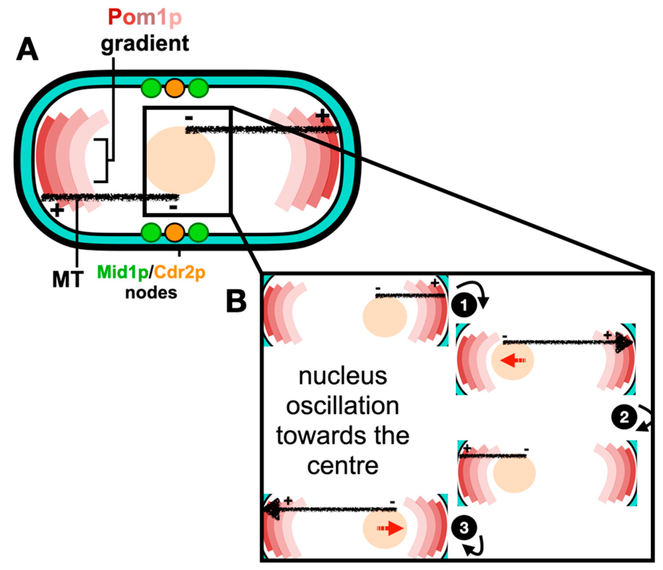
2.1. Positioning of the Cell Division Plane

In S. pombe, cellular growth occurs throughout a longer interphase period, with this ceasing during the shorter mitosis and cytokinesis periods after a certain cell length is achieved. During the cell cycle, the “middle” and “end or tip” locations are specified by two spatial axes. The “middle” location is defined by the nucleus, which is positioned at the cell center by a microtubule-pushing mechanism, where a force is produced by the cytoplasmic microtubule bundles and acts on the nucleus [10][11]. Furthermore, this force is able to efficiently re-center the nuclei of cells exposed to nuclear displacement [12][13]. The dynamic interplay between the nucleus and the microtubule cytoskeleton is illustrated and reviewed by Gallardo et al. [14]. The “end or tip” location is defined by a formin-mediated actin assembly mechanism at cell tips [15], and polarity factors including the DYRK kinase Pom1p gradient at cell poles [16]. Pom1p gradients are tightly controlled at the “end or tip”, with the dephosphorylation of Pom1p enhancing a lipid-binding activity, whereas autophosphorylation promotes Pom1p’s detachment from the plasma membrane [16]. Pom1p has an established role in regulating the timing of mitotic entry, as it phosphorylates the membrane-binding C-terminal region of the ACR scaffold protein, Cdr2p, at the cell “middle” through preventing its plasma membrane binding and the formation of nodes [17]. Research has revealed a strong correlation between the cell size at division and Pom1p medial levels; however, such a correlation between the cytosolic or cell tip levels of Pom1p and the cell length is inconsistent, indicating that Pom1p may interact with Cdr2p in the cytosol or at the cell tips [18]. For example, the Pom1p gradient model [16] is opposed by the findings of Pan et al. [19], in which they proposed a novel cell size control mechanism in which cells use Cdr2p to monitor their size. This model therefore implicates a relationship between both Pom1p and Cdr2p with cell size.
Much evidence shows that the Anillin-like protein Mid1p localizes to the “middle” location and initiates ACR assembly [20][21][22][23]. Mid1p has two membrane binding domains, the pleckstrin homology domain (PH) and the cryptic domain (C2) [24]. However, it only binds the plasma membrane after it is activated and released from the nucleus [20][25]. The roles of Mid1p in positioning the ACR are now well understood in S. pombe and are reviewed in Rezig et al. [26], with the mechanism of the medial positioning of the ACR schematically described in Figure 1.
Figure 1. Medial positioning of the ACR in fission yeast. (A) Pom1p gradient at the cell tips restricts the division site to the cell center. Mitotic entry is controlled by Pom1p phosphorylation of Cdr2p, preventing Cdr2p from binding the plasma membrane and the subsequent formation of cortical nodes. Upon mitotic entry, both active Mid1p and Cdr2p scaffold the formation of medial cortical nodes. (B) Upon microtubule–cortex contact, polymerization at the microtubule plus end generates a pushing force (large arrowhead) (1) that displaces the nucleus in the opposite direction (nuclear movement demonstrated by red arrows) (2). The antiparallel direction of the microtubule bundle ensures that, over time, the nucleus oscillates back and forth toward the center (3). References within the main text.
In contrast, the comprehension of the organization of proteins that assemble the complex cytokinetic machinery during cytokinesis is still relatively rudimentary. In S. pombe, cytokinesis proteins are recruited to the cell center, pre-determining the future division plane; these are organized as cortical spots, named “nodes” [27][28].

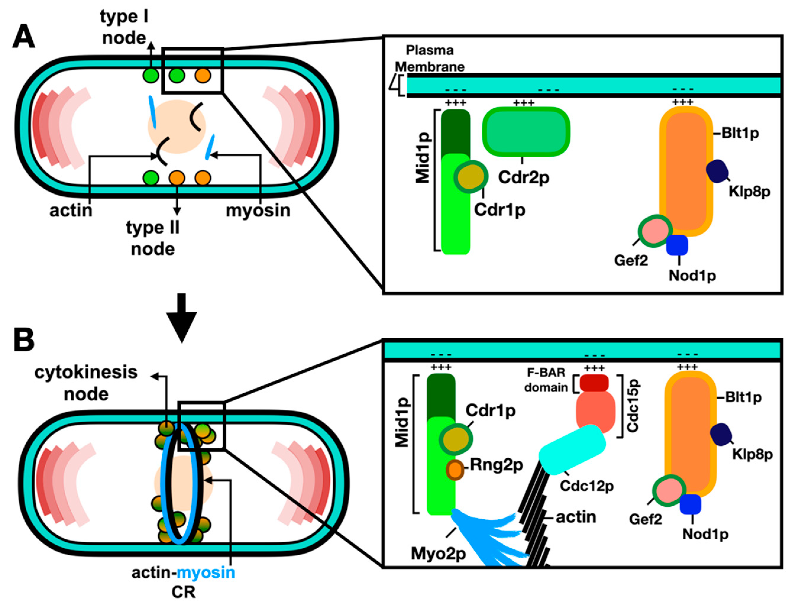
2.2. Molecular Organization of Nodes within the ACR

The current model for ACR assembly includes the formation of two types of interphase nodes: type 1 “stationary” nodes containing Mid1p, Cdr1p and Cdr2p; and type 2 “anchoring” nodes containing Blt1p, Klp8p, Gef2p and Nod1p [27][29]. Type 2 “anchoring” nodes diffuse into the cell cortex, and at mitotic onset, they are captured by type 1 “stationary” nodes to form cytokinesis nodes. Next, Mid1p recruits the myosin-II, Myo2p, Cdc15p, Rng2p and Cdc12p [30]. The cytokinesis nodes then merge into a ring-like structure, named the actin–myosin contractile ring (ACR) and, as its name implies, it is composed of actin filaments and myosin-II motors in addition to various classes of cytokinesis proteins [31].
Live cell imaging, high-speed fluorescence photo-activation localization microscopy (FPALM), and fluorescence resonance energy transfer (FRET) have been shown to be excellent methods to dissect ACR nodes. Recent findings have revealed that nodes are discrete units with stoichiometric ratios and a specific distribution of constituent proteins [28][30][31][32]. Furthermore, the localization of the ACR constituents is thought to be arranged in several layers relative to the plasma membrane, starting with the plasma-membrane-binding proteins and the tail of myosin-II, to the intermediate cytokinesis proteins, and farthest from the plasma membrane lies the myosin motor domains, F-actin and its cross-linkers [32]. Advances in laser scanning microscopy, such as Airyscanning using a very low laser power to acquire high-quality images, have increased the resolution and signal-to-noise ratio and enabled the detection and measurement of even faint individual cytokinesis nodes [33]. The coalescence of nodes leads to ACR assembly through the search, capture, pull and release (SCPR) mechanism, whereby Cdc12p nucleates actin filaments as Myo2p pulls actin filaments, thus producing the force required to pull the individual nodes into the ACR [28][34][35]. Such an assembly of the ACR from node precursors is schematically described in Figure 2.
Figure 2. Assembly of the ACR from node precursors. (A) During interphase, type 1 “stationary” (green: Mid1p, Cdr1p and Cdr2p) nodes and type 2 “anchoring” (orange: Blt1p, Klp8p, Gef2p and Nod1p) nodes bind the plasma membrane and scaffold other cytokinesis proteins. (B) Coalescence of type 1 and type 2 nodes leads to their maturation into cytokinesis nodes (green–orange gradient). Maturation of cytokinesis nodes leads to the recruitment of Myo2p, Cdc15p and Cdc12p and nucleation of actin filaments. Interactions between myosin-II and actin promote ACR formation. References within the main text.

2.3. Anchorage of the ACR to the Plasma Membrane

In the assembled ACR, the Myo2p tails and Cdc15p anchor to the plasma membrane, with the Myo2p heads, Myp2p and the bundle of actin filaments localizing 60 nm away from the plasma membrane [36]. It is suggested that this organization connects the bundle of actin filaments to the plasma membrane [37]. Cdc15p next recruits Cdc12p to the ACR, and this interaction is thought to be essential for ACR organization and stability [38].
The phospho-status of Cdc15p influences its ability to bind the plasma membrane, with the phosphorylation of Cdc15p by Pom1p inhibiting its binding to the plasma membrane at the cell tips [39]. Additionally, the p21-activated protein kinase (Pak1p), another polarity kinase, was found to regulate the function of Mid1p and Cdc15p [40]. Cdc15p has three regulatory components: an N-terminal Fre/Cip4 homology Bin/Amphiphysin/Rvs domain (F-BAR), a medial intrinsically disordered region (IDR) and a C-terminal Src homology 3 domain (SH3). While the F-BAR domain enables protein oligomerization and concentration on the plasma membrane to scaffold protein assemblies resulting in membrane deformation [41], it was recently found that the phosphorylation of Cdc15p induces the separation of the Cdc15p IDR region resulting in an inhibition of Cdc15p phase separation, and the formation of condensate on the plasma membrane [42].
Moshtohry et al. [43] recently used laser ablation, a technique based on photodamage in which cellular structures could be degraded using a focused pulsed laser, to investigate the mechanical role of Cdc15p during S. pombe cytokinesis and found that the ACR recoils after being severed. However, this recoil profile was greater and slower in the ablated ACR of Cdc15-depleted cells, suggesting that the loss of Cdc15p decreases the stiffness of the ACR material. Furthermore, another F-BAR protein, Imp2p, was found to contribute to the stiffness of the ACR [37].

References

  1. Pollard, T.D.; Wu, J.-Q. Understanding cytokinesis: Lessons from fission yeast. Nat. Rev. Mol. Cell Biol. 2010, 11, 149–155.
  2. Hartwell, L.H.; Culotti, J.; Reid, B. Genetic Control of the Cell-Division Cycle in Yeast, I. Detection of Mutants. Proc. Natl. Acad. Sci. USA 1970, 66, 352–359.
  3. Hartwell, L.H.; Mortimer, R.K.; Culotti, J.; Culotti, M. Genetic control of the cell division cycle in yeast: V. genetic analysis of cdc mutants. Genetics 1973, 74, 267–286.
  4. Nurse, P. Genetic control of cell size at cell division in yeast. Nature 1975, 256, 547–551.
  5. Nurse, P.; Thuriaux, P.; Nasmyth, K. Genetic control of the cell division cycle in the fission yeast Schizosaccharomyces pombe. Mol. Gen. Genet. 1976, 146, 167–178.
  6. Chang, F.; Nurse, P. Finishing the cell cycle: Control of mitosis and cytokinesis in fission yeast. Trends Genet. 1993, 9, 333–335.
  7. Chang, F.; Nurse, P. How fission yeast fission in the middle. Cell 1996, 84, 191–194.
  8. Nurse, P. Fission yeast cell cycle mutants and the logic of eukaryotic cell cycle control. Mol. Biol. Cell 2020, 31, 2871–2873.
  9. Chang, F. Forces that shape fission yeast cells. Mol. Biol. Cell 2017, 28, 1819–1824.
  10. Piel, M.; Tran, P.T. Cell shape and cell division in fission yeast. Curr. Biol. 2009, 19, R823–R827.
  11. Tran, P.T.; Marsh, L.; Doye, V.; Inoue, S.; Chang, F. A mechanism for nuclear positioning in fission yeast based upon microtubule pushing. J. Cell Biol. 2001, 153, 397–411.
  12. Daga, R.R.; Chang, F. Dynamic positioning of the fission yeast cell division plane. Proc. Natl. Acad. Sci. USA 2005, 102, 8228–8232.
  13. Daga, R.R.; Yonetani, A.; Chang, F. Asymmetric Microtubule Pushing Forces in Nuclear Centering. Curr. Biol. 2006, 16, 1544–1550.
  14. Gallardo, P.; Barrales, R.R.; Daga, R.R.; Salas-Pino, S. Nuclear Mechanics in the Fission Yeast. Cells 2019, 8, 1285.
  15. Martin, S.G.; McDonald, W.H.; Yates, J.R., 3rd; Chang, F. Tea4p links microtubule plus ends with the formin for3p in the establishment of cell polarity. Dev. Cell. 2005, 8, 479–491.
  16. Hersch, M.; Hachet, O.; Dalessi, S.; Ullal, P.; Bhatia, P.; Bergmann, S.; Martin, S.G. Pom1 gradient buffering through intermolecular auto-phosphorylation. Mol. Syst. Biol. 2015, 11, 818.
  17. Moseley, J.B.; Mayeux, A.; Paoletti, A.; Nurse, P. A spatial gradient coordinates cell size and mitotic entry in fission yeast. Nature. 2009, 7248, 857–860.
  18. Gerganova, V.; Floderer, C.; Archetti, A.; Michon, L.; Carlini, L.; Reichler, T.; Manley, S.; Martin, S.G. Author response: Multi-phosphorylation reaction and clustering tune Pom1 gradient mid-cell levels according to cell size. Elife 2019, 3, e45983.
  19. Pan, K.Z.; Saunders, T.E.; Flor-Parra, I.; Howard, M.; Chang, F. Cortical regulation of cell size by a sizer cdr2p. Elife 2014, 3, e02040.
  20. Almonacid, M.; Celton-Morizur, S.; Jakubowski, J.L.; Dingli, F.; Loew, D.; Mayeux, A.; Chen, J.-S.; Gould, K.L.; Clifford, D.M.; Paoletti, A. Temporal control of contractile ring assembly by Plo1 regulation of Myosin II recruitment by Mid1/Anillin. Curr. Biol. 2011, 21, 473–479.
  21. Paoletti, A.; Chang, F. Analysis of mid1p, a protein required for placement of the cell division site, reveals a link between the nucleus and the cell surface in fission Yeast. Mol. Biol. Cell 2000, 11, 2757–2773.
  22. Rezig, I.M.; Yaduma, W.G.; Gould, G.W.; McInerny, C.J. Anillin/Mid1p interacts with the ESCRT-associated protein Vps4p and mitotic kinases to regulate cytokinesis in fission yeast. Cell Cycle 2021, 20, 1845–1860.
  23. Saha, S.; Pollard, T.D.; Akamatsu, M.; Lin, Y.; Bewersdorf, J.; Lidke, M.E.D.; Sherlekar, A.; Rikhy, R.; Montell, M.E.D.; Goss, J.W.; et al. Anillin-related protein Mid1p coordinates the assembly of the cytokinetic contractile ring in fission yeast. Mol. Biol. Cell 2012, 23, 3982–3992.
  24. Chatterjee, M.; Pollard, T.D. The functionally important N-terminal half of fission yeast mid1p anillin is intrinsically disordered and undergoes phase separation. Biochemistry 2019, 58, 3031–3041.
  25. Steever, A.; Pringle, J.; Bähler, J.; Wang, Y.-L.; Gould, K.; McCollum, D.; Wheatley, S. Role of polo kinase and mid1p in determining the site of cell division in fission yeast. J. Cell Biol. 1998, 143, 1603–1616.
  26. Rezig, I.M.; Yaduma, W.G.; Gould, G.W.; McInerny, C.J. The role of anillin/Mid1p during medial division and cytokinesis: From fission yeast to cancer cells. Cell Cycle 2022, 22, 633–644.
  27. Akamatsu, M.; Berro, J.; Pu, K.-M.; Tebbs, I.R.; Pollard, T.D. Cytokinetic nodes in fission yeast arise from two distinct types of nodes that merge during interphase. J. Cell Biol. 2014, 204, 977–988.
  28. Laplante, C.; Huang, F.; Tebbs, I.R.; Bewersdorf, J.; Pollard, T.D. Molecular organization of cytokinesis nodes and contractile rings by super-resolution fluorescence microscopy of live fission yeast. Proc. Natl. Acad. Sci. USA 2016, 113, E5876–E5885.
  29. Zhu, Y.-H.; Ye, Y.; Wu, Z.; Wu, J.-Q. Cooperation between Rho-GEF Gef2 and its binding partner Nod1 in the regulation of fission yeast cytokinesis. Mol. Biol. Cell 2013, 24, 3187–3204.
  30. Akamatsu, M.; Lin, Y.; Bewersdorf, J.; Pollard, T.D.; Saha, S.; Wang, M.E.Y.-L.; Wills, R.C.; Goulden, B.D.; Hammond, G.R.V.; Kozminski, M.E.K.G.; et al. Analysis of interphase node proteins in fission yeast by quantitative and superresolution fluorescence microscopy. Mol. Biol. Cell 2017, 28, 3203–3214.
  31. Malla, M.; Pollard, T.D.; Chen, Q. Counting actin in contractile rings reveals novel contributions of cofilin and type II myosins to fission yeast cytokinesis. Mol. Biol. Cell 2022, 33, ar51.
  32. McDonald, N.A.; Lind, A.L.; Smith, S.E.; Li, R.; Gould, K.L. Nanoscale architecture of the Schizosaccharomyces pombe contractile ring. Elife 2017, 15, e28865.
  33. Sayyad, W.A.; Pollard, T.D. The number of cytokinesis nodes in mitotic fission yeast scales with cell size. Elife 2022, 12, e76249.
  34. Vavylonis, D.; Wu, J.-Q.; Hao, S.; O’Shaughnessy, B.; Pollard, T.D. Assembly mechanism of the contractile ring for cytokinesis by fission yeast. Science 2008, 319, 97–100.
  35. Zimmermann, D.; Homa, K.E.; Hocky, G.M.; Pollard, L.W.; De La Cruz, E.M.; Voth, G.A.; Trybus, K.M.; Kovar, D.R. Mechanoregulated inhibition of formin facilitates contractile actomyosin ring assembly. Nat. Commun. 2017, 8, 703.
  36. Swulius, M.T.; Nguyen, L.T.; Ladinsky, M.S.; Ortega, D.R.; Aich, S.; Mishra, M.; Jensen, G.J. Structure of the fission yeast actomyosin ring during constriction. Proc. Natl. Acad. Sci. USA 2018, 115, E1455–E1464.
  37. Bellingham-Johnstun, K.; Anders, E.C.; Ravi, J.; Bruinsma, C.; Laplante, C. Molecular organization of cytokinesis node predicts the constriction rate of the contractile ring. J. Cell Biol. 2021, 220, e202008032.
  38. Snider, C.E.; Chandra, M.; McDonald, N.A.; Willet, A.H.; Collier, S.E.; Ohi, M.D.; Jackson, L.P.; Gould, K.L. Opposite surfaces of the Cdc15 F-BAR domain create a membrane platform that coordinates cytoskeletal and signaling components for cytokinesis. Cell Rep. 2020, 33, 108526.
  39. Bhattacharjee, R.; Mangione, M.C.; Wos, M.; Chen, J.-S.; Snider, C.E.; Roberts-Galbraith, R.H.; McDonald, N.A.; Presti, L.L.; Martin, S.G.; Gould, K.L. DYRK kinase Pom1 drives F-BAR protein Cdc15 from the membrane to promote medial division. Mol. Biol. Cell 2020, 31, 917–929.
  40. Magliozzi, J.O.; Sears, J.; Cressey, L.; Brady, M.; Opalko, H.E.; Kettenbach, A.N.; Moseley, J.B. Fission yeast Pak1 phosphorylates anillin-like Mid1 for spatial control of cytokinesis. J. Cell Biol. 2020, 219, e201908017.
  41. Snider, C.E.; Noor, W.N.I.W.M.; Nguyen, N.T.H.; Gould, K.L.; Suetsugu, S. The state of F-BAR domains as membrane-bound oligomeric platforms. Trends Cell Biol. 2021, 31, 644–655.
  42. Bhattacharjee, R.; Hall, A.R.; Mangione, M.C.; Igarashi, M.G.; Roberts-Galbraith, R.H.; Chen, J.-S.; Vavylonis, D.; Gould, K.L. Author response: Multiple polarity kinases inhibit phase separation of F-BAR protein Cdc15 and antagonize cytokinetic ring assembly in fission yeast. Elife 2023, 7, e83062.
  43. Moshtohry, M.; Bellingham-Johnstun, K.; Elting, M.W.; Laplante, C. Laser ablation reveals the impact of Cdc15p on the stiffness of the contractile ring. Mol. Biol. Cell 2022, 33, br9.
More
Information
Subjects: Cell Biology
Contributors MDPI registered users' name will be linked to their SciProfiles pages. To register with us, please refer to https://encyclopedia.pub/register : , ,
View Times: 60
Revisions: 2 times (View History)
Update Date: 07 Mar 2024
1000/1000