Your browser does not fully support modern features. Please upgrade for a smoother experience.
Submitted Successfully!
 +1 credit
Thank you for your contribution! You can also upload a video entry or images related to this topic.
For video creation, please contact our Academic Video Service.
| Version | Summary | Created by | Modification | Content Size | Created at | Operation |
|---|---|---|---|---|---|---|
| 1 | Grzegorz Zieliński | -- | 4430 | 2024-03-06 10:32:01 | | | |
| 2 | Rita Xu | Meta information modification | 4430 | 2024-03-06 10:38:41 | | | | |
| 3 | Rita Xu | Meta information modification | 4430 | 2024-03-06 10:58:53 | | |
Video Upload Options
We provide professional Academic Video Service to translate complex research into visually appealing presentations. Would you like to try it?
Cite
If you have any further questions, please contact Encyclopedia Editorial Office.
Zieliński, G.; Gawda, P. Surface Electromyography in Dentistry. Encyclopedia. Available online: https://encyclopedia.pub/entry/55912 (accessed on 25 June 2026).
Zieliński G, Gawda P. Surface Electromyography in Dentistry. Encyclopedia. Available at: https://encyclopedia.pub/entry/55912. Accessed June 25, 2026.
Zieliński, Grzegorz, Piotr Gawda. "Surface Electromyography in Dentistry" Encyclopedia, https://encyclopedia.pub/entry/55912 (accessed June 25, 2026).
Zieliński, G., & Gawda, P. (2024, March 06). Surface Electromyography in Dentistry. In Encyclopedia. https://encyclopedia.pub/entry/55912
Zieliński, Grzegorz and Piotr Gawda. "Surface Electromyography in Dentistry." Encyclopedia. Web. 06 March, 2024.
Copy Citation
Surface electromyography (sEMG) is a technique for measuring and analyzing the electrical signals of muscle activity using electrodes placed on the skin’s surface.
electromyography
electrodiagnosis
temporomandibular joint
history of dentistry
1. Introduction
Surface electromyography (sEMG) is a technique for measuring (development and recording) and analyzing the electrical signals of muscle activity using electrodes placed on the skin’s surface [1][2]. sEMG works by detecting and analyzing electrical signals that result from physiological changes in the cell membranes of muscle fibers [1]. A key aspect of sEMG is the understanding that human tissue, particularly muscle, has the ability to generate and conduct electrical impulses that are fundamental to the process of muscle contraction [3]. When a muscle is at rest, it is in a state of electrical equilibrium known as the resting potential. However, during contraction, depolarization of the muscle membrane occurs, which means that there is a flow of ions between the inside and outside of the muscle membrane, generating an electrical signal that is recorded [1][4][5].
2. Surface Electromyography in Dentistry—History
2.1. The 17th Century
The concept of “animal spirits” has a rich history in the realm of physiology and psychology. It was a concept that worked for 1500 years (Figure 1) [6]. Originating in ancient times, it was initially proposed by Greek philosophers like Galen, who believed that these spirits were responsible for sensation and movement. During the Middle Ages, this idea was further developed by Islamic and European scholars, who postulated that animal spirits were fine, vaporous substances flowing through the body’s nerves and brain [7]. People have become increasingly familiar with human anatomy. In 1543, Andreas Vesalius created seven books under the title De Humani Corporis Fabrica Libri Septem on anatomy [8].
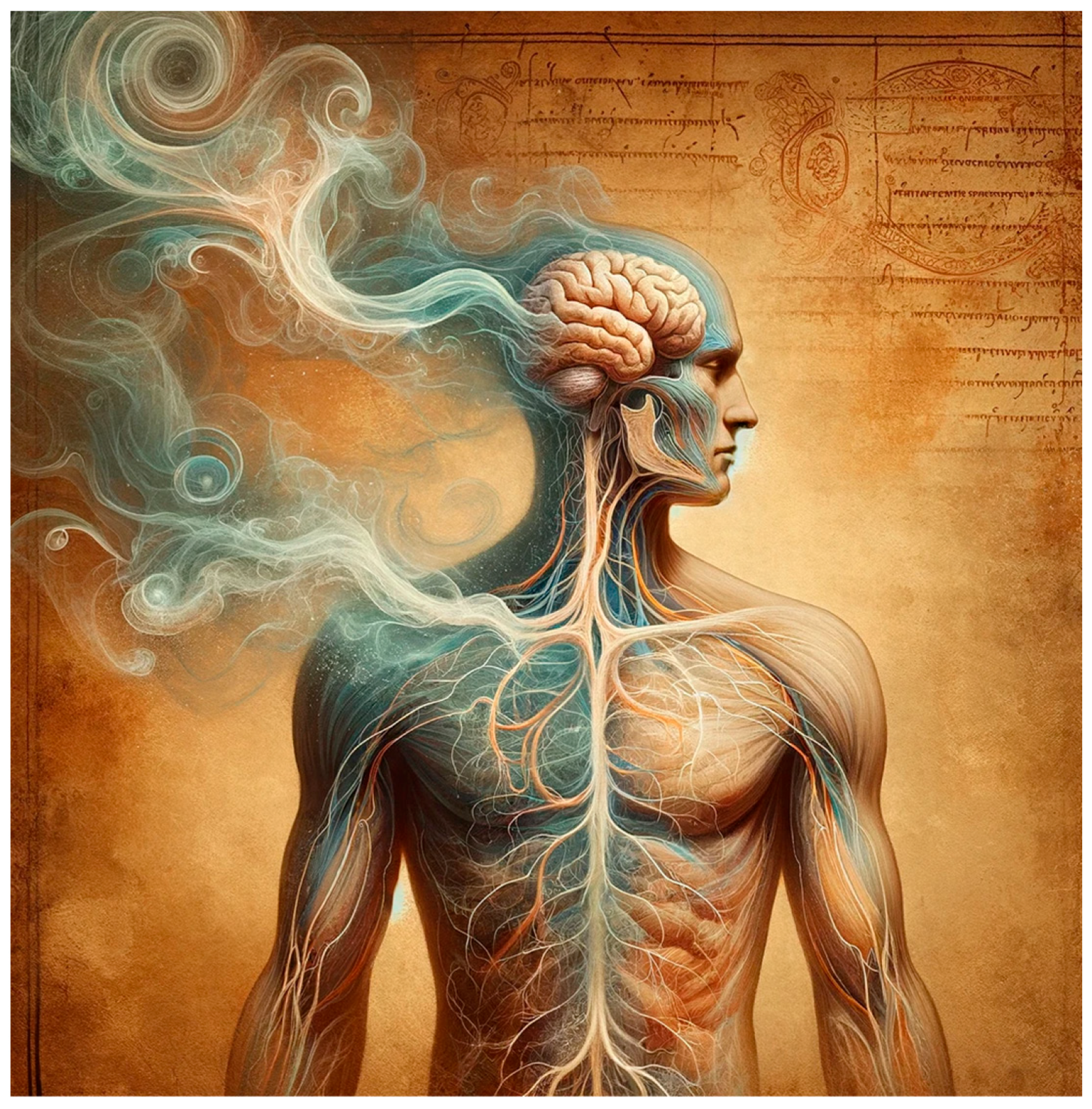
Figure 1. An artistic interpretation of the historical concept of ‘animal spirits’ in human physiology. Derived on 2 January 2024 by DALL-E 2 program (OpenAI, Inc., San Francisco, CA, United States).
Jan Swammerdam was a pioneer in the study of muscle anatomy and the nervous system, although his work focused primarily on insects and other small organisms. His contribution to the understanding of muscle function was significant, particularly in relation to his experiments on muscle contraction [6][9].
-
1658. Jan Swammerdam showed the Duke of Tuscany how to contract a muscle simply by irritating a nerve [10].
-
1662. Jan Swammerdam, while dissecting dogs, showed that movement could occur without any connection between the muscle and brain, thus putting an end to part of Descartes’ theory [6].
-
1681. The earliest evidence of research into the strength of the masticatory muscles comes from the work of Giovanni Alfonso Borelli. He recorded a “biting force” of up to 200 kg (about 430 pounds) [12][13]. The test method used by Borelli consisted of placing a weight under the patient’s mandible [14].
2.2. The 18th Century
The 18th century was crucial for the development of electricity, marking a transformative period in its understanding and application. This era witnessed the birth of modern electrical science. These advancements paved the way for the electrical innovations of the 19th century, which revolutionized society.
-
1745–1746. The origins of the development of modern electrophysiology can be traced back to the creation of an early condenser. The Leyden jar was invented independently by Dean von Kleist and Petrus van Musschenbroek. This device was used to collect and store electric charge and was one of the first discoveries in the field of electricity [15][16].
-
1746. Jean-Antoine Nollet conducted an experiment in which the test group consisted of about 200 monks who held hands to form a long chain over a kilometer long (Figure 2). At one end of the chain, Nollet used a Leyden jar to create an electric charge. When the charge was released, it passed through the entire chain of monks. Each of the monks felt a shock as the charge passed through their bodies. The monks’ reactions to the electric shock were instantaneous and simultaneous, demonstrating that electricity can be transmitted through the human body over great distances at tremendous speed. This was the first human study of electricity on such a scale [17].
-
1748. Jean-Antoine Nollet invented the first device to detect and measure the presence of an electric charge. As an early tool for the study of electricity, Nollet’s electroscope played an important role in experiments related to electrostatics [18].
-
1752. Benjamin Franklin conducted the famous kite experiment. This experiment was an important step in the understanding of electricity [19].
-
1772. John Walsh’s research on electric fish, such as the electric eel (torpedo), proved that the shocks given off by these fish were a form of electricity. Using a Leyden jar, a device for storing electric charge, Walsh collected and measured electric discharges, providing direct evidence of the electrical nature of the shocks; his work was published in 1773 [20][21]. Walsh’s experiments probably contributed to Luigi Galvani’s decision to begin his experiments on frog muscle contraction [22]. However, Galvani’s experiments focused on a completely different aspect—the electricity generated by the muscles. Luigi Galvani’s discovery, which took place at the end of the 18th century, is often described as a series of coincidences and experimental observations.
-
1791. Luigi Galvani’s accidental experiment took place in 1791. Galvani was performing anatomical experiments on a frog in his laboratory. At the same time, some of his colleagues were playing with a Leyden jar. While Galvani was working, an electric spark was unexpectedly produced by the Leyden jar. At the exact moment the spark was emitted, Galvani touched the frog’s nerve with a metal knife. The result was a violent contraction of the frog’s muscles, which shocked and fascinated Galvani [15]. The results were published in 1791 [23].
-
1792. George Adams published the paper “An Essay on Electricity: Explaining the Theory and Practice of That Useful Science, and the Mode of Applying it to Medical Purposes” considering the application of electricity in medicine [19].
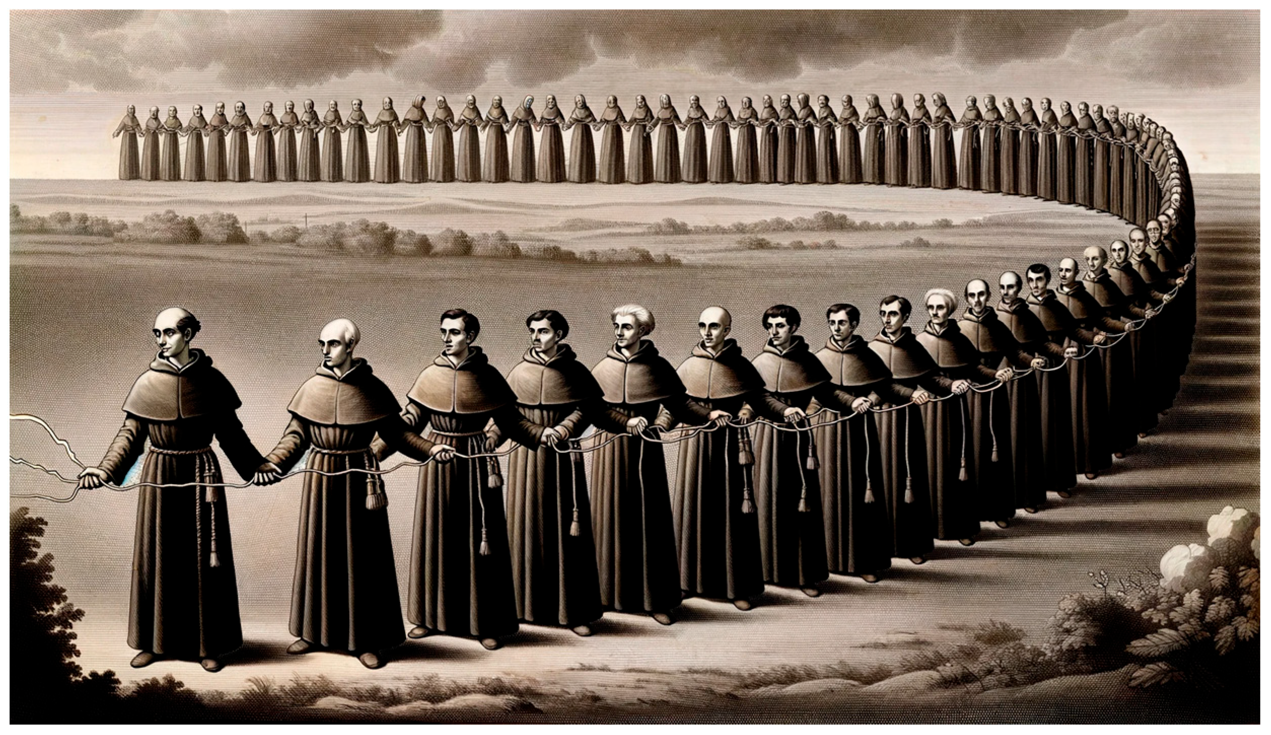
Figure 2. An illustration of Jean-Antoine Nollet’s experiment. Derived on 13 January 2024 by DALL-E 2 program (OpenAI, Inc., San Francisco, CA, United States) (slightly corrected by the author G.Z.).
2.3. The 19th Century
In the 19th century, researchers inspired by the previous century continued to investigate electricity, nerve conduction and the study of the strength of the masticatory muscles.
-
1803. Inspired by the work of his uncle Luigi Galvani, Giovanni Aldini was the first to demonstrate the reactions of the muscles of dead men to electrical stimulation from a battery of galvanic cells. His most famous experiment took place at the Royal College of Surgeons in London in 1803, on a hanged man called George Forster [24].
-
1825. Leopoldo Nobili built the astatic galvanometer [25]. An astatic galvanometer is a special type of galvanometer designed to minimize the influence of an external magnetic field on its readings. The main purpose of such a solution is to increase the sensitivity and accuracy of electric current measurements.
-
1838. Inspired by Luigi Galvani, Carlo Matteucci, in an 1838 study, observed the frog’s muscle responses to different types of electrical stimulation, which allowed him to understand the basic mechanisms of bioelectricity in living tissues. He used a complete frog leg, cut off below the knee, with only an isolated nerve above [15]. Carlo Matteucci used the astatic galvanometer invented by Leopoldo Nobili for his research [26]. This discovery was key to understanding the mechanisms by which nerve impulses cause muscle contractions [27].
-
1849. Emil du Bois-Reymond was inspired by the work of Carlo Matteucci. One of du Bois-Reymond’s most important achievements during this period was his demonstration that stimulation of a nerve causes an electrical change, which he called an ‘action potential’. This discovery was fundamental to understanding how nerve impulses transmit information in the body. He described it in 1849 in his work ‘Untersuchungen über thierische Elektricität’ [28]. Du Bois-Reymond also discovered that it was possible to record (using a galvanometer) electrical activity during voluntary muscle contraction [2]. In the same year, Hermann von Helmholtz measured at what speed a nerve signal is transmitted through the nerve fibers of a frog and reported transmission rates ranging from 24.6to 38.4 m per second [29][30].
-
1861. Wilhelm Erb identified specific points on the body that were particularly sensitive to electrical stimulation, known as Erb’s points [31].
-
1862. Guillaume Benjamin Duchenne conducted research into the transcutaneous electrical stimulation of muscles and was able to do so as early as 1833 [15]. However, it was not until 1862 that Duchenne published the work “Mécanisme de la Physionomie Humaine” [32] that he included the results of his research on facial expressions, which he carried out using electrical muscle stimulation, and also included the identification of motor points.
-
1868. Julius Bernstein, in his work from 1868, theorized that a living cell’s interior consists of an electrolytic substance and that the cell is separated from its surrounding environment by a membrane. This membrane is characterized by its limited permeability to ions. Consequently, he suggested that the cell membrane would be the primary resistive component within a cell [33].
-
1870. In the 1870s, Hermann Muller contributed to the classification of the electrical properties of tissues. His work included observing the directionality of the flow of electric charges through biological tissues, such as muscles [19].
-
1895. Greene Vardiman Black conducted a bite force study. He reported that in 1895, he tested the bite force of several thousand people and found that the average was 175 pounds [35]. Black achieved his results by designing a new gnathodynamometer. His device was used to measure intraoral forces caused by vertical jaw movements [36].
-
1898. Napoleon Cybulski proposed the idea that tissue currents could have their own origin. Cybulski concluded that undamaged muscle was the source of the current. Some sources claim that he is the founder of the theory of electric currents in tissues [37].
2.4. The 20th Century
The 20th century was critical for the development of surface electromyography. In this period, significant advancements were made in both the theoretical and practical aspects of sEMG. Early in the century, the foundational principles of muscle electricity were established, leading to the development of more sophisticated sEMG equipment. These advancements allowed for the non-invasive recording of muscle electrical activity, providing valuable insights into muscle physiology, movement disorders and rehabilitation. The technological advancements in electronics and computing during the latter half of the century enabled the more precise, real-time analysis of sEMG data, further enhancing its diagnostic and research capabilities. The integration of sEMG with other technologies, like biofeedback and computerized analysis systems, opened new avenues in medical research.
-
1912. Hermann Piper was the first to describe the phenomenon known as the H-wave. The H-wave is the electrical response of a muscle to nerve stimulation. It is a form of reflex that can be recorded by electromyography and its measurement is used to assess peripheral nerve function and neuromuscular integration. Piper analyzed the electrical activity in the muscles in response to electrical stimulation of the ulnar nerve. He published his results in the paper ‘’Elektrophysiologie menschlicher muskeln” [38]. It is worth noting that Hermann Piper is considered to be the first scientist to study the surface electrical signal. This was the first study of surface electromyography. He did this using a string galvanometer [10].
Julius Bernstein published the book “Elektrobiologie”, summarizing his electrophysiological work and expanding his theoretical concepts [39]. This book has made a significant contribution to the field [40].
-
1913–1915. Napoleon Cybulski suggested that the carrier of electricity in skeletal muscle could be ions flowing across the semi-permeable membrane in his work entitled “Prądy czynnościowe nerwów i ich stosunek do temperatur” and in a subsequent work of the same year entitled “Model prądów czynnościowych w mięśniach” [41][42]. In 1915, Napoleon Cybulski recorded the first electrical signal in mammalian (dogs and rabbits) muscle fibers [37][43].
-
1917. Frederick Haven Pratt showed in his work that the amount of energy associated with muscle contraction was due to the recruitment of individual muscle fibers rather than the size of the nerve impulse [44].
-
1919. Gildemeister highlighted the resemblance between the electrical conductivity characteristics of skin and those found in a polarization cell [45].
-
1921. Philippson carried out a significant research project where he measured the electrical impedances of a range of biological substances. This included packed blood cells, muscle and liver tissue from guinea pigs, as well as potato tuber [46].
-
1924. Hans Berger made the first recording of electroencephalography (EEG) in humans and introduced the name that is still used today. He also described the types of waves and rhythms present in a properly functioning brain. This influenced the development of the EMG, sEMG and paved the way for research into human bioelectricity [47].
Hugo Fricke published a technical paper “A Mathematical Treatment of the Electric Conductivity and Capacity of Disperse Systems” that delves into the mathematical analysis of the electrical properties of disperse systems [48]. This work focuses on understanding and calculating the electrical conductivity and capacity of materials composed of dispersed particles or elements. It provides theoretical insights and mathematical models for analyzing these properties, contributing significantly to the field of materials science and electrical engineering.
-
1925. E. K. Liddell and Charles Sherrington made significant contributions to the understanding of motor units in muscle physiology. They conducted experiments on the flexor muscles in the cat’s knee, providing crucial insights into how motor units function. Their work helped to establish the concept of the motor unit, defining it as a single motor neuron and the group of muscle fibers it innervates. This research was fundamental in understanding muscle contraction and the role of the nervous system in controlling muscle movement [49].
In 1925, H. Fricke and S. Morse conducted a study titled “The Electric Resistance and Capacity of Blood for Frequencies between 800 and 4(1/2) Million Cycles”. This research explored the electrical properties of blood, specifically its resistance and capacity over a wide range of frequencies. This study was significant in advancing the understanding of bioelectrical properties of biological materials [50].
-
1937. Joseph Erlanger and Herbert Spencer Gasser published the paper entitled “Electrical Signs of Nervous Activity” [52]. In this work, Gasser and Erlanger described in detail their findings regarding the conduction velocity of nerve impulses and the various properties of nerve fibers. Using a cathode-ray oscilloscope, they were able to accurately record and analyze the electrical signals generated by nerves. Their research contributed to a deeper understanding of the mechanisms of nerve conduction and had a major impact on the development of neurology and electrophysiology.
-
1938. Edmund Jacobson realized that there was a deep connection between muscle tension and emotional state. He conducted numerous experiments using surface electromyography to monitor muscle tension in people in different situations. Through these studies he proved that stress, anxiety and negative emotions lead to an increase in muscle tension. His most important paper was published in 1938—“Progressive Relaxation” [53].
In the same year, Derek Denny-Brown and Pennybacker published the first EMG study in patients with neurological disorders [54].
-
1940. Emergence of surface electromyography [55].
-
1941. Kenneth S. Cole and Robert H. Cole published a research paper “Dispersion and Absorption in Dielectrics I. Alternating Current Characteristics” that delves into the behavior of dielectrics (insulating materials) when subjected to alternating current. It primarily explores how these materials disperse and absorb electrical energy, focusing on their alternating current (AC) characteristics. This study is foundational in understanding the dielectric properties of materials, which has implications in various fields of electrical and materials engineering [56].
-
1943. Graham Weddell published a paper entitled: “Electromyography in Clinical Medicine” [57].
-
1944. V. T. Inman, J B Saunders and L. C. Abbott analyzed the superficial electromyography of shoulder movements [58]. In the same year, Joseph Erlanger and Herbert Spencer Gasser won the Nobel Prize in Physiology or Medicine “for their discoveries relating to the highly differentiated functions of single nerve fibers” [59].
-
1947. Edward Lambert established the first electromyography laboratory in the United States and the first EMG training program. His research began in 1948 and focused on the use of EMG in the diagnosis of myasthenia gravis. Lambert is often referred to as the “father of electromyography” [60]. His early research established EMG as a fundamental neurological tool.
This year also saw the founding of the Dansk Industri Syndikat (DISA), which later developed an analogue electromyograph [61].
-
1948. Price et al. found that back pain affected changes in bioelectrical activity [62].
-
1950. DISA introduces a three-channel analogue EMG recorder (model 13A67). Developed in collaboration with Buchthal at Copenhagen University Hospital [65].
-
1952. Alan Lloyd Hodgkin and Andrew Fielding Huxley published five papers describing a series of experiments and an action potential model [10]. Amongst others, entitled “A Quantitative Description of Membrane Current and its Application to Conduction and Excitation in Nerve”. This work describes in detail how changes in the conductance of sodium and potassium ions across the cell membrane of an axon affect the generation and conduction of an action potentials [66]. Among others, they published another paper entitled: “Currents Carried by Sodium and Potassium Ions through the Membrane of the Giant Axon of Loligo”. This work focused on the study of ionic currents flowing through the membrane of the axon and their role in nerve conduction [67]. Published mainly in the 1950s, it was of great importance to neurophysiology, and its results are still fundamental to understanding the electrical activity of neurons. The Hodgkin–Huxley model is still used today in modern neuroscience research and mathematical models of neuronal activity.
O. C. J. Lippold published a paper entitled: “The relation between integrated action potentials in a human muscle and its isometric tension”. The study explored how the isometric tension produced by a human muscle during voluntary contraction relates to its integrated electromyogram [68].
-
1953. The American Association of Neuromuscular & Electrodiagnostic Medicine was founded [69].
-
1954. Fritz Buchthal published a paper entitled: “Action potential parameters in normal human muscle and their physiological determinants” [70]. This work had a significant impact on the development of electromyography as a diagnostic tool in medicine. Buchthal is therefore considered one of the pioneers of clinical electromyography [43].
Brenda Bigland and O. C. J. Lippold published a paper entitled: “The relation between force, velocity and integrated electrical activity in human muscles” [71]. Among other things, the authors proved that tension, velocity, and electrical activity are closely linked. The integrated electrical record offers a combined measure reflecting both the number of muscle fibers in action and the rate at which they are stimulated [71].
-
1956. Greenfield and Wyke published a paper in which they determine the placement of surface or needle electrodes when studying the masticatory muscles. This is one of the steps in the reproducibility of electromyographic studies [72].
That same year, Joseph R. Jarabak published “An Electromyographic Analysis of Muscular And Temporomandibular Joint Disturbances Due to Imbalances in Occlusion” [73].
-
1959. The American Association of Neuromuscular & Electrodiagnostic Medicine was registered as nonprofit [69].
-
1962. Basmajian, and G. Stecko created a new bipolar electrode for electromyography [74].
-
1963. John Basmajian demonstrated that he could isolate motor units and control their isolated contractions [75].
Peter Vig published the first review paper on electromyography in dental science. In this paper, he described the techniques and their limitations in electromyographic studies of mandibular movements [64].
-
1965. The International Society of Electrophysiological Kinesiology was founded as an international organization. During the International Congress of Anatomy in the Rhein-Maine-Halle, Wiesbaden, Germany in the summer of 1965, a group of anatomists convened to discuss the formation of a small society dedicated to electrophysiological kinesiology. Among those present were notable individuals like J.V. Basmajian, S. Carlsøø, B. Jonsson, M.A. MacConaill, J. Pauly and L. Scheving [76].
In the same year, Elwood Henneman, George Somjen and David O. Carpenter published a paper entitled: “Functional Significance Of Cell Size In Spinal Motoneurons” [77]. They published a further four studies describing in detail the activation properties of motor units or motoneurons. Their findings can be summarized in a single thesis, which they called the “size principle”. This thesis states that the smaller motoneurons in the anterior horn of the spinal cord are likely to correspond to the smallest motor units [10][77].
-
1966. Moller published a paper entitled: “The chewing apparatus: An electromyographic study of the action of the muscles of mastication and its correlation to facial morphology”. It was one of the first papers of its kind on the action of the masticatory muscles and its correlation to facial morphology [78].
-
1968. Herman P. Schwan published a paper entitled: ‘’Electrode Polarization Impedance And Measurements In Biological Materials” [79]. It appeared in the Annals of the New York Academy of Sciences. This study is significant in the field of bioimpedance, particularly focusing on the challenges and methodologies related to measuring the electrical properties of biological materials.
-
1969. R. Yemm conducted some of the first more sophisticated research on the bioelectrical activity of the human masseter muscle associated with emotional stress [80]. In the following years, Yemm continued his research on the above topics.
J. M. Bernstein, N. D. Mohl and H. Spiller published papers on temporomandibular joint dysfunction and its symptoms manifested as diseases of the ear, nose and throat [81].
In the same year, E Moller published a paper entitled “Clinical electromyography in dentistry” [82]. The paper presents electromyography as a diagnostic method in dentistry.
-
1971. C. J. Griffin and R. R. Munro published papers on the direct effect of TMDs on the electrical activity of the masticatory muscles [83].
-
1975. DISA produced the first complete digital EMG system and all modules are equipped with digital components (Model 1500) [65].
In the same year, H. S. Milner-Brown and R. B. Stein published a paper entitled: “The relation between the surface electromyogram and muscular force” [84]. This work focuses on the study of motor units in the first dorsal interosseous muscle of the healthy subject. It explore how the wave form contributed by each motor unit to the sEMG is determined and relates to muscle force. This includes an analysis of the amplitude and duration of the wave forms and their relationship to muscle recruitment and force levels. The study discussed the distribution of motor units in the muscle and the relation of sEMG to muscle force production [84].
-
1978. G. J. Pruim, J. J. Ten Bosch and H. J. de Jongh published a paper entitled: ‘’Jaw muscle EMG-activity and static loading of the mandible” [85]. The study outlines a method for correlating EMG activity in jaw muscles with static biting forces. It supports the linear relationship between integrated EMG activity and force produced by muscles in isometric conditions. It also notes distinct functional behaviors in the anterior and posterior parts of the temporal muscle. The study emphasizes the significant role of opening muscles as antagonists, suggesting their importance should not be overlooked in muscle force analysis [85].
-
1979. Bernard Jankelson, with an international group of clinicians and dental educators, founded the International College of Craniomandibular Orthopedics (ICCMO). Bernard Jankelson’s motto is “If you can measure it is a fact. If you cannot measure it is an opinion” [86].
-
1980. M. Bakke and E. Møller conducted a study entitled ‘’Distortion of maximal elevator activity by unilateral premature tooth contact” [87]. On the basis of this, they note that a one-sided premature contact resulted in a notable imbalance of action across all the muscles being examined, showing increased activity on the side of the contact. As the thickness of the overlay was enhanced, there was a corresponding decrease in the mean voltage on both sides. This asymmetry is thought to be due to heightened spindle afferent activity on the side of the contact compared to the opposite side. Furthermore, the overall decrease in muscle activity is attributed to a progressive reduction in activity stemming from the periodontal pressure receptors [87].
-
1981. M. Yardin studied the effect of head displacement on the change in electrical activity of the masticatory muscles [88].
-
1982. InterSoft introduced the first fully computerized EMG system, the package included motor unit analysis and interference pattern analysis [65].
In the same year, Erik Stålberg and Lars Antoni developed a mini-computerized EMG system. The software allowed, among other things, analysis of motor units based on template matching and scanning EMG [89].
In the same year, C. Riise and A. Sheikholeslam proved that the anterior temporal muscles and sometimes the masseter muscles exhibit postural activity. Additionally, it appears that occlusal interferences, similar to those commonly created in routine dental treatments like fillings, crowns, and bridges, can influence the neuromuscular pattern of the mandibular elevators when they are at rest. This impact is observed in the context of occlusal rehabilitation practices [90].
-
1983. S. E. Widmalm and S. G. Ericsson observed changes in the bioelectrical activity of the masticatory muscles in response to a visual stimulus [91].
-
1984. Robert Jankelson and Mary Lynn Pulley published the textbook ‘’Electromyography in Clinical Dentistry’’ [92].
-
1985. BIOPAC (BioResearch Associates, Inc., Milwaukee, WI, USA) was founded, a company that manufactures equipment for electromyographic testing, including sEMG of the masticatory muscles [93] (Figure 3).
-
1986. A. Sheikholeslam, K. Holmgren, and C. Riise suggested that using an occlusal splint can alleviate or reduce the signs and symptoms of functional disorders. It can also normalize and lessen the postural activity in the temporal and masseter muscles. This improvement may aid in procedures like functional analysis and occlusal adjustment by establishing more balanced muscle activity [94].
-
1989. M. Naeije, R. S. Mccarroll, W. A. Weijs created activity and asymmetry patterns for the masseter and the anterior temporal muscles [95].
-
The 1980s/1990s. Noraxon, a company developing software for biomechanical testing, including electromyography, is founded. It is currently the most advanced equipment for sEMG testing [96].
-
1990. Jeffrey R. Cram published the book “Clinical EMG for Surface Recordings”, which focuses on the clinical use of surface electromyography [97].
-
1991. The group of V.F. Ferrario, C. Sforza, A. D’addona, A. Miani Jr., using the BIO-PAK system (Bio-Research Associates Inc., Milwaukee, WI, USA), stated that the electromyographic system and protocol used allowed good reproducibility of measurements in chewing muscle testing. The authors stated that electromyographic testing has potential applications in clinical and research settings [98].
-
1996. Under the leadership of Dick Stegeman, the development of the HD-sEMG technique (High-Density Surface Electromyography) took place. This is a more sophisticated version of classical surface electromyography (sEMG), with more electrodes and higher spatial resolution [10].
-
1998. Jeffrey R. Cram produced another textbook on electromyography entitled “Introduction To Surface Electromyography” [99].
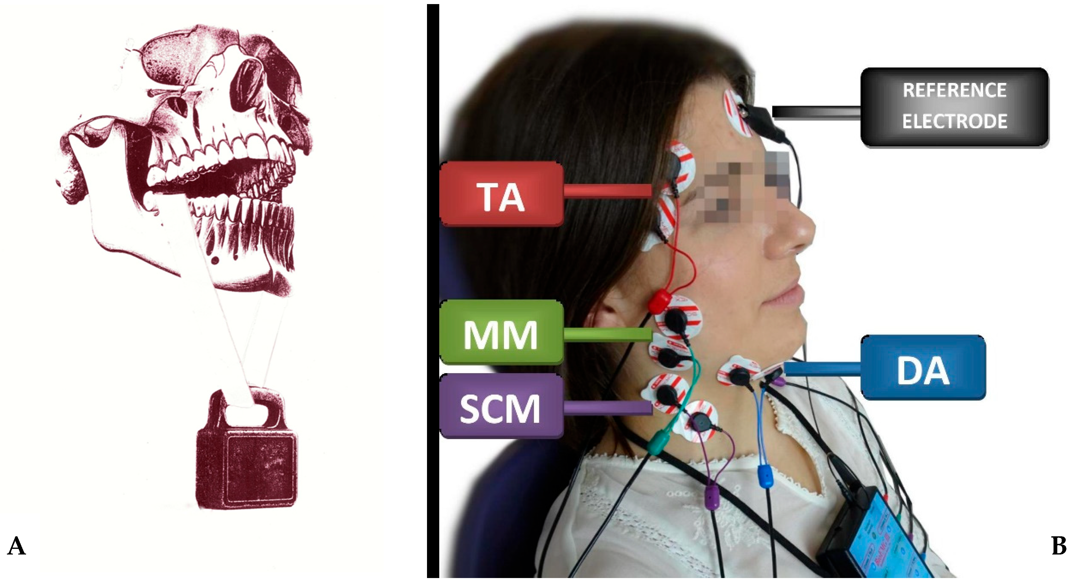
Figure 3. The difference of 340 years of research on masticatory muscles between the discoveries of Giovanni Alfonso Borelli and modern sEMG studies (8-channel BioEMG III electromyography, BioResearch Associates, Inc., Milwaukee, WI, USA). (A) shows the assumptions of the 1681 Giovanni Alfonso Borelli study and (B) the 2021 study. TA—the temporalis anterior muscle; MM—the superficial part of the masseter muscle; DA—the anterior belly of the digastric muscle; SCM—the middle part of the sternocleidomastoid muscle. The elements for graphic A were generated on 2.01.2024 using the DALL-E 2 program (OpenAI, Inc., San Francisco, CA, USA), and merged by the author G.Z.
References
- Konrad, P. The Abc of Emg—A Practical Introduction to Kinesiological Electromyography; Noraxon Inc.: Scottsdale, AZ, USA, 2005; Volume 1.
- Raez, M.B.I.; Hussain, M.S.; Mohd-Yasin, F. Techniques of EMG Signal Analysis: Detection, Processing, Classification and Applications. Biol. Proced. Online 2006, 8, 11.
- del Olmo, M.; Domingo, R. EMG Characterization and Processing in Production Engineering. Materials 2020, 13, 5815.
- Chrysafides, S.M.; Bordes, S.J.; Sharma, S. Physiology, Resting Potential. In StatPearls; StatPearls Publishing: Treasure Island, FL, USA, 2023.
- Brull, S.J.; Silverman, D.G.; Naguib, M. Chapter 15—Monitoring Neuromuscular Blockade. In Anesthesia Equipment, 2nd ed.; Ehrenwerth, J., Eisenkraft, J.B., Berry, J.M., Eds.; W.B. Saunders: Philadelphia, PA, USA, 2013; pp. 307–327. ISBN 978-0-323-11237-6.
- Cobb, M. Exorcizing the Animal Spirits: Jan Swammerdam on Nerve Function. Nat. Rev. Neurosci. 2002, 3, 395–400.
- Clower, W.T. The Transition from Animal Spirits to Animal Electricity: A Neuroscience Paradigm Shift. J. Hist. Neurosci. 1998, 7, 201–218.
- Steele, F.R. Body of Evidence Supports New Anatomical Finding. Nat. Med. 1996, 2, 506.
- Hoerni, B. Jan Swammerdam, physician and naturalist of the 17th century. Hist. Sci. Medicales 2015, 49, 75–80.
- Drost, G. High-Density Surface EMG. Pathophysiological Insights and Clinical Applications; Radboud University: Nijmegen, The Netherlands, 2007; ISBN 978-90-90-21609-6.
- Swammerdam, J. Tractatus Physico-Enatomico-Medicus de Respiratione Usuque Pulmonum; Gaasbeeck: Leiden, The Netherlands, 1667.
- Brekhus, P.J.; Armstrong, W.D.; Simon, W.J. Stimulation of the Muscles of Mastication. J. Dent. Res. 1941, 20, 87–92.
- Brawley, R.E.; Sedwick, H.J. Gnathodynamometer. Am. J. Orthod. Oral Surg. 1938, 24, 256–258.
- Konstantinova, D.; Dimova, M. Historical Review Of Gnathodynamometric Methods Used For The Assessment Of Masticatory Function. J IMAB 2016, 22, 1226–1229.
- Kazamel, M.; Warren, P.P. History of Electromyography and Nerve Conduction Studies: A Tribute to the Founding Fathers. J. Clin. Neurosci. 2017, 43, 54–60.
- van Musschenbroek, P. Quoted in JA Nollet. In Memoires of the French Academy of Sciences; JA Nollet: Paris, France, 1746.
- Zitzewitz, P.W. The Handy Physics Answer Book; Visible Ink Press: Metro Detroit, MI, USA, 2011; ISBN 978-1-57859-357-6.
- Bard, A.J.; Inzelt, G.; Scholz, F. Electrochemical Dictionary; Springer Science & Business Media: Berlin/Heidelberg, Germany, 2012; ISBN 978-3-642-29550-8.
- Tsadok, S. The Historical Evolution of Bioimpedance. AACN Clin. Issues 1999, 10, 371–384.
- Walsh, J.; Seignette, S. Of the Electric Property of the Torpedo. In a Letter from John Walsh, Esq; F. R. S. to Benjamin Franklin, Esq; LL.D., F.R.S., Ac. R. Par. Soc. Ext., &c. Philos. Trans. 1683–1775 1773, 63, 461–480.
- Edwards, P. The Shocking Discovery of Electric Fish: 250th Anniversary; University of Canberra: Canberra, Australia, 2022.
- Piccolino, M.; Bresadola, M. Drawing a Spark from Darkness: John Walsh and Electric Fish. Trends Neurosci. 2002, 25, 51–57.
- Galvani, L. De Viribus Electricitatis in Motu Musculari Commentarius. Bononiensi Sci. Artium Inst. Acad. Comment. 1791, 7, 363–418.
- AIM25 Text-Only Browsing: Royal College of Surgeons of England: Aldini, Giovanni: Notebook. Available online: https://web.archive.org/web/20160303220239/http://www.aim25.ac.uk/cats/9/10190.htm (accessed on 28 December 2023).
- Nobili, L. Bibliothèque Universelle Des Sciences, Belles-Lettres et Arts; Forgotten Books: London, UK, 1825; Volume 29.
- Gallone, P. Galvani’s Frog: Harbinger of a New Era. Electrochimica Acta 1986, 31, 1485–1490.
- Moruzzi, G. The Electrophysiological Work of Carlo Matteucci. Brain Res. Bull. 1996, 40, 69–91.
- du Bois-Reymond, E. Untersuchungen Über Thierische Elektricität. Ann. Phys. 1848, 151, 463–464.
- von Helmholtz, H. Vorläufiger Bericht über die Fortpflanzungs-Geschwindigkeit der Nervenreizung. Arch. Für Anat. Physiol. Wiss. Med. 1850, 71–73. Available online: https://echo.mpiwg-berlin.mpg.de/ECHOdocuView?url=/permanent/vlp/lit29168/index.meta (accessed on 28 December 2023).
- von Helmholtz, H. Messungen über den zeitlichen Verlauf der Zuckung animalischer Muskeln und die Fortpflanzungsgeschwindigkeit der Reizung in den Nerven. Arch. Für Anat. Physiol. Wiss. Med. 1850, 276–364.
- Erb, W.H. Handbuch der Elektrotherapie; F.C.W. Vogel: Hamburg, Germany, 1886.
- Duchenne, G. Mécanisme de la Physionomie Humaine, ou Analyse Électro-Physiologique de L’expression des Passions; Ve Jules Renouard, Libraire: Paris, France, 1862.
- Bernstein, J. Ukr Den Zeitlichen Verlauf Der Negative” Schwdung Des Nensvoms. Arch. Ges. Physiol. 1868, 1, 173–207.
- Pecolt, S.; Blazejewski, A.; Królikowski, T.; Młyński, B. Conversion of Bioelectric sEMG Signals into Analog Form for the BLDC Motors Control. Procedia Comput. Sci. 2022, 207, 3840–3849.
- Black, G. An Investigation of the Physical Characters of the Human Teeth in Relation to their Diseases, and to Practical Dental Operations, together with the Physical Characters of Filling-Materials. Dental Cosmos. 1895, 37, 469–484.
- Ortuğ, G. A New Device for Measuring Mastication Force (Gnathodynamometer). Ann. Anat.-Anat. Anz. 2002, 184, 393–396.
- Czubalski, F. Napoleon Cybulski (1854–1919) w Stuletnią Rocznicę Urodzin. Acta Physiol. Pol. 1953, 5, 3–13.
- Piper, H. Elektrophysiologie Menschlicher Muskeln; Springer: Berlin/Heidelberg, Germany, 1912; ISBN 3-642-50634-8.
- Bernstein, J. Elektrobiologie: Die Lehre von den Elektrischen Vorgängen im Organismus auf Moderner Grundlage Dargestellt; Vieweg + Teubner Verlag: Wiesbaden, Germany, 1912; ISBN 978-3-663-01628-1.
- Seyfarth, E.-A. Julius Bernstein (1839–1917): Pioneer Neurobiologist and Biophysicist. Biol. Cybern. 2006, 94, 2–8.
- Beck, A. Prof. Napoleon Cybulski Wspomnienia Pośmiertne i Ocena Działalności Naukowej; Odbitka z Gazety Lekarskiej: Warszawa, Poland, 1919; pp. 22–23.
- Rola, R. Badania neurograficzne i elektromiograficzne w praktyce klinicznej. Neurol. Po Dyplomie 2012, 7, 7–18.
- Trzmiel, G.; Kurz, D.; Smoczyński, W. Mikroprocesorowy Układ Sterowania Ramieniem Robota Sygnałem Elektromiograficznym. Pozn. Univ. Technol. Acad. J. Electr. Eng. 2019, 99, 179–190.
- Pratt, F.H. The All-or-None Principle in Graded Response of Skeletal Muscle. Am. J. Physiol.-Leg. Content 1917, 44, 517–542.
- McAdams, E.; Jossinet, J. Tissue Impedance: A Historical Overview. Physiol. Meas. 1995, 16, A1–A13.
- Philippson, M. Les Lois de La Résistance Électrique Des Tissus Vivants. Bull Acad. Roy Belg. 1921, 7, 387–403.
- Kułak, W.; Sobaniec, W. Historia odkrycia EEG. Neurol. Dziecięca 2006, 15, 53–56.
- Fricke, H. A Mathematical Treatment of the Electric Conductivity and Capacity of Disperse Systems I. The Electric Conductivity of a Suspension of Homogeneous Spheroids. Phys. Rev. 1924, 24, 575–587.
- Liddell, E.G.T.; Sherrington, C.S. Recruitment and Some Other Features of Reflex Inhibition. Proc. R. Soc. Lond. Ser. B Contain. Pap. Biol. Character 1925, 97, 488–518.
- Fricke, H.; Morse, S. The Electric Resistance and Capacity of Blood for Frequencies between 800 and 4(1/2) Million Cycles. J. Gen. Physiol. 1925, 9, 153–167.
- Adrian, E.; Bronk, D. The discharge of impulses in motor nerve fibres. J. Physiol. 1929, 67, 81–101.
- Erlanger, J.; Gasser, H.S. Electrical Signs of Nervous Activity; Electrical signs of nervous activity; University Pennsylvania Press: Oxford, UK, 1937; p. 221.
- Jacobson, E. Progressive Relaxation, 2nd ed.; Progressive relaxation; University Chicago Press: Oxford, UK, 1938; p. 494.
- Mayer, R.F. The Motor Unit and Electromyography—The Legacy of Derek Denny-Brown. J. Neurol. Sci. 2001, 189, 7–11.
- Cram, J.R. The History of Surface Electromyography. Appl. Psychophysiol. Biofeedback 2003, 28, 81–91.
- Cole, K.S.; Cole, R.H. Dispersion and Absorption in Dielectrics I. Alternating Current Characteristics. J. Chem. Phys. 2004, 9, 341–351.
- Weddell, G. Electromyography in Clinical Medicine. Proc. R. Soc. Med. 1943, 36, 513–514.
- Inman, V.T.; Saunders, J.B.; Abbott, L.C. Observations of the Function of the Shoulder Joint. 1944. Clin. Orthop. 1996, 330, 3–12.
- The Nobel Prize in Physiology or Medicine 1944. Available online: https://www.nobelprize.org/prizes/medicine/1944/summary/ (accessed on 28 December 2023).
- Mayo Clinic Alumni Association. Neuro then and No. Alumni Mayo Clin. 2018, 4, 1–47.
- The History of Dantec Dynamics. Dantec Dyn. Precis. Meas. Syst. Sens. Available online: https://www.dantecdynamics.com/about/history/ (accessed on 7 January 2024).
- Price, J.P.; Clare, M.H.; Ewerhardt, F.H. Studies in Low Backache with Persistent Muscle Spasm. Arch. Phys. Med. Rehabil. 1948, 29, 703–709.
- Moyers, R.E. Temporomandibular Muscle Contraction Patterns in Angle Class II, Division 1 Malocclusions; an Electromyographic Analysis. Am. J. Orthod. 1949, 35, 837–857.
- Vig, P. Electromyography in Dental Science: A Review. Aust. Dent. J. 1963, 8, 315–322.
- Ladegaard, J. Story of Electromyography Equipment. Muscle Nerve. Suppl. 2002, 11, S128–S133.
- Hodgkin, A.L.; Huxley, A.F. A Quantitative Description of Membrane Current and Its Application to Conduction and Excitation in Nerve. J. Physiol. 1952, 117, 500–544.
- Hodgkin, A.L.; Huxley, A.F. Currents Carried by Sodium and Potassium Ions through the Membrane of the Giant Axon of Loligo. J. Physiol. 1952, 116, 449–472.
- Lippold, O.C.J. The Relation between Integrated Action Potentials in a Human Muscle and Its Isometric Tension. J. Physiol. 1952, 117, 492–499.
- History. Available online: https://www.aanem.org/header-utility-items/about-aanem/history (accessed on 30 December 2023).
- Buchthal, F.; Pinell, P.; Rosenfalck, P. Action Potential Parameters in Normal Human Muscle and Their Physiological Determinants. Acta Physiol. Scand. 1954, 32, 219–229.
- Bigland, B.; Lippold, O.C.J. The Relation between Force, Velocity and Integrated Electrical Activity in Human Muscles. J. Physiol. 1954, 123, 214–224.
- Greenfield, B.; Wyke, B. Electromyographic Studies of Come of the Muscles of Mastication. Brit. J. 1956, 100, 129–143.
- Jarabak, J.R. An Electromyographic Analysis of Muscular And Temporomandibular Joint Disturbances Due to Imbalances in Occlusion. Angle Orthod. 1956, 26, 170–190.
- Basmajian, J.V.; Stecko, G. A New Bipolar Electrode for Electromyography. J. Appl. Physiol. 1962, 17, 849.
- Basmajian, J.V. Control and Training of Individual Motor Units. Science 1963, 141, 440–441.
- Birth of ISEK. Int. Soc. Electrophysiol. Kinesiol. ISEK. Available online: https://isek.org/announcement/birth-of-isek/ (accessed on 7 January 2024).
- Henneman, E.; Somjen, G.; Carpenter, D.O. Functional Significance of Cell Size in Spinal Motoneurons. J. Neurophysiol. 1965, 28, 560–580.
- Moller, E. The Chewing Apparatus. An Electromyographic Study of the Action of the Muscles of Mastication and Its Correlation to Facial Morphology. Acta Physiol. Scand. Suppl. 1966, 280, 1–229.
- Schwan, H.P. Electrode Polarization Impedance and Measurements in Biological Materials*. Ann. N. Y. Acad. Sci. 1968, 148, 191–209.
- Yemm, R. Variations in the Electrical Activity of the Human Masseter Muscle Occurring in Association with Emotional Stress. Arch. Oral Biol. 1969, 14, 873–878.
- Bernstein, J.M.; Mohl, N.D.; Spiller, H. Temporomandibular Joint Dysfunction Masquerading as Disease of Ear, Nose, and Throat. Trans. Am. Acad. Ophthalmol. Otolaryngol. Am. Acad. Ophthalmol. Otolaryngol. 1969, 73, 1208–1217.
- Moller, E. Clinical Electromyography in Dentistry. Int. Dent. J. 1969, 19, 250–266.
- Griffin, C.J.; Munro, R.R. Electromyography of the Masseter and Anterior Temporalis Muscles in Patients with Temporomandibular Dysfunction. Arch. Oral Biol. 1971, 16, 929–949.
- Milner-Brown, H.S.; Stein, R.B. The Relation between the Surface Electromyogram and Muscular Force. J. Physiol. 1975, 246, 549–569.
- Pruim, G.J.; Ten Bosch, J.J.; de Jongh, H.J. Jaw Muscle EMG-Activity and Static Loading of the Mandible. J. Biomech. 1978, 11, 389–395.
- ICCMO. Mission and History. Available online: https://iccmo.org/who-we-are (accessed on 3 January 2024).
- Bakke, M.; Møller, E. Distortion of Maximal Elevator Activity by Unilateral Premature Tooth Contact. Scand. J. Dent. Res. 1980, 88, 67–75.
- Yardin, M. Effect of head posture on muscle tonus of the masticatory muscles. J. Biol. Buccale 1981, 9, 99–107.
- Stålberg, E. Computer Aided EMG Analysis in Clinical Routine. Electroencephalogr. Clin. Neurophysiol. 1985, 61, S223.
- Riise, C.; Sheikholeslam, A. The Influence of Experimental Interfering Occlusal Contacts on the Postural Activity of the Anterior Temporal and Masseter Muscles in Young Adults. J. Oral Rehabil. 1982, 9, 419–425.
- Widmalm, S.E.; Ericsson, S.G. The Influence of Eye Closure on Muscle Activity in the Anterior Temporal Region. J. Oral Rehabil. 1983, 10, 25–29.
- Jankelson, R.; Pulley, M. Electromyography in Clinical Dentistry; Myo-Tronics Research: Seattle, DC, USA, 1984.
- BIOPAC Syst. Inc. About | U.S. | International | Contact | Jobs | Policies. Available online: https://www.biopac.com/corporate/about-biopac/ (accessed on 29 December 2023).
- Sheikholeslam, A.; Holmgren, K.; Riise, C. A Clinical and Electromyographic Study of the Long-Term Effects of an Occlusal Splint on the Temporal and Masseter Muscles in Patients with Functional Disorders and Nocturnal Bruxism. J. Oral Rehabil. 1986, 13, 137–145.
- Naeije, M.; McCarroll, R.S.; Weijs, W.A. Electromyographic Activity of the Human Masticatory Muscles during Submaximal Clenching in the Inter-Cuspal Position. J. Oral Rehabil. 1989, 16, 63–70.
- Home. Available online: https://www.noraxon.com (accessed on 29 December 2023).
- Cram, J. Clinical EMG for Surface Recordings, Volume 2; Clinical Resources: Atlanta, GA, USA, 1990.
- Ferrario, V.F.; Sforza, C.; D’Addona, A.; Miani, A. Reproducibility of Electromyographic Measures: A Statistical Analysis. J. Oral Rehabil. 1991, 18, 513–521.
- Criswell, E.; Cram, J.R. (Eds.) Cram’s Introduction to Surface Electromyography, 2nd ed.; Jones and Bartlett: Sudbury, MA, USA, 2011; ISBN 978-0-7637-3274-5.
More
Information
Subjects:
Dentistry, Oral Surgery & Medicine
Contributors
MDPI registered users' name will be linked to their SciProfiles pages. To register with us, please refer to https://encyclopedia.pub/register
:
View Times:
401
Revisions:
3 times
(View History)
Update Date:
06 Mar 2024
Table of Contents


Notice
You are not a member of the advisory board for this topic. If you want to update advisory board member profile, please contact office@encyclopedia.pub.
OK
Confirm
Only members of the Encyclopedia advisory board for this topic are allowed to note entries. Would you like to become an advisory board member of the Encyclopedia?
Yes
No
${ textCharacter }/${ maxCharacter }
Submit
Cancel
Back
Comments
${ item }
|
${ item.createdUser.fullName }
${ item.createdAt }
${ item.vote }
${ item.reply }
Delete
${ reply.createdUser.fullName }
${ reply.createdAt }
${ reply.vote }
Delete
There is no reply to this comment~
${ item.replyTextCharacter }/${ item.replyMaxCharacter }
Submit
Cancel
More
No more~
There is no comment~
${ textCharacter }/${ maxCharacter }
Submit
Cancel
${ selectedItem.replyTextCharacter }/${ selectedItem.replyMaxCharacter }
Submit
Cancel
Confirm
Are you sure to Delete?
Yes
No