Submitted Successfully!
To reward your contribution, here is a gift for you: A free trial for our video production service.
Thank you for your contribution! You can also upload a video entry or images related to this topic.
Version Summary Created by Modification Content Size Created at Operation
1 -- 2872 2024-03-04 14:27:49 |
2 Reference format revised. Meta information modification 2872 2024-03-05 01:58:28 |

Video Upload Options

Do you have a full video?

Confirm

Are you sure to Delete?
Cite
If you have any further questions, please contact Encyclopedia Editorial Office.
Terriaca, S.; Ferlosio, A.; Scioli, M.G.; Coppa, F.; Bertoldo, F.; Pisano, C.; Belmonte, B.; Balistreri, C.R.; Orlandi, A. miRNA Regulation in Atherosclerotic and Non-Atherosclerotic Aortic Aneurysms. Encyclopedia. Available online: https://encyclopedia.pub/entry/55831 (accessed on 09 May 2024).
Terriaca S, Ferlosio A, Scioli MG, Coppa F, Bertoldo F, Pisano C, et al. miRNA Regulation in Atherosclerotic and Non-Atherosclerotic Aortic Aneurysms. Encyclopedia. Available at: https://encyclopedia.pub/entry/55831. Accessed May 09, 2024.
Terriaca, Sonia, Amedeo Ferlosio, Maria Giovanna Scioli, Francesca Coppa, Fabio Bertoldo, Calogera Pisano, Beatrice Belmonte, Carmela Rita Balistreri, Augusto Orlandi. "miRNA Regulation in Atherosclerotic and Non-Atherosclerotic Aortic Aneurysms" Encyclopedia, https://encyclopedia.pub/entry/55831 (accessed May 09, 2024).
Terriaca, S., Ferlosio, A., Scioli, M.G., Coppa, F., Bertoldo, F., Pisano, C., Belmonte, B., Balistreri, C.R., & Orlandi, A. (2024, March 04). miRNA Regulation in Atherosclerotic and Non-Atherosclerotic Aortic Aneurysms. In Encyclopedia. https://encyclopedia.pub/entry/55831
Terriaca, Sonia, et al. "miRNA Regulation in Atherosclerotic and Non-Atherosclerotic Aortic Aneurysms." Encyclopedia. Web. 04 March, 2024.
miRNA Regulation in Atherosclerotic and Non-Atherosclerotic Aortic Aneurysms
Edit

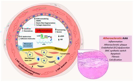
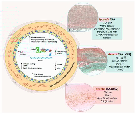
Aortic aneurysms are a serious health concern as their rupture leads to high morbidity and mortality. Abdominal aortic aneurysms (AAAs) and thoracic aortic aneurysms (TAAs) exhibit differences and similarities in their pathophysiological and pathogenetic features. AAA is a multifactorial disease, mainly associated with atherosclerosis, characterized by a relevant inflammatory response and calcification. TAA is rarely associated with atherosclerosis and in some cases is associated with genetic mutations such as Marfan syndrome (MFS) and bicuspid aortic valve (BAV). MFS-related and non-genetic or sporadic TAA share aortic degeneration with endothelial-to-mesenchymal transition (End-Mt) and fibrosis, whereas in BAV TAA, aortic degeneration with calcification prevails. microRNA (miRNAs) contribute to the regulation of aneurysmatic aortic remodeling. miRNAs are a class of non-coding RNAs, which post-transcriptionally regulate gene expression. 

abdominal aortic aneurysm thoracic aortic aneurysm atherosclerosis

1. Introduction

The aorta is the largest arterial blood vessel in the body. It is subjected to high pressure when a large volume of blood is pumped out of the heart with each contraction, exposing it to the risk of wall degeneration and aneurysms [1]. The latter are categorized in two main types: thoracic aortic aneurysms (TAAs) and abdominal aortic aneurysms (AAAs) [2]. Aortic aneurysms are often asymptomatic until the aortic media undergoes dissection or rupture, with a high degree of morbidity and mortality [3]. Currently, the available pharmacological treatments for aortic aneurysms are not specific and aim to delay disease progression and surgical intervention [3]. TAAs and AAAs have different developmental origins as well as pathogenetic factors that induce different structural degeneration of the aortic wall [3]. Atherosclerosis and chronic inflammation are associated with AAAs [3], whereas TAAs are characterized by an increased accumulation of proteoglycans [4]. The histopathological abnormality of TAAs is named cystic medial degeneration, characterized by loss of smooth muscle cells (SMCs) and elastic fiber degeneration that leads to a weakened wall and the consequent high risk of dilatation and aneurysm formation [4][5]. Cystic medial degeneration occurs normally with aging, in particular in the presence of hypertension [3]. As mentioned above, AAAs are often associated with atherosclerosis, but as for TAAs, AAAs show SMC loss and fragmentation of elastic fibers [4]. Smoking, male sex, advanced age and atherosclerosis are mostly associated with AAAs and some sporadic (non-genetic) TAAs [1]. In fact, the majority of sporadic TAAs are mainly associated with hypertension and aging. However, there are some TAAs associated with genetic conditions, such as Marfan syndrome (MFS) and bicuspid aortic valve (BAV) [6]. All genetic TAAs occur at an early age but their underlying pathogenetic mechanisms can be very different. In fact, MFS TAAs display fibrosis [7], whereas BAV TAAs show mainly calcification [8].

2. Vascular Remodeling in Abdominal Aortic Aneurysms

As mentioned above, AAAs are often associated with atherosclerosis, and some evidence suggests that their formation starts from occlusive disease [9]. In particular, the formation of atheroma in the abdominal aorta starts in the subendothelial intimal space of medium- and large-sized arteries, through a multistep process. First, the inflammatory stimulus induces endothelial dysfunction and permeabilization with the consequent formation of a fatty streak. The latter evolves into an atheromatous or fibroatheromatous plaque that can rupture and provoke thrombosis [10]. During this process, endothelial cells increase the expression of vascular cell adhesion molecule-1 and intercellular adhesion molecule-1 and reduce nitric oxide synthesis [10]. The increased expression of those molecules leads to a recruitment of monocytes to the site of injury. Macrophages and T lymphocytes, in turn, express growth factors, cytokines and matrix metalloproteinases (MMPs) that stimulate the migration and proliferation of aortic SMCs into the tunica intima and the formation of atherosclerotic plaque with the accumulation of foam cells. [10]. With time, SMCs and inflammatory cells undergo apoptosis, contributing to the development of atherosclerotic lesions [11][12][13]. Crystallized calcium concentrates around the apoptotic bodies derived from dead SMCs, leading to aortic micro-calcification [3].

3. Phenotypic Alterations and Vascular Remodeling in Thoracic Aortic Aneurysms

In contrast to AAAs, TAAs are rarely associated with atherosclerosis and are divided into two main categories: sporadic (non-genetic) and genetic TAAs [14]. Sporadic TAAs may occur with aging, and their progression is accelerated by hypertension [14]. In young patients, TAA formation is associated with genetic conditions such as MFS and BAV.
Similarly to AAAs, TAAs are characterized by an extensive remodeling of the ECM [15]. Endothelial dysfunction is also present in TAAs, but the formation of fibro-atherosclerotic plaque is rarely evidenced [16]. In addition, endothelial cells can undergo endothelial-to-mesenchymal transition (End-MT), a process in which cells switch from an endothelial to a mesenchymal phenotype, losing cell-to-cell contact and cell polarity [17]. The main pathway responsible for the End-MT is the canonical Transforming Growth Factor β (TGF-β) signaling in association with NOTCH and Wnt/β catenin signaling [8][17][18]. End-Mt has been observed in sporadic TAAs but appears to be more accentuated in MFS TAAs [7]. At the same time, SMCs in the tunica media can dedifferentiate in myofibroblasts, switching from a contractile phenotype to a proliferative/synthetic one. Myofibroblasts deposit alcianophilic material like Glycosaminoglycans (GAGs) and collagen, determining fibrosis [19]. This process is well evidenced in sporadic TAAs and strongly exacerbated in MFS TAAs [7]. SMCs can also acquire an osteoblast-like phenotype, promoting calcification, especially in BAV TAAs [8][20]. Those alterations are associated with the deregulation of NOTCH and BMP signaling [21][22].

4. miRNAs’ Biogenesis and Their Role in Aortic Remodeling

In the last few decades, many studies have demonstrated that about 97% of the human genome consists of non-coding sequences that are not transcribed in mRNAs but in non-coding RNAs [23]. Non-coding RNAs regulate gene expression at the post-transcriptional level [24]. miRNAs are a class of non-coding RNAs, which act as epigenetic regulators without affecting the chromatin architecture [25]. In the human genome, miRNAs are encoded by introns of "host genes" and also enriched within unique miRNA clusters [25]. miRNA biogenesis starts with RNA polymerases II/III, which transcribe a primary miRNA (pri-miRNA) molecule from the intergenic or intragenic region [24]. Successively, pri-miRNAs are processed by a multi-component microprocessor complex (Drosha/DGCR8) to precursor miRNAs (pre-miRNAs) with specific hairpin structures [24]. Pre-miRNAs are exported from the nucleus to the cytoplasm via the transport factor exportin-5 using GTP as a cofactor [24]. Within the cytoplasm, pre-miRNAs are released from exportin-5 following GTP hydrolysis and are further processed by an RNase III, called “Dicer”, generating double-stranded miRNAs of approximately 22 nucleotides [24]. This product is incorporated into the RNA-induced silencing complex multiprotein complex (RISC) in which a helicase ensures that only one of the miRNA duplex strands remains in the complex to control the post-transcriptional expression of target genes [26]. miRNAs are known as negative regulators of gene expression [26]. Precisely, miRNAs induce the cleavage and degradation of target mRNAs or the inhibition of the translation process [26]. miRNAs have been demonstrated to play a critical role in several physiological processes, such as cell proliferation, apoptosis, fat metabolism, neuronal development, cell differentiation, hormone secretion and the development of multiple diseases [27]. In the last few years, many studies have reported an aberrant miRNA expression in different cardiovascular diseases such as atherosclerosis, cardiac remodeling, myocardial infarction and aneurysms [28].

5. The Regulatory Role of miRNAs in AAAs

Different studies have reported that inflammation, endothelial dysfunction, ECM remodeling and SMC proliferation/apoptosis, which characterize the AAA aortic wall, are associated with specific miRNA deregulations [29]. ECM proteins such as collagen and elastin are mainly produced by SMCs, and the apoptosis of those cells has been demonstrated to be implicated in the development of AAA [30]. It has been reported that SMAD3, a key intracellular mediator of the fibrotic process [31], is down-regulated in AAA [32][33]. It was also found that miR-195, an important regulator of ECM proteins, was up-regulated in AAA patients [34]. Bioinformatics analysis revealed that miR-195 has a binding site in the 3′-UTR for SMAD3, and their interaction was confirmed by in vitro studies [35]. miR-195 overexpression in vitro inhibited SMC proliferation, inducing apoptosis, whereas SMAD3 overexpression blocked those effects [35]. Therefore, miR-195 displays a crucial role in AAA pathogenesis and represents a potential therapeutic target [35].
As mentioned above, AAA is characterized by an increased degree of calcification, which can lead to rupture [36]. During AAA progression, SMCs switch from the contractile to the synthetic phenotype and synthetize osteogenic factors, such as the osteogenic transcription factor Runt-related gene (RUNX). The latter is functionally associated with SMAD2/3, which, in turn, are activated by angiotensin II (AngII) signaling [37]. The activation of the SMAD-RUNX2 signaling pathway induces osteogenic differentiation of SMCs [37]. It has been demonstrated that miR-424 has a key role in cardiovascular pathologies. miR-424 is also called “osteomir” as it regulates vascular calcification [38]. Bioinformatics analysis revealed that AAA patients express high levels of RUNX2 and exhibit down-regulation of miR-424 compared with control subjects [37]
Another miRNA, reported to be involved in the chronic inflammation that characterizes the AAA aortic wall, is miR-33-5p [39]. Some studies reported that this miRNA regulates the innate immune response by the adenosine triphosphate-binding cassette transporter A1 (ABCA1) [40][41]. ABCA1 is a protein that transports free cholesterol and phospholipids from intracellular compartments to the cell membrane by using ATP as a source of energy, so this protein is fundamental in macrophage cholesterol efflux and reverse cholesterol transport [42][43]. It has been reported that ABCA1 acts as an anti-inflammatory receptor, and the enhancement of its function could be a beneficial therapeutic approach [44]
Another study correlated miR-21 expression to inflammatory response and aortic remodeling in AAA. Precisely, Yu et al. investigated the effects of dexmedetomidine (Dex) on miR-21 expression [45]. It has been reported that Dex suppresses the activities of inflammatory mediators and maintains a balanced myocardial function and coronary blood flow [46]. miR-21 is reported to control the inflammatory responses, ECM remodeling and lipid accumulation in cerebral aneurysms [47][48]. Moreover, it has been demonstrated that miR-21 inhibition blocks AAA development. Programmed cell death 4 (PDCD4), an inflammation- and apoptosis-related gene, has been proven to be a gene target of miR-21 [49][50]. In order to evaluate the effects of Dex on miR-21 expression and consequently on AAA progression, rat models of AAA were injected with Dex. The authors discovered that Dex administration in AAA rat models down-regulated the expression of inflammatory factors and MMPs as well as up-regulating miR-21 expression [45]

6. The Regulatory Role of miRNAs in Sporadic TAAs

As previously reported, TAA formation is associated with progressive pathological remodeling of the aortic wall that leads to structural parietal degeneration, with rearrangement of hemodynamic loads, and finally rupture [51][52]. During this remodeling, both endothelial cells and SMCs undergo phenotypic changes in response to pathogenetic stimuli [16][53][54]. Recently, miRNA deregulation has been reported to be associated with vascular cell phenotypical changes. The phenotypic switching of SMCs from a contractile to a synthetic phenotype has been suggested to be involved in the development of aortic aneurysm and its dissection [53][54]. As already mentioned, aortic SMCs of sporadic TAA generally switch into a myofibroblast phenotype with the increased collagen synthesis and consequent aortic fibrosis [55][56]. During this process, SMCs reduced the expression of functional markers such as smooth muscle 22 α (SM22α), smooth muscle cell-specific myosin heavy chain (MYH11) and α-smooth muscle actin (α-SMA) [53]. However, the molecular mechanisms underlying the SMC phenotypic switch is not completely understood. The analysis of aortic tissues derived from patients with severe TAA evidenced a strong miR-335-5p down-regulation and Specificity Protein 1 (SP1) up-regulation; the latter is involved in SMC proliferation and phenotype switching [57]. However, cultured human SMCs overexpressing miR-335-5p showed SP1 down-regulation with reduced proliferation and migration as well as increased expression of contractile markers, such as SM22α, α-SMA and CNN1 [57]. In addition, the authors documented SP1 as a gene target of miR-335-5p. In a mouse model of aortic dissection, the administration of miR-335-5p clearly suppressed aorta dilatation and vascular media degeneration [57]. Aberrant down-regulation of miR-134-5p has been evidenced in aortic tissues derived from sporadic TAA patients [58]. In vitro studies, on aortic SMCs, revealed that miR-134-5p overexpression promoted differentiation and expression of contractile markers, suggesting a crucial role of miRNA in aortic SMC homeostasis [58]. PDGF and TGFβ are involved in SMC differentiation, vascular remodeling and aortic aneurysms [59][60].
Endothelial cells play a fundamental role during pathological aortic aneurysmatic remodeling [16]. Several studies identified the aberrant expression of different miRNAs in TAA and their role in the regulation of endothelial cell phenotyping and functions [61]. The activation of endoplasmic reticulum stress (ERS) contributes to the pathogenesis of cardiovascular diseases, in particular endothelial dysfunction [62][63][64]. The mechanism through which ERS mediates vascular cell dysfunction is not completely understood. miRNAs regulate the ERS response by targeting specific genes [65]. In particular, miR-204 seems to be associated with ERS by targeting sirtuin1 lysine deacetylase (SIRT1) [66][67]. Kassan et al. investigated the role of miR-204 in ERS and endothelial dysfunction by targeting SIRT1 [68]. Overexpression of miR-204 in HUVECs induced ERS by up-regulation of specific stress markers, such as glucose-regulated protein, C/-EBP homologous protein and Activating Transcription Factor 6, as well as phosphorylation of PKR-like ER kinase and eukaryotic initiation factor 2. 

7. miRNA Regulation of Vascular Cell Phenotype in Genetic TAAs

As reported above, TAAs are, in some cases, associated with genetic mutations [69]. BAV (bicuspid aortic valve) is a congenital cardiovascular malformation leading to an increased risk for severe cardiovascular events, such as TAA [70][71][72]. The BAV condition is associated with different genetic mutations including the NOTCH1, TGFBR2, FBN1, SMAD6, GATA5 and GATA6 genes [70][71][72]. Several studies described the differential expression of miRNAs between BAV and TAV (tricuspid aortic valve) patients [73]. Some studies reported that BAV aortopathy is also characterized by a lower expression of End-Mt markers compared with sporadic TAAs [8][74]. However, some studies demonstrated that, before dilation, BAV aortas showed an activation of the End-Mt process [75]. Using a systemic biological approach, a strong association between the miR-200 family, which targets the End-Mt transcription factors zinc-finger E homeobox-binding transcription factors 1 and 2 (ZEB1 and ZEB2) [76][77], with the BAV signaling network was highlighted [75].
The differential expression of ERG and its transcription factor miR-126-5p has also been demonstrated, in the vascular phenotypic changes occurring in BAV and TAV aortas [8]. The expression levels of the protein ERG and miR-126-5p were up-regulated in TAA samples derived from BAV compared to TAV patients. Moreover, this up-regulation in BAV TAA was shown to be associated with a down-regulation of SMAD2/3 proteins [8], whose activation induces End-MT [78]. Therefore, the down-regulation of SMAD2/3 could explain the different phenotypic changes observed in the endothelium of BAV TAA. Similarly, the tunica media of BAV TAA showed an up-regulation of ERG and miR-126-5p in association with an evident aortic calcification compared with TAV tunica media; the latter were mainly characterized by a marked fibrosis [8].

8. Conclusions

Aortic aneurysms remain a serious health concern with many clinical complications as the associated ruptures can cause significant morbidity and mortality. The onset and development of AAA and TAA are associated with different risk factors. AAA is generally associated with atherosclerosis, hypertension and aging, whereas TAA frequently occurs in patients with genetic diseases. Both types of aortic aneurysms are characterized by pathological aortic remodeling. In that light, AAA shows a more marked inflammatory parietal response compared with TAA. miRNAs have been identified to be involved in the regulation of vascular cell phenotypic transformation, inflammation and SMC apoptosis. In AAA, miR-195 and miR-21 deregulation are associated with SMC apoptosis; miR-424 down-regulation is associated with calcification of the aortic wall; miR-33b and miR-33-5p deregulation is involved in the parietal inflammatory response. In TAA, miRNA deregulation induces a vascular cell phenotype switch. In sporadic TAA, miR-335-5p, miR-134, miR-155b-5p, miR-122-3p and miR-23b-5p down-regulation lead to a reduction in SMC cytoplasmic contractile cytoskeletal filaments with a switch into a synthetic phenotype promoting medial fibrosis. In TAA, endothelial cells characteristically lose physiological endothelial markers, with an End-Mt phenotypic switch and consequent endothelial dysfunction. For genetic TAA, MFS TAA shares histopathological features with sporadic TAA such as End-Mt and fibrosis, but in a more accentuated manner. Deregulation of specific miRNAs such as miR-29, miR-122 and miR-632 plays a key role in the regulation of the phenotypical cell changes and parietal remodeling observed in MFS TAA. Aortic remodeling in BAV TAA differs from sporadic and MFS TAAs, and miRNAs appear crucial in the regulation of aortic remodeling. Deregulation of miR-126, miR-200 and miR-423-5p seems to inhibit the End-Mt process and favor the calcification of the tunica media observed in BAV TAA. Schematic representations of the phenotypic alterations and aortic remodeling of AAA and TAA regulated by miRNAs are shown in Figure 1 and Figure 2.
Figure 1. Schematic representation of miRNA deregulation-related pathogenic mechanisms in atherosclerotic abdominal aortic aneurysms. Atherosclerotic process prevails in the progression of abdominal aortic aneurysm (AAA) and is characterized at least in part by miRNA deregulation-driven endothelial dysfunction, inflammation, smooth muscle cell (SMC) proliferation, migration and apoptosis, fibroatheromatous plaque formation and thrombosis, with medial fibrosis and calcification (insert, Hematoxylin and Eosin staining).
Figure 2. Schematic representation of miRNA deregulation-related pathogenic mechanisms in thoracic aortic aneurysms. Thoracic aortic aneurysms (TAAs) are sporadic or frequently related to genetic diseases. Sporadic and Marfan syndrome disease (MFS) TAAs show an aortic cell differentiative process, in which endothelial cells switch into a mesenchymal phenotype, whereas medial SMCs switch into a myofibroblastic phenotype and synthetize collagen and glycosaminoglycan with consequent aortic degeneration and fibrosis (inserts (a,b), Masson’s Trichrome Goldner staining). Those processes are precocious, much more marked and strongly accentuated in MFS TAAs, in which TGF-β signaling is hyperactivated. Genetic bicuspid aortic valve (BAV) TAA displays a specific degenerative process, in which SMCs dedifferentiate into osteoblast-like cells promoting medial calcification (insert (c), Alizarin Red staining) by miRNA deregulation-mediated activation of NOTCH and BMP signaling.

References

  1. Qian, G.; Adeyanju, O.; Olajuyin, A.; Guo, X. Abdominal Aortic Aneurysm Formation with a Focus on Vascular Smooth Muscle Cells. Life 2022, 12, 191.
  2. Quintana, R.A.; Taylor, W.R. Cellular Mechanisms of Aortic Aneurysm Formation. Circ. Res. 2019, 124, 607–618.
  3. Guo, D.C.; Papke, C.L.; He, R.; Milewicz, D.M. Pathogenesis of thoracic and abdominal aortic aneurysms. Ann. N. Y. Acad. Sci. 2006, 1085, 339–352.
  4. Jauhiainen, S.; Kiema, M.; Hedman, M.; Laakkonen, J.P. Large Vessel Cell Heterogeneity and Plasticity: Focus in Aortic Aneurysms. Arterioscler. Thromb. Vasc. Biol. 2022, 42, 811–818.
  5. Romaniello, F.; Mazzaglia, D.; Pellegrino, A.; Grego, S.; Fiorito, R.; Ferlosio, A.; Chiariello, L.; Orlandi, A. Aortopathy in Marfan syndrome: An update. Cardiovasc. Pathol. 2014, 23, 261–266.
  6. Salmasi, M.Y.; Alwis, S.; Cyclewala, S.; Jarral, O.A.; Mohamed, H.; Mozalbat, D.; Nienaber, C.A.; Athanasiou, T.; Morris-Rosendah, D. The genetic basis of thoracic aortic disease: The future of aneurysm classification? Hell. J. Cardiol. 2023, 69, 41–50.
  7. Terriaca, S.; Scioli, M.G.; Pisano, C.; Ruvolo, G.; Ferlosio, A.; Orlandi, A. miR-632 Induces DNAJB6 Inhibition Stimulating Endothelial-to-Mesenchymal Transition and Fibrosis in Marfan Syndrome Aortopathy. Int. J. Mol. Sci. 2023, 24, 15133.
  8. Pisano, C.; Terriaca, S.; Scioli, M.G.; Nardi, P.; Altieri, C.; Orlandi, A.; Ruvolo, G.; Balistreri, C.R. The Endothelial Transcription Factor ERG Mediates a Differential Role in the Aneurysmatic Ascending Aorta with Bicuspid or Tricuspid Aorta Valve: A Preliminary Study. Int. J. Mol. Sci. 2022, 23, 10848.
  9. Koch, A.E.; Haines, G.K.; Rizzo, R.J.; Radosevich, J.A.; Pope, R.M.; Robinson, P.G.; Pearce, W.H. Human abdominal aortic aneurysms. Immunophenotypic analysis suggesting an immune-mediated response. Am. J. Pathol. 1990, 137, 1199–1213.
  10. Tabas, I.; García-Cardeña, G.; Owens, G.K. Recent insights into the cellular biology of atherosclerosis. J. Cell Biol. 2015, 209, 13–22.
  11. Shimizu, K.; Mitchell, R.N.; Libby, P. Inflammation and cellular immune responses in abdominal aortic aneurysms. Arterioscler. Thromb. Vasc. Biol. 2006, 26, 987–994.
  12. Rateri, D.L.; Howatt, D.A.; Moorleghen, J.J.; Charnigo, R.; Cassis, L.A.; Daugherty, A. Prolonged infusion of angiotensin II in apoE(−/−) mice promotes macrophage recruitment with continued expansion of abdominal aortic aneurysm. Am. J. Pathol. 2011, 179, 1542–1548.
  13. Ait-Oufella, H.; Wang, Y.; Herbin, O.; Bourcier, S.; Potteaux, S.; Joffre, J.; Loyer, X.; Ponnuswamy, P.; Esposito, B.; Dalloz, M.; et al. Natural regulatory T cells limit angiotensin II-induced aneurysm formation and rupture in mice. Arterioscler. Thromb. Vasc. Biol. 2013, 33, 2374–2379.
  14. Ince, H.; Nienaber, C.A. Etiology, pathogenesis and management of thoracic aortic aneurysm. Nat. Clin. Pract. Cardiovasc. Med. 2007, 4, 418–427.
  15. D’Amico, F.; Doldo, E.; Pisano, C.; Scioli, M.G.; Centofanti, F.; Proietti, G.; Falconi, M.; Sangiuolo, F.; Ferlosio, A.; Ruvolo, G.; et al. Specific miRNA and Gene Deregulation Characterize the Increased Angiogenic Remodeling of Thoracic Aneurysmatic Aortopathy in Marfan Syndrome. Int. J. Mol. Sci. 2020, 21, 6886.
  16. Verstraeten, A.; Fedoryshchenko, I.; Loeys, B. The emerging role of endothelial cells in the pathogenesis of thoracic aortic aneurysm and dissection. Eur. Heart J. 2023, 44, 1262–1264.
  17. Souilhol, C.; Harmsen, M.C.; Evans, P.C.; Krenning, G. Endothelial-mesenchymal transition in atherosclerosis. Cardiovasc. Res. 2018, 114, 565–577.
  18. Zhong, A.; Mirzaei, Z.; Simmons, C.A. The Roles of Matrix Stiffness and ß-Catenin Signaling in Endothelial-to-Mesenchymal Transition of Aortic Valve Endothelial Cells. Cardiovasc. Eng. Technol. 2018, 9, 158–167.
  19. Cao, H.; Wang, C.; Chen, X.; Hou, J.; Xiang, Z.; Shen, Y.; Han, X. Inhibition of Wnt/β-catenin signaling suppresses myofibroblast differentiation of lung resident mesenchymal stem cells and pulmonary fibrosis. Sci. Rep. 2018, 8, 13644.
  20. Ignatieva, E.; Kostina, D.; Irtyuga, O.; Uspensky, V.; Golovkin, A.; Gavriliuk, N.; Moiseeva, O.; Kostareva, A.; Malashicheva, A. Mechanisms of Smooth Muscle Cell Differentiation Are Distinctly Altered in Thoracic Aortic Aneurysms Associated with Bicuspid or Tricuspid Aortic Valves. Front. Physiol. 2017, 8, 536.
  21. Balistreri, C.R.; Crapanzano, F.; Schirone, L.; Allegra, A.; Pisano, C.; Ruvolo, G.; Forte, M.; Greco, E.; Cavarretta, E.; Marullo, G.M.; et al. Deregulation of Notch1 pathway and circulating endothelial progenitor cell (EPC) number in patients with bicuspid aortic valve with and without ascending aorta aneurysm. Sci. Rep. 2018, 8, 13834.
  22. Kazik, H.B.; Kandail, H.S.; La Disa, J.F., Jr.; Lincoln, J. Molecular and Mechanical Mechanisms of Calcification Pathology Induced by Bicuspid Aortic Valve Abnormalities. Front. Cardiovasc. Med. 2021, 8, 677977.
  23. Vartak, T.; Kumaresan, S.; Brennan, E. Decoding microRNA drivers in atherosclerosis. Biosci. Rep. 2022, 42, BSR20212355.
  24. Martínez-Micaelo, N.; Beltrán-Debón, R.; Baiges, I.; Faiges, M.; Alegret, J.M. Specific circulating microRNA signature of bicuspid aortic valve disease. J. Transl. Med. 2017, 15, 76.
  25. Rodriguez, A.; Griffiths-Jones, S.; Ashurst, J.L.; Bradley, A. Identification of mammalian microRNA host genes and transcription units. Genome Res. 2004, 14, 1902–1910.
  26. Hayder, H.; O’Brien, J.; Nadeem, U.; Peng, C. MicroRNAs: Crucial regulators of placental development. Reproduction 2018, 155, R259–R271.
  27. Wang, G.; Luo, Y.; Gao, X.; Liang, Y.; Yang, F.; Wu, J.; Fang, D.; Lou, M. MicroRNA regulation of phenotypic transformations in vascular smooth muscle: Relevance to vascular remodeling. Cell. Mol. Life Sci. 2023, 80, 144.
  28. Zhou, S.S.; Jin, J.P.; Wang, J.Q.; Zhang, Z.G.; Freedman, J.H.; Zheng, Y.; Cai, L. miRNAS in cardiovascular diseases: Potential biomarkers, therapeutic targets and challenges. Acta Pharmacol. Sin. 2018, 39, 1073–1084.
  29. Romaine, S.P.; Tomaszewski, M.; Condorelli, G.; Samani, N.J. MicroRNAs in cardiovascular disease: An introduction for clinicians. Heart 2015, 101, 921–928.
  30. Sorokin, V.; Vickneson, K.; Kofidis, T.; Woo, C.C.; Lin, X.Y.; Foo, R.; Shanahan, C.M. Role of Vascular Smooth Muscle Cell Plasticity and Interactions in Vessel Wall Inflammation. Front. Immunol. 2020, 11, 599415.
  31. Dai, X.; Shen, J.; Annam, N.P.; Jiang, H.; Levi, E.; Schworer, C.M.; Tromp, G.; Arora, A.; Higgins, M.; Wang, X.F.; et al. SMAD3 deficiency promotes vessel wall remodeling, collagen fiber reorganization and leukocyte infiltration in an inflammatory abdominal aortic aneurysm mouse model. Sci. Rep. 2015, 5, 10180.
  32. Leask, A.; Abraham, D.J. TGF-beta signaling and the fibrotic response. FASEB J. 2004, 18, 816–827.
  33. Flanders, K.C. Smad3 as a mediator of the fibrotic response. Int. J. Exp. Pathol. 2004, 85, 47–64.
  34. Zampetaki, A.; Attia, R.; Mayr, U.; Gomes, R.S.; Phinikaridou, A.; Yin, X.; Langley, S.R.; Willeit, P.; Lu, R.; Fanshawe, B.; et al. Role of miR-195 in aortic aneurysmal disease. Circ. Res. 2014, 115, 857–866.
  35. Liang, B.; Che, J.; Zhao, H.; Zhang, Z.; Shi, G. MiR-195 promotes abdominal aortic aneurysm media remodeling by targeting Smad3. Cardiovasc. Ther. 2017, 35, e12286.
  36. Youssef, G.; Guo, M.; McClelland, R.L.; Shavelle, D.M.; Nasir, K.; Rivera, J.; Carr, J.J.; Wong, D.N.; Budoff, M.j. Risk Factors for the Development and Progression of Thoracic Aorta Calcification: The Multi-Ethnic Study of Atherosclerosis. Acad. Radiol. 2015, 22, 1536–1545.
  37. Tsai, H.Y.; Wang, J.C.; Hsu, Y.J.; Chiu, Y.L.; Lin, C.Y.; Lu, C.Y.; Tsai, S.H. miR-424/322 protects against abdominal aortic aneurysm formation by modulating the Smad2/3/runt-related transcription factor 2 axis. Mol. Ther. Nucleic Acids 2022, 27, 656–669.
  38. Baptista, R.; Marques, C.; Catarino, S.; Enguita, F.J.; Costa, M.C.; Matafome, P.; Zuzarte, M.; Castro, G.; Reis, A.; Monteiro, P.; et al. MicroRNA-424(322) as a new marker of disease progression in pulmonary arterial hypertension and its role in right ventricular hypertrophy by targeting SMURF1. Cardiovasc. Res. 2018, 114, 53–64.
  39. Zhao, L.; Huang, J.; Zhu, Y.; Han, S.; Qing, K.; Wang, J.; Feng, Y. miR-33-5p knockdown attenuates abdominal aortic aneurysm progression via promoting target adenosine triphosphate-binding cassette transporter A1 expression and activating the PI3K/Akt signaling pathway. Perfusion 2020, 35, 57–65.
  40. Chen, W.M.; Sheu, W.H.; Tseng, P.C.; Lee, T.S.; Lee, W.J.; Chang, P.J.; Chiang, A.N. Modulation of microRNA Expression in Subjects with Metabolic Syndrome and Decrease of Cholesterol Efflux from Macrophages via microRNA-33-Mediated Attenuation of ATP-Binding Cassette Transporter A1 Expression by Statins. PLoS ONE 2016, 11, e0154672.
  41. Zhang, N.; Lei, J.; Lei, H.; Ruan, X.; Liu, Q.; Chen, Y.; Huang, W. MicroRNA-101 overexpression by IL-6 and TNF-α inhibits cholesterol efflux by suppressing ATP-binding cassette transporter A1 expression. Exp. Cell Res. 2015, 336, 33–42.
  42. Bowden, K.L.; Dubland, J.A.; Chan, T.; Xu, Y.H.; Grabowski, G.A.; Du, H.; Francis, G.A. LAL (Lysosomal Acid Lipase) Promotes Reverse Cholesterol Transport In Vitro and In Vivo. Arterioscler. Thromb. Vasc. Biol. 2018, 38, 1191–1201.
  43. D’Amore, S.; Härdfeldt, J.; Cariello, M.; Graziano, G.; Copetti, M.; Di Tullio, G.; Scialpi, N.; Sabbà, C.; Palasciano, G.; Vacca, M.; et al. Identification of miR-9-5p as direct regulator of ABCA1 and HDL-driven reverse cholesterol transport in circulating CD14+ cells of patients with metabolic syndrome. Cardiovasc. Res. 2018, 114, 1154–1164.
  44. Tang, C.; Liu, Y.; Kessler, P.S.; Vaughan, A.M.; Oram, J.F. The macrophage cholesterol exporter ABCA1 functions as an anti-inflammatory receptor. J. Biol. Chem. 2009, 284, 32336–32343.
  45. Yu, Q.; Li, Q.; Yang, X.; Liu, Q.; Deng, J.; Zhao, Y.; Hu, R.; Dai, M. Dexmedetomidine suppresses the development of abdominal aortic aneurysm by downregulating the mircoRNA-21/PDCD 4 axis. Int. J. Mol. Med. 2021, 47, 90.
  46. Jiang, L.; Hu, M.; Lu, Y.; Cao, Y.; Chang, Y.; Dai, Z. The protective effects of dexmedetomidine on ischemic brain injury: A meta-analysis. J. Clin. Anesth. 2017, 40, 25–32.
  47. Sheedy, F.J. Turning 21: Induction of miR-21 as a Key Switch in the Inflammatory Response. Front. Immunol. 2015, 6, 19.
  48. Bekelis, K.; Kerley-Hamilton, J.S.; Teegarden, A.; Tomlinson, C.R.; Kuintzle, R.; Simmons, N.; Singer, R.J.; Roberts, D.W.; Kellis, M.; Hendrix, D.A. MicroRNA and gene expression changes in unruptured human cerebral aneurysms. J. Neurosurg. 2016, 125, 1390–1399.
  49. Maegdefessel, L.; Azuma, J.; Toh, R.; Deng, A.; Merk, D.R.; Raiesdana, A.; Leeper, N.J.; Raaz, U.; Schoelmerich, A.M.; McConnell, M.V.; et al. MicroRNA-21 blocks abdominal aortic aneurysm development and nicotine-augmented expansion. Sci. Transl. Med. 2012, 4, 122ra22.
  50. Zhang, J.; Zhang, M.; Yang, Z.; Huang, S.; Wu, X.; Cao, L.; Wang, X.; Li, Q.; Li, N.; Gao, F. PDCD4 deficiency ameliorates left ventricular remodeling and insulin resistance in a rat model of type 2 diabetic cardiomyopathy. BMJ Open Diabetes Res. Care 2020, 8, e001081.
  51. Iliopoulos, D.C.; Kritharis, E.P.; Giagini, A.T.; Papadodima, S.A.; Sokolis, D.P. Ascending thoracic aortic aneurysms are associated with compositional remodeling and vessel stiffening but not weakening in age-matched subjects. J. Thorac. Cardiovasc. Surg. 2009, 137, 101–109.
  52. Bennett, M.R.; Sinha, S.; Owens, G.K. Vascular Smooth Muscle Cells in Atherosclerosis. Circ. Res. 2016, 118, 692–702.
  53. Rombouts, K.B.; van Merrienboer, T.A.R.; Ket, J.C.F.; Bogunovic, N.; van der Velden, J.; Yeung, K.K. The role of vascular smooth muscle cells in the development of aortic aneurysms and dissections. Eur. J. Clin. Investig. 2022, 52, e13697.
  54. Orlandi, A.; Ferlosio, A.; Gabbiani, G.; Spagnoli, L.G.; Ehrlich, P.H. Phenotypic heterogeneity influences the behavior of rat aortic smooth muscle cells in collagen lattice. Exp. Cell Res. 2005, 311, 317–327.
  55. Wong, L.; Kumar, A.; Gabela-Zuniga, B.; Chua, J.; Singh, G.; Happe, C.L.; Engler, A.J.; Fan, Y.; McCloskey, K.E. Substrate stiffness directs diverging vascular fates. Acta Biomater. 2019, 96, 321–329.
  56. Schnellmann, R.; Ntekoumes, D.; Choudhury, M.I.; Sun, S.; Wei, Z.; Gerecht, S. Stiffening Matrix Induces Age-Mediated Microvascular Phenotype Through Increased Cell Contractility and Destabilization of Adherens Junctions. Adv. Sci. 2022, 9, e2201483.
  57. Ma, R.; Zhang, D.; Song, Y.; Kong, J.; Mu, C.; Shen, P.; Gui, W. miR-335-5p regulates the proliferation, migration and phenotypic switching of vascular smooth muscle cells in aortic dissection by directly regulating SP1. Acta Biochim. Biophys. Sin. 2022, 54, 961–973.
  58. Wang, Y.; Dong, C.Q.; Peng, G.Y.; Huang, H.Y.; Yu, Y.S.; Chun Ji, Z.; Shen, Z.Y. MicroRNA-134-5p Regulates Media Degeneration through Inhibiting VSMC Phenotypic Switch and Migration in Thoracic Aortic Dissection. Mol. Ther. Nucleic Acids 2019, 16, 284–294.
  59. Tallquist, M.; Kazlauskas, A. PDGF signaling in cells and mice. Cytokine Growth Factor. Rev. 2004, 15, 205–213.
  60. Orlandi, A.; Ropraz, P.; Gabbiani, G. Proliferative activity and alpha-smooth muscle actin expression in cultured rat aortic smooth muscle cells are differently modulated by transforming growth factor-beta 1 and heparin. Exp. Cell Res. 1994, 214, 528–536.
  61. Gimbrone, M.A., Jr.; García-Cardeña, G. Endothelial Cell Dysfunction and the Pathobiology of Atherosclerosis. Circ. Res. 2016, 118, 620–636.
  62. Sanson, M.; Augé, N.; Vindis, C.; Muller, C.; Bando, Y.; Thiers, J.C.; Marachet, M.A.; Zarkovic, K.; Sawa, Y.; Salvayre, R.; et al. Oxidized low-density lipoproteins trigger endoplasmic reticulum stress in vascular cells: Prevention by oxygen-regulated protein 150 expression. Circ. Res. 2009, 104, 328–336.
  63. Wang, Y.I.; Bettaieb, A.; Sun, C.; De Verse, J.S.; Radecke, C.E.; Mathew, S.; Edwards, C.M.; Haj, F.G.; Passerini, A.G.; Simon, S.I. Triglyceride-rich lipoprotein modulates endothelial vascular cell adhesion molecule (VCAM)-1 expression via differential regulation of endoplasmic reticulum stress. PLoS ONE 2013, 8, e78322.
  64. Karali, E.; Bellou, S.; Stellas, D.; Klinakis, A.; Murphy, C.; Fotsis, T. VEGF Signals through ATF6 and PERK to promote endothelial cell survival and angiogenesis in the absence of ER stress. Mol. Cell 2014, 54, 559–572.
  65. Maurel, M.; Chevet, E. Endoplasmic reticulum stress signaling: The microRNA connection. Am. J. Physiol. Cell Physiol. 2013, 304, C1117–C1126.
  66. Vikram, A.; Kim, Y.R.; Kumar, S.; Li, Q.; Kassan, M.; Jacobs, J.S.; Irani, K. Vascular microRNA-204 is remotely governed by the microbiome and impairs endothelium-dependent vasorelaxation by downregulating Sirtuin1. Nat. Commun. 2016, 7, 12565.
  67. Zhang, L.; Huang, G.; Li, X.; Zhang, Y.; Jiang, Y.; Shen, J.; Liu, J.; Wang, Q.; Zhu, J.; Feng, X.; et al. Hypoxia induces epithelial-mesenchymal transition via activation of SNAI1 by hypoxia-inducible factor -1α in hepatocellular carcinoma. BMC Cancer 2013, 13, 108.
  68. Kassan, M.; Vikram, A.; Li, Q.; Kim, Y.R.; Kumar, S.; Gabani, M.; Liu, J.; Jacobs, J.S.; Irani, K. MicroRNA-204 promotes vascular endoplasmic reticulum stress and endothelial dysfunction by targeting Sirtuin1. Sci. Rep. 2017, 7, 9308.
  69. Brownstein, A.J.; Kostiuk, V.; Ziganshin, B.A.; Zafar, M.A.; Kuivaniemi, H.; Body, S.C.; Bale, A.E.; Elefteriades, J.A. Genes Associated with Thoracic Aortic Aneurysm and Dissection: 2018 Update and Clinical Implications. Aorta 2018, 6, 13–20.
  70. Yassine, N.M.; Shahram, J.T.; Body, S.C. Pathogenic Mechanisms of Bicuspid Aortic Valve Aortopathy. Front. Physiol. 2017, 8, 687.
  71. Gillis, E.; Kumar, A.A.; Luyckx, I.; Preuss, C.; Cannaerts, E.; van de Beek, G.; Wieschendorf, B.; Alaerts, M.; Bolar, N.; Vandeweyer, G.; et al. Candidate Gene Resequencing in a Large Bicuspid Aortic Valve-Associated Thoracic Aortic Aneurysm Cohort: SMAD6 as an Important Contributor. Front. Physiol. 2017, 8, 400.
  72. Ma, M.; Li, Z.; Mohamed, M.A.; Liu, L.; Wei, X. Aortic root aortopathy in bicuspid aortic valve associated with high genetic risk. BMC Cardiovasc. Disord. 2021, 21, 413.
  73. Junco-Vicente, A.; Del Río-García, Á.; Martín, M.; Rodríguez, I. Update in Biomolecular and Genetic Bases of Bicuspid Aortopathy. Int. J. Mol. Sci. 2021, 22, 5694.
  74. Zhang, H.; Liu, D.; Zhu, S.; Wang, F.; Sun, X.; Yange, S.; Wang, C. Plasma Exosomal Mir-423-5p Is Involved in the Occurrence and Development of Bicuspid Aortopathy via TGF-β/SMAD2 Pathway. Front. Physiol. 2021, 12, 759035.
  75. Maleki, S.; Cottrill, K.A.; Poujade, F.A.; Bhattachariya, A.; Bergman, O.; Gådin, J.R.; Simon, N.; Lundströmer, K.; Cereceda, A.F.; Björck, H.M.; et al. The mir-200 family regulates key pathogenic events in ascending aortas of individuals with bicuspid aortic valves. J. Intern. Med. 2019, 285, 102–114.
  76. Hill, L.; Browne, G.; Tulchinsky, E. ZEB/miR-200 feedback loop: At the crossroads of signal transduction in cancer. Int. J. Cancer 2013, 132, 745–754.
  77. Brabletz, S.; Brabletz, T. The ZEB/miR-200 feedback loop—A motor of cellular plasticity in development and cancer? EMBO Rep. 2010, 11, 670–677.
  78. Zeisberg, E.M.; Tarnavski, O.; Zeisberg, M.; Dorfman, A.L.; McMullen, J.R.; Gustafsson, E.; Chandraker, A.; Yuan, X.; Pu, W.T.; Roberts, A.B.; et al. Endothelial-to-mesenchymal transition contributes to cardiac fibrosis. Nat. Med. 2007, 13, 952–961.
More
Information
Subjects: Pathology
Contributors MDPI registered users' name will be linked to their SciProfiles pages. To register with us, please refer to https://encyclopedia.pub/register : , , , , , , , ,
View Times: 58
Revisions: 2 times (View History)
Update Date: 05 Mar 2024
1000/1000