Submitted Successfully!
To reward your contribution, here is a gift for you: A free trial for our video production service.
Thank you for your contribution! You can also upload a video entry or images related to this topic.
Version Summary Created by Modification Content Size Created at Operation
1 -- 2754 2024-02-29 15:31:42 |
2 format change Meta information modification 2754 2024-03-04 03:47:02 |

Video Upload Options

Do you have a full video?

Confirm

Are you sure to Delete?
Cite
If you have any further questions, please contact Encyclopedia Editorial Office.
Oláh, J.; Norris, V.; Lehotzky, A.; Ovádi, J. Tubulin Polymerization Promoting Protein for Interventions in Parkinsonism. Encyclopedia. Available online: https://encyclopedia.pub/entry/55744 (accessed on 09 May 2024).
Oláh J, Norris V, Lehotzky A, Ovádi J. Tubulin Polymerization Promoting Protein for Interventions in Parkinsonism. Encyclopedia. Available at: https://encyclopedia.pub/entry/55744. Accessed May 09, 2024.
Oláh, Judit, Vic Norris, Attila Lehotzky, Judit Ovádi. "Tubulin Polymerization Promoting Protein for Interventions in Parkinsonism" Encyclopedia, https://encyclopedia.pub/entry/55744 (accessed May 09, 2024).
Oláh, J., Norris, V., Lehotzky, A., & Ovádi, J. (2024, February 29). Tubulin Polymerization Promoting Protein for Interventions in Parkinsonism. In Encyclopedia. https://encyclopedia.pub/entry/55744
Oláh, Judit, et al. "Tubulin Polymerization Promoting Protein for Interventions in Parkinsonism." Encyclopedia. Web. 29 February, 2024.
Tubulin Polymerization Promoting Protein for Interventions in Parkinsonism
Edit

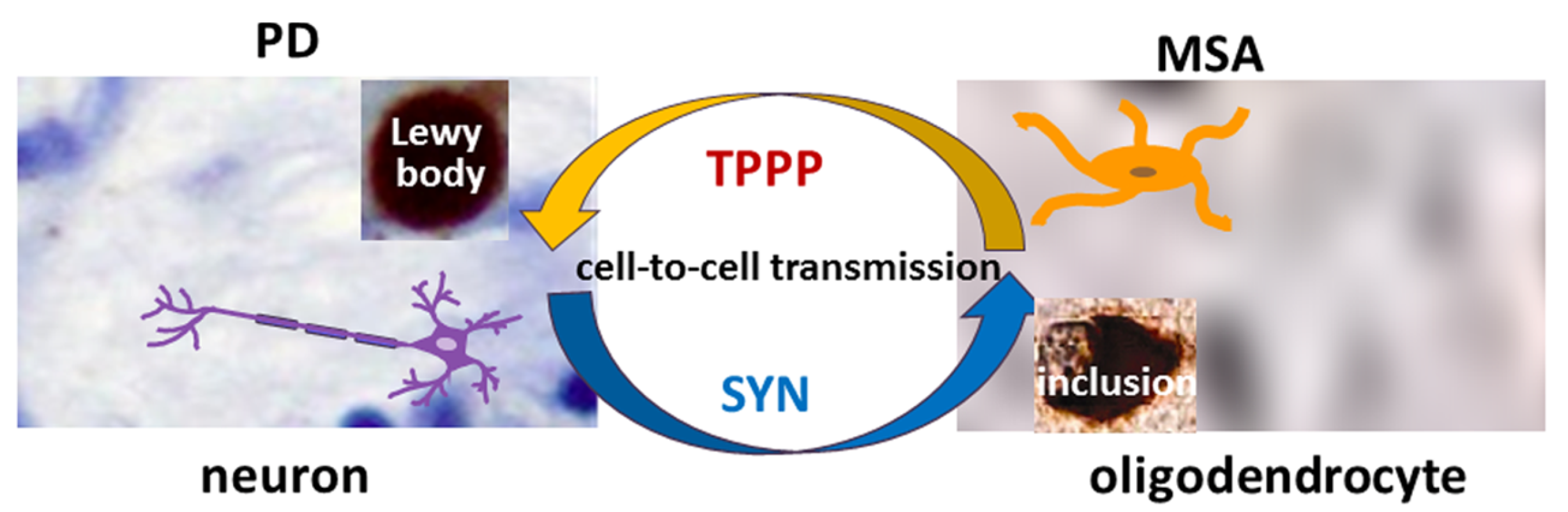
Neurological disorders such as Parkinsonism cause serious socio-economic problems as there are only therapies that treat their symptoms. The well-established hallmark alpha-synuclein (SYN) is enriched in the inclusion bodies characteristic of Parkinsonism. A prominent partner of SYN was discovered, termed Tubulin Polymerization Promoting Protein (TPPP), which has important physiological and pathological activities such as the regulation of the microtubule network and the promotion of SYN aggregation. The role of TPPP in Parkinsonism is often neglected in research. In the normal brain, SYN and TPPP are expressed endogenously in neurons and oligodendrocytes, respectively, whilst, at an early stage of Parkinsonism, soluble hetero-associations of these proteins are found in both cell types. The cell-to-cell transmission of these proteins, which is central to disease progression, provides a unique situation for specific drug targeting.

Parkinsonism methodologies alpha-synuclein TPPP drug targeting

1. Introduction

There are millions of people suffering from Parkinsonism in the world and there is neither a medicine to cure the disease nor an appropriate test to detect the illness at an early stage and to follow the disease/treatment processes at the clinical level [1][2]. Alpha-synuclein (SYN) plays a key role in Parkinsonism, which includes Parkinson’s disease (PD), dementia with Lewy bodies (DLB), and multiple system atrophy (MSA) [3][4][5][6][7]. Parkinsonism can be characterized by the classical motor symptoms (such as resting tremor, rigidity, bradykinesia, and freezing of gait) as well as non-motor features (including constipation, depression, sleep disorder, cognitive impairment, and dementia) [6]. In the case of MSA, patients may exhibit either Parkinsonism (MSA-P) or cerebellar ataxia (MSA-C). Despite the many shared symptoms, these diseases differ in the rate of disease progression. Median survival is much shorter for DLB and MSA patients than for PD ones. Current treatments include different therapies (mostly affecting dopamine metabolism, for example, by levodopa), deep brain stimulation, active and passive immunotherapy, and other options for transformative treatment (such as stem cell transplants or gene-targeted treatments) [1][8][9]. Levodopa relieves the motor symptoms through the replacement of lost dopamine; however, MSA-P patients are usually less responsive to levodopa therapy, and it can also worsen neuropsychiatric symptoms [6].
A major characteristic of these neurological disorders is the atypical protein assembly (proteopathy) leading to cell death, which is one of the key mechanisms of many neurodegenerative diseases. In pathological conditions, the aggregation of SYN results in the formation of insoluble fibrils, which had long been considered the primary structural components of synucleinopathies [3][4][10]. However, there are also striking differences between the different synucleinopathies as there are differences in the brain regions affected and in the cell types with inclusion bodies [6]. SYN aggregation is typically observed in the substantia nigra pars compacta in PD and in the cerebral cortex and hippocampus in DLB, while the olivopontocerebellar, nigrostriatal, and autonomic systems are affected in MSA. In PD and DLB, SYN-bearing Lewy bodies and Lewy neurites can be observed in neurons, while MSA is characterized by aggregated SYN in glial cytoplasmic inclusions [3][4][5]. The pathological SYN strains from Lewy bodies and glial cytoplasmic inclusions seem to be conformationally and biologically distinct [11].
In the last decade, however, Tubulin Polymerization Promoting Protein (TPPP) has also emerged as one of the principal actors in the processes underlying neurological disorders [12][13][14]. Pathological interactions of TPPP with SYN can induce SYN assembly that leads to the formation of inclusions as pathological hallmarks characteristic of PD, DLB, and MSA [12][15]. TPPP, as a Neomorphic Moonlighting Protein, plays an important role in both physiological and pathological conditions without alterations at the gene level [16]. TPPP is expressed specifically in oligodendrocytes (OLGs) in the normal brain during differentiation of the progenitor cells, where it is crucial to the formation of projections that results in the ensheathment of the axons [17]. This ensheathment enables highly efficient signal transmission; moreover, OLGs also provide metabolic and trophic support to the neurons [18]. TPPP is not present in astroglia or microglia [17][19]. In physiological conditions, TPPP modulates the dynamics and stability of the cytoskeletal microtubule system via its bundling and tubulin acetylation-promoting activities [13]. These physiological functions are mediated by its direct associations with tubulin/microtubules as well as with tubulin deacetylases such as histone deacetylase 6 and sirtuin-2 (SIRT2) [20][21]. The bundling and stabilization of microtubules result from the dimerization of the tubulin-attached monomeric TPPP [22]. The disordered TPPP has a zinc-finger motif and also has a Mg2+-dependent GTPase activity [23][24].

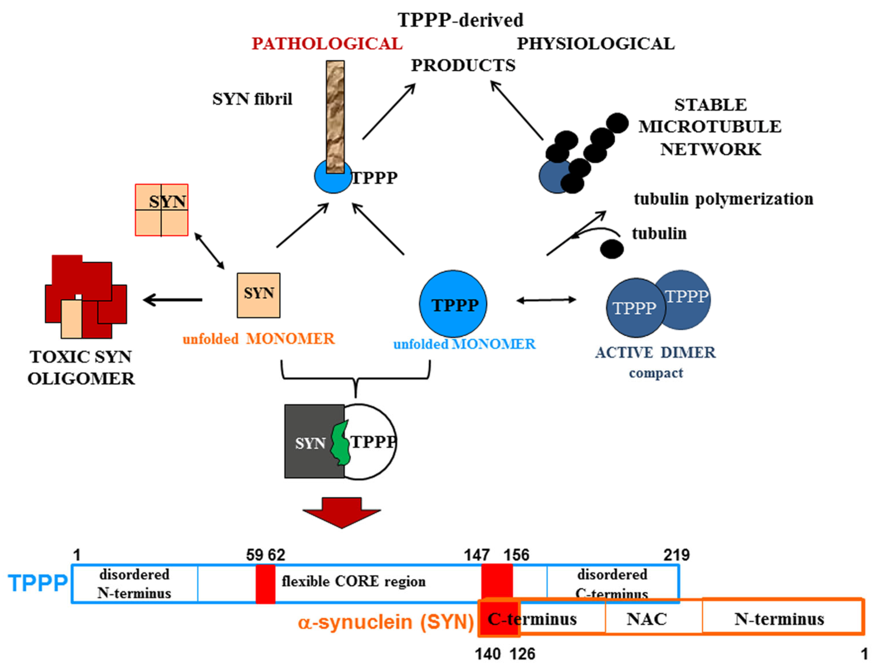
2. Pathological Interaction between TPPP and SYN

An important aspect of Parkinsonism is that the partner proteins in the pathological assembly are expressed in distinct cell types in normal brain: SYN in neurons and TPPP in OLGs, respectively [17][25][26][27]. This raises the question as to which mechanism can be responsible for their co-localization.
One possibility is the cell-to-cell transmission of SYN from neurons via the extracellular space [28][29]. Exosomes, classical exocytosis, and endocytosis as well as direct penetration can be involved in the cell-to-cell transmission of SYN [30]. The levels of exosomal total and oligomeric SYN increased in plasma in PD patients as compared to healthy controls [30]. Using immunofluorescence confocal microscopy on its own or coupled with Bifunctional Fluorescence Complementation, the researchers' experiments revealed the co-location/co-enrichment of these two proteins in living human HeLa cells transiently transfected with TPPP and SYN [31]. The assembly of SYN and TPPP could be observed as well in the cells after their uptake from the medium [16][32]. This experimental setup mimics the pathological situation occurring in human brain where these proteins are transmitted via the extracellular space. The mechanisms involved in the cell-to-cell transmission of TPPP are not known. Interestingly, TPPP was detected in exosomes isolated from SH-SY5Y cells by LC-MS/MS-based label-free quantitative proteomics analysis (http://exocarta.org/gene_summary?gene_id=11076, accessed on 18 December 2023) [33].
In pathological conditions, another possibility of the inappropriate presence of SYN and TPPP in OLGs and neurons, respectively, might be the translation of their mRNAs. According to the data in the Human Protein Atlas, both SYN mRNA (SNCA: 243.0 nTPM and 51.5 nTPM in excitatory and inhibitory neurons, 89.6 nTPM and 181.8 nTPM in OLG precursor cells and OLGs, respectively) and TPPP mRNA (TPPP: 43.7 nTPM and 31.2 nTPM in excitatory and inhibitory neurons, 6.1 nTPM and 97.5 nTPM in OLG precursor cells and OLGs, respectively) are detected in OLGs and neurons in normal human brains (https://www.proteinatlas.org/ENSG00000145335-SNCA/single+cell+type, https://www.proteinatlas.org/ENSG00000171368-TPPP/single+cell+type, accessed on 27 October 2023). Recently, single-nucleus RNA sequencing revealed a high level of SNCA transcripts in inhibitory neurons and OLG progenitor cells but only a low level of these transcripts in excitatory neurons and mature OLGs [34]. In the case of TPPP, its mRNA is present in neurons but the protein itself is only present in OLGs [17][35]. Hence, possible pathological factors include a failure to inhibit translation of the inappropriate mRNA (an inhibition that should occur in physiological conditions) and/or a failure to degrade the newly synthesized protein; these possibilities would be consistent with the absence of the SYN protein in mature OLGs despite the presence of its mRNA. Another possibility would be a failure to put the protein in a place where it would not cause a problem.
The results of recent research are consistent with the above possibilities: the addition of human preformed SYN fibrils to mouse OLGs triggered an increase in endogenous SYN, which was critical for its aggregation with TPPP [14]; in agreement with this, in rat OLN-AS7 and human MO3.13 OLG models of MSA, TPPP transfection significantly increased the levels of both SYN mRNA and SYN protein [36]. Moreover, SYN fibrils interfered with the production of proteins associated with neuromodulation and myelination [37]. Regardless of the exact contribution of these mechanisms, SYN-TPPP assembly is characteristic of PD, DLB, and MSA and is only pathological [12].

3. The Role of TPPP in the Pathomechanism of Synucleinopathies: Protein Aggregation

TPPP is co-localized with SYN in Lewy bodies in PD and DLB as well as in glial cytoplasmic inclusions in MSA [12][15] (Figure 1). Therefore, TPPP—like SYN—is considered a hallmark of synucleinopathies [12]. A TPPP-induced SYN strain had different structure and enhanced in vivo prodegenerative properties as compared to the SYN strain; moreover, injection of the preformed fibrils of the SYN-TPPP strain resulted in a shortened lifespan in a mouse model [38].
Figure 1. The role of SYN and TPPP in the formation of Lewy bodies and glial cytoplasmic inclusions in PD and MSA.
Analysis of the role of TPPP also demonstrated that TPPP is enriched in SYN-bearing Lewy bodies in both PD and DLB [12][39]. The oligodendroglial pathology in DLB is generally considered less significant than in MSA. Comparison of DLB and MSA did, however, reveal both shared and distinct patterns. The immunoreactivity of TPPP in the oligodendroglia cytoplasm differed more from the controls for MSA than for DLB; a disintegration of myelin with loss of TPPP nuclear staining was characteristic of MSA, while DLB was more similar to the controls without showing a significant loss of nuclear TPPP.

4. The Role of TPPP in the Dysregulation of Protein Degradation in Parkinsonism

In normal human brain, levels of unwanted proteins as well as aggregates are controlled by the ubiquitin–proteasome system (UPS) and the autophagy–lysosome pathways, which can remove troublesome proteins. One target of these degradative processes is the set of intrinsically disordered proteins (IDPs) involved in the formation of toxic aggregates. Monomeric proteins such as SYN and TPPP are usually degraded by the UPS, while macroautophagy is able to degrade oligomers or aggregates ([40] and references therein). SYN can also be degraded by chaperone-mediated autophagy and endo-lysosomal degradation. Dysregulation of the cellular degradative pathways has been reported in Parkinsonism, and autophagy modulation has been suggested as a possible strategy for therapeutic intervention [41].

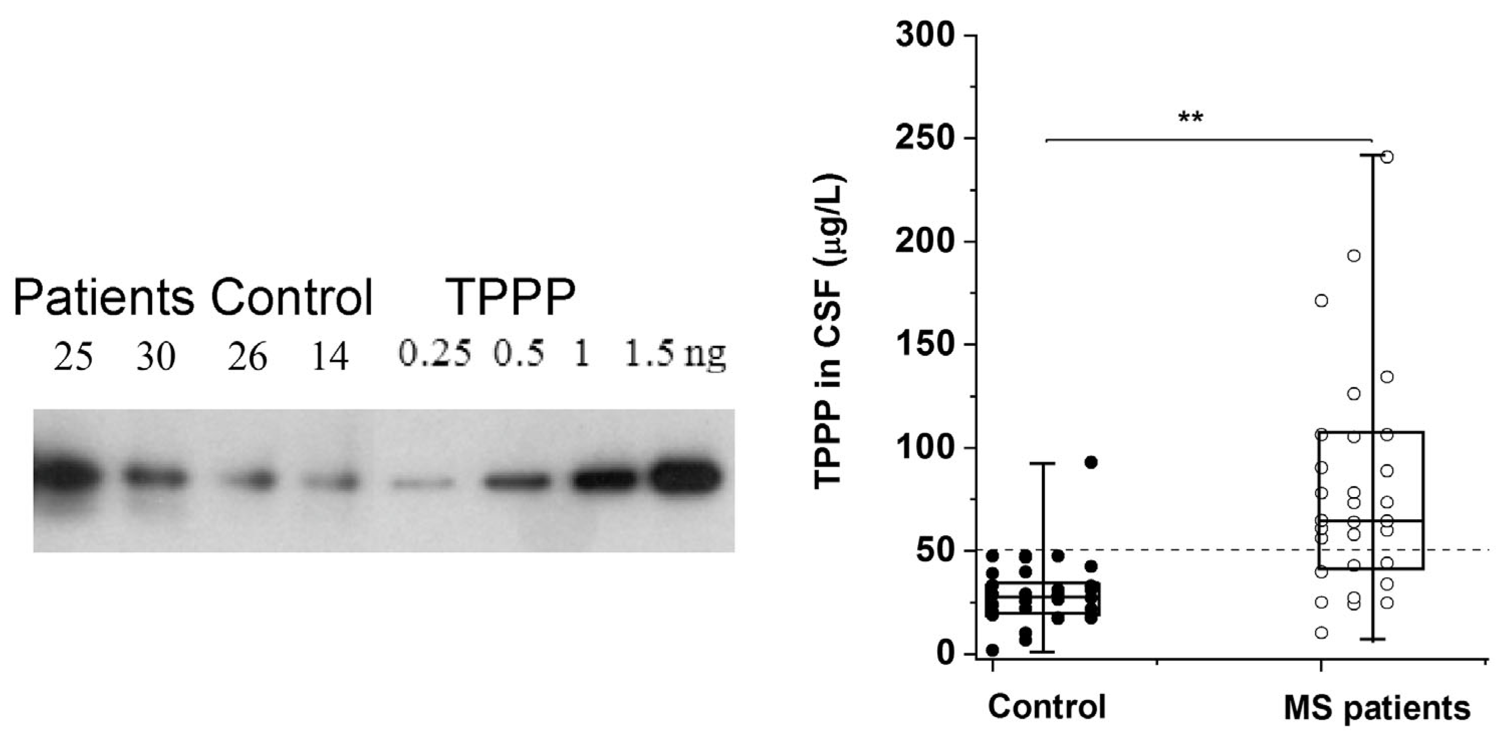
5. SYN and TPPP as Biomarkers

Synaptic dysfunction and degeneration are central contributors to the pathogenesis and progression of Parkinsonian disorders. Therefore, identification and validation of biomarkers reflecting pathological synaptic alterations are greatly needed [42]. Human cerebrospinal fluid (CSF) has a composition similar to that of the brain extracellular fluid (ECF), and the two fluids circulate freely together in the brain [43].
In a recent webinar conference organized by the Michael J. Fox Foundation entitled “Major Research Breakthrough: A New Biomarker for Parkinson’s” (20 April 2023) the significance of the SYN seed amplification assay (SAA) using CSF was highlighted [44]. This new assay may allow the aggregation of SYN to be used as a biomarker in order to distinguish people with PD from healthy controls even at an early stage of the disease. The cross-sectional analysis included 1123 participants; the assay classified PD patients with high sensitivity and specificity, and could detect low-abundancy SYN. That said, application of this assay for routine laboratory tests may prove challenging. Nevertheless, use of the SYN SAA for biochemical diagnosis of PD and MSA reveals the crucial role it could play in therapeutic development [44].
In the context of the exploration of candidate biomarkers for MSA, disregarding its close relationship with PD does not seem justifiable. Several recent studies have shown that SYN might be a potential diagnostic biomarker for PD in CSF though the results are inconsistent [45][46][47]. The results of a meta-analysis investigating the diagnostic and differential diagnosis efficacy of CSF SYN in PD have shown that its median concentration is significantly lower in PD compared to controls, but significantly higher in PD as compared to MSA [48][49]. However, the median concentration of CSF SYN oligomers was significantly higher in PD than in controls [48]. This could be due to the fact that the presence of TPPP in the neurons promotes the SYN assembly where these proteins are co-enriched and co-located in the well-separated, individual Lewy bodies.
The altered regulation/location of TPPP results in its pathological assembly with SYN in MSA, which leads to demyelination as in the case of Multiple Sclerosis (MS). MS is a chronic, inflammatory, demyelinating disease with a variable extent of remyelination depending on the differentiation of OLGs [50]. The researchers have developed and validated a sensitive assay based on Western blotting coupled with chemiluminescent detection using human recombinant TPPP and CSF for the quantification of TPPP in the case of different MS patients and controls [51] (Figure 2). According to this assay, the median TPPP content of the CSF was 64.7 μg/L for patients, while it was 27.9 μg/L for non-MS patients. The higher level of TPPP in the MS patients was independent of age, gender, and the time between lumbar puncture and relapse [51]. These results suggest that (i) TPPP-based assay with CSF of the patients may be suitable for the diagnostic testing of MS patients and (ii) this assay could be used to measure TPPP levels in the CSF of MSA and PD patients.
Figure 2. TPPP levels in the CSF of MS and non-MS patients as quantified by Western blot [51]. A representative Western blot using a specific TPPP antibody is shown for different CSF samples of patients (25 and 30, ○) and the corresponding controls (non-MS patients, ●) (14 and 26). Each box extends from the 25th to the 75th percentile with the middle line representing the median. The vertical bars indicate the full range of TPPP levels. The p values were determined by Mann–Whitney U tests. ** p < 0.000005. The dashed line corresponds to 50 μg/L [51].

6. Targeting the Interface of the Pathological SYN-TPPP Complex

Efficient therapies and disease-modifying treatments are under intense research. A search for Parkinsonism produced 100 clinical trials, 57 of which were interventional (https://clinicaltrials.gov, accessed on 1 December 2023).
A potential strategy to eliminate the excess of these hallmark proteins as well as their toxic aggregates includes the use of small molecules (such as Epigallocatechin gallate, Anle138b, or SynuClean-D), which have the advantages of their usually low cost, high stability, and bioavailability [52][53][54]. However, IDPs such as SYN and TPPP often lack the pocket needed to bind these molecules; an alternative strategy to inhibit protein–protein interactions is to use peptides and peptidomimetics, which is now a rapidly progressing field. Short peptides may have high specificity and low toxicity, although poor bioavailability and limited cellular uptake may pose problems [55]. Peptides that target different SYN regions have indeed been identified [55]. For example, Nim and co-workers found PDpep1.3 by a high-throughput, proteome-wide peptide screen; this peptide reduced the accumulation of SYN in a rat model of PD [56]. A neurohormone peptide and a cerebral dopamine neurotrophic factor (CDNF)-derived peptidomimetic also displayed beneficial effects in PD models [57][58].
A therapeutic strategy based on the targeting of either TPPP or SYN is not without risks. One reason for this is that, as IDPs, the structures of these ‘chameleon’ proteins can adopt many conformations, some of which can perturb the drug target [31][59]. As demonstrated previously, the SYN protein functions physiologically as a monomer; however, its occurrence as a metastable tetramer in a dynamic equilibrium with the monomers has also been suggested [60][61]. Another reason is that they are responsible for essential physiological functions that must be preserved.
A strategy that the researchers have developed and reported is the drug targeting of the hetero-complexes of SYN and TPPP [16][31][32]; this is because these complexes, which are only pathological, are the initiators of the neurodegenerative processes [12][14][62][63] (Figure 3). This strategy is based on targeting the interface where SYN and TPPP are in contact so as to trigger the disassembly of the pathological complexes [16][31][32]. The segments of the interface of both SYN and TPPP that are involved in the formation of their assemblies have been identified by studying the effects of several mutated and truncated forms of the proteins on their association [16][31][32]. The researchers' studies revealed that the truncation of the C-terminal segment of SYN did prevent its hetero-association with TPPP [32]. This suggests that the SYN126-140 fragment may function as a competitive inhibitor of the association of the two proteins in vitro as well as in a human cell model, where the proteins were added to the medium of CHO cells [32]. Taken together, these findings substantiate drug-targeting strategies based upon (i) elimination of the toxic species; (ii) proteolytic degradation of the excess and/or pathological partner; and (iii) maintenance/recovery of the physiological function of SYN/TPPP. Moreover, strategies for selecting drugs must also take into account the physiological functions of the validated sites of the hallmark proteins.
Figure 3. Homo- and hetero-associations of TPPP and SYN in physiological and pathological conditions and the interface segment of the SYN-TPPP complex as a potential drug target.
One of the novelties of the strategy is that it is based on the crucial role of TPPP in the formation of SYN assemblies and in the deregulation of their cellular proteolysis; hence, targeting the SYN-TPPP interaction could eliminate the toxic SYN-TPPP assemblies. For this strategy to work, the researchers suggest that autophagy modulation by itself may not be sufficient and that the inhibition/destruction of SYN-TPPP assemblies is also necessary. Such elimination of these assemblies may require adopting and adapting new and emergent techniques.

References

  1. Kalia, L.V.; Lang, A.E. Parkinson’s disease. Lancet 2015, 386, 896–912.
  2. Soni, R.; Delvadia, P.; Joharapurkar, A.; Shah, J. Uncovering Novel Therapeutic Targets for Parkinson’s Disease. ACS Chem. Neurosci. 2023, 14, 1935–1949.
  3. Spillantini, M.G.; Schmidt, M.L.; Lee, V.M.; Trojanowski, J.Q.; Jakes, R.; Goedert, M. Alpha-synuclein in Lewy bodies. Nature 1997, 388, 839–840.
  4. Wakabayashi, K.; Yoshimoto, M.; Tsuji, S.; Takahashi, H. Alpha-synuclein immunoreactivity in glial cytoplasmic inclusions in multiple system atrophy. Neurosci. Lett. 1998, 249, 180–182.
  5. Koga, S.; Sekiya, H.; Kondru, N.; Ross, O.A.; Dickson, D.W. Neuropathology and molecular diagnosis of Synucleinopathies. Mol. Neurodegener. 2021, 16, 83.
  6. Graves, N.J.; Gambin, Y.; Sierecki, E. alpha-Synuclein Strains and Their Relevance to Parkinson’s Disease, Multiple System Atrophy, and Dementia with Lewy Bodies. Int. J. Mol. Sci. 2023, 24, 12134.
  7. Stefanova, N.; Wenning, G.K. Multiple system atrophy: At the crossroads of cellular, molecular and genetic mechanisms. Nat. Rev. Neurosci. 2023, 24, 334–346.
  8. Hill, D.R.; Huters, A.D.; Towne, T.B.; Reddy, R.E.; Fogle, J.L.; Voight, E.A.; Kym, P.R. Parkinson’s Disease: Advances in Treatment and the Syntheses of Various Classes of Pharmaceutical Drug Substances. Chem. Rev. 2023, 123, 13693–13712.
  9. Stott, S.R.W.; Wyse, R.K.; Brundin, P. Novel approaches to counter protein aggregation pathology in Parkinson’s disease. Prog. Brain Res. 2020, 252, 451–492.
  10. Morris, H.R.; Spillantini, M.G.; Sue, C.M.; Williams-Gray, C.H. The pathogenesis of Parkinson’s disease. Lancet 2024, 403, 293–304.
  11. Peng, C.; Gathagan, R.J.; Covell, D.J.; Medellin, C.; Stieber, A.; Robinson, J.L.; Zhang, B.; Pitkin, R.M.; Olufemi, M.F.; Luk, K.C.; et al. Cellular milieu imparts distinct pathological alpha-synuclein strains in alpha-synucleinopathies. Nature 2018, 557, 558–563.
  12. Kovacs, G.G.; Laszlo, L.; Kovacs, J.; Jensen, P.H.; Lindersson, E.; Botond, G.; Molnar, T.; Perczel, A.; Hudecz, F.; Mezo, G.; et al. Natively unfolded tubulin polymerization promoting protein TPPP/p25 is a common marker of alpha-synucleinopathies. Neurobiol. Dis. 2004, 17, 155–162.
  13. Olah, J.; Lehotzky, A.; Szunyogh, S.; Szenasi, T.; Orosz, F.; Ovadi, J. Microtubule-Associated Proteins with Regulatory Functions by Day and Pathological Potency at Night. Cells 2020, 9, 357.
  14. Mavroeidi, P.; Arvanitaki, F.; Karakitsou, A.K.; Vetsi, M.; Kloukina, I.; Zweckstetter, M.; Giller, K.; Becker, S.; Sorrentino, Z.A.; Giasson, B.I.; et al. Endogenous oligodendroglial alpha-synuclein and TPPP/p25alpha orchestrate alpha-synuclein pathology in experimental multiple system atrophy models. Acta Neuropathol. 2019, 138, 415–441.
  15. Kovacs, G.G.; Gelpi, E.; Lehotzky, A.; Hoftberger, R.; Erdei, A.; Budka, H.; Ovadi, J. The brain-specific protein TPPP/p25 in pathological protein deposits of neurodegenerative diseases. Acta Neuropathol. 2007, 113, 153–161.
  16. Tokesi, N.; Olah, J.; Hlavanda, E.; Szunyogh, S.; Szabo, A.; Babos, F.; Magyar, A.; Lehotzky, A.; Vass, E.; Ovadi, J. Identification of motives mediating alternative functions of the neomorphic moonlighting TPPP/p25. Biochim. Biophys. Acta 2014, 1842, 547–557.
  17. Lehotzky, A.; Lau, P.; Tokesi, N.; Muja, N.; Hudson, L.D.; Ovadi, J. Tubulin polymerization-promoting protein (TPPP/p25) is critical for oligodendrocyte differentiation. Glia 2010, 58, 157–168.
  18. Han, S.; Gim, Y.; Jang, E.H.; Hur, E.M. Functions and dysfunctions of oligodendrocytes in neurodegenerative diseases. Front. Cell. Neurosci. 2022, 16, 1083159.
  19. Ota, K.; Obayashi, M.; Ozaki, K.; Ichinose, S.; Kakita, A.; Tada, M.; Takahashi, H.; Ando, N.; Eishi, Y.; Mizusawa, H.; et al. Relocation of p25alpha/tubulin polymerization promoting protein from the nucleus to the perinuclear cytoplasm in the oligodendroglia of sporadic and COQ2 mutant multiple system atrophy. Acta Neuropathol. Commun. 2014, 2, 136.
  20. Tokesi, N.; Lehotzky, A.; Horvath, I.; Szabo, B.; Olah, J.; Lau, P.; Ovadi, J. TPPP/p25 promotes tubulin acetylation by inhibiting histone deacetylase 6. J. Biol. Chem. 2010, 285, 17896–17906.
  21. Szabo, A.; Olah, J.; Szunyogh, S.; Lehotzky, A.; Szenasi, T.; Csaplar, M.; Schiedel, M.; Low, P.; Jung, M.; Ovadi, J. Modulation of Microtubule Acetylation by the Interplay Of TPPP/p25, SIRT2 and New Anticancer Agents with Anti-SIRT2 Potency. Sci. Rep. 2017, 7, 17070.
  22. Olah, J.; Szenasi, T.; Szunyogh, S.; Szabo, A.; Lehotzky, A.; Ovadi, J. Further evidence for microtubule-independent dimerization of TPPP/p25. Sci. Rep. 2017, 7, 40594.
  23. Zotter, A.; Bodor, A.; Olah, J.; Hlavanda, E.; Orosz, F.; Perczel, A.; Ovadi, J. Disordered TPPP/p25 binds GTP and displays Mg2+-dependent GTPase activity. FEBS Lett. 2011, 585, 803–808.
  24. Zotter, A.; Olah, J.; Hlavanda, E.; Bodor, A.; Perczel, A.; Szigeti, K.; Fidy, J.; Ovadi, J. Zn(2)+-induced rearrangement of the disordered TPPP/p25 affects its microtubule assembly and GTPase activity. Biochemistry 2011, 50, 9568–9578.
  25. Maroteaux, L.; Campanelli, J.T.; Scheller, R.H. Synuclein: A neuron-specific protein localized to the nucleus and presynaptic nerve terminal. J. Neurosci. Off. J. Soc. Neurosci. 1988, 8, 2804–2815.
  26. Bates, C.A.; Zheng, W. Brain disposition of alpha-Synuclein: Roles of brain barrier systems and implications for Parkinson’s disease. Fluids Barriers CNS 2014, 11, 17.
  27. Takahashi, M.; Tomizawa, K.; Fujita, S.C.; Sato, K.; Uchida, T.; Imahori, K. A brain-specific protein p25 is localized and associated with oligodendrocytes, neuropil, and fiber-like structures of the CA hippocampal region in the rat brain. J. Neurochem. 1993, 60, 228–235.
  28. Valdinocci, D.; Radford, R.A.; Siow, S.M.; Chung, R.S.; Pountney, D.L. Potential Modes of Intercellular alpha-Synuclein Transmission. Int. J. Mol. Sci. 2017, 18, 469.
  29. Bras, I.C.; Outeiro, T.F. Alpha-Synuclein: Mechanisms of Release and Pathology Progression in Synucleinopathies. Cells 2021, 10, 375.
  30. Xia, Y.; Zhang, G.; Han, C.; Ma, K.; Guo, X.; Wan, F.; Kou, L.; Yin, S.; Liu, L.; Huang, J.; et al. Microglia as modulators of exosomal alpha-synuclein transmission. Cell Death Dis. 2019, 10, 174.
  31. Szenasi, T.; Olah, J.; Szabo, A.; Szunyogh, S.; Lang, A.; Perczel, A.; Lehotzky, A.; Uversky, V.N.; Ovadi, J. Challenging drug target for Parkinson’s disease: Pathological complex of the chameleon TPPP/p25 and alpha-synuclein proteins. Biochim. Biophys. Acta. Mol. Basis Dis. 2017, 1863, 310–323.
  32. Szunyogh, S.; Olah, J.; Szenasi, T.; Szabo, A.; Ovadi, J. Targeting the interface of the pathological complex of alpha-synuclein and TPPP/p25. Biochim. Biophys. Acta 2015, 1852, 2653–2661.
  33. Keerthikumar, S.; Gangoda, L.; Liem, M.; Fonseka, P.; Atukorala, I.; Ozcitti, C.; Mechler, A.; Adda, C.G.; Ang, C.S.; Mathivanan, S. Proteogenomic analysis reveals exosomes are more oncogenic than ectosomes. Oncotarget 2015, 6, 15375–15396.
  34. Kon, T.; Forrest, S.L.; Lee, S.; Martinez-Valbuena, I.; Li, J.; Nassir, N.; Uddin, M.J.; Lang, A.E.; Kovacs, G.G. Neuronal SNCA transcription during Lewy body formation. Acta Neuropathol. Commun. 2023, 11, 185.
  35. Gould, R.; Brady, S. Identifying mRNAs Residing in Myelinating Oligodendrocyte Processes as a Basis for Understanding Internode Autonomy. Life 2023, 13, 945.
  36. Cui, H.; Kilpelainen, T.; Zouzoula, L.; Auno, S.; Trontti, K.; Kurvonen, S.; Norrbacka, S.; Hovatta, I.; Jensen, P.H.; Myohanen, T.T. Prolyl oligopeptidase inhibition reduces alpha-synuclein aggregation in a cellular model of multiple system atrophy. J. Cell. Mol. Med. 2021, 25, 9634–9646.
  37. Kaji, S.; Maki, T.; Kinoshita, H.; Uemura, N.; Ayaki, T.; Kawamoto, Y.; Furuta, T.; Urushitani, M.; Hasegawa, M.; Kinoshita, Y.; et al. Pathological Endogenous alpha-Synuclein Accumulation in Oligodendrocyte Precursor Cells Potentially Induces Inclusions in Multiple System Atrophy. Stem Cell Rep. 2018, 10, 356–365.
  38. Ferreira, N.; Gram, H.; Sorrentino, Z.A.; Gregersen, E.; Schmidt, S.I.; Reimer, L.; Betzer, C.; Perez-Gozalbo, C.; Beltoja, M.; Nagaraj, M.; et al. Multiple system atrophy-associated oligodendroglial protein p25alpha stimulates formation of novel alpha-synuclein strain with enhanced neurodegenerative potential. Acta Neuropathol. 2021, 142, 87–115.
  39. Rohan, Z.; Milenkovic, I.; Lutz, M.I.; Matej, R.; Kovacs, G.G. Shared and Distinct Patterns of Oligodendroglial Response in alpha-Synucleinopathies and Tauopathies. J. Neuropathol. Exp. Neurol. 2016, 75, 1100–1109.
  40. Lehotzky, A.; Olah, J.; Fekete, J.T.; Szenasi, T.; Szabo, E.; Gyorffy, B.; Varady, G.; Ovadi, J. Co-Transmission of Alpha-Synuclein and TPPP/p25 Inhibits Their Proteolytic Degradation in Human Cell Models. Front. Mol. Biosci. 2021, 8, 666026.
  41. Sanchez-Mirasierra, I.; Ghimire, S.; Hernandez-Diaz, S.; Soukup, S.F. Targeting Macroautophagy as a Therapeutic Opportunity to Treat Parkinson’s Disease. Front. Cell Dev. Biol. 2022, 10, 921314.
  42. Nilsson, J.; Constantinescu, J.; Nellgard, B.; Jakobsson, P.; Brum, W.S.; Gobom, J.; Forsgren, L.; Dalla, K.; Constantinescu, R.; Zetterberg, H.; et al. Cerebrospinal Fluid Biomarkers of Synaptic Dysfunction are Altered in Parkinson’s Disease and Related Disorders. Mov. Disord. Off. J. Mov. Disord. Soc. 2023, 38, 267–277.
  43. Vendel, E.; Rottschafer, V.; de Lange, E.C.M. The need for mathematical modelling of spatial drug distribution within the brain. Fluids Barriers CNS 2019, 16, 12.
  44. Siderowf, A.; Concha-Marambio, L.; Lafontant, D.E.; Farris, C.M.; Ma, Y.; Urenia, P.A.; Nguyen, H.; Alcalay, R.N.; Chahine, L.M.; Foroud, T.; et al. Parkinson’s Progression Markers, I. Assessment of heterogeneity among participants in the Parkinson’s Progression Markers Initiative cohort using alpha-synuclein seed amplification: A cross-sectional study. Lancet. Neurol. 2023, 22, 407–417.
  45. Kwon, D.H.; Hwang, J.S.; Kim, S.G.; Jang, Y.E.; Shin, T.H.; Lee, G. Cerebrospinal Fluid Metabolome in Parkinson’s Disease and Multiple System Atrophy. Int. J. Mol. Sci. 2022, 23, 1879.
  46. Schulz, I.; Kruse, N.; Gera, R.G.; Kremer, T.; Cedarbaum, J.; Barbour, R.; Zago, W.; Schade, S.; Otte, B.; Bartl, M.; et al. Systematic Assessment of 10 Biomarker Candidates Focusing on alpha-Synuclein-Related Disorders. Mov. Disord. Off. J. Mov. Disord. Soc. 2021, 36, 2874–2887.
  47. Mavroudis, I.; Petridis, F.; Chatzikonstantinou, S.; Kazis, D. Alpha-synuclein Levels in the Differential Diagnosis of Lewy Bodies Dementia and Other Neurodegenerative Disorders: A Meta-analysis. Alzheimer Dis. Assoc. Disord. 2020, 34, 220–224.
  48. Zhou, B.; Wen, M.; Yu, W.F.; Zhang, C.L.; Jiao, L. The Diagnostic and Differential Diagnosis Utility of Cerebrospinal Fluid alpha-Synuclein Levels in Parkinson’s Disease: A Meta-Analysis. Park. Dis. 2015, 2015, 567386.
  49. Xiang, C.; Cong, S.; Tan, X.; Ma, S.; Liu, Y.; Wang, H.; Cong, S. A meta-analysis of the diagnostic utility of biomarkers in cerebrospinal fluid in Parkinson’s disease. NPJ Park. Dis. 2022, 8, 165.
  50. Hoftberger, R.; Fink, S.; Aboul-Enein, F.; Botond, G.; Olah, J.; Berki, T.; Ovadi, J.; Lassmann, H.; Budka, H.; Kovacs, G.G. Tubulin polymerization promoting protein (TPPP/p25) as a marker for oligodendroglial changes in multiple sclerosis. Glia 2010, 58, 1847–1857.
  51. Vincze, O.; Olah, J.; Zadori, D.; Klivenyi, P.; Vecsei, L.; Ovadi, J. A new myelin protein, TPPP/p25, reduced in demyelinated lesions is enriched in cerebrospinal fluid of multiple sclerosis. Biochem. Biophys. Res. Commun. 2011, 409, 137–141.
  52. Rodger, A.T.; ALNasser, M.; Carter, W.G. Are Therapies That Target alpha-Synuclein Effective at Halting Parkinson’s Disease Progression? A Systematic Review. Int. J. Mol. Sci. 2023, 24, 11022.
  53. Pujols, J.; Pena-Diaz, S.; Lazaro, D.F.; Peccati, F.; Pinheiro, F.; Gonzalez, D.; Carija, A.; Navarro, S.; Conde-Gimenez, M.; Garcia, J.; et al. Small molecule inhibits alpha-synuclein aggregation, disrupts amyloid fibrils, and prevents degeneration of dopaminergic neurons. Proc. Natl. Acad. Sci. USA 2018, 115, 10481–10486.
  54. Wagner, J.; Ryazanov, S.; Leonov, A.; Levin, J.; Shi, S.; Schmidt, F.; Prix, C.; Pan-Montojo, F.; Bertsch, U.; Mitteregger-Kretzschmar, G.; et al. Anle138b: A novel oligomer modulator for disease-modifying therapy of neurodegenerative diseases such as prion and Parkinson’s disease. Acta Neuropathol. 2013, 125, 795–813.
  55. Allen, S.G.; Meade, R.M.; White Stenner, L.L.; Mason, J.M. Peptide-based approaches to directly target alpha-synuclein in Parkinson’s disease. Mol. Neurodegener. 2023, 18, 80.
  56. Nim, S.; O’Hara, D.M.; Corbi-Verge, C.; Perez-Riba, A.; Fujisawa, K.; Kapadia, M.; Chau, H.; Albanese, F.; Pawar, G.; De Snoo, M.L.; et al. Disrupting the alpha-synuclein-ESCRT interaction with a peptide inhibitor mitigates neurodegeneration in preclinical models of Parkinson’s disease. Nat. Commun. 2023, 14, 2150.
  57. Simon, C.; Soga, T.; Ahemad, N.; Bhuvanendran, S.; Parhar, I. Kisspeptin-10 Rescues Cholinergic Differentiated SHSY-5Y Cells from alpha-Synuclein-Induced Toxicity In Vitro. Int. J. Mol. Sci. 2022, 23, 5193.
  58. Kulesskaya, N.; Bhattacharjee, A.; Holmstrom, K.M.; Vuorio, P.; Henriques, A.; Callizot, N.; Huttunen, H.J. HER-096 is a CDNF-derived brain-penetrating peptidomimetic that protects dopaminergic neurons in a mouse synucleinopathy model of Parkinson’s disease. Cell Chem. Biol. 2023.
  59. Uversky, V.N. A protein-chameleon: Conformational plasticity of alpha-synuclein, a disordered protein involved in neurodegenerative disorders. J. Biomol. Struct. Dyn. 2003, 21, 211–234.
  60. Sulzer, D.; Edwards, R.H. The physiological role of alpha-synuclein and its relationship to Parkinson’s Disease. J. Neurochem. 2019, 150, 475–486.
  61. Selkoe, D.; Dettmer, U.; Luth, E.; Kim, N.; Newman, A.; Bartels, T. Defining the native state of alpha-synuclein. Neuro-Degener. Dis. 2014, 13, 114–117.
  62. Cascella, R.; Bigi, A.; Cremades, N.; Cecchi, C. Effects of oligomer toxicity, fibril toxicity and fibril spreading in synucleinopathies. Cell. Mol. Life Sci. CMLS 2022, 79, 174.
  63. Winner, B.; Jappelli, R.; Maji, S.K.; Desplats, P.A.; Boyer, L.; Aigner, S.; Hetzer, C.; Loher, T.; Vilar, M.; Campioni, S.; et al. In vivo demonstration that alpha-synuclein oligomers are toxic. Proc. Natl. Acad. Sci USA 2011, 108, 4194–4199.
More
Information
Contributors MDPI registered users' name will be linked to their SciProfiles pages. To register with us, please refer to https://encyclopedia.pub/register : , , ,
View Times: 81
Revisions: 2 times (View History)
Update Date: 04 Mar 2024
1000/1000