 +1 credit
| Version | Summary | Created by | Modification | Content Size | Created at | Operation |
|---|---|---|---|---|---|---|
| 1 | University of Oviedo | -- | 6512 | 2024-02-26 10:17:55 | | | |
| 2 | Sirius Huang | Meta information modification | 6512 | 2024-02-27 02:01:37 | | |
Video Upload Options
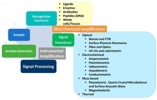
One of the most consumed foods is milk and milk products, and guaranteeing the suitability of these products is one of the major concerns in our society. This has led to the development of numerous sensors to enhance quality controls in the food chain. However, this is not a simple task, because it is necessary to establish the parameters to be analyzed and often, not only one compound is responsible for food contamination or degradation. To attempt to address this problem, a multiplex analysis together with a non-directed (e.g., general parameters such as pH) analysis are the most relevant alternatives to identifying the safety of dairy food. In recent years, the use of new technologies in the development of devices/platforms with optical or electrochemical signals has accelerated and intensified the pursuit of systems that provide a simple, rapid, cost-effective, and/or multiparametric response to the presence of contaminants, markers of various diseases, and/or indicators of safety levels.
1. Introduction
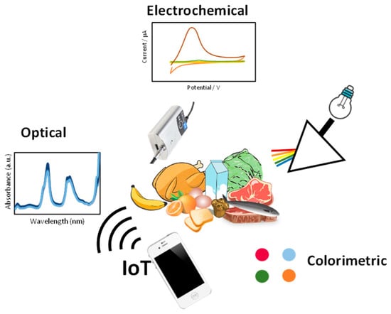
2. Optical Sensors
2.1. Surface-Enhanced Raman Spectroscopy
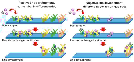
2.2. Lateral Flow Assays (LFAs)
2.3. Immunoassays with Optical Detection
2.4. Chemiluminescence
2.5. Label-Free Assays
2.6. Dairy and Allergens
| Sample | Detection | |||||
|---|---|---|---|---|---|---|
| Food | Allergens | Technique | Sensing System | Total Assay Time | LOD | Ref. |
| Multiplex detection of allergens in milk, milk-containing products, and dairy products | ||||||
| Milk, (*) cookies, ice cream | Gliadin, Ara h 1, Cor a1 (hazelnut), casein, ovalbumin | Amperometry | Magnet/SPE antibody-tagged immunomagnetic beads | <10 min | Ranging from 0.003 to 0.170 mg/kg | [40] |
| Powdered milk, (*) cookies, sponge cake | Hazelnut, peanut, soybean | Optical (laser) | Digoxin-labeled PCR products detected by hybridization on modified DVD surface | 5 h 20 min | 1 μg/g | [34] |
| MoniQA milk, NIST SRM 1549a milk, (*) Nutella hazelnut spread, 2% milk | Ana o 3 (cashew), Ara h 3/Ara h 6 (peanut), Cor a 9 (hazelnut), Gal d 1/Gal d 2 (egg), Gly m 5 (soy), Bos d 5 (milk), tropomyosin (shrimp) | Fluorescent multiplex array | Monoclonal or polyclonal antibodies covalently coupled to Luminex xMAP® system | 30 min | Ranging from 0.02 to 1.95 ng/mL | [41] |
| (*) Cookies | Casein, soy protein, gluten | Flow cytometry | Fluorescent microsphere-based immunoassay | 1 h 10 min | 0.4 ppm | [42] |
| (*) Oatmeal cookies, milk chocolate, chocolate ice cream | Hazelnut, Brazil nut, peanut | Colorimetry | Enzyme immunoassay system with chromogenic substrate | 4 h 10 min | Ranging from 0.1 to 1.0 μg/g | [35] |
| (*) Cookies | Hazelnut, peanut | Colorimetry | Lab-on-chip/Carbon dot label for lateral flow immunoassay | 15 min | 0.1 ppm | [43] |
| Multiplex detection of milk allergens in food stuff | ||||||
| Allergen-free probiotics | Gliadin, β-lactoglobulin, hazelnut, almond, peanut, soy | Colorimetry | DVD functionalized with the capture bioreceptors in microarray format | 20 samples in 70 min | Ranging from 0.1 to 143.4 ng/mL | [44] |
| A panel of 38 food commodities based on AOAC recommendations and milk from six different animal sources | Casein, β-lactoglobulin | Colorimetry/Visual | Antibodies coupled to red and blue Carboxyl-dyed, antibody-modified latex beads | 10 min | 0.5 ppm β-lactoglobulin, 2 ppm for caseins | [31] |
| Infant jar food, apple juice | Gliadin, casein, β-lactoglobulin, ovalbumin | Optical (laser) | Immunoassay developed on DVD surface | 1 h 25 min | 31 μg/L (casein), 120 μg/L (β-lactoglobulin) | [33] |
3. Electrochemical Devices/Platforms
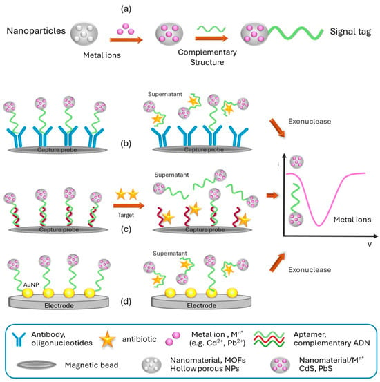
3.1. Multiplex Electrochemical Platform for Antibiotics
3.2. Multiplex Devices for Bacterial Recognition
3.3. Platforms for Other Targets of Interest
| Target | Analyte | Electrode/Modification/Label | Analytical Signal | Linear Range | LOD | Reference |
|---|---|---|---|---|---|---|
| Antibiotics | Sulphapyridine (SPY), Oxytetracycline (OTC) |
SPCE/protein G/– | Amperometric | 0.39, 1.93 nM |
1.92–454 nM, – |
[43] |
| Streptomycin (STR), Chloramphenicol (CHL), Tetracycline (TC) |
Gold electrode | SWV | – – |
10 nM, 5 nM, 20 nM |
[44] | |
| Kanamycin (KAN), Streptomycin (STR) |
SPCE/carbon nanofibers, carbon–gold nanoparticles/CdS, PbS | DPV | 10−1–103 nM | 87.3 pM, 45.0 pM |
[48] | |
| Kanamycin (KAN), Tobramycin (TOB) |
Au electrode/gold nanoshells/SCd, SPb | DPV | 1–4 × 10² nM, 1–1 × 10⁴ nM |
0.12 nM, 0.49 nM |
[49] | |
| Pathogens | Escherichia coli, Campylobacter, Salmonella |
SPCE/multiwall carbon nanotube–polyallylamine/CdS, PbS, CuS QDs | SWASV | 103–5 × 10⁵ cells/mL | 400 cells/mL, 400 cells/mL, 800 cells/mL |
[50] |
| Listeria monocytogenes, Staphylococcus aureus |
SPCE/gold nanoparticle–streptavidin/magnetic nanoparticles | SWV | 10–107 CFU/mL, 10–107 CFU/mL |
9 CFU/mL, 3 CFU/mL |
[51] | |
| Aeromonas hydrophile (Ah), Pseudomonas aeruginosa (Ps) |
GCE/ZIF-8-gold nanoparticles/thionine, ferrocene | SWV | 101–103 CFU/mL, 101–105 CFU/mL |
3.60 CFU/mL, 8095 CFU/mL |
[52] | |
| Listeria monocytogenes (Lm), Enterobacter cloacae (Ec) |
GCE electrodes/carbon nanotubes, Au nanoparticles anti-Lm, anti-Ec/thionine ferrocene |
SWV | 101–107 CFU/mL, 101–106 CFU/mL |
3.22 CFU/mL, 4.17 CFU/mL |
[53] | |
| Pesticide | Malathion (MAL), chlorpyrifos (CLO) |
GCE/Au nanoparticles, cDNA/Ce(III)–Ce(V)–MOF | SWV | 1−1 pM–1 μM | 0.045 pM, 0.038 pM |
[54] |
| Immunoglobulins | Bovine casein, bovine immunoglobulin G |
Graphite/bismuth layer/CdS, PbS QDs | ASV | 1–102% v/v | 0.04 μg/mL, 0.02 μg/mL |
[56] |
| Bovine immunoglobulin G, bovine immunoglobulin G, caprine immunoglobulin G |
SPCE/–/HRP, hydrogen peroxide, hydroquinone | Amperometric | 2.6–250 ng/mL, 2.7–250 ng/mL, 2.2–250 ng/mL |
0.74 ng/mL, 0.82 ng/mL, 0.66 ng/mL |
[57] | |
| mi-RNA | mi-RNAs | SPCE/MoS2 nanosheets, CuFe2O4/MoS2 nanosheets, ferrocene | SWV | 1 pM to 1.5 nM | 0.48 pM | [58] |
| Others | Glucose, galactose, lactose, urea | Gold thin-film interdigitated sensors/AgNPs/enzymes | Impedance spectroscopy | PCA discrimination of milks with different nutritional characteristics | [63] | |
| Enrofloxacin Melamine |
SPCE into fluidic microarray/Au@PtNPs | Impedance spectroscopy/Cyclic voltammetry | 0.1–1000 ng/mL 0.1–500 ng/mL |
18.97 pg/mL 26.80 pg/mL |
[55] | |
References
- Juronen, D.; Kuusk, A.; Kivirand, K.; Rinken, A.; Rinken, T. Immunosensing system for rapid multiplex detection of mastitis-causing pathogens in milk. Talanta 2018, 178, 949–954.
- Duan, N.; Gong, W.; Wang, Z.; Wu, S. An aptasensor based on fluorescence resonance energy transfer for multiplexed pathogenic bacteria determination. Anal. Methods 2016, 8, 1390–1395.
- Huang, Y.; Zhang, H.; Chen, X.; Wang, X.; Duan, N.; Wu, S.; Xu, B.; Wang, Z. A multicolor time-resolved fluorescence aptasensor for the simultaneous detection of multiplex Staphylococcus aureus enterotoxins in the milk. Biosens. Bioelectron. 2015, 74, 170–176.
- Shriver-Lake, L.C.; Erickson, J.S.; Sapsford, K.E.; Ngundi, M.M.; Shaffer, K.M.; Kulagina, N.V.; Hu, J.E.; Gray III, S.A.; Golden, J.P.; Ligler, F.S.; et al. Blind laboratory trials for multiple pathogens in spiked food matrices. Anal. Lett. 2007, 40, 3219–3231.
- Cho, I.H.; Mauer, L.; Irudayaraj, J. In-situ fluorescent immunomagnetic multiplex detection of foodborne pathogens in very low numbers. Biosens. Bioelectron. 2014, 57, 143–148.
- Wang, C.; Xiao, R.; Wang, S.; Yang, X.; Bai, Z.; Li, X.; Rong, Z.; Shen, B.; Wang, S. Magnetic quantum dot based lateral flow assay biosensor for multiplex and sensitive detection of protein toxins in food samples. Biosens. Bioelectron. 2019, 146, 111754.
- Shi, Q.; Tao, C.; Kong, D. Multiplex SERS-based lateral flow assay for one-step simultaneous detection of neomycin and lincomycin in milk. Eur. Food Res. Technol. 2022, 248, 2157–2165.
- Li, X.; Wang, X.; Wang, L.; Yang, T.; Wang, D. Duplex Detection of Antibiotics in Milk Powder Using Lateral-Flow Assay Based on Surface-Enhanced Raman Spectroscopy. Food Anal. Methods 2021, 14, 165–171.
- Li, Y.; Jin, G.; Liu, L.; Kuang, H.; Jing, X.; Chuanlai, X. A portable fluorescent microsphere-based lateral flow immunosensor for the simultaneous detection of colistin and bacitracin in milk. Analyst 2020, 145, 7884–7892.
- Li, Z.; Li, Z.; Zhao, D.; Wen, F.; Jiang, J.; Xu, D. Smartphone-based visualized microarray detection for multiplexed harmful substances in milk. Biosens. Bioelectron. 2017, 87, 874–880.
- Zhou, C.; Huang, C.; Zhang, H.; Yang, W.; Jiang, F.; Chen, G.; Liu, S.; Chen, Y. Machine-Learning-driven optical immunosensor based on microespheres-encoded signal transduction for the rapid and multiplexed detection of antibiotics in milk. Food Chem. 2024, 437, 137740.
- Shi, Q.; Huang, J.; Sun, Y.; Yin, M.; Hu, M.; Hu, X.; Zhang, Z.; Zhang, G. Utilization of a lateral flow colloidal gold immunoassay strip based on surface-enhanced Raman spectroscopy for ultrasensitive detection of antibiotics in milk. Spectrochim. Acta A Mol. Biomol. Spectrosc. 2018, 197, 107–113.
- Jiang, R.; Lin, D.; Zhang, Q.; Li, L.; Yang, L. Multiplex chroma-response based fluorescent smartphone sensing platform for rapid and visual quantitaive determination of antibiotic. Sens. Actuators B. Chem. 2022, 350, 130902.
- Lu, L.; Xu, L.; Zhang, Y.; Jiang, T. Multiplexed surface-enhanced Raman scattering detection of melamine and dicyandiamide in dairy food enabled by three-dimensional polystyrene@silver@graphene oxide hybrid substrate. Appl. Surf. Sci. 2022, 603, 154419.
- Zhang, H.; Ma, X.; Liu, Y.; Duan, N.; Wu, S.; Wang, Z.; Xu, B. Gold nanoparticles enhanced SERS aptasensor for the simultaneous detection of Salmonella typhimurium and Staphylococcus aureus. Biosens. Bioelectron. 2015, 74, 872–877.
- Zhang, Z.; Tang, S.; Jin, Y.; Yang, C.; He, L.; Wang, J.; Chen, Y. Multiplex SERS-based lateral flow immunosensor for the detection of major mycotoxins in maize utilizing dual Raman labels and triple test lines. J. Hazard. Mater. 2020, 393, 122348.
- Yahaya, M.L.; Zakaria, N.D.; Noordin, R.; Abdul Razak, K. Development of rapid gold nanoparticles based lateral flow assays for simultaneous detection of Shigella and Salmonella genera. Biotechnol. Appl. Biochem. 2021, 68, 1095–1106.
- Tao, X.; Wang, J.; Xie, Y.; Zuo, X.; Mo, F.; Zhou, S.; Li, H. Dual-Label Chemiluminescence Strategy for Multiplexed Immunoassay of 20 Fluoroquinolones, 15 β-Lactams. 15 Sulfonamides, and CAP in Milk. Food Anal. Methods 2017, 10, 3009–3022.
- Yu, X.; Tao, X.; Shen, J.; Zhang, S.; Cao, X.; Chen, M.; Wang, W.; Wang, Z.; Wen, K. A one-step chemiluminescence immunoassay for 20 fluoroquinolone residues in fish and shrimp based on a single chain Fv–alkaline phosphatase fusion protein. Anal. Methods 2015, 7, 9032–9039.
- Zhang, Y.; Chang, X.; Wang, X.; Tao, X. A quadruple-labeling luminescence strategy for multiplexed immunoassay of 51 drugs in milk with an automated pretreatment system. Anal. Methods 2019, 11, 5055–5063.
- Singh, A.K.; Sun, X.; Bai, X.; Kim, H.; Abdalhaseib, M.U.; Bae, E.; Bhunia, A.K. Label-free, non-invasive light scattering sensor for rapid screening of Bacillus colonies. J. Microbiol. Methods 2015, 109, 56–66.
- Raz, S.R.; Bremer, M.G.E.G.; Haasnoot, W.; Norde, W. Label-Free and Multiplex Detection of Antibiotic Residues in Milk Using Imaging Surface Plasmon Resonance-Based Immunosensor. Anal. Chem. 2009, 81, 7743–7749.
- Suarez, G.; Jin, Y.H.; Auerswald, J.; Berchtold, S.; Knapp, H.F.; Diserens, J.M.; Leterrier, Y.; Manson, J.A.E.; Voirin, G. Lab-on-a-chip for multiplexed biosensing of residual antibiotics in milk. Lab Chip 2009, 9, 1625–1630.
- Angelopoulou, M.; Petrou, P.S.; Makarona, E.; Haasnoot, W.; Moser, I.; Jobst, G.; Goustouridis, D.; Lees, M.; Kalatzi, K.; Raptis, I.; et al. Ultrafast Multiplexed-Allergen Detection through Advanced Fluidic Design and Monolithic Interferometric Silicon Chips. Anal. Chem. 2018, 90, 9559–9567.
- Yang, Z.; Xu, G.; Reboud, J.; Ali, S.A.; Kaur, G.; McGiven, J.; Boby, N.; Gupta, P.K.; Chaudhuri, P.; Cooper, J.M. Rapid Veterinary Diagnosis of Bovine Reproductive Infectious Diseases from Semen Using Paper-Origami DNA Microfluidics. ACS Sens. 2018, 3, 403–409.
- Prasad, A.; Tran, T.; Gartia, M.R. Multiplexed Paper Microfluidics for Titration and Detection of Ingredients in Beverages. Sensors 2019, 19, 1286.
- Chen, X.; Yao, C.; Li, Z. Microarray-based chemical sensors and biosensors: Fundamentals and food safety applications, TrAC. Trends Anal. Chem. 2023, 158, 116785.
- Ashley, J.; D’Aurelio, R.; Piekarska, M.; Temblay, J.; Pleasants, M.; Trinh, L.; Rodgers, T.L.; Tothill, I.E. Development of a β-Lactoglobulin Sensor Based on SPR for Milk Allergens Detection. Biosensors 2018, 8, 32.
- Bojcukova, J.; Vlas, T.; Forstenlechner, P.; Panzner, P. Comparison of two multiplex arrays in the diagnostics of allergy. Clin. Transl. Allergy 2019, 9, 31.
- Scala, E.; Caprini, E.; Abeni, D.; Meneguzzi, G.; Buzzulini, F.; Cecchi, L.; Villalta, D.; Asero, R. A qualitative and quantitative comparison of IgE antibody profiles with two multiplex platforms for component-resolved diagnostics in allergic patients. Clin. Exp. Allergy 2021, 51, 1603–1612.
- ImmunoCAP ISAC Test. Available online: https://www.thermofisher.com/phadia/wo/en/our-solutions/immunocap-allergy-solutions/specific-ige-multiplex.html (accessed on 14 February 2024).
- ELISA Based In-Vitro Multiplex Allergy Test. Available online: https://www.macroarraydx.com/products/alex (accessed on 14 February 2024).
- Monroe, M.R.; Daaboul, G.G.; Tuysuzoglu, A.; López, C.A.; Little, F.F.; Ünlü, M.S. Single Nanoparticle Detection for Multiplexed Protein Diagnostics with Attomolar Sensitivity in Serum and Unprocessed Whole Blood. Anal. Chem. 2013, 85, 3698–3706.
- Galan-Malo, P.; Pellicer, S.; Pérez, M.D.; Sánchez, L.; Razquin, P.; Mata, L. Development of a novel duplex lateral flow test for simultaneous detection of casein and β-lactoglobulin in food. Food Chem. 2019, 293, 41–48.
- Masiri, J.; Barrios-López, B.; Benoit, L.; Tamayo, J.; Day, J.; Nadala, C.; Sung, S.L.; Samadpour, M. Development and Validation of a Lateral Flow Immunoassay Test Kit for Dual Detection of Casein and β-Lactoglobulin Residues. J. Food Prot. 2016, 79, 477–483.
- Badran, A.A.; Morais, S.; Maquieira, Á. Simultaneous determination of four food allergens using compact disc immunoassaying technology. Anal. Bioanal. Chem. 2017, 409, 2261–2268.
- Raz, S.R.; Liu, H.; Norde, W.; Bremer, M.G.E.G. Food Allergens Profiling with an Imaging Surface Plasmon Resonance-Based Biosensor. Anal. Chem. 2010, 82, 8485–8491.
- Tortajada-Genaro, L.A.; Santiago-Felipe, S.; Morais, S.; Gabaldón, J.A.; Puchades, R.; Maquieira, Á. Multiplex DNA Detection of Food Allergens on a Digital Versatile Disk. J. Agric. Food Chem. 2012, 60, 36–43.
- Blais, B.W.; Gaudreault, M.; Phillippe, L.M. Multiplex enzyme immunoassay system for the simultaneous detection of multiple allergens in foods. Food Control 2003, 14, 43–47.
- Lin, H.Y.; Huang, C.H.; Park, J.; Pathania, D.; Castro, C.M.; Fasano, A.; Weissleder, R.; Lee, H. Integrated Magneto-Chemical Sensor for On-Site Food Allergen Detection. ACS Nano 2017, 11, 10062–10069.
- Filep, S.C.; Black, K.R.; Smith, B.R.E.; Block, D.S.; Kuklinska-Pijanka, A.; Bermingham, M.; Oliver, M.A.; Thorpe, C.M.; Schuhmacher, Z.P.; Agah, S.; et al. Simultaneous quantification of specific food allergen proteins using a fluorescent multiplex array. Food Chem. 2022, 389, 132986.
- Gomaa, A.; Boye, J. Simultaneous detection of multi-allergens in an incurred food matrix using ELISA, multiplex flow cytometry and liquid chromatography mass spectrometry (LC–MS). Food Chem. 2015, 175, 585–592.
- Ross, G.M.S.; Filippini, D.; Nielen, M.W.F.; Salentijn, G.I.J. Interconnectable solid-liquid protein extraction unit and chip-based dilution for multiplexed consumer immunodiagnostics. Anal. Chim. Acta 2020, 1140, 190–198.
- Sena-Torralba, A.; Smits, N.G.E.; Blázquez, D.; Albero-Pérez, C.; Pallás-Tamarit, Y.; Maquieira, Á.; Morais, S. Simultaneous quantification of six major allergens in commercial foods for children using a multiplex array on a digital versatile disc. Food Chem. 2023, 404, 134570.
- Grabowska, I.; Hepel, M.; Kurzątkowska-Adaszyńska, K. Advances in Design Strategies of Multiplex Electrochemical Aptasensors. Sensors 2022, 22, 161.
- Rapini, R.; Marrazza, G. Electrochemical aptasensors for contaminants detection in food and environment: Recent advances. Bioelectrochemistry 2017, 118, 47–61.
- Conzuelo, F.; Campuzano, S.; Gamella, M.; Pinacho, D.G.; Reviejo, A.J.; Marco, M.P.; Pingarrón, J.M. Integrated disposable electrochemical immunosensors for the simultaneous determination of sulfonamide and tetracycline antibiotics residues in milk. Biosens. Bioelectron. 2013, 50, 100–105.
- Xue, J.; Liu, J.; Wang, C.; Tiana, Y.; Zhou, N. Simultaneous electrochemical detection of multiple antibiotic residues in milk based on aptamers and quantum dots. Anal. Methods 2016, 8, 1981–1988.
- Chen, M.; Gan, N.; Zhou, Y.; Li, T.; Xua, Q.; Cao, Y.; Chen, Y. An electrochemical aptasensor for multiplex antibiotics detection based on metal ions doped nanoscale MOFs as signal tracers and RecJf exonuclease assisted targets recycling amplification. Talanta 2016, 161, 867–874.
- Chen, M.; Gan, N.; Zhou, Y.; Li, T.; Xua, Q.; Cao, Y.; Chen, Y. A novel aptamer- metal ions- nanoscale MOF based electrochemicalbiocodes for multiple antibiotics detection and signal amplification. Sens. Actuators B Chem. 2017, 242, 1201–1209.
- Yan, Z.; Gan, N.; Li, T.; Cao, Y.; Chen, Y. A sensitive electrochemical aptasensor for multiplex antibiotics detection based on high-capacity magnetic hollow porous nanotracers coupling exonuclease-assisted cascade target recycling. Biosens. Bioelectron. 2016, 78, 51–57.
- Li, F.; Wang, X.; Sun, X.; Guo, Y. Multiplex electrochemical aptasensor for detecting multipleantibiotics residues based on carbon fiber and mesoporouscarbon-gold nanoparticles. Sens. Actuators B Chem. 2018, 265, 217–226.
- Li, F.; Wu, Y.; Chen, D.; Guo, Y.; Wang, X.; Sun, X. Sensitive dual-labeled electrochemical aptasensor for simultaneous detection of multi-antibiotics in milk. Int. J. Hydrogen Energy 2021, 46, 23301–23309.
- Viswanathan, S.; Rani, C.; Ho, L.A. Electrochemical immunosensor for multiplexed detection of food-borne pathogens using nanocrystal bioconjugates and MWCNT screen-printed electrode. Talanta 2012, 94, 315–319.
- Eissa, S.; Zourob, M. Ultrasensitive peptide-based multiplexed electrochemical biosensor for the simultaneous detection of Listeria monocytogenes and Staphylococcus aureus. Mikrochim. Acta 2020, 187, 486.
- Viswanath, K.B.; Krithiga, N.; Jayachitra, A.; Mideen, A.K.S.; Amali, A.J.; Vasantha, V.S. Enzyme-Free Multiplex Detection of Pseudomonas aeruginosa and Aeromonas hydrophila with Ferrocene and Thionine-Labeled Antibodies Using ZIF-8/Au NPs as a Platform. ACS Omega 2018, 3, 17010–17022.
- Viswanath, K.B.; Suganya, K.; Krishnamoorthy, G.; Marudhamuthu, M.; Selvan, S.T.; Vasantha, V.S. Enzyme-Free Multiplex Detection of Foodborne Pathogens Using Au Nanoparticles-Decorated Multiwalled Carbon Nanotubes. ACS Food Sci. Technol. 2021, 1, 1236–1246.
- Ma, D.; Liu, J.; Liu, H.; Yi, J.; Xia, F.; Tian, D.; Zhou, C. Multiplexed electrochemical aptasensor based on mixed valence Ce (III, IV)-MOF for simultaneous determination of malathion and chlorpyrifos. Anal. Chim. Acta 2022, 1230, 340364.
- Gu, Y.; Wang, J.; Pan, M.; Yun, Y.; Wen, W.; Fang, G.; Wang, S. On-chip multiplex electrochemical immunosensor based on disposable 24-site fluidic micro-array screen printing analytical device for multi-component quantitative analysis. Sens. Actuat. B Chem. 2018, 260, 499–507.
- Kokkinos, C.; Angelopoulou, M.; Economou, A.; Prodromidis, M.; Florou, A.; Haasnoot, W.; Petrou, P.; Kakabakos, S. Lab-on-a-Membrane Foldable Devices for Duplex Drop-Volume Electrochemical Biosensing Using Quantum Dot Tags. Anal. Chem. 2016, 88, 6897–6904.
- Ruiz-Valdepeñas Montiel, V.; Povedano, E.; Benedé, S.; Mata, L.; Galán-Malo, P.; Gamella, M.; Reviejo, A.J.; Campuzano, S.; Pingarrón, J.M. Disposable Amperometric Immunosensor for the Detection of Adulteration in Milk through Single or Multiplexed Determination of Bovine, Ovine, or Caprine Immunoglobulins G. Anal. Chem. 2019, 91, 11266–11274.
- Chand, R.; Ramalingam, S.; Neethirajan, S. A 2D transition-metal dichalcogenide MoS2 based novel nanocomposite and nanocarrier for multiplex miRNA detection. Nanoscale 2018, 10, 8217–8225.
- Perez-Gonzalez, C.; Salvo-Comino, C.; Martin-Pedrosa, F.; Rodriguez-Mendez, M.L.; García-Cabezón, C. A new data analysis approach for an AgNPs-modified impedimetric bioelectronic tongue for dairy analysis. Food Control 2024, 156, 110136.

