Your browser does not fully support modern features. Please upgrade for a smoother experience.
Submitted Successfully!
 +1 credit
Thank you for your contribution! You can also upload a video entry or images related to this topic.
For video creation, please contact our Academic Video Service.
| Version | Summary | Created by | Modification | Content Size | Created at | Operation |
|---|---|---|---|---|---|---|
| 1 | Adelina Vlad | -- | 3723 | 2024-02-17 10:32:59 | | | |
| 2 | Lindsay Dong | Meta information modification | 3723 | 2024-02-18 02:27:43 | | |
Video Upload Options
We provide professional Academic Video Service to translate complex research into visually appealing presentations. Would you like to try it?
Cite
If you have any further questions, please contact Encyclopedia Editorial Office.
Grădinaru, T.; Vlad, A.; Gilca, M. Bitter Phytochemicals in Skin Disease Treatment. Encyclopedia. Available online: https://encyclopedia.pub/entry/55111 (accessed on 28 July 2026).
Grădinaru T, Vlad A, Gilca M. Bitter Phytochemicals in Skin Disease Treatment. Encyclopedia. Available at: https://encyclopedia.pub/entry/55111. Accessed July 28, 2026.
Grădinaru, Teodora-Cristiana, Adelina Vlad, Marilena Gilca. "Bitter Phytochemicals in Skin Disease Treatment" Encyclopedia, https://encyclopedia.pub/entry/55111 (accessed July 28, 2026).
Grădinaru, T., Vlad, A., & Gilca, M. (2024, February 17). Bitter Phytochemicals in Skin Disease Treatment. In Encyclopedia. https://encyclopedia.pub/entry/55111
Grădinaru, Teodora-Cristiana, et al. "Bitter Phytochemicals in Skin Disease Treatment." Encyclopedia. Web. 17 February, 2024.
Copy Citation
Skin diseases represent a global healthcare challenge due to their rising incidence and substantial socio-economic burden. While biological, immunological, and targeted therapies have brought a revolution in improving quality of life and survival rates for certain dermatological conditions, there remains a stringent demand for new remedies. Nature has long served as an inspiration for drug development. Recent studies have identified bitter taste receptors (TAS2Rs) in both skin cell lines and human skin. Additionally, bitter natural compounds have shown promising benefits in addressing skin aging, wound healing, inflammatory skin conditions, and even skin cancer. Thus, TAS2Rs may represent a promising target in all these processes.
bitter
TAS2R
phytochemicals
skin
skin inflammation
skin cancer
1. Introduction
Traditionally viewed as merely a mechanical barrier separating the body from its surroundings, the skin is now recognized as a dynamic, multi-layered interface with diverse functions (e.g., keratogenesis, melanogenesis, hydrolipidic layer formation, sudoral and sebaceous secretion, pilogenesis, thermoregulation, metabolic, endocrine, elastic, plastic, resistance, immunological, psychosocial, and communication functions) [1]. Some of these roles are attributed to the living cells in the skin (e.g., keratinocytes, melanocytes) or to the skin infiltrating cells (e.g., T lymphocytes), while others are achieved in connection with other organs/systems.
Rated as the fourth most prevalent health concern, dermatologic diseases are a substantial healthcare challenge [2]. Inflammatory skin conditions hold a significant weight among them, since approximately one in four individuals will experience a chronic inflammatory skin disease at some point [3]. This category covers various ailments, with psoriasis and atopic dermatitis among the most prevalent [4]. Both conditions profoundly impact the patients’ quality of life and also affect their families [5].
Alongside ultraviolet radiation skin exposure [6], viruses [7], immunodeficiency [8][9], genetic predisposition [10][11], existence of multiple nevi [12], smoking [13], chemical [14] or ionizing radiation exposure [15], chronic cutaneous inflammation is an important risk factor for skin carcinogenesis [16][17].
The incidence of skin cancer has increased in the past few years. In 2020, over 1.5 million new cases of skin cancers (except basal cell carcinoma) have been diagnosed globally [18]. Given the socio-economic impact of inflammatory skin diseases and skin cancers, it becomes imperative to seize every possible opportunity for their prevention and treatment.
A better understanding of the mechanisms by which skin inflammation and cancer are developed may lead to effective therapies. Many of the reported molecules involved in skin inflammation and cancer, such as C–C chemokine receptor type4 (CCR4), C–C chemokine receptor type10 (CCR10), involved in skin lymphocyte recruitment [19], G-protein-coupled estrogen receptor 1 (GPER1) [20], and melanocortin 1 receptor (MC1R), involved in melanocyte proliferation, pigmentary variation, sun sensitivity, and susceptibility to skin cancers [21][22], are G-coupled protein receptors (GPCRs). Based on this, scientists have directed their interest toward other proteins within the same class, identifying them as potential targets for drug development in treating skin diseases. Among them, taste receptors (TASRs), especially bitter taste receptors (TAS2Rs), discovered relatively recently to have widespread extraoral expression (including in the skin), are a novel class of GPCRs that became attractive for scientists in dermatology.
Exploring natural resources through bioprospecting, an important research trend in dermatology [23][24][25], may be a useful approach for identifying new ligands of TASRs that could serve as potential templates for the development of innovative drugs and therapeutic strategies for various skin diseases. Many natural compounds exert anti-inflammatory effects, acting on specific molecular pathways involved in the development of skin inflammation and/or its progression to cancer [26][27]. Among them, bitter phytochemicals showed a greater probability of possessing both anti-inflammatory and anticancer properties than other plant-derived tastants [28][29].
2. Bitter Taste Receptors (TAS2R)-Types and Signaling Pathways
Bitter taste represents one of the five basic tastes (bitter, sweet, sour, salty, umami). From an evolutionary point of view, bitter taste is thought to play an important role in avoiding potentially harmful substances, which most frequently taste bitter [30][31].
To date, 26 human TAS2Rs are known [32]. These receptors belong to the type A G protein-coupled receptor cluster [33]. The binding of bitter tastants to TAS2R induces a conformational modification of this receptor, followed by the dissociation of G-protein in Gα-gustducin and Gβγ [34][35]. Gβγ activates the β2 isoform of phospholipase, capable of converting phosphatidylinositol 4,5-bisphosphate (PIP2) into inositol 1,4,5-trisphosphate (IP3) and diacylglycerol (DAG) [34][35]. Upon binding to its receptor located on the surface of the endoplasmic reticulum (ER), IP3 triggers calcium efflux from the ER, leading to an increase in the cytosolic concentration of Ca2+ [34][35]. Subsequent molecular events depend on the specific cell type. In the taste buds, a rise in cytosolic Ca2+ concentration determines membrane depolarization through transient receptor potential cation channel subfamily M member 4/5 (TRPM4/5), prompts ATP release from calcium homeostasis modulator 1/3 (CALHM1/3), and initiates the activation of nerve fibers that transmit the gustatory signal to the brain [34][35].
In the skin, the TAS2R signaling pathway is not yet completely elucidated. Activation of this receptor in skin cells leads to an intracellular increase in Ca2+ concentration [36].
Among the 26 known human bitter taste receptors, four are considered orphan receptors (TAS2R42, TAS2R45, TAS2R48, and TAS2R60), indicating that their specific activating compounds or ligands have not yet been identified [37][38][39][40][41][42]. Some of the remaining 22 exhibit broad tuning, displaying varying levels of promiscuity, such as TAS2R10 [37], TAS2R14 [37][43], TAS2R39 [37], and TAS2R46 [37][44]. Others demonstrate narrow modulation, responding to a limited set of agonists; for example, TAS2R3 recognizes chloroquine and theaflavin-3′-O-gallate [37][45], while TAS2R41 acts as a ‘specialist’ receptor for chloramphenicol [40].
3. Extraoral Bitter Taste Receptors and Their Biological Roles
TAS2Rs are expressed in virtually all human systems, including cardiovascular [46][47], digestive [48][49][50], endocrine [51], immune [52][53], integumentary [36][54][55][56], muscular [57], nervous [58][59][60][61], renal [62][63], reproductive [64][65][66], respiratory [67][68][69], and skeletal systems [70][71][72][73]. Evidence shows an increasing number of cells expressing TAS2Rs. Interestingly, recent studies have highlighted differential expression of TAS2Rs in inflammatory versus non-inflammatory states [74][75], as well as in normal versus cancerous cells [76][77].
The discovery of extraoral taste receptors has raised questions regarding their biological roles in these cvasi-ectopic locations. Beyond their primordial attribute of detecting bitter taste, TAS2Rs have various non-sensorial functions. They are involved in regulating innate and adaptive immunity [53][78][79][80][81], inflammation [82][83], endocrine or exocrine secretion [51][84][85], or the contraction/relaxation process [86][87]. As well, they are engaged in the pathogenesis of various diseases, such as cancer [66][88][89][90], metabolic disorders [91][92][93], and infections [94][95].
4. Differential Expression of Bitter Taste Receptors in Human Skin
The expression of TAS2Rs in human skin varies significantly based on diverse factors such as the degree of sun exposure, physiological characteristics (e.g., sex, age), or the presence of pathological conditions (e.g., inflammation) in the tested specimens. For instance, in sun-exposed skin samples (lower leg) compared to non-exposed areas (suprapubic), TAS2R14, TAS2R30, and TAS2R42 are notably less expressed, while TAS2R60 exhibits a significant increased expression [54]. Moreover, a positive correlation occurs in non-exposed skin areas between aging and TAS2R5 expression levels [54]. Women display significantly higher expression levels of TAS2R3, TAS2R4, and TAS2R8 in the suprapubic area (considered non-exposed skin), while exhibiting lower levels of TAS2R3, TAS2R9, and TAS2R14 in sun-exposed regions like the lower leg; TAS2R60, however, has a significantly higher expression [54].
Scientists also reported high inter-individual variability in TAS2Rs expression, with some having universal expression across individuals, although at varying levels, while others were expressed selectively [54][55]. In addition, TAS2Rs show differential expression based on the investigated sample types, including whole skin samples, cell lines, or primary cells. For instance, the TAS2R14 transcript had a notably higher expression in human skin samples compared to primary basal keratinocytes and both differentiated and undifferentiated N/TERT-1 keratinocytes [36]. Immunofluorescent analysis using anti-TAS2R14 antibodies displayed the strongest signal in the stratum granulosum of skin samples, whereas N/TERT-1 keratinocytes presented a signal dispersed throughout the cytoplasm [36].
5. Functionality of Bitter Taste Receptors in Skin
There is substantial evidence regarding the functionality of TAS2Rs expressed in the skin. Their activation by corresponding agonists (e.g., (-)-α-thujone for TAS2R14 [36], amarogentin for TAS2R1 [56]) is associated with a dose-dependent increase in intracellular calcium concentration. Several studies have indicated potential ligands for skin TAS2Rs, suggesting that besides exogenous compounds (both natural and pathogenic) [96], endogenous substances could also activate these receptors, including a keratinocyte-derived product not yet identified [97] or bitter amino acids derived from natural moisturizing factors [96].
Skin TAS2Rs are believed to display diverse biological roles as chemosensory receptors, regulators of keratinocyte differentiation, skin barrier protein expression and lipid synthesis, inhibitors of hair growth, modulators of skin aging, wound healing, adipocyte functions, migration within the skin, and oral microbiota [36][98][99][100][101].
5.1. Bitter Taste Receptors as Chemosensory Receptors
Alongside Merkel cells, keratinocytes have the ability to receive external sensory stimuli and trigger skin sensations, including nociception [102][103]. There is a suggestion among scientists that TAS2Rs might act as chemosensory receptors in skin cells, allowing them to recognize noxious compounds that may have breached a damaged epidermal barrier.
5.2. Bitter Taste Receptors as Regulators of Keratinocyte Differentiation and Skin Barrier Structural and Functional Integrity
Certain bitter compounds, such as amarogentin (a non-selective TAS2R1 agonist) and diphenidol (an agonist for TAS2R1 and TAS2R38), have been found to induce the expression of both early and late differentiation markers in human primary keratinocytes and HaCaT cells (markers including Keratin 10, involucrin, and transglutaminase-1) [56]. These compounds not only influence skin barrier proteins but also impact skin lipids.
5.3. Influence of Bitter Taste Receptors on Aging and Wound Healing
The aging process in the skin can be induced by D-galactose through various molecular mechanisms involving oxidative stress: downregulation of antioxidant enzymes; formation of advanced glycation end products (AGEs) that target extracellular matrix proteins, such as collagen and elastin, diminish their quality and quantity and subsequently cause reduced skin flexibility; activation of NADPH oxidase; and increase of mitochondrial DNA damage, among others [104]. Intriguingly, in D-galactose-induced aged HaCaT keratinocytes, TAS2R10 and TAS2R16, along with downstream proteins (TRPM5 and PLCβ2), had increased expression compared to normal HaCaT cells [99].
5.4. Bitter Taste Receptors as Regulators of Hair Follicle Growth
The hair follicle growth cycle comprises three phases: 1. anagen, characterized by active cell division and hair growth, lasting 3–10 years; 2. catagen, marked by the cessation of cell division and hair growth, completed in 2–3 weeks; 3. telogen, the resting phase, with a duration of 3–4 months [105]. Keratinocytes from the outer root sheath of scalp hair follicles express functional TAS2R4.. Stimulation of TAS2R4 with rebaudioside A has been found to prematurely induce the catagen phase through TGF-β2, thus inhibiting hair growth [98].
5.5. Bitter Taste Receptors as Modulators of Skin Immunity and Oral Microbiome
Bitter taste receptors play a role in skin immunity. Functional TAS2R38 receptors are expressed by skin-infiltrating lymphocytes [97]. Both mRNA and protein expression levels were significantly higher in atopic dermatitis compared to healthy skin [97]. This receptor can be considered a marker for the severity of atopic dermatitis based on the positive correlations established between serum thymus and activation-regulated chemokine (TARC), or serum IgE and TAS2R38 mRNA levels [97]. Stimulation of TAS2R38 with phenylthiocarbamide (PTC) and 6-n-propylthiouracil (PROP) induces a dose-dependent inhibition of the migration signal in TAS2R38-transduced Jurkat cells in response to CXCL12 [97].
5.6. Bitter Taste Receptors as Regulators of Adipocyte Functions
Chronic exposure of subcutaneous adipocytes to the bitter compound propylthiouracil increased TAS2R38 expression while concurrently decreasing the expression of three genes involved in adipocyte differentiation (FASN, GLUT4, and PPAR-γ) [106]. In a separate study, two other bitter agonists, denatonium benzoate and quinine, hampered the differentiation of 3T3-F442A pre-adipocytes into mature adipocytes [107]. This inhibition resulted in reduced expression levels of several differentiation markers, such as leptin, adiponectin, PPAR-γ, adipocyte protein 2, fatty acid synthase, and uncoupling protein 2 [107].
5.7. Involvement of Bitter Taste Receptors in Skin and Oral Cancers
5.7.1. Skin Melanoma
In a study conducted by Ryan Carey et al., it was found that among all types of cancer studied, skin melanoma exhibited the highest rate of non-silent mutations for TAS1R and TAS2R [77]. Among TAS2R types, the most frequent non-silent mutations involved TAS2R38, TAS2R41, and TAS2R60 [77]. This study also investigated TAS1R and TAS2R gene expression and their impact on survival rates [77]. Interestingly, increased expression of TAS2R14 was associated with notably longer survival (over 7 years) in distantly metastatic skin melanoma cases [77]. Even though melanoma originates from melanocytes [108], current genome-wide analysis of tissue-specific RNA and protein expression does not provide evidence of TAS2R expression in melanocytic cells (https://www.proteinatlas.org, accessed on 1, October 2023) [109].
5.7.2. Oral Squamous Cell Carcinoma
Oral squamous cell carcinoma, much like skin squamous cell carcinoma, originates from keratinocytes but exhibits certain differences in evolution and management [110][111]. In some samples from oral cavity squamous cell carcinoma and corresponding contralateral normal locations, intraindividual variations in TAS2R expression were observed [112]. Differential expressions for TAS2Rs were also observed in oral squamous cell carcinoma cell lines [112].
5.8. Other Potential Functions of Bitter Taste Receptors
Indirect evidence suggests various potential functions of TAS2Rs. For example, recent research demonstrated the involvement of several TAS2Rs (TAS2R3, TAS2R4, TAS2R14, TAS2R19, and TAS2R43) expressed in follicular granulosa cells in gonadal steroidogenesis [113]. Considering the established elevated expression of TAS2R3 and TAS2R4 in the female suprapubic area, an area abundant in sexual hair follicles [54], and the recognition of skin and hair follicles as sites for extra-adrenal and extra-gonadal steroidogenesis [114], scholars can estimate a potential role for TAS2R3 and TAS2R4 in cutaneous steroidogenesis and hair follicle growth.
6. Chemical and Orosensorial Complexity of Bitter Phytochemicals
Bitter phytochemicals are characterized by significant structural heterogeneity, belonging to various chemical classes: alkaloids, aminoacids, carotenoids, coumarins, flavonoids, steroids, terpenoids, etc. [115]. The majority of bitter compounds are characterized by their relatively small molecular size and high hydrophobicity, notably distinct from sweet compounds, which are generally larger and more polar [116].
A single phytochemical may present a complex orosensory profile, inducing multiple tastes or orosensations. Compounds that display simultaneous bitter taste and astringency are often found in the tannin class (e.g., castalagin activates TAS2R7 [117]) or within flavonoids (e.g., myricetin activates TAS2R14 and TAS2R39 [118]). Camphor, a well-known pungent monoterpenoid, also activates several bitter taste receptors: TAS2R4, TAS2R10, TAS2R14, and TAS2R47 [37].
Although the majority of saccharides are sweet, a few display bitterness (e.g., gentiobiose [119], gentianose [120], beta-D-mannose) [121]). Rebaudiosides, belonging to the class of steviol glycosides, are well known as sweet compounds, but many of them also exhibit slight bitterness, acting as agonists for various bitter taste receptors (e.g., rebaudiosides A, B, and C are agonists of TAS2R4 and TAS2R14 [122]).
7. Bitter Phytochemicals Active on Skin Inflammation, Skin Carcinogenesis, and Wound Healing
Recently, scholars have demonstrated that taste serves as a more significant predictor of anti-inflammatory and anti-cancer activity than the chemical class itself [28][29][123]. Bitter taste was positively correlated, while sweet taste showed a negative correlation with both activities [29]. As outlined in a recent review, anti-inflammatory activity emerged as the most frequently cited biological property of natural compounds beneficial in wound healing [124]. This finding is reasonable considering that inflammation represents the second phase of the wound healing process [124]. Therefore, bitter phytochemicals show promising therapeutic potential in treating skin inflammatory diseases, skin cancer, and skin ulcers.
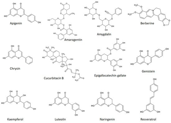
Figure 1 illustrates the chemical structures of a select group of bitter phytochemicals, which will be briefly discussed in connection with TAS2Rs.
Figure 1. Chemical structures of some bitter phytochemicals.
Apigenin, also known as 4′,5,7-trihydroxyflavone, is a bitter flavonoid present in fruits, vegetables, seasonings, medicinal plants, or plant-derived beverages (e.g., grapefruit, lettuce, celery, parsley, oregano, rosemary, red and white wine [125][126][127]). Apigenin is a well-known agonist of TAS2R14 [118][128] and TAS2R39 [118]. This flavonoid has been associated with various pharmacological activities, such as anti-oxidant [129], anti-inflammatory [130][131][132], anti-cancer [133][134], anti-bacterial [135], anti-viral [136][137], cardioprotective [138], and anti-obesity effects [139].
In the skin, apigenin showed beneficial effects in addressing inflammatory conditions and certain types of skin cancer. In a mouse model of imiquimod-induced psoriasis-like skin lesions, apigenin decreased erythema, scaling, and Psoriasis Area and Severity Index (PASI) score, and it also inhibited NF-kB activation and the IL-23/STAT3/IL-17A pathway [140].
Amarogentin, identified as an agonist of seven TAS2Rs (TAS2R1 [37], TAS2R4 [37], TAS2R39 [37], TAS2R43 [37], TAS2R46 [37], TAS2R47 [37], TAS2R50 [37]), is a secoiridoid glycoside that can be found in various plants from the Gentianaceae family and ranks among the most bitter natural substances [141][142][143].
Amygdalin, also known as D-mandelonitrile-β-D-gentiobioside or vitamin B17, is a cyanogenic glucoside notably present in the seeds of various species within the Rosaceae family (e.g., apricot, peach, bitter almond) [144] that acts as an agonist of TAS2R16 [37][145]. Amygdalin proved to have good skin penetration, making it a good candidate for skin diseases [146]. In a study using burn-induced skin wounds in diabetic rats, amygdalin improved the time and quality of wound healing [147].
Systemic administration of these amygdalin analogues in a xenograft transplantation model, where human psoriatic skin was transplanted onto immunodeficient mice, significantly improved psoriatic lesions [144]. This improvement was reflected in the reduction of clinical psoriasis score, epidermal thickness, parakeratosis, and Munro’s abscesses [144]. Also, topical application of a cream based on an amygdalin analogue ameliorated psoriasis-like disease in a mouse model [148]. It achieved this by reducing keratinocyte proliferation, skin inflammation, and the levels of systemic pro-inflammatory cytokines typically increased in psoriasis subjects, such as IL-17A, IL6, or G-CSF [148].
Berberine, a bitter alkaloid found in various plants, acts as an agonist for TAS2R38 [149] and TAS2R46 [150]. In an animal model of atopic dermatitis, berberine displayed anti-inflammatory effects [151]. In the context of skin cancer, a growing body of evidence supports the antitumor actions of berberine. In both melanoma and squamous cell carcinoma cells, berberine inhibited cell proliferation, migration, and invasion, inhibited epithelial-mesenchymal transition, and induced apoptosis [152][153][154].
Chrysin, a flavonoid also known as 5,7-dihydroxyflavone, is a TAS2R14 [118] and TAS2R39 [118] agonist. It can be found in certain medicinal plants, such as Opuntia ficus indica L and Viburnum opulus L [155][156], as well as in honey [157] and propolis [158]. In an imiquimod-induced mouse psoriasis model, chrysin demonstrated an ability to alleviate inflammation [159].
Cucurbitacin B, a triterpenoid known to act as an agonist for TAS2R10 [37] and TAS2R14 [37], shows promising anti-inflammatory and anti-cancer actions specific to the skin. In a model of imiquimod-induced skin inflammation, cucurbitacin B inhibited psoriatic cytokines (e.g., IL-8, CCL-20) and keratinocyte proliferation [160].
Epigallocatechin gallate (EGCG), a flavonoid and known agonist for TAS2R14 [118][161], TAS2R31 [161], and TAS2R39 [118][161], is the major component in green tea [162]. It demonstrates beneficial effects concerning skin inflammation and cutaneous tumors. In a mouse model induced by psoriasis-like inflammation, EGCG reduced the serum level of pro-inflammatory cytokines and mitigated dermal T cell infiltration [163].
Genistein, a soy isoflavone [164] and a recognized agonist for TAS2R14 [118] and TAS2R39 [118], has proven anticancer effects on skin cancer cell lines. It inhibited growth, proliferation, and migration, reduced cell survival and invasion, induced apoptosis, and led to cell cycle arrest [165][166][167][168][169][170][171].
Kaempferol, a known agonist of TAS2R14 [118] and TAS2R39 [118], is found abundantly in green leafy vegetables [172]. In a mouse model of imiquimod-induced psoriasis, kaempferol reduced T cell infiltration in the skin and the gene expression of inflammatory cytokines [173].
Luteolin is a recognized agonist of TAS2R14 [118] and TAS2R39 [118]. In a murine model of atopic dermatitis, luteolin reduced inflammation, oxidative stress, and serum IgE levels [174]. Similarly, in a mouse psoriasis model, luteolin inhibited the infiltration of inflammatory cells in the skin, decreased levels of proinflammatory cytokines, and lowered inflammatory mediators [175].
Naringenin, also known as 4′,5,7-trihydroxyflavone, is a citrus flavonoid identified as a TAS2R14 agonist [176][177]. In a mouse model of induced atopic dermatitis, naringenin inhibited T cell production of IFN-γ, immune cell infiltration into skin lesions, and decreased serum IgE concentration [178]. In skin cancer cell lines (both human and murine), naringenin reduced cell viability and migration and induced apoptosis [179]. In a two-stage mouse skin carcinogenesis model, naringenin decreased the number and size of tumors [180].
Resveratrol, a phytoalexin known for its activation of TAS2R14 [118] and TAS2R39 [118], can be found in grapes, wines, and peanuts [181]. It exerts various pharmacological actions, including effects at the cutaneous level. Resveratrol significantly diminished skin inflammation in a mouse model of induced psoriasis [182], and numerous studies highlight its anti-inflammatory roles in models of atopic dermatitis [183][184].
8. Phytochemical Bitter Taste Receptors agonists in Skin Aging, Inflammation, and Cancer: Insights into mammalian Target of rapamycin (mTOR) Signaling Pathways
Skin aging is a risk factor for skin carcinogenesis, attributed not only to photoaging or environmental exposure but also to intrinsic aging processes [185]. With age, proteins like keratin 10 and involucrin exhibit reduced expression in the epidermis, among other changes [185]. The mTOR pathway plays a pivotal role in aging, including skin aging [186]. This pathway is implicated in both skin inflammatory diseases (e.g., psoriasis) and skin cancers (e.g., melanoma, cutaneous T cell lymphomas) [187].
Numerous bitter phytochemicals exhibit the ability to inhibit the mTOR signaling pathway, thereby influencing skin aging, inflammation, and cancer. For instance, fisetin, an agonist of TAS2R [118], demonstrates anti-inflammatory, anti-proliferative, and pro-differentiation effects in keratinocytes by modulating the mTOR pathway [188]. Resveratrol, by inhibiting the PI3K/AKT/mTOR pathway in melanoma cells (human-A375 cells, mouse-B16-F10 cells), promotes autophagy [189].
9. Direct Involvement of Bitter Taste Receptors in the Bitter Phytochemicals’ Anti-Inflammatory and Anti-Cancer Effects
Although numerous studies demonstrate the beneficial effects of bitter phytochemicals on inflammation and cancer, and there is increasing evidence of TAS2Rs expressed across human cells, the direct connection between bitter phytochemicals and TAS2Rs in anti-inflammatory or anti-cancer actions remains relatively unexplored.
Evidence supporting the anti-inflammatory action of bitter phytochemicals through TAS2R involvement is limited. For instance, specific agonists activating TAS2Rs on mast cells have shown inhibitory effects on histamine and prostaglandin D2 (PGD2) release [190].
10. Conclusions
TAS2Rs are expressed in all the layers of human skin in a personalized manner. Differential skin expression of TAS2Rs depends on many inter-individual or intra-individual factors, including age, sex, body map, and sun exposure. Some TAS2Rs have confirmed functionality within the skin and its cell lines. Experimental studies have shown promising effects of bitter phytochemicals in skin aging, wound healing, inflammatory skin conditions, and skin cancers, both in vitro and in animal models.
Exploring the therapeutic potential of bitter phytochemicals in inflammatory skin conditions and their influence on the risk of skin cancer development in patients with skin inflammation may be an interesting research area. Another avenue of investigation could assess combinations of two or more bitter phytocompounds targeting the same TAS2R, aiming to identify potential synergistic effects in animal models of skin conditions.
The drawbacks associated with bitter phytochemicals remain relatively underexplored and necessitate comprehensive research.
References
- McGrath, J.A.; Uitto, J. Structure and Function of the Skin. In Rook’s Textbook of Dermatology, 9th ed.; Griffiths, C.E.M., Barker, J., Bleiker, T.O., Chalmers, R., Creamer, D., Eds.; John Wiley & Sons Ltd.: Chichester, UK, 2016; pp. 1–52. ISBN 9781118441213.
- Flohr, C.; Hay, R. Putting the Burden of Skin Diseases on the Global Map. Br. J. Dermatol. 2021, 184, 189–190.
- Ujiie, H.; Rosmarin, D.; Schön, M.P.; Ständer, S.; Boch, K.; Metz, M.; Maurer, M.; Thaci, D.; Schmidt, E.; Cole, C.; et al. Unmet Medical Needs in Chronic, Non-Communicable Inflammatory Skin Diseases. Front. Med. 2022, 9, 875492.
- Carretero, M.; Guerrero-Aspizua, S.; Illera, N.; Galvez, V.; Navarro, M.; García-García, F.; Dopazo, J.; Jorcano, J.L.; Larcher, F.; del Rio, M. Differential Features between Chronic Skin Inflammatory Diseases Revealed in Skin-Humanized Psoriasis and Atopic Dermatitis Mouse Models. J. Investig. Dermatol. 2016, 136, 136–145.
- Snyder, A.M.; Brandenberger, A.U.; Taliercio, V.L.; Rich, B.E.; Webber, L.B.; Beshay, A.P.; Biber, J.E.; Hess, R.; Rhoads, J.L.W.; Secrest, A.M. Quality of Life Among Family of Patients with Atopic Dermatitis and Psoriasis. Int. J. Behav. Med. 2023, 30, 409–415.
- Narayanan, D.L.; Saladi, R.N.; Fox, J.L. Review: Ultraviolet Radiation and Skin Cancer. Int. J. Dermatol. 2010, 49, 978–986.
- Becerril, S.; Corchado-Cobos, R.; García-Sancha, N.; Revelles, L.; Revilla, D.; Ugalde, T.; Román-Curto, C.; Pérez-Losada, J.; Cañueto, J. Viruses and Skin Cancer. Int. J. Mol. Sci. 2021, 22, 5399.
- Rollan, M.P.; Cabrera, R.; Schwartz, R.A. Current Knowledge of Immunosuppression as a Risk Factor for Skin Cancer Development. Crit. Rev. Oncol. Hematol. 2022, 177, 103754.
- Facciolà, A.; Venanzi Rullo, E.; Ceccarelli, M.; D’Andrea, F.; Coco, M.; Micali, C.; Cacopardo, B.; Marino, A.; Cannavò, S.P.; Di Rosa, M.; et al. Malignant Melanoma in HIV: Epidemiology, Pathogenesis, and Management. Dermatol. Ther. 2020, 33, e13180.
- Nikolaou, V.; Stratigos, A.J.; Tsao, H. Hereditary Nonmelanoma Skin Cancer. Semin. Cutan. Med. Surg. 2012, 31, 204–210.
- Zocchi, L.; Lontano, A.; Merli, M.; Dika, E.; Nagore, E.; Quaglino, P.; Puig, S.; Ribero, S. Familial Melanoma and Susceptibility Genes: A Review of the Most Common Clinical and Dermoscopic Phenotypic Aspect, Associated Malignancies and Practical Tips for Management. J. Clin. Med. 2021, 10, 3760.
- Shreberk-Hassidim, R.; Ostrowski, S.M.; Fisher, D.E. The Complex Interplay between Nevi and Melanoma: Risk Factors and Precursors. Int. J. Mol. Sci. 2023, 24, 3541.
- Arafa, A.; Mostafa, A.; Navarini, A.A.; Dong, J.-Y. The Association between Smoking and Risk of Skin Cancer: A Meta-Analysis of Cohort Studies. Cancer Causes Control 2020, 31, 787–794.
- Matthews, N.H.; Fitch, K.; Li, W.-Q.; Morris, J.S.; Christiani, D.C.; Qureshi, A.A.; Cho, E. Exposure to Trace Elements and Risk of Skin Cancer: A Systematic Review of Epidemiologic Studies. Cancer Epidemiol. Biomark. Prev. 2019, 28, 3–21.
- Bennardo, L.; Passante, M.; Cameli, N.; Cristaudo, A.; Patruno, C.; Nisticò, S.P.; Silvestri, M. Skin Manifestations after Ionizing Radiation Exposure: A Systematic Review. Bioengineering 2021, 8, 153.
- Neagu, M.; Constantin, C.; Caruntu, C.; Dumitru, C.; Surcel, M.; Zurac, S. Inflammation: A Key Process in Skin Tumorigenesis (Review). Oncol. Lett. 2018, 17, 4068–4084.
- Ju, T.; Hernandez, L.; Mohsin, N.; Labib, A.; Frech, F.; Nouri, K. Evaluation of Risk in Chronic Cutaneous Inflammatory Conditions for Malignant Transformation. J. Eur. Acad. Dermatol. Venereol. 2023, 37, 231–242.
- Sung, H.; Ferlay, J.; Siegel, R.L.; Laversanne, M.; Soerjomataram, I.; Jemal, A.; Bray, F. Global Cancer Statistics 2020: GLOBOCAN Estimates of Incidence and Mortality Worldwide for 36 Cancers in 185 Countries. CA Cancer J. Clin. 2021, 71, 209–249.
- Sackstein, R.; Schatton, T.; Barthel, S.R. T-Lymphocyte Homing: An Underappreciated yet Critical Hurdle for Successful Cancer Immunotherapy. Lab. Investig. 2017, 97, 669–697.
- Spałkowska, M.; Dyduch, G.; Broniatowska, E.; Damiani, G.; Wojas-Pelc, A. Molecular Proof of a Clinical Concept: Expression of Estrogen Alpha-, Beta-Receptors and G Protein-Coupled Estrogen Receptor 1 (GPER) in Histologically Assessed Common Nevi, Dysplastic Nevi and Melanomas. Medicina 2021, 57, 1228.
- Rees, J.L. The Genetics of Sun Sensitivity in Humans. Am. J. Hum. Genet. 2004, 75, 739–751.
- Yamaguchi, Y.; Passeron, T.; Watabe, H.; Yasumoto, K.; Rouzaud, F.; Hoashi, T.; Hearing, V.J. The Effects of Dickkopf 1 on Gene Expression and Wnt Signaling by Melanocytes: Mechanisms Underlying Its Suppression of Melanocyte Function and Proliferation. J. Investig. Dermatol. 2007, 127, 1217–1225.
- Labes, A. Marine Resources Offer New Compounds and Strategies for the Treatment of Skin and Soft Tissue Infections. Mar. Drugs 2023, 21, 387.
- Liu, J.-K. Natural Products in Cosmetics. Nat. Prod. Bioprospect. 2022, 12, 40.
- Nicholas-Haizelden, K.; Murphy, B.; Hoptroff, M.; Horsburgh, M.J. Bioprospecting the Skin Microbiome: Advances in Therapeutics and Personal Care Products. Microorganisms 2023, 11, 1899.
- Yoon, J.H.; Kim, M.-Y.; Cho, J.Y. Apigenin: A Therapeutic Agent for Treatment of Skin Inflammatory Diseases and Cancer. Int. J. Mol. Sci. 2023, 24, 1498.
- Peterle, L.; Sanfilippo, S.; Borgia, F.; Li Pomi, F.; Vadalà, R.; Costa, R.; Cicero, N.; Gangemi, S. The Role of Nutraceuticals and Functional Foods in Skin Cancer: Mechanisms and Therapeutic Potential. Foods 2023, 12, 2629.
- Dragoș, D.; Petran, M.; Gradinaru, T.-C.; Gilca, M. Phytochemicals and Inflammation: Is Bitter Better? Plants 2022, 11, 2991.
- Grădinaru, T.-C.; Gilca, M.; Vlad, A.; Dragoș, D. Relevance of Phytochemical Taste for Anti-Cancer Activity: A Statistical Inquiry. Int. J. Mol. Sci. 2023, 24, 16227.
- Reed, D.R.; Knaapila, A. Genetics of Taste and Smell. In Progress in Molecular Biology and Translational Science; Elsevier: Amsterdam, The Netherlands, 2010; Volume 94, pp. 213–240.
- Lindemann, B. Taste Reception. Physiol. Rev. 1996, 76, 718–766.
- Lang, T.; Di Pizio, A.; Risso, D.; Drayna, D.; Behrens, M. Activation Profile of TAS2R2, the 26th Human Bitter Taste Receptor. Mol. Nutr. Food Res. 2023, 67, e2200775.
- Sanematsu, K.; Yoshida, R.; Shigemura, N.; Ninomiya, Y. Structure, Function, and Signaling of Taste G-Protein-Coupled Receptors. Curr. Pharm. Biotechnol. 2014, 15, 951–961.
- Ahmad, R.; Dalziel, J.E. G Protein-Coupled Receptors in Taste Physiology and Pharmacology. Front. Pharmacol. 2020, 11, 587664.
- Talmon, M.; Pollastro, F.; Fresu, L.G. The Complex Journey of the Calcium Regulation Downstream of TAS2R Activation. Cells 2022, 11, 3638.
- Ho, H.K.-Y.; Bigliardi, P.L.; Stelmashenko, O.; Ramasamy, S.; Postlethwaite, M.; Bigliardi-Qi, M. Functionally Expressed Bitter Taste Receptor TAS2R14 in Human Epidermal Keratinocytes Serves as a Chemosensory Receptor. Exp. Dermatol. 2021, 30, 216–225.
- Meyerhof, W.; Batram, C.; Kuhn, C.; Brockhoff, A.; Chudoba, E.; Bufe, B.; Appendino, G.; Behrens, M. The Molecular Receptive Ranges of Human TAS2R Bitter Taste Receptors. Chem. Senses 2010, 35, 157–170.
- Ji, M.; Su, X.; Su, X.; Chen, Y.; Huang, W.; Zhang, J.; Gao, Z.; Li, C.; Lu, X. Identification of Novel Compounds for Human Bitter Taste Receptors. Chem. Biol. Drug Des. 2014, 84, 63–74.
- Di Pizio, A.; Niv, M.Y. Promiscuity and Selectivity of Bitter Molecules and Their Receptors. Bioorg. Med. Chem. 2015, 23, 4082–4091.
- Thalmann, S.; Behrens, M.; Meyerhof, W. Major Haplotypes of the Human Bitter Taste Receptor TAS2R41 Encode Functional Receptors for Chloramphenicol. Biochem. Biophys. Res. Commun. 2013, 435, 267–273.
- Lossow, K.; Hubner, S.; Roudnitzky, N.; Slack, J.P.; Pollastro, F.; Behrens, M.; Meyerhof, W. Comprehensive Analysis of Mouse Bitter Taste Receptors Reveals Different Molecular Receptive Ranges for Orthologous Receptors in Mice and Humans. J. Biol. Chem. 2016, 291, 15358–15377.
- Tuzim, K.; Korolczuk, A. An Update on Extra-Oral Bitter Taste Receptors. J. Transl. Med. 2021, 19, 440.
- Behrens, M.; Brockhoff, A.; Kuhn, C.; Bufe, B.; Winnig, M.; Meyerhof, W. The Human Taste Receptor HTAS2R14 Responds to a Variety of Different Bitter Compounds. Biochem. Biophys. Res. Commun. 2004, 319, 479–485.
- Brockhoff, A.; Behrens, M.; Massarotti, A.; Appending, G.; Meyerhof, W. Broad Tuning of the Human Bitter Taste Receptor HTAS2R46 to Various Sesquiterpene Lactones, Clerodane and Labdane Diterpenoids, Strychnine, and Denatonium. J. Agric. Food Chem. 2007, 55, 6236–6243.
- Yamazaki, T.; Sagisaka, M.; Ikeda, R.; Nakamura, T.; Matsuda, N.; Ishii, T.; Nakayama, T.; Watanabe, T. The Human Bitter Taste Receptor HTAS2R39 Is the Primary Receptor for the Bitterness of Theaflavins. Biosci. Biotechnol. Biochem. 2014, 78, 1753–1756.
- Foster, S.R.; Porrello, E.R.; Purdue, B.; Chan, H.-W.; Voigt, A.; Frenzel, S.; Hannan, R.D.; Moritz, K.M.; Simmons, D.G.; Molenaar, P.; et al. Expression, Regulation and Putative Nutrient-Sensing Function of Taste GPCRs in the Heart. PLoS ONE 2013, 8, e64579.
- Kertesz, Z.; Harrington, E.O.; Braza, J.; Guarino, B.D.; Chichger, H. Agonists for Bitter Taste Receptors T2R10 and T2R38 Attenuate LPS-Induced Permeability of the Pulmonary Endothelium in Vitro. Front. Physiol. 2022, 13, 794370.
- Kaji, I.; Karaki, S.; Fukami, Y.; Terasaki, M.; Kuwahara, A. Secretory Effects of a Luminal Bitter Tastant and Expressions of Bitter Taste Receptors, T2Rs, in the Human and Rat Large Intestine. Am. J. Physiol. Gastrointest. Liver Physiol. 2009, 296, G971–G981.
- Jeon, T.-I.; Seo, Y.-K.; Osborne, T.F. Gut Bitter Taste Receptor Signalling Induces ABCB1 through a Mechanism Involving CCK. Biochem. J. 2011, 438, 33–37.
- Liszt, K.I.; Wang, Q.; Farhadipour, M.; Segers, A.; Thijs, T.; Nys, L.; Deleus, E.; Van der Schueren, B.; Gerner, C.; Neuditschko, B.; et al. Human Intestinal Bitter Taste Receptors Regulate Innate Immune Responses and Metabolic Regulators in Obesity. J. Clin. Investig. 2022, 132, 1–16.
- Clark, A.A.; Dotson, C.D.; Elson, A.E.T.; Voigt, A.; Boehm, U.; Meyerhof, W.; Steinle, N.I.; Munger, S.D. TAS2R Bitter Taste Receptors Regulate Thyroid Function. FASEB J. 2015, 29, 164–172.
- Malki, A.; Fiedler, J.; Fricke, K.; Ballweg, I.; Pfaffl, M.W.; Krautwurst, D. Class I Odorant Receptors, TAS1R and TAS2R Taste Receptors, Are Markers for Subpopulations of Circulating Leukocytes. J. Leukoc. Biol. 2015, 97, 533–545.
- Tran, H.T.T.; Herz, C.; Ruf, P.; Stetter, R.; Lamy, E. Human T2R38 Bitter Taste Receptor Expression in Resting and Activated Lymphocytes. Front. Immunol. 2018, 9, 2949.
- Shaw, L.; Mansfield, C.; Colquitt, L.; Lin, C.; Ferreira, J.; Emmetsberger, J.; Reed, D.R. Personalized Expression of Bitter ‘Taste’ Receptors in Human Skin. PLoS ONE 2018, 13, e0205322.
- Reszka, E.; Nowakowska-Swirta, E.; Kupczyk, M.; Dudek, W.; Swierczynska-Machura, D.; Wittczak, T.; Rykała, J.; Przybek, M. Expression of Bitter Taste Receptors in the Human Skin In Vitro. J. Clin. Res. Bioeth. 2015, 6, 1000218.
- Wolfle, U.; Elsholz, F.A.; Kersten, A.; Haarhaus, B.; Muller, W.E.; Schempp, C.M. Expression and Functional Activity of the Bitter Taste Receptors TAS2R1 and TAS2R38 in Human Keratinocytes. Skin Pharmacol. Physiol. 2015, 28, 137–146.
- Talmon, M.; Massara, E.; Quaregna, M.; De Battisti, M.; Boccafoschi, F.; Lecchi, G.; Puppo, F.; Bettega Cajandab, M.A.; Salamone, S.; Bovio, E.; et al. Bitter Taste Receptor (TAS2R) 46 in Human Skeletal Muscle: Expression and Activity. Front. Pharmacol. 2023, 14, 1205651.
- Garcia-Esparcia, P.; Schluter, A.; Carmona, M.; Moreno, J.; Ansoleaga, B.; Torrejon-Escribano, B.; Gustincich, S.; Pujol, A.; Ferrer, I. Functional Genomics Reveals Dysregulation of Cortical Olfactory Receptors in Parkinson Disease: Novel Putative Chemoreceptors in the Human Brain. J. Neuropathol. Exp. Neurol. 2013, 72, 524–539.
- Wölfle, U.; Haarhaus, B.; Kersten, A.; Fiebich, B.; Hug, M.J.; Schempp, C.M. Salicin from Willow Bark Can Modulate Neurite Outgrowth in Human Neuroblastoma SH-SY5Y Cells. Phyther. Res. 2015, 29, 1494–1500.
- Ansoleaga, B.; Garcia-Esparcia, P.; Pinacho, R.; Haro, J.M.; Ramos, B.; Ferrer, I. Decrease in Olfactory and Taste Receptor Expression in the Dorsolateral Prefrontal Cortex in Chronic Schizophrenia. J. Psychiatr. Res. 2015, 60, 109–116.
- Ansoleaga, B.; Garcia-Esparcia, P.; Llorens, F.; Moreno, J.; Aso, E.; Ferrer, I. Dysregulation of Brain Olfactory and Taste Receptors in AD, PSP and CJD, and AD-Related Model. Neuroscience 2013, 248, 369–382.
- Zheng, M.; Simon, R.; Mirlacher, M.; Maurer, R.; Gasser, T.; Forster, T.; Diener, P.A.; Mihatsch, M.J.; Sauter, G.; Schraml, P. TRIO Amplification and Abundant MRNA Expression Is Associated with Invasive Tumor Growth and Rapid Tumor Cell Proliferation in Urinary Bladder Cancer. Am. J. Pathol. 2004, 165, 63–69.
- Wölfle, U.; Elsholz, F.; Kersten, A.; Haarhaus, B.; Schumacher, U.; Schempp, C. Expression and Functional Activity of the Human Bitter Taste Receptor TAS2R38 in Human Placental Tissues and JEG-3 Cells. Molecules 2016, 21, 306.
- Governini, L.; Semplici, B.; Pavone, V.; Crifasi, L.; Marrocco, C.; De Leo, V.; Arlt, E.; Gudermann, T.; Boekhoff, I.; Luddi, A.; et al. Expression of Taste Receptor 2 Subtypes in Human Testis and Sperm. J. Clin. Med. 2020, 9, 264.
- Semplici, B.; Luongo, F.P.; Passaponti, S.; Landi, C.; Governini, L.; Morgante, G.; De Leo, V.; Piomboni, P.; Luddi, A. Bitter Taste Receptors Expression in Human Granulosa and Cumulus Cells: New Perspectives in Female Fertility. Cells 2021, 10, 3127.
- Martin, L.T.P.; Nachtigal, M.W.; Selman, T.; Nguyen, E.; Salsman, J.; Dellaire, G.; Dupré, D.J. Bitter Taste Receptors Are Expressed in Human Epithelial Ovarian and Prostate Cancers Cells and Noscapine Stimulation Impacts Cell Survival. Mol. Cell. Biochem. 2019, 454, 203–214.
- Shah, A.S.; Ben-Shahar, Y.; Moninger, T.O.; Kline, J.N.; Welsh, M.J. Motile Cilia of Human Airway Epithelia Are Chemosensory. Science 2009, 325, 1131–1134.
- Grassin-Delyle, S.; Abrial, C.; Fayad-Kobeissi, S.; Brollo, M.; Faisy, C.; Alvarez, J.-C.; Naline, E.; Devillier, P. The Expression and Relaxant Effect of Bitter Taste Receptors in Human Bronchi. Respir. Res. 2013, 14, 134.
- Barham, H.P.; Cooper, S.E.; Anderson, C.B.; Tizzano, M.; Kingdom, T.T.; Finger, T.E.; Kinnamon, S.C.; Ramakrishnan, V.R. Solitary Chemosensory Cells and Bitter Taste Receptor Signaling in Human Sinonasal Mucosa. Int. Forum Allergy Rhinol. 2013, 3, 450–457.
- Lund, T.C.; Kobs, A.J.; Kramer, A.; Nyquist, M.; Kuroki, M.T.; Osborn, J.; Lidke, D.S.; Low-Nam, S.T.; Blazar, B.R.; Tolar, J. Bone Marrow Stromal and Vascular Smooth Muscle Cells Have Chemosensory Capacity via Bitter Taste Receptor Expression. PLoS ONE 2013, 8, e58945.
- Cheng, W.; Yao, M.; Liu, F. Bitter Taste Receptor as a Therapeutic Target in Orthopaedic Disorders. Drug Des. Devel. Ther. 2021, 15, 895–903.
- Wang, Y.; Zajac, A.L.; Lei, W.; Christensen, C.M.; Margolskee, R.F.; Bouysset, C.; Golebiowski, J.; Zhao, H.; Fiorucci, S.; Jiang, P. Metal Ions Activate the Human Taste Receptor TAS2R7. Chem. Senses 2019, 44, 339–347.
- Gaida, M.; Dapunt, U.; Hansch, G.M. Sensing Developing Biofilms: The Bitter Receptor T2R38 on Myeloid Cells. Pathog. Dis. 2016, 74, ftw004.
- Kang, W.; Wang, Y.; Li, J.; Xie, W.; Zhao, D.; Wu, L.; Wang, H.; Xie, S. TAS2R Supports Odontoblastic Differentiation of Human Dental Pulp Stem Cells in the Inflammatory Microenvironment. Stem Cell Res. Ther. 2022, 13, 374.
- Orsmark-Pietras, C.; James, A.; Konradsen, J.R.; Nordlund, B.; Söderhäll, C.; Pulkkinen, V.; Pedroletti, C.; Daham, K.; Kupczyk, M.; Dahlén, B.; et al. Transcriptome Analysis Reveals Upregulation of Bitter Taste Receptors in Severe Asthmatics. Eur. Respir. J. 2013, 42, 65–78.
- Singh, N.; Chakraborty, R.; Bhullar, R.P.; Chelikani, P. Differential Expression of Bitter Taste Receptors in Non-Cancerous Breast Epithelial and Breast Cancer Cells. Biochem. Biophys. Res. Commun. 2014, 446, 499–503.
- Carey, R.M.; Kim, T.; Cohen, N.A.; Lee, R.J.; Nead, K.T. Impact of Sweet, Umami, and Bitter Taste Receptor (TAS1R and TAS2R) Genomic and Expression Alterations in Solid Tumors on Survival. Sci. Rep. 2022, 12, 8937.
- Lee, R.J.; Cohen, N.A. Taste Receptors in Innate Immunity. Cell. Mol. Life Sci. 2015, 72, 217–236.
- Patel, N.N.; Workman, A.D.; Cohen, N.A. Role of Taste Receptors as Sentinels of Innate Immunity in the Upper Airway. J. Pathog. 2018, 2018, 9541987.
- Xi, R.; Zheng, X.; Tizzano, M. Role of Taste Receptors in Innate Immunity and Oral Health. J. Dent. Res. 2022, 101, 759–768.
- Tran, H.T.T.; Stetter, R.; Herz, C.; Spöttel, J.; Krell, M.; Hanschen, F.S.; Schreiner, M.; Rohn, S.; Behrens, M.; Lamy, E. Allyl Isothiocyanate: A TAS2R38 Receptor-Dependent Immune Modulator at the Interface Between Personalized Medicine and Nutrition. Front. Immunol. 2021, 12, 669005.
- Grassin-Delyle, S.; Salvator, H.; Mantov, N.; Abrial, C.; Brollo, M.; Faisy, C.; Naline, E.; Couderc, L.-J.; Devillier, P. Bitter Taste Receptors (TAS2Rs) in Human Lung Macrophages: Receptor Expression and Inhibitory Effects of TAS2R Agonists. Front. Physiol. 2019, 10, 1267.
- Zhou, Z.; Xi, R.; Liu, J.; Peng, X.; Zhao, L.; Zhou, X.; Li, J.; Zheng, X.; Xu, X. TAS2R16 Activation Suppresses LPS-Induced Cytokine Expression in Human Gingival Fibroblasts. Front. Immunol. 2021, 12, 726546.
- Liszt, K.I.; Ley, J.P.; Lieder, B.; Behrens, M.; Stöger, V.; Reiner, A.; Hochkogler, C.M.; Köck, E.; Marchiori, A.; Hans, J.; et al. Caffeine Induces Gastric Acid Secretion via Bitter Taste Signaling in Gastric Parietal Cells. Proc. Natl. Acad. Sci. USA 2017, 114, E6260–E6269.
- Grau-Bové, C.; Miguéns-Gómez, A.; González-Quilen, C.; Fernández-López, J.-A.; Remesar, X.; Torres-Fuentes, C.; Ávila-Román, J.; Rodríguez-Gallego, E.; Beltrán-Debón, R.; Blay, M.T.; et al. Modulation of Food Intake by Differential TAS2R Stimulation in Rat. Nutrients 2020, 12, 3784.
- Zagorchev, P.; Petkov, G.V.; Gagov, H.S. Bitter Taste Receptors as Regulators of Abdominal Muscles Contraction. Physiol. Res. 2019, 68, 991–995.
- Zhou, Y.-W.; Sun, J.; Wang, Y.; Chen, C.-P.; Tao, T.; Ma, M.; Chen, X.; Zhang, X.-N.; Yang, L.-Y.; Zhang, Z.-L.; et al. Tas2R Activation Relaxes Airway Smooth Muscle by Release of Gα t Targeting on AChR Signaling. Proc. Natl. Acad. Sci. USA 2022, 119, e2121513119.
- Costa, A.R.; Duarte, A.C.; Costa-Brito, A.R.; Gonçalves, I.; Santos, C.R.A. Bitter Taste Signaling in Cancer. Life Sci. 2023, 315, 121363.
- Zehentner, S.; Reiner, A.T.; Grimm, C.; Somoza, V. The Role of Bitter Taste Receptors in Cancer: A Systematic Review. Cancers 2021, 13, 5891.
- Seo, Y.; Kim, Y.-S.; Lee, K.E.; Park, T.H.; Kim, Y. Anti-Cancer Stemness and Anti-Invasive Activity of Bitter Taste Receptors, TAS2R8 and TAS2R10, in Human Neuroblastoma Cells. PLoS ONE 2017, 12, e0176851.
- Pham, H.; Hui, H.; Morvaridi, S.; Cai, J.; Zhang, S.; Tan, J.; Wu, V.; Levin, N.; Knudsen, B.; Goddard, W.A.; et al. A Bitter Pill for Type 2 Diabetes? The Activation of Bitter Taste Receptor TAS2R38 Can Stimulate GLP-1 Release from Enteroendocrine L-Cells. Biochem. Biophys. Res. Commun. 2016, 475, 295–300.
- Choi, J.-H. Variation in the TAS2R38 Bitterness Receptor Gene Was Associated with Food Consumption and Obesity Risk in Koreans. Nutrients 2019, 11, 1973.
- Sun, S.; Yang, Y.; Xiong, R.; Ni, Y.; Ma, X.; Hou, M.; Chen, L.; Xu, Z.; Chen, L.; Ji, M. Oral Berberine Ameliorates High-Fat Diet-Induced Obesity by Activating TAS2Rs in Tuft and Endocrine Cells in the Gut. Life Sci. 2022, 311, 121141.
- Lee, R.J.; Cohen, N.A. Role of the Bitter Taste Receptor T2R38 in Upper Respiratory Infection and Chronic Rhinosinusitis. Curr. Opin. Allergy Clin. Immunol. 2015, 15, 14–20.
- Luo, X.-C.; Chen, Z.-H.; Xue, J.-B.; Zhao, D.-X.; Lu, C.; Li, Y.-H.; Li, S.-M.; Du, Y.-W.; Liu, Q.; Wang, P.; et al. Infection by the Parasitic Helminth Trichinella Spiralis Activates a Tas2r-Mediated Signaling Pathway in Intestinal Tuft Cells. Proc. Natl. Acad. Sci. USA 2019, 116, 5564–5569.
- Wölfle, U.; Haarhaus, B.; Schempp, C.M. Amarogentin Displays Immunomodulatory Effects in Human Mast Cells and Keratinocytes. Mediat. Inflamm. 2015, 2015, 630128.
- Sakakibara, M.; Sumida, H.; Yanagida, K.; Miyasato, S.; Nakamura, M.; Sato, S. Bitter Taste Receptor T2R38 Is Expressed on Skin-Infiltrating Lymphocytes and Regulates Lymphocyte Migration. Sci. Rep. 2022, 12, 11790.
- Gherardini, J.; Rouille, T.; Ferholz, M.; Funk, W.; Rodríguez-Feliz, J.; Bauman, A.J.; Bíró, T.; Chéret, J.; Paus, R. 571 Human Hair Follicles Can “Taste”: Stimulation of the Bitter Taste Receptor TAS2R4 Inhibits Hair Growth Ex Vivo by Up-Regulating TGF-Β2. J. Investig. Dermatol. 2022, 142, S279.
- Chung, M.G.; Kim, Y.; Cha, Y.K.; Park, T.H.; Kim, Y. Bitter Taste Receptors Protect against Skin Aging by Inhibiting Cellular Senescence and Enhancing Wound Healing. Nutr. Res. Pract. 2022, 16, 1.
- Wölfle, U.; Haarhaus, B.; Seiwerth, J.; Cawelius, A.; Schwabe, K.; Quirin, K.-W.; Schempp, C. The Herbal Bitter Drug Gentiana Lutea Modulates Lipid Synthesis in Human Keratinocytes In Vitro and In Vivo. Int. J. Mol. Sci. 2017, 18, 1814.
- de Jesus, V.C.; Mittermuller, B.-A.; Hu, P.; Schroth, R.J.; Chelikani, P. Genetic Variants in Taste Genes Play a Role in Oral Microbial Composition and Severe Early Childhood Caries. iScience 2022, 25, 105489.
- Lumpkin, E.A.; Caterina, M.J. Mechanisms of Sensory Transduction in the Skin. Nature 2007, 445, 858–865.
- Keppel Hesselink, J.M.; Kopsky, D.J.; Bhaskar, A.K. Skin Matters! The Role of Keratinocytes in Nociception: A Rational Argument for the Development of Topical Analgesics. J. Pain Res. 2016, 10, 1–8.
- Umbayev, B.; Askarova, S.; Almabayeva, A.; Saliev, T.; Masoud, A.-R.; Bulanin, D. Galactose-Induced Skin Aging: The Role of Oxidative Stress. Oxidative Med. Cell. Longev. 2020, 2020, 7145656.
- Schwartzenfeld, D.M.; Karamikian, J. Chapter 18—Hair Transplantation. In Plastic Surgery Secrets Plus, 2nd ed.; Weinzweig, J., Ed.; Mosby: Philadelphia, PA, USA, 2010; pp. 123–127. ISBN 978-0-323-03470-8.
- Cancello, R.; Micheletto, G.; Meta, D.; Lavagno, R.; Bevilacqua, E.; Panizzo, V.; Invitti, C. Expanding the Role of Bitter Taste Receptor in Extra Oral Tissues: TAS2R38 Is Expressed in Human Adipocytes. Adipocyte 2020, 9, 7–15.
- Avau, B.; Bauters, D.; Steensels, S.; Vancleef, L.; Laermans, J.; Lesuisse, J.; Buyse, J.; Lijnen, H.R.; Tack, J.; Depoortere, I. The Gustatory Signaling Pathway and Bitter Taste Receptors Affect the Development of Obesity and Adipocyte Metabolism in Mice. PLoS ONE 2015, 10, e0145538.
- Shain, A.H.; Bastian, B.C. From Melanocytes to Melanomas. Nat. Rev. Cancer 2016, 16, 345–358.
- Uhlen, M.; Fagerberg, L.; Hallstrom, B.M.; Lindskog, C.; Oksvold, P.; Mardinoglu, A.; Sivertsson, A.; Kampf, C.; Sjostedt, E.; Asplund, A.; et al. Proteomics. Tissue-Based Map of the Human Proteome. Science 2015, 347, 1260419.
- Kallini, J.R.; Hamed, N.; Khachemoune, A. Squamous Cell Carcinoma of the Skin: Epidemiology, Classification, Management, and Novel Trends. Int. J. Dermatol. 2015, 54, 130–140.
- Almangush, A.; Mäkitie, A.A.; Triantafyllou, A.; de Bree, R.; Strojan, P.; Rinaldo, A.; Hernandez-Prera, J.C.; Suárez, C.; Kowalski, L.P.; Ferlito, A.; et al. Staging and Grading of Oral Squamous Cell Carcinoma: An Update. Oral Oncol. 2020, 107, 104799.
- Carey, R.M.; McMahon, D.B.; Miller, Z.A.; Kim, T.; Rajasekaran, K.; Gopallawa, I.; Newman, J.G.; Basu, D.; Nead, K.T.; White, E.A.; et al. T2R Bitter Taste Receptors Regulate Apoptosis and May Be Associated with Survival in Head and Neck Squamous Cell Carcinoma. Mol. Oncol. 2022, 16, 1474–1492.
- Luongo, F.P.; Passaponti, S.; Haxhiu, A.; Raeispour, M.; Belmonte, G.; Governini, L.; Casarini, L.; Piomboni, P.; Luddi, A. Bitter Taste Receptors and Endocrine Disruptors: Cellular and Molecular Insights from an In Vitro Model of Human Granulosa Cells. Int. J. Mol. Sci. 2022, 23, 15540.
- Slominski, A.; Zbytek, B.; Nikolakis, G.; Manna, P.R.; Skobowiat, C.; Zmijewski, M.; Li, W.; Janjetovic, Z.; Postlethwaite, A.; Zouboulis, C.C.; et al. Steroidogenesis in the Skin: Implications for Local Immune Functions. J. Steroid Biochem. Mol. Biol. 2013, 137, 107–123.
- Gradinaru, T.-C.; Petran, M.; Dragos, D.; Gilca, M. PlantMolecularTasteDB: A Database of Taste Active Phytochemicals. Front. Pharmacol. 2022, 12, 751712.
- Dagan-Wiener, A.; Nissim, I.; Ben Abu, N.; Borgonovo, G.; Bassoli, A.; Niv, M.Y. Bitter or Not? BitterPredict, a Tool for Predicting Taste from Chemical Structure. Sci. Rep. 2017, 7, 12074.
- Soares, S.; Silva, M.S.; García-Estevez, I.; Groβmann, P.; Brás, N.; Brandão, E.; Mateus, N.; de Freitas, V.; Behrens, M.; Meyerhof, W. Human Bitter Taste Receptors Are Activated by Different Classes of Polyphenols. J. Agric. Food Chem. 2018, 66, 8814–8823.
- Roland, W.S.U.; Van Buren, L.; Gruppen, H.; Driesse, M.; Gouka, R.J.; Smit, G.; Vincken, J.P. Bitter Taste Receptor Activation by Flavonoids and Isoflavonoids: Modeled Structural Requirements for Activation of HTAS2R14 and HTAS2R39. J. Agric. Food Chem. 2013, 61, 10454–10466.
- Sakurai, T.; Misaka, T.; Ueno, Y.; Ishiguro, M.; Matsuo, S.; Ishimaru, Y.; Asakura, T.; Abe, K. The Human Bitter Taste Receptor, HTAS2R16, Discriminates Slight Differences in the Configuration of Disaccharides. Biochem. Biophys. Res. Commun. 2010, 402, 595–601.
- Chialva, F.; Dada, G. Bitterness in Alcoholic Beverages. In Bitternes in Foods and Beverages. Developments in Food Science; Rouseff, R.L., Ed.; Elsevier: Amsterdam, The Netherlands, 1990; Volume 25, pp. 103–122.
- Steinhardt, R.G.; Calvin, A.D.; Dodd, E.A. Taste-Structure Correlation with α-D-Mannose and β-D-Mannose. Science 1962, 135, 367–368.
- Hellfritsch, C.; Brockhoff, A.; Stähler, F.; Meyerhof, W.; Hofmann, T. Human Psychometric and Taste Receptor Responses to Steviol Glycosides. J. Agric. Food Chem. 2012, 60, 6782–6793.
- Dragos, D.; Gilca, M. Taste of Phytocompounds: A Better Predictor for Ethnopharmacological Activities of Medicinal Plants Than The Phytochemical Class? J. Ethnopharmacol. 2018, 220, 129–146.
- Trinh, X.-T.; Long, N.-V.; Van Anh, L.T.; Nga, P.T.; Giang, N.N.; Chien, P.N.; Nam, S.-Y.; Heo, C.-Y. A Comprehensive Review of Natural Compounds for Wound Healing: Targeting Bioactivity Perspective. Int. J. Mol. Sci. 2022, 23, 9573.
- Ali, F.; Rahul; Naz, F.; Jyoti, S.; Siddique, Y.H. Health Functionality of Apigenin: A Review. Int. J. Food Prop. 2017, 20, 1197–1238.
- Shankar, E.; Goel, A.; Gupta, K.; Gupta, S. Plant Flavone Apigenin: An Emerging Anticancer Agent. Curr. Pharmacol. Rep. 2017, 3, 423–446.
- Hostetler, G.L.; Ralston, R.A.; Schwartz, S.J. Flavones: Food Sources, Bioavailability, Metabolism, and Bioactivity. Adv. Nutr. 2017, 8, 423–435.
- Behrens, M.; Meyerhof, W. The Vertebrate Gustatory System. In Flavour; Guichard, E., Salles, C., Morzel, M., Le Bon, A.M., Eds.; John Wiley & Sons, Ltd.: Chichester, UK, 2016; pp. 57–78. ISBN 9781118929384.
- Kashyap, P.; Shikha, D.; Thakur, M.; Aneja, A. Functionality of Apigenin as a Potent Antioxidant with Emphasis on Bioavailability, Metabolism, Action Mechanism and in Vitro and in Vivo Studies: A Review. J. Food Biochem. 2022, 46, e13950.
- Ginwala, R.; Bhavsar, R.; Chigbu, D.I.; Jain, P.; Khan, Z.K. Potential Role of Flavonoids in Treating Chronic Inflammatory Diseases with a Special Focus on the Anti-Inflammatory Activity of Apigenin. Antioxidants 2019, 8, 35.
- Zhang, X.; Wang, G.; Gurley, E.C.; Zhou, H. Flavonoid Apigenin Inhibits Lipopolysaccharide-Induced Inflammatory Response through Multiple Mechanisms in Macrophages. PLoS ONE 2014, 9, e107072.
- Lee, J.-H.; Zhou, H.Y.; Cho, S.Y.; Kim, Y.S.; Lee, Y.S.; Jeong, C.S. Anti-Inflammatory Mechanisms of Apigenin: Inhibition of Cyclooxygenase-2 Expression, Adhesion of Monocytes to Human Umbilical Vein Endothelial Cells, and Expression of Cellular Adhesion Molecules. Arch. Pharm. Res. 2007, 30, 1318–1327.
- Imran, M.; Aslam Gondal, T.; Atif, M.; Shahbaz, M.; Batool Qaisarani, T.; Hanif Mughal, M.; Salehi, B.; Martorell, M.; Sharifi-Rad, J. Apigenin as an Anticancer Agent. Phyther. Res. 2020, 34, 1812–1828.
- Rahmani, A.H.; Alsahli, M.A.; Almatroudi, A.; Almogbel, M.A.; Khan, A.A.; Anwar, S.; Almatroodi, S.A. The Potential Role of Apigenin in Cancer Prevention and Treatment. Molecules 2022, 27, 6051.
- Nayaka, H.B.; Londonkar, R.L.; Umesh, M.K.; Tukappa, A. Antibacterial Attributes of Apigenin, Isolated from Portulaca oleracea L. Int. J. Bacteriol. 2014, 2014, 175851.
- Xu, X.; Miao, J.; Shao, Q.; Gao, Y.; Hong, L. Apigenin Suppresses Influenza A Virus-induced RIG-I Activation and Viral Replication. J. Med. Virol. 2020, 92, 3057–3066.
- Khandelwal, N.; Chander, Y.; Kumar, R.; Riyesh, T.; Dedar, R.K.; Kumar, M.; Gulati, B.R.; Sharma, S.; Tripathi, B.N.; Barua, S.; et al. Antiviral Activity of Apigenin against Buffalopox: Novel Mechanistic Insights and Drug-Resistance Considerations. Antivir. Res. 2020, 181, 104870.
- Thomas, S.D.; Jha, N.K.; Jha, S.K.; Sadek, B.; Ojha, S. Pharmacological and Molecular Insight on the Cardioprotective Role of Apigenin. Nutrients 2023, 15, 385.
- Qiao, Y.; Zhang, Z.; Zhai, Y.; Yan, X.; Zhou, W.; Liu, H.; Guan, L.; Peng, L. Apigenin Alleviates Obesity-Associated Metabolic Syndrome by Regulating the Composition of the Gut Microbiome. Front. Microbiol. 2021, 12, 805827.
- Meng, X.; Zheng, S.; Yin, Z.; Wang, X.; Yang, D.; Zou, T.; Li, H.; Chen, Y.; Liao, C.; Xie, Z.; et al. Apigenin Ameliorates Imiquimod-Induced Psoriasis in C57BL/6J Mice by Inactivating STAT3 and NF-ΚB. Food Sci. Hum. Wellness 2024, 13, 211–224.
- Fiorito, S.; Epifano, F.; Marchetti, L.; Palumbo, L.; Mascioli, F.; Bastianini, M.; Cardellini, F.; Spogli, R.; Genovese, S. An Improved Method for the Isolation of Amarogentin, the Bitter Principle of Yellow Gentian Roots. Food Chem. 2021, 364, 130383.
- Zhang, L.; Ulriksen, E.S.; Hoel, H.; Sandvik, L.; Malterud, K.E.; Inngjerdingen, K.T.; Inngjerdingen, M.; Wangensteen, H. Phytochemical Characterization and Anti-Inflammatory Activity of a Water Extract of Gentiana Purpurea Roots. J. Ethnopharmacol. 2023, 301, 115818.
- Kumar, V.; Sood, H.; Chauhan, R.S. Detection of Intermediates through High-Resolution Mass Spectrometry for Constructing Biosynthetic Pathways for Major Chemical Constituents in a Medicinally Important Herb, Swertia Chirayita. Nat. Prod. Res. 2015, 29, 1449–1455.
- Perez, J.J. Amygdalin Analogs for the Treatment of Psoriasis. Future Med. Chem. 2013, 5, 799–808.
- Bufe, B.; Hofmann, T.; Krautwurst, D.; Raguse, J.-D.; Meyerhof, W. The Human TAS2R16 Receptor Mediates Bitter Taste in Response to β-Glucopyranosides. Nat. Genet. 2002, 32, 397–401.
- Hui, K.; Huihua, Q.; Baoping, Q.; Wenhao, Z.; Yan, Z.; Xueqian, W.; Qingguo, W. Correlation between the Transdermal Characteristics of Pseudoephedrine and Amygdalin in Majiepingchuan in Vitro. J. Tradit. Chin. Med. 2016, 36, 238–242.
- Haidar, H.; Lauricella, M.; Jurjus, R.; Daouk, H.; Leone, A.; Capello, F.; Eid, A.; Jurjus, A. Amygdalin Improves Burn Wound Healing in Diabetic Rats. FASEB J. 2019, 33, 662.17.
- Gago-López, N.; Lagunas Arnal, C.; Perez, J.J.; Wagner, E.F.; Gago-López, N.; Lagunas Arnal, C.; Perez, J.J.; Wagner, E.F. Topical Application of an Amygdalin Analogue Reduces Inflammation and Keratinocyte Proliferation in a Psoriasis Mouse Model. Exp. Dermatol. 2021, 30, 1662–1674.
- Yu, Y.; Hao, G.; Zhang, Q.; Hua, W.; Wang, M.; Zhou, W.; Zong, S.; Huang, M.; Wen, X. Berberine Induces GLP-1 Secretion through Activation of Bitter Taste Receptor Pathways. Biochem. Pharmacol. 2015, 97, 173–177.
- Behrens, M.; Gu, M.; Fan, S.; Huang, C.; Meyerhof, W. Bitter Substances from Plants Used in Traditional Chinese Medicine Exert Biased Activation of Human Bitter Taste Receptors. Chem. Biol. Drug Des. 2018, 91, 422–433.
- Andoh, T.; Yoshihisa, Y.; Rehman, M.U.; Tabuchi, Y.; Shimizu, T. Berberine Induces Anti-Atopic Dermatitis Effects through the Downregulation of Cutaneous EIF3F and MALT1 in NC/Nga Mice with Atopy-like Dermatitis. Biochem. Pharmacol. 2021, 185, 114439.
- Li, D.X.; Zhang, J.; Zhang, Y.; Zhao, P.W.; Yang, L.M. Inhibitory Effect of Berberine on Human Skin Squamous Cell Carcinoma A431 Cells. Genet. Mol. Res. 2015, 14, 10553–10568.
- Kou, Y.; Li, L.; Li, H.; Tan, Y.; Li, B.; Wang, K.; Du, B. Berberine Suppressed Epithelial Mesenchymal Transition through Cross-Talk Regulation of PI3K/AKT and RARα/RARβ in Melanoma Cells. Biochem. Biophys. Res. Commun. 2016, 479, 290–296.
- Liu, J.-F.; Lai, K.; Peng, S.-F.; Maraming, P.; Huang, Y.-P.; Huang, A.-C.; Chueh, F.-S.; Huang, W.-W.; Chung, J.-G. Berberine Inhibits Human Melanoma A375.S2 Cell Migration and Invasion via Affecting the FAK, UPA, and NF-ΚB Signaling Pathways and Inhibits PLX4032 Resistant A375.S2 Cell Migration In Vitro. Molecules 2018, 23, 2019.
- Ammam, A.; Zemour, H.; Kaid, M.; Villemin, D.; Soufan, W.; Belhouadjeb, F.A. Assessment of the Anti-Inflammatory and Analgesic Effects of Opuntia Ficus Indica L. Cladodes Extract. Libyan J. Med. 2023, 18, 2275417.
- Goławska, S.; Łukasik, I.; Chojnacki, A.A.; Chrzanowski, G. Flavonoids and Phenolic Acids Content in Cultivation and Wild Collection of European Cranberry Bush Viburnum opulus L. Molecules 2023, 28, 2285.
- Chan, C.W.; Deadman, B.J.; Manley-Harris, M.; Wilkins, A.L.; Alber, D.G.; Harry, E. Analysis of the Flavonoid Component of Bioactive New Zealand Mānuka (Leptospermum Scoparium) Honey and the Isolation, Characterisation and Synthesis of an Unusual Pyrrole. Food Chem. 2013, 141, 1772–1781.
- Woźniak, M.; Mrówczyńska, L.; Kwaśniewska-Sip, P.; Waśkiewicz, A.; Nowak, P.; Ratajczak, I. Effect of the Solvent on Propolis Phenolic Profile and Its Antifungal, Antioxidant, and In Vitro Cytoprotective Activity in Human Erythrocytes Under Oxidative Stress. Molecules 2020, 25, 4266.
- Li, H.-J.; Wu, N.-L.; Pu, C.-M.; Hsiao, C.-Y.; Chang, D.-C.; Hung, C.-F. Chrysin Alleviates Imiquimod-Induced Psoriasis-like Skin Inflammation and Reduces the Release of CCL20 and Antimicrobial Peptides. Sci. Rep. 2020, 10, 2932.
- Li, Z.J.; Shin, J.-M.; Choi, D.-K.; Lim, S.K.; Yoon, T.-J.; Lee, Y.H.; Sohn, K.-C.; Im, M.; Lee, Y.; Seo, Y.-J.; et al. Inhibitory Effect of Cucurbitacin B on Imiquimod-Induced Skin Inflammation. Biochem. Biophys. Res. Commun. 2015, 459, 673–678.
- Yamazaki, T.; Narukawa, M.; Mochizuki, M.; Misaka, T.; Watanabe, T. Activation of the HTAS2R14 Human Bitter-Taste Receptor by (−)-Epigallocatechin Gallate and (−)-Epicatechin Gallate. Biosci. Biotechnol. Biochem. 2013, 77, 1981–1983.
- Khan, N.; Afaq, F.; Saleem, M.; Ahmad, N.; Mukhtar, H. Targeting Multiple Signaling Pathways by Green Tea Polyphenol (−)-Epigallocatechin-3-Gallate. Cancer Res. 2006, 66, 2500–2505.
- Zhang, S.; Liu, X.; Mei, L.; Wang, H.; Fang, F. Epigallocatechin-3-Gallate (EGCG) Inhibits Imiquimod-Induced Psoriasis-like Inflammation of BALB/c Mice. BMC Complement. Altern. Med. 2016, 16, 334.
- Fukutake, M.; Takahashi, M.; Ishida, K.; Kawamura, H.; Sugimura, T.; Wakabayashi, K. Quantification of Genistein and Genistin in Soybeans and Soybean Products. Food Chem. Toxicol. 1996, 34, 457–461.
- Record, I.R.; Broadbent, J.L.; King, R.A.; Dreosti, I.E.; Head, R.J.; Tonkin, A.L. Genistein Inhibits Growth of B16 Melanoma Cellsin Vivo Andin Vitro and Promotes Differentiationin Vitro. Int. J. Cancer 1997, 72, 860–864.
- Yan, C.; Han, R. Genistein Suppresses Adhesion-Induced Protein Tyrosine Phosphorylation and Invasion of B16-BL6 Melanoma Cells. Cancer Lett. 1998, 129, 117–124.
- Yan, C.-H.; Chen, X.-G.; Li, V.; Han, R. Effects of Genistein, A Soybean-Derived Isoflavone, on Proliferation and Differentiation of B16-BL6 Mouse Melanoma Cells. J. Asian Nat. Prod. Res. 1999, 1, 285–299.
- Wang, H.-Z.; Zhang, Y.; Xie, L.-P.; Yu, X.-Y.; Zhang, R.-Q. Effects of Genistein and Daidzein on the Cell Growth, Cell Cycle, and Differentiation of Human and Murine Melanoma Cells1 1The Abbreviations Used Are: BSA, Bovine Serum Albumin; FBS, Fetal Bovine Serum; ECM, Extracellular Matrix; DMSO, Dimethyl Sulfoxide. J. Nutr. Biochem. 2002, 13, 421–426.
- Farina, H.G.; Pomies, M.; Alonso, D.F.; Gomez, D.E. Antitumor and Antiangiogenic Activity of Soy Isoflavone Genistein in Mouse Models of Melanoma and Breast Cancer. Oncol. Rep. 2006, 16, 885–891.
- Wang, H.; Zhu, Y.; Li, C.; Xie, L.; Chen, G.; Nie, Y.; Zhang, R. Effects of Genistein on Cell Cycle and Apoptosis of Two Murine Melanoma Cell Lines. Tsinghua Sci. Technol. 2007, 12, 372–380.
- Cui, S.; Wang, J.; Wu, Q.; Qian, J.; Yang, C.; Bo, P. Genistein Inhibits the Growth and Regulates the Migration and Invasion Abilities of Melanoma Cells via the FAK/Paxillin and MAPK Pathways. Oncotarget 2017, 8, 21674–21691.
- Dabeek, W.M.; Marra, M.V. Dietary Quercetin and Kaempferol: Bioavailability and Potential Cardiovascular-Related Bioactivity in Humans. Nutrients 2019, 11, 2288.
- Liu, C.; Liu, H.; Lu, C.; Deng, J.; Yan, Y.; Chen, H.; Wang, Y.; Liang, C.-L.; Wei, J.; Han, L.; et al. Kaempferol Attenuates Imiquimod-Induced Psoriatic Skin Inflammation in a Mouse Model. Clin. Exp. Immunol. 2019, 198, 403–415.
- Wang, J.; Li, G.; Zhang, H.; Xie, Y. Protective Effect of Luteolin on Atopic Dermatitis Murine Model via IgE Mediated Immune Response. Isr. J. Plant Sci. 2021, 68, 99–106.
- Zhou, W.; Hu, M.; Zang, X.; Liu, Q.; Du, J.; Hu, J.; Zhang, L.; Du, Z.; Xiang, Z. Luteolin Attenuates Imiquimod–Induced Psoriasis-like Skin Lesions in BALB/c Mice via Suppression of Inflammation Response. Biomed. Pharmacother. 2020, 131, 110696.
- Dagan-Wiener, A.; Di Pizio, A.; Nissim, I.; Bahia, M.S.; Dubovski, N.; Margulis, E.; Niv, M.Y. BitterDB: Taste Ligands and Receptors Database in 2019. Nucleic Acids Res. 2019, 47, D1179–D1185.
- Fierro, F.; Peri, L.; Hübner, H.; Tabor-Schkade, A.; Waterloo, L.; Löber, S.; Pfeiffer, T.; Weikert, D.; Dingjan, T.; Margulis, E.; et al. Inhibiting a Promiscuous GPCR: Iterative Discovery of Bitter Taste Receptor Ligands. Cell. Mol. Life Sci. 2023, 80, 114.
- Kim, T.-H.; Kim, G.-D.; Ahn, H.-J.; Cho, J.-J.; Park, Y.S.; Park, C.-S. The Inhibitory Effect of Naringenin on Atopic Dermatitis Induced by DNFB in NC/Nga Mice. Life Sci. 2013, 93, 516–524.
- Choi, J.; Lee, D.-H.; Jang, H.; Park, S.-Y.; Seol, J.-W. Naringenin Exerts Anticancer Effects by Inducing Tumor Cell Death and Inhibiting Angiogenesis in Malignant Melanoma. Int. J. Med. Sci. 2020, 17, 3049–3057.
- Kumar, R.; Bhan Tiku, A. Naringenin Suppresses Chemically Induced Skin Cancer in Two-Stage Skin Carcinogenesis Mouse Model. Nutr. Cancer 2020, 72, 976–983.
- Burns, J.; Yokota, T.; Ashihara, H.; Lean, M.E.J.; Crozier, A. Plant Foods and Herbal Sources of Resveratrol. J. Agric. Food Chem. 2002, 50, 3337–3340.
- Kjær, T.N.; Thorsen, K.; Jessen, N.; Stenderup, K.; Pedersen, S.B. Resveratrol Ameliorates Imiquimod-Induced Psoriasis-Like Skin Inflammation in Mice. PLoS ONE 2015, 10, e0126599.
- Shen, Y.; Xu, J. Resveratrol Exerts Therapeutic Effects on Mice With Atopic Dermatitis. Wounds A Compend. Clin. Res. Pract. 2019, 31, 279–284.
- Karuppagounder, V.; Arumugam, S.; Thandavarayan, R.A.; Pitchaimani, V.; Sreedhar, R.; Afrin, R.; Harima, M.; Suzuki, H.; Nomoto, M.; Miyashita, S.; et al. Resveratrol Attenuates HMGB1 Signaling and Inflammation in House Dust Mite-Induced Atopic Dermatitis in Mice. Int. Immunopharmacol. 2014, 23, 617–623.
- Assaf, H.; Adly, M.A.; Hussein, M.R. Aging and Intrinsic Aging: Pathogenesis and Manifestations. In Textbook of Aging Skin; Farage, M.A., Miller, K.W., Maibach, H.I., Eds.; Springer: Berlin/Heidelberg, Germany, 2010; pp. 129–138. ISBN 978-3-540-89656-2.
- Stallone, G.; Infante, B.; Prisciandaro, C.; Grandaliano, G. MTOR and Aging: An Old Fashioned Dress. Int. J. Mol. Sci. 2019, 20, 2774.
- Karagianni, F.; Pavlidis, A.; Malakou, L.S.; Piperi, C.; Papadavid, E. Predominant Role of MTOR Signaling in Skin Diseases with Therapeutic Potential. Int. J. Mol. Sci. 2022, 23, 1693.
- Chamcheu, J.C.; Esnault, S.; Adhami, V.M.; Noll, A.L.; Banang-Mbeumi, S.; Roy, T.; Singh, S.S.; Huang, S.; Kousoulas, K.G.; Mukhtar, H. Fisetin, a 3,7,3′,4′-Tetrahydroxyflavone Inhibits the PI3K/Akt/MTOR and MAPK Pathways and Ameliorates Psoriasis Pathology in 2D and 3D Organotypic Human Inflammatory Skin Models. Cells 2019, 8, 1089.
- Gong, C.; Xia, H. Resveratrol Suppresses Melanoma Growth by Promoting Autophagy through Inhibiting the PI3K/AKT/MTOR Signaling Pathway. Exp. Ther. Med. 2019, 19, 1878–1886.
- Ekoff, M.; Choi, J.-H.; James, A.; Dahlén, B.; Nilsson, G.; Dahlén, S.-E. Bitter Taste Receptor (TAS2R) Agonists Inhibit IgE-Dependent Mast Cell Activation. J. Allergy Clin. Immunol. 2014, 134, 475–478.
More
Information
Subjects:
Dermatology
Contributors
MDPI registered users' name will be linked to their SciProfiles pages. To register with us, please refer to https://encyclopedia.pub/register
:
View Times:
473
Revisions:
2 times
(View History)
Update Date:
18 Feb 2024
Table of Contents


Notice
You are not a member of the advisory board for this topic. If you want to update advisory board member profile, please contact office@encyclopedia.pub.
OK
Confirm
Only members of the Encyclopedia advisory board for this topic are allowed to note entries. Would you like to become an advisory board member of the Encyclopedia?
Yes
No
${ textCharacter }/${ maxCharacter }
Submit
Cancel
Back
Comments
${ item }
|
${ item.createdUser.fullName }
${ item.createdAt }
${ item.vote }
${ item.reply }
Delete
${ reply.createdUser.fullName }
${ reply.createdAt }
${ reply.vote }
Delete
There is no reply to this comment~
${ item.replyTextCharacter }/${ item.replyMaxCharacter }
Submit
Cancel
More
No more~
There is no comment~
${ textCharacter }/${ maxCharacter }
Submit
Cancel
${ selectedItem.replyTextCharacter }/${ selectedItem.replyMaxCharacter }
Submit
Cancel
Confirm
Are you sure to Delete?
Yes
No