Submitted Successfully!
To reward your contribution, here is a gift for you: A free trial for our video production service.
Thank you for your contribution! You can also upload a video entry or images related to this topic.
Version Summary Created by Modification Content Size Created at Operation
1 -- 2533 2024-02-16 10:21:51 |
2 layout & references Meta information modification 2533 2024-02-17 02:30:35 |

Video Upload Options

Do you have a full video?

Confirm

Are you sure to Delete?
Cite
If you have any further questions, please contact Encyclopedia Editorial Office.
Litz, R.J.; Feigl, G.C.; Radny, D.; Weiß, T.; Schwarzkopf, P.; Mäcken, T. Continuous Interscalene Brachial Plexus Blocks. Encyclopedia. Available online: https://encyclopedia.pub/entry/55087 (accessed on 19 May 2024).
Litz RJ, Feigl GC, Radny D, Weiß T, Schwarzkopf P, Mäcken T. Continuous Interscalene Brachial Plexus Blocks. Encyclopedia. Available at: https://encyclopedia.pub/entry/55087. Accessed May 19, 2024.
Litz, Rainer J., Georg C. Feigl, Daniel Radny, Thomas Weiß, Peter Schwarzkopf, Tim Mäcken. "Continuous Interscalene Brachial Plexus Blocks" Encyclopedia, https://encyclopedia.pub/entry/55087 (accessed May 19, 2024).
Litz, R.J., Feigl, G.C., Radny, D., Weiß, T., Schwarzkopf, P., & Mäcken, T. (2024, February 16). Continuous Interscalene Brachial Plexus Blocks. In Encyclopedia. https://encyclopedia.pub/entry/55087
Litz, Rainer J., et al. "Continuous Interscalene Brachial Plexus Blocks." Encyclopedia. Web. 16 February, 2024.
Continuous Interscalene Brachial Plexus Blocks
Edit

Brachial plexus blocks at the interscalene level are frequently chosen by physicians and recommended by textbooks for providing regional anesthesia and analgesia to patients scheduled for shoulder surgery. Published data concerning interscalene single-injection or continuous brachial plexus blocks report good analgesic effects. The principle of interscalene catheters is to extend analgesia beyond the duration of the local anesthetic’s effect through continuous infusion, as opposed to a single injection. However, in addition to the recognized beneficial effects of interscalene blocks, whether administered as a single injection or through a catheter, there have been reports of consequences ranging from minor side effects to severe, life-threatening complications. 

interscalene brachial plexus block interscalene catheter ultrasound scalenovertebral triangle shoulder surgery catheter malposition

1. Introduction

The use of interscalene brachial plexus blocks has been an established procedure for decades to provide intra- and postoperative analgesia for patients undergoing shoulder surgery. Although often regarded as a simple procedure [1], this technique is associated with specific side effects and complications. The underlying reasons are certainly not known to all users [2][3]. Typical undesired side effects such as phrenic nerve palsy, Horner’s syndrome, or recurrent nerve palsy are commonly observed [3]. Among them, the frequently observed ipsilateral phrenic nerve block and hemidiaphragmatic paralysis (HDP) are considered the most undesirable side effects. The use of ultrasound allows clinicians to reduce local anesthetic (LA) volumes [4][5][6], concentrations [7][8], and/or LA injections [9] outside the interscalene gap. All these strategies can reduce the incidence of HDP but not completely negate it. Consequently, numerous alternative techniques, such as C7 nerve root [10], suprascapular [11], upper-trunk [12], and costoclavicular brachial plexus block [13], have been described and extensively discussed [14][15]. They all provide acceptable postoperative analgesia but not to the same degree as interscalene blocks (ISBs), which are conceptional from an anatomical view. Furthermore, they cannot reduce the risk of HDP to zero, which is in demand for patients with severe pre-existing pulmonary disease. The side effects of a single-injection interscalene block (ISB) can be explained by uncontrolled LA spread, especially with larger LA volumes, high injection pressure, or a medially directed canula placement toward the neuroforamina. Besides relatively mild side effects, serious complications such as intrathecal [16] or intravascular injections [17] or damage to the spinal cord [18] have been reported in relation to single-injection ISB. Catheter techniques for continuous postoperative analgesia are even more challenging. Despite uneventful initial LA injection before catheter placement, life-threatening and fatal complications have been reported following intrathecal [2][19][20], epidural [21], or intravascular [22][23] catheter positioning. Symptom onset for misplaced catheters can occur immediately after injection but also with a delay in non-monitored patients [24]. The underlying mechanisms can be explained by extensive catheter threading beyond the correctly placed canula [2] or by late dislocation [25][26]. Because of the good analgesic effects of interscalene brachial plexus blocks, whether administered as a single injection or continuously, they remain the standard of care for many anesthesiologists. To understand the mechanisms of potentially severe side effects, profound anatomical knowledge of the posterior triangle and the adjacent space of the neck is mandatory. For example, the epineurium of the ventral rami of the spinal nerves is the continuation of the dura mater. One could argue that the interscalene brachial plexus block may be considered a central neuraxial rather than a peripheral nerve block (Figure 1). 
Figure 1. Transverse section of the lateral neck at the level of the larynx illustrating the close relationship between the spinal canal and the ventral rami of C5, C6, and C7. Catheter advancement along the nerve course toward the neuroforamen may explain intrathecal misplacements. The black circle indicates the close relationship of the ventral rami to the epidural space and the spinal cord. Dotted black arrows mark the dura mater. Non-dotted black arrow: vertebral artery; C5: ventral ramus of the 5th spinal nerve; C6: ventral ramus of the 6th spinal nerve; C7: ventral ramus of the 7th spinal nerve; CCA: common carotid artery; ES: epidural space; aSM: anterior scalene muscle; mSM: middle scalene muscle; SCM: sternocleidomastoid muscle; SC: spinal cord; TP C6: transverse process of the 6th cervical vertebra; iJV: internal jugular vein.

2. Relevant Anatomy for Interscalene Brachial Plexus Blocks

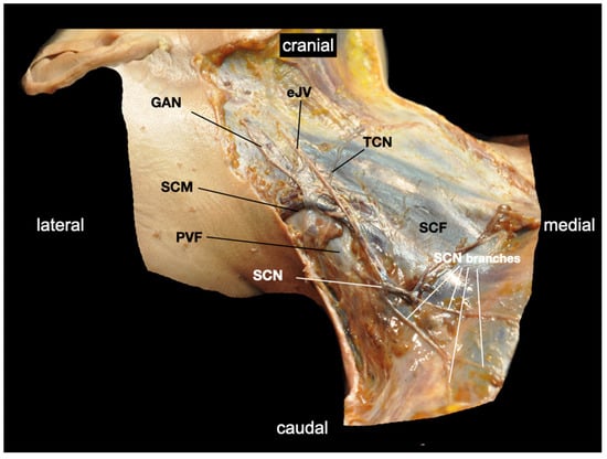
2.1. Innervation and Formation

The brachial plexus originates from spinal segments C5 to Th1 and innervates the upper extremity and parts of the trunk wall. The plexus runs through four defined anatomical regions according to its course from proximal to distal directions: (1) the scalene hiatus (the hiatus scalenorum posterius), (2) the posterior or lateral neck triangle (trigonum colli posterius sive laterale, the posterior cervical triangle), (3) the infraclavicular region (trigonum deltoideopectorale sive, Mohrenheim’s fossa or groove), and (4) the axillary fossa.
The fila radicularia arise from the spinal cord and form the radix anterior and radix posterior, which then form the actual spinal nerves in the spinal canal. In the cervical region, the spinal nerves emerge, after their formation, from the spinal canal and on through the neuroforamina (intervertebral foramina). Immediately after their ascent from the neuroforamen, they split into a larger ventral ramus and a smaller dorsal ramus (Figure 2). The brachial plexus is formed in the posterior scalene gap by the ventral rami of the spinal nerves. The dorsal rami supply the neck muscles with motor activity and the skin of the neck and occiput with sensory activity. They are not involved in the formation of the brachial plexus. The distance to the plexus formation is often less than 1 cm after the splitting of the spinal nerves.
Figure 2. Transverse section of the lateral neck region showing the anatomical relationships within the compartment of the brachial plexus. A muscular connection separates the ventral rami from C5, C6, and C7. The phrenic nerve is depicted deep to the prevertebral fascia within the same compartment as the brachial plexus. The gap is narrowed by fat and connective tissue. Red arrows mark the prevertebral fascia; black arrows mark the vertebral artery; white arrows mark the sympathetic trunk; white circle: phrenic nerve; black circle: dorsal ramus of the 6th spinal nerve; C5: ventral ramus of the 5th spinal nerve; C6: ventral ramus of the 6th spinal nerve; C7: ventral ramus of the 7th spinal nerve; CCA: common carotid artery; Mb: muscle bridges; LCM: longus colli muscle; aSM: anterior scalene muscle; mSM: middle scalene muscle; SCM: sternocleidomastoid muscle; TP C7: transverse process of the 7th cervical vertebra; TG: thyroid gland; iJV: internal jugular vein. Note: the continuity of the prevertebral fascia of the body donor was accidentally interrupted via dissection (dotted red arrows). This location represents the connection to the Danger Space [27]
In the scalene gap, the brachial plexus is located deep to the prevertebral fascia between the anterior and middle scalene muscles in a compartment that is rich in nerves and vessels. When observed in ultrasound images, the ventral rami are often described as roots; however, the anatomical term roots describes the anterior and posterior radices occurring before the formation of the spinal nerves. They are located more centrally and are not visible through ultrasound as they are covered by the vertebral arch of the bony spinal column. After they exit the neuroforamina, the ventral rami can be visualized sonographically.

2.2. Anatomical Variations in the Scalene Gap

As a consequence of embryonic development and the principle of segmental innervation, fibers of C4 (cranial fixation) and T2 (caudal fixation) may be involved in plexus formation as well. In addition to variants of plexus formation, variants of the scalene musculature [28][29][30][31][32] and/or vessels [33] can frequently be found. There are often muscular connections between the anterior and middle scalene muscles. Another scalene muscle, the scalene minimus muscle, referred to in older nomenclature as the levator pleurae muscle [32], is frequently present. Such muscles may separate the plexus into different parts. In practice, such muscle formations, as well as the loose connective and fatty tissue located in the scalene gap (Figure 1), narrow the gap and make spatial LA distribution unpredictable or even prevent it from spreading around the plexus (Figure 3).
Figure 3. Sonogram of the lateral neck region at the level of the 7th cervical vertebra captured to demonstrate problems of LA distribution. Nerves of the brachial plexus are deep to the prevertebral fascia (red arrows), and the supraclavicular nerves from the cervical plexus (not depicted) are superficial to it. Muscle bridges between anterior (aSM) and medius scalene muscle (mSM) act as mechanical barriers to LA spread (blue arrows). In this patient, local anesthetic would likely cause phrenic nerve (white circle) palsy if injected close to the ventral ramus of C5 due to its direct proximity to the phrenic nerve within the same compartment. The yellow circle marks a supraclavicular nerve. The vertebral vessels (VA: vertebral artery; VV: vertebral vein) are in close proximity to the 7th spinal nerve (C7). They and the spinal nerve at the level of the neuroforamen will be subject to injuries if a block with a needle trajectory planned according to Winnie is carried out. C6: ventral ramus of the 6th spinal nerve; SCM: sternocleidomastoid muscle; TPC7: transverse process of the 7th cervical vertebra; pTC7: posterior tubercle of the 7th transverse process; LN: lymph node; CCA: common carotid artery; iJV: internal jugular vein.

2.3. Variants of the Course of the Ventral Rami

In the scalene gap, the cranial plexus parts (C5 and C6) frequently disperse between the anterior and middle scalene muscles and often do not intersect the caudal parts of C7, C8, and Th1 (Figure 3). C5 may run anteriorly or through the anterior scalene muscle (Figure 3 and Figure 4), and C5 and C6 may also pierce the anterior scalene muscle together [32]. This explains the clinical observation of incomplete interscalene blocks regardless of whether neurostimulation or ultrasound guide techniques are used (Figure 3 and Figure 4).
Figure 4. Sonogram of the lateral neck region at the level of the 6th cervical vertebra, demonstrating the variable course of the ventral rami (spinal nerves). In this patient, the ventral ramus of the 5th spinal nerve (C5) passes superficially to the anterior scalene muscle (aSM), while the ventral ramus of the 6th spinal nerve (C6) does not. Nerves of the brachial plexus were deep to the prevertebral fascia (red arrows), with the supraclavicular nerves from C4 running superficially to it (yellow circle). The phrenic nerve (white circle) is located deep to the prevertebral fascia on top of the fascia of the aSM. A phrenic-nerve-sparing brachial plexus block at this level is simply not possible. The ventral ramus of the 6th spinal nerve (C6) is depicted at its course out of the neuroforamen between the anterior tubercle (aT C6) and posterior tubercle (pT C6). Any needle advancement toward the neuroforamen carries the risk of nerve injury since the nerve at this position is not movable between the bony structures. SCM: sternocleidomastoid muscle; TPC6: transverse process of the 6th cervical vertebra; mSM: middle scalene muscle; iJV: internal jugular vein; CCA: common carotid artery.

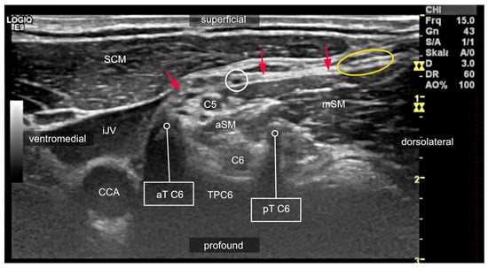
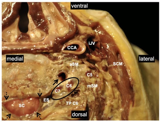
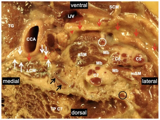
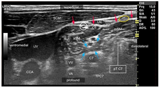
2.4. Compartments of the Lateral Neck and Their Relevance to Side Effects
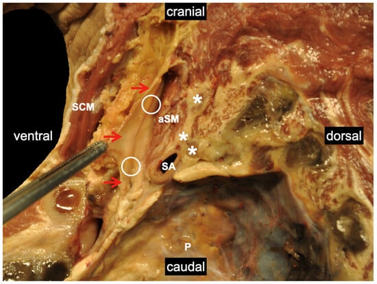
In the interscalene gap, the brachial plexus lies deep to the prevertebral fascia, as do the ventral rami of C1 to C4 that form the cervical plexus. The phrenic nerve usually arises from C4 and occasionally, in part from C3 or C5 or even lower cervical segments. The nerve then runs covered by the prevertebral fascia on the ventral surface of the anterior scalene muscle. Figure 5 demonstrates the close vicinity of the brachial plexus to the phrenic nerve.
Figure 5. Sagittal section through the left lateral neck region (scalenovertebral triangle) including the pleural cavity. The phrenic nerve (white circles) is deep to the prevertebral fascia (red arrows). White stars mark the ventral rami of the spinal nerves, pointing out their close vicinity to the phrenic nerve, which explains why phrenic-nerve-sparing injection techniques are so difficult and why ventro-medial LA distributions should be avoided. SA: subclavian artery; P: pleura; aSM: anterior scalene muscle; SCM: sternocleidomastoid muscle. 
The sympathetic trunk is located medial to the scalene gap on the surface of the longus colli muscle. It is anteriorly attached and partially enveloped by the prevertebral fascia. If LA spreads medially deep or anterior to the anterior scalene muscle, it can disperse into the scalenovertebral triangle [31][34]. The triangle is bounded laterally by the anterior scalene muscle and medially by the longus colli muscle. The tip of the triangle is marked by the carotid tubercle (the anterior tubercle) of the sixth cervical vertebra. The base is defined by the subclavian artery or the cupula pleurae [35]. The large vessels, the common carotid artery, and the internal jugular vein, as well as the vagal nerve, are enveloped in this position by the carotid vagina and mark the ventral border.
Due to the presence of the pleural dome, the scalenovertebral triangle corresponds to a pyramidally shaped three-dimensional space. The prevertebral fascia surrounds the anterior scalene and the longus colli muscle. However, the prevertebral fascia does not separate these two muscles. Consequently, there is a connection from the scalenovertebral triangle to the dorsal prevertebral, the so-called Danger Space [27], which connects to the mediastinum.

2.5. Danger Space

The Danger Space constitutes a loose connective tissue space with a craniocaudal extension from the skull base to the diaphragm. This space contains the recurrent laryngeal nerve, parts of the sympathetic trunk and the vertebral vessels, autonomic fibers of the heart, and the phrenic nerve [27] (Figure 5). Therefore, LA spreading into this space can cause unwanted side effects like Horner’s syndrome or hoarseness.

2.6. Are Brachial Plexus Blocks Sufficient for Shoulder Surgery?

ISBs are usually applied for shoulder surgery, which ranks among the most painful surgical procedures [36]. The suprascapular nerve and the axillary nerve, both originating from the ventral rami from C5 and C6, which form the superior trunk, are the main suppliers of the shoulder joint. The lateral pectoral nerve, which originates from the lateral cord, is involved, and so too are the subscapular nerves [37][38]. Since the suprascapular nerve originates from the most proximal section of the brachial plexus (the superior trunk), the target for shoulder surgery must be the brachial plexus at the level of its trunks. This explains the superior analgesic effects of an interscalene or supraclavicular block. Furthermore, the cutaneous innervation of the shoulder cape is also mediated by the supraclavicular nerves originating from C4, like the phrenic nerve. They have a short course of up to 2 cm underneath the prevertebral fascia; they then penetrate the fascia and become superficial (Figure 6). After passing through the prevertebral fascia, they run in a different compartment, superficial to the brachial plexus. These anatomical facts indicate why they are unlikely to be blocked using peripheral techniques (such as a costoclavicular block). Therefore, if shoulder surgery is carried out solely with peripheral nerve blocks, brachial plexus blocks must be combined with a supraclavicular nerve block to anesthetize the skin for incision or closure.
Figure 6. The dissection of the right lateral neck (ventrolateral view) demonstrates the peripheral course of the supraclavicular nerves after their exit through the prevertebral fascia (PVF). The PVF is depicted at the lateral border of the sternocleidomastoid muscle (SCM). This image clearly depicts the different compartments of the cervical and brachial plexus. Analgesia for skin incision or closure in shoulder surgery can only be achieved if the supraclavicular nerves are blocked superficial to the PVF, provided that the phrenic nerve can be spared. eJV: external jugular vein; GAN: greater auricular nerve; SCF: superficial cervical fascia; SCN: common trunk of the supraclavicular nerves; TCN: transverse nerve of the neck.

References

  1. Ermert, T.; Schnabel, A. Ultrasound in regional anesthesia. Anasthesiol. Intensivmed. Notfallmed. Schmerzther. 2011, 46, 182–187; quiz 188.
  2. Gaus, P.; Kutz, P.; Bachtler, J.A.; Lindner, R.; Saur, P. Cave: Interscalene catheters. Anaesthesist 2017, 66, 961–968.
  3. Liu, S.S.; Gordon, M.A.; Shaw, P.M.; Wilfred, S.; Shetty, T.; Yadeau, J.T. A prospective clinical registry of ultrasound-guided regional anesthesia for ambulatory shoulder surgery. Anesth. Analg. 2010, 111, 617–623.
  4. Riazi, S.; Carmichael, N.; Awad, I.; Holtby, R.M.; McCartney, C.J. Effect of local anaesthetic volume (20 vs. 5 mL) on the efficacy and respiratory consequences of ultrasound-guided interscalene brachial plexus block. Br. J. Anaesth. 2008, 101, 549–556.
  5. Stundner, O.; Meissnitzer, M.; Brummett, C.M.; Moser, S.; Forstner, R.; Koköfer, A.; Danninger, T.; Gerner, P.; Kirchmair, L.; Fritsch, G. Comparison of tissue distribution, phrenic nerve involvement, and epidural spread in standard- vs. low-volume ultrasound-guided interscalene plexus block using contrast magnetic resonance imaging: A randomized, controlled trial. Br. J. Anaesth. 2016, 116, 405–412.
  6. Lee, J.H.; Cho, S.H.; Kim, S.H.; Chae, W.S.; Jin, H.C.; Lee, J.S.; Kim, Y.I. Ropivacaine for ultrasound-guided interscalene block: 5 mL provides similar analgesia but less phrenic nerve paralysis than 10 mL. Can. J. Anaesth. 2011, 58, 1001–1006.
  7. Thackeray, E.M.; Swenson, J.D.; Gertsch, M.C.; Phillips, K.M.; Steele, J.W.; Burks, R.T.; Tashjian, R.Z.; Greis, P.E. Diaphragm function after interscalene brachial plexus block: A double-blind, randomized comparison of 0.25% and 0.125% bupivacaine. J. Shoulder Elb. Surg. 2013, 22, 381–386.
  8. Wong, A.K.; Keeney, L.G.; Chen, L.; Williams, R.; Liu, J.; Elkassabany, N.M. Effect of Local Anesthetic Concentration (0.2% vs. 0.1% Ropivacaine) on Pulmonary Function, and Analgesia After Ultrasound-Guided Interscalene Brachial Plexus Block: A Randomized Controlled Study. Pain Med. 2016, 17, 2397–2403.
  9. Palhais, N.; Brull, R.; Kern, C.; Jacot-Guillarmod, A.; Charmoy, A.; Farron, A.; Albrecht, E. Extrafascial injection for interscalene brachial plexus block reduces respiratory complications compared with a conventional intrafascial injection: A randomized, controlled, double-blind trial. Br. J. Anaesth. 2016, 116, 531–537.
  10. Renes, S.H.; van Geffen, G.J.; Rettig, H.C.; Gielen, M.J.; Scheffer, G.J. Minimum effective volume of local anesthetic for shoulder analgesia by ultrasound-guided block at root C7 with assessment of pulmonary function. Reg. Anesth. Pain Med. 2010, 35, 529–534.
  11. Siegenthaler, A.; Moriggl, B.; Mlekusch, S.; Schliessbach, J.; Haug, M.; Curatolo, M.; Eichenberger, U. Ultrasound-guided suprascapular nerve block, description of a novel supraclavicular approach. Reg. Anesth. Pain Med. 2012, 37, 325–328.
  12. Burckett-St Laurent, D.; Chan, V.; Chin, K.J. Refining the ultrasound-guided interscalene brachial plexus block: The superior trunk approach. Can. J. Anaesth. 2014, 61, 1098–1102.
  13. Karmakar, M.K.; Sala-Blanch, X.; Songthamwat, B.; Tsui, B.C. Benefits of the costoclavicular space for ultrasound-guided infraclavicular brachial plexus block: Description of a costoclavicular approach. Reg. Anesth. Pain Med. 2015, 40, 287–288.
  14. Tran, D.Q.; Layera, S.; Bravo, D.; Cristi-Sánchez, I.; Bermúdez, L.; Aliste, J. Diaphragm-sparing nerve blocks should spare the diaphragm. Reg. Anesth. Pain Med. 2020, 45, 752–753.
  15. Hussain, N.; Goldar, G.; Ragina, N.; Banfield, L.; Laffey, J.G.; Abdallah, F.W. Suprascapular and Interscalene Nerve Block for Shoulder Surgery: A Systematic Review and Meta-analysis. Anesthesiology 2017, 127, 998–1013.
  16. Nöske, E.; Stolzer, M.; Racher, M.; Baumann, H.; Lehmann, K.J.; Henrich, M. Central neurological complication of an interscalene plexus catheter. Anaesthesist 2021, 70, 937–941.
  17. Spitzer, D.; Wenger, K.J.; Neef, V.; Divé, I.; Schaller-Paule, M.A.; Jahnke, K.; Kell, C.; Foerch, C.; Burger, M.C. Local Anesthetic-Induced Central Nervous System Toxicity during Interscalene Brachial Plexus Block: A Case Series Study of Three Patients. J. Clin. Med. 2021, 10, 1013.
  18. Benumof, J.L. Permanent loss of cervical spinal cord function associated with interscalene block performed under general anesthesia. Anesthesiology 2000, 93, 1541–1544.
  19. Walter, M.; Rogalla, P.; Spies, C.; Kox, W.J.; Volk, T. Intrathecal misplacement of an interscalene plexus catheter. Anaesthesist 2005, 54, 215–219.
  20. Frasca, D.; Clevenot, D.; Jeanny, A.; Laksiri, L.; Petitpas, F.; Debaene, B. Total spinal anesthesia after interscalenic plexus block. Ann. Fr. Anesth. Reanim. 2007, 26, 994–998.
  21. Gaus, P.; Heb, B.; Tanyay, Z.; Müller-Breitenlohner, H. Epidural malpositioning of an interscalene plexus catheter. Anaesthesist 2011, 60, 850–853.
  22. Hohner, M.; von Knobelsdorff, G.; Andresen-Streichert, H.; Püschel, K.; Schulz, F. Tödlicher Ausgang einer Katheterfehllage bei Plexusanästhesie. Rechtsmedizin 2013, 23, 38–43.
  23. Tuominen, M.K.; Pere, P.; Rosenberg, P.H. Unintentional arterial catheterization and bupivacaine toxicity associated with continuous interscalene brachial plexus block. Anesthesiology 1991, 75, 356–358.
  24. Yanovski, B.; Gaitini, L.; Volodarski, D.; Ben-David, B. Catastrophic complication of an interscalene catheter for continuous peripheral nerve block analgesia. Anaesthesia 2012, 67, 1166–1169.
  25. Marhofer, D.; Marhofer, P.; Triffterer, L.; Leonhardt, M.; Weber, M.; Zeitlinger, M. Dislocation rates of perineural catheters: A volunteer study. Br. J. Anaesth. 2013, 111, 800–806.
  26. Maecken, T.; Lohr, D.; Zahn, P.K.; Litz, R.J. Sonographic comparison of two different peripheral nerve catheters for continuous interscalene nerve blocks. EJA 2017, 34 (Suppl. S55), 89.
  27. Grodinsky, M.; Holyoke, E.A. The fasciae and fascial spaces of the head, neck and adjacent regions. Am. J. Anat. 1938, 63, 367–408.
  28. Feigl, G.C.; Pixner, T. Combination of variations of the interscalene gap as a pitfall for ultrasound-guided brachial plexus block. Reg. Anesth. Pain Med. 2011, 36, 523–524.
  29. Feigl, G.C.; Dreu, M. Important structures to be known for a safe ultrasound-guided supraclavicular plexus block. Reg. Anesth. Pain Med. 2010, 35, 317–318.
  30. Feigl, G.C.; Pixner, T. The cleidoatlanticus muscle: A potential pitfall for the practice of ultrasound guided interscalene brachial plexus block. Surg. Radiol. Anat. 2011, 33, 823–825.
  31. Hafferl, A. Lehrbuch der Topographischen Anatomie, 3rd ed.; Springer: Berlin/Heidelberg, Germany; New York, NY, USA, 1969.
  32. Harry, W.G.; Bennett, J.D.; Guha, S.C. Scalene muscles and the brachial plexus: Anatomical variations and their clinical significance. Clin. Anat. 1997, 10, 250–252.
  33. Weiglein, A.H.; Moriggl, B.; Schalk, C.; Künzel, K.H.; Müller, U. Arteries in the posterior cervical triangle in man. Clin. Anat. 2005, 18, 553–557.
  34. Feigl, G.; Hammer, G.P.; Litz, R.; Kachlik, D. The intercarotid or alar fascia, other cervical fascias, and their adjacent spaces—A plea for clarification of cervical fascia and spaces terminology. J. Anat. 2020, 237, 197–207.
  35. Hafferl, A. (Ed.) Die Anatomie der Pleurakuppel; Springer Verlag GmbH: Berlin/Heidelberg, Germany, 1939.
  36. Gerbershagen, H.J.; Aduckathil, S.; van Wijck, A.J.; Peelen, L.M.; Kalkman, C.J.; Meissner, W. Pain intensity on the first day after surgery: A prospective cohort study comparing 179 surgical procedures. Anesthesiology 2013, 118, 934–944.
  37. Aszmann, O.C.; Dellon, A.L.; Birely, B.T.; McFarland, E.G. Innervation of the human shoulder joint and its implications for surgery. Clin. Orthop. Relat. Res. 1996, 330, 202–207.
  38. Gardner, E. The innervation of the shoulder joint. Anat. Rec. 1948, 102, 1–18.
More
Information
Subjects: Anesthesiology
Contributors MDPI registered users' name will be linked to their SciProfiles pages. To register with us, please refer to https://encyclopedia.pub/register : , , , , ,
View Times: 76
Revisions: 2 times (View History)
Update Date: 17 Feb 2024
1000/1000