Submitted Successfully!
To reward your contribution, here is a gift for you: A free trial for our video production service.
Thank you for your contribution! You can also upload a video entry or images related to this topic.
Version Summary Created by Modification Content Size Created at Operation
1 -- 2767 2024-02-08 08:13:23 |
2 Reference format revised. Meta information modification 2767 2024-02-08 08:58:33 |

Video Upload Options

Do you have a full video?

Confirm

Are you sure to Delete?
Cite
If you have any further questions, please contact Encyclopedia Editorial Office.
Gravina, A.G.; Pellegrino, R.; Esposito, A.; Cipullo, M.; Romeo, M.; Palladino, G.; Iodice, P.; Federico, A.; Troiani, T. JAK-STAT Pathway in Immune Checkpoint Inhibitor-Induced Colitis Patients. Encyclopedia. Available online: https://encyclopedia.pub/entry/54902 (accessed on 05 July 2024).
Gravina AG, Pellegrino R, Esposito A, Cipullo M, Romeo M, Palladino G, et al. JAK-STAT Pathway in Immune Checkpoint Inhibitor-Induced Colitis Patients. Encyclopedia. Available at: https://encyclopedia.pub/entry/54902. Accessed July 05, 2024.
Gravina, Antonietta Gerarda, Raffaele Pellegrino, Alfonso Esposito, Marina Cipullo, Mario Romeo, Giovanna Palladino, Patrizia Iodice, Alessandro Federico, Teresa Troiani. "JAK-STAT Pathway in Immune Checkpoint Inhibitor-Induced Colitis Patients" Encyclopedia, https://encyclopedia.pub/entry/54902 (accessed July 05, 2024).
Gravina, A.G., Pellegrino, R., Esposito, A., Cipullo, M., Romeo, M., Palladino, G., Iodice, P., Federico, A., & Troiani, T. (2024, February 08). JAK-STAT Pathway in Immune Checkpoint Inhibitor-Induced Colitis Patients. In Encyclopedia. https://encyclopedia.pub/entry/54902
Gravina, Antonietta Gerarda, et al. "JAK-STAT Pathway in Immune Checkpoint Inhibitor-Induced Colitis Patients." Encyclopedia. Web. 08 February, 2024.
JAK-STAT Pathway in Immune Checkpoint Inhibitor-Induced Colitis Patients
Edit

Immunotherapy has emerged as a pivotal component in the treatment of various malignancies, encompassing lung, skin, gastrointestinal, and head and neck cancers. The foundation of this therapeutic approach lies in immune checkpoint inhibitors (ICI). While ICIs have demonstrated remarkable efficacy in impeding the neoplastic progression of these tumours, their use may give rise to substantial toxicity, notably in the gastrointestinal domain, where ICI colitis constitutes a significant aspect. The optimal positioning of Janus kinase (JAK)–signal transducer and activator of transcription (STAT) pathway inhibitors in the therapeutic management of ICI colitis remains unclear. Numerous reports have highlighted notable improvements in ICI colitis through the application of pan-JAK-STAT inhibitors, with tofacitinib, in particular, reporting evident clinical remission of colitis. 

immunotherapy immune checkpoint inhibitors colitis JAK-STAT

1. Introduction

Immunotherapy is indicated in several malignancies, such as lung cancer, colorectal, melanoma, and head and neck neoplasms. Among the immune checkpoint inhibitors (ICI) are agents acting on cytotoxic T-cell lymphocyte-4 (CTLA-4) and programmed cell death-1/programmed cell death-ligand 1 (PD-1/PD-L1). Among Anti-CTLA-4 agents is ipilimumab, while among anti-PD-1 are pembrolizumab, nivolumab, and durvalumab, and among anti-PD-L1 are avelumab and atezolizumab.
Immunotherapy can result in immune-related adverse events (IrAEs) at the skin, gastroenterological, neurological, endocrinological, pulmonological, and cardiological levels. From a gastroenterological perspective, autoimmune diarrheal and colitis, i.e., immune-mediated diarrhoea and colitis (IMDC), can occur, the clinical manifestations of which include abdominal pain, nausea, diarrhoea, mucorrhea, rectorrhagia, peritoneal signs, up to and including conditions that can be life threatening [1][2]. Regarding frequency, gastrointestinal side effects occur most often in combination treatments between anti-CTLA-4 and anti-PD-1/PD-L1, followed by a single treatment with anti-CTLA-4 and a single therapy with anti-PD-1/PD-L1 [3].
Thus, side effects are mainly represented by inflammatory and autoimmune complications [4]. Indeed, ICI are monoclonal antibodies acting on the balance between immune system cells and tumour cells [4][5]. IrAEs can be highly variable, as mild, or life-threatening reactions are possible. The development of colitis is associated with an increased risk of ileus, toxic megacolon, and perforations [6].
From an endoscopic point of view, the colitis characteristics include the presence of erosions, ulcerations, erythema, reduction to loss of vascular pattern, mucosal friability, oedematous walls, and areas of exudation. An endoscopic negative examination (i.e., a normal colonic mucosa appearance) is also possible [7]. Faecal inflammatory markers, such as faecal calprotectin and lactoferrin, and serum markers, such as C-reactive protein, are helpful for risk stratification [8].
In patients refractory to steroid therapy, anti-tumour necrosis factor (TNF) α drugs such as infliximab can be administered by intravenous infusion, starting at 5 mg/Kg [6][9]. An alternative is treatment with vedolizumab, a monoclonal antibody acting on α4β7 integrin [9][10], administered as an intravenous infusion at a dose of 300 mg [6]. Faecal microbiota transplantation is another option for refractory patients [1][11]. Finally, surgery represents the last treatment option. An emergency colectomy is indicated in severe complications, such as perforation or toxic megacolon [11].

2. Immunotherapy, Generalities, and Toxicity: The Dimensions That Matter

The discovery of the basis of immunotherapy earned James Allison and Tasuku Honjo the Nobel Prize in Physiology or Medicine in 2018 [12].
Their research has elucidated the pivotal inhibitory role of PD-1 and CTLA-4 in immune function, revealing their capacity to effectively reinvigorate T cells and efficiently stimulate anti-cancer immunity [13].
CTLA-4 is a transmembrane receptor protein with inhibitory activity belonging to the immunoglobulin superfamily and localized on the surface of activated T lymphocytes [14]. Its physiological role is to regulate immune activity to prevent potential organ harm and immunity imbalances [14]. CTLA-4 expression on the lymphocyte’s surface appears in the early stages of its activation, approximately 48 h after the binding of costimulatory molecules B7-1 or B7-2 (also known as CD80 and CD86) to the CD28 receptor [14][15]. CTLA-4 competes with CD28 for binding with B7-1 and B7-2 [14][16]. Once CD28 is displaced, it binds to the costimulatory molecules, suppressing T-cell proliferation and survival signals [14]. This action occurs centrally within lymph nodes before T cells encounter tumour cells in the peripheral tissues and before any immune specificity develops [15][16].
Two monoclonal antibodies, ipilimumab and tremelimumab, can bind CTLA-4 and inhibit its normal function [17]. This inhibition prevents CTLA-4 from binding to B7-1 or B7-2 receptors, allowing these receptors to bind the co-stimulatory molecule CD28, leading to sustained activation of T lymphocytes. Consequently, this activation triggers an anti-tumour response [17].
On the other hand, PD-1 also belongs to the immunoglobulin superfamily, and it is found on the surface of activated T cells and pro-B cells [18][19]. It has a receptor function and binds two ligands (i.e., PDL-1 and PDL-2), which are part of the B7 costimulatory molecules expressed on antigen-presenting cells and epithelial cells [20]. The primary role of the PD-1-PDL-1 receptor–ligand interaction is to induce apoptosis in T cells activated against an antigen and enhance the efficiency of anti-apoptotic mechanisms in regulatory T cells [18][21]. This activity mainly occurs peripherally, within the tumour microenvironment, where activated T lymphocytes are already involved in immune responses. However, when they encounter tumour cells expressing PDL-1, their anti-tumour immune function is inhibited and disrupted [22].
Dermatologic irAEs are the most frequent but also among the earliest (i.e., they also arise more than two weeks after the start of treatment), with an incidence of about 44–68% in patients on anti-CTLA4 therapy and 37–42% in patients on anti-PD1 therapy and rates higher at 58% in patients on combination therapy [23]. Gastrointestinal tract toxicity, on the other hand, is pervasive in patients with melanoma, and lower gastrointestinal toxicity (i.e., ICI colitis) is prevalent with the use of anti-CTLA4 agents (i.e., 10–25% incidence) compared with anti-PD1 ICI (i.e., 1–5%) and, finally, with a considerable incidence when combination therapy is considered (i.e., about 20%) [23]. ICI can result in toxicity to other organs and systems. IrAEs encompass a large group of more than fifty different clinical entities that can affect almost any system, including the skin and gastrointestinal and the pulmonary, cardiovascular, hepatobiliary, and genitourinary systems [24].
There is often a delicate relationship between toxicity and oncologic response to immunotherapy. Khan et al., for example, reported how dermatologic toxicity (e.g., vitiligo, psoriasis) could predict oncologic response and overall survival in the case of bladder cancer [25].

3. Clinical Evidence Concerning JAK-STAT Inhibitors and ICI Colitis

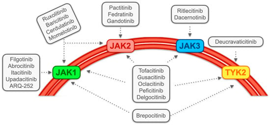
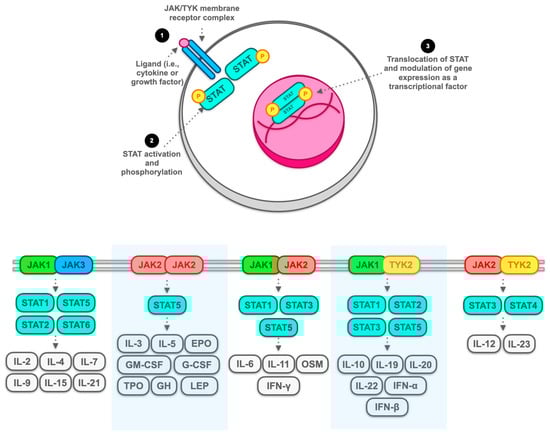
3.1. JAK Inhibitors: Generality and Classification

JAK inhibitors, over time, are classified according to their selectivity profile for different molecules of the JAK family.
In accordance with this definition, a diverse array of JAK inhibitors is identified, encompassing pan-JAK inhibitors such as tofacitinib, oclacitinib, and gusaticinib. The term “pan-JAK” implies the potential for these inhibitors to bind to all members of the JAK family. Additionally, there are JAK1,2-selective inhibitors like baricitinib and ruxolitinib, JAK1-selective inhibitors including filgotinib, upadacitinib, and itacitinib, and JAK2-selective inhibitors like gandotinib and fedratinib. Further classifications consist of JAK1/TYK2-selective inhibitors (e.g., brepocitinib), JAK3-selective inhibitors (e.g., ritlecitinib), and TYK2-selective inhibitors such as BMS-986165 and NDI-031301 [26][27] (see Figure 1).
Figure 1. Main selectivity profiles of major Janus kinases (JAK) and JAK-related tyrosine kinase-2 (TYK2) inhibitors. Several JAK inhibitors and TYK2 inhibitors have been made over time. The broad affinity spectrum combining these molecules with JAK/TYK2 allows the blockade of an extensive range of immunologic phenomena aimed at blocking inflammation. The greater the selectivity profile, the greater the specific inhibition of the biological function to be controlled, as well as the spectrum of controlled pro-inflammatory cytokines. This underlies the wide variety of clinical indications that such molecules have received over time, encompassing different branches of medicine (e.g., gastroenterology, rheumatology, immunology, etc.).
JAK inhibitors have recognized a particular explosion in inflammatory diseases over time. They have been used in a broad spectrum of, for example, dermatologic diseases (such as alopecia areata, atopic dermatitis, erythema multiforme, vitiligo, and psoriasis), as well as in other inflammatory vascular disorders (such as polyarteritis nodosa), and rheumatologic and muscular diseases (such as rheumatoid arthritis, lupus erythematosus, hypereosinophilic syndrome, and dermatomyositis) [28].
A debated element on the use of JAK inhibitors has been that of safety. In particular, for some molecules (e.g., tofacitinib and upadacitinib), some red flags have been raised for cardiovascular events, venous thromboembolism, infections, and cancer; however, this issue must be related to the individual patient’s risk factors [29][30].
The use of JAK inhibitors, therefore, should be the focus of decision making that weighs the benefits and risks from the use of a moderate-to-severe immunosuppressant [31], taking into account the individual patient’s comorbidities and individual risk and the conventional and biologic therapy alternatives that are offered [32].
Certainly, JAK inhibitors are also gaining prominence in gastroenterology for managing IBD. While sharing commonalities with ICI colitis, the pathogenesis of IBD does exhibit differences, including a self-sustained inflammatory process characterized by a relapsing–remitting course independent of any previous patient treatment [33][34].
Tofacitinib [35][36], upadacitinib [37][38], and filgotinib [39] have now received indications of ulcerative colitis. Encouraging results appear to be provided in Crohn’s disease by upadacitinib [40][41] and filgotinib [42].
These results, produced from solid randomized clinical trials, demonstrate that in each case, JAK inhibitors can adequately control colonic inflammatory processes by leading to relevant rates of mucosal healing in settings of even moderate-to-severe intestinal inflammation, such as those of IBD. What is more, tofacitinib seems to be able to play a role even in cases of devastating inflammation, such as in acute severe ulcerative colitis, where its potential is increasingly emerging over the traditional use, in this setting, of parenteral steroids, cyclosporine, and infliximab [43][44][45][46].
Although these findings are not automatically applicable to ICI colitis precisely in light of the pathogenetic and etiologic differences between these entities, they nevertheless show a glimmer of the potential in ICI colitis of JAK-STAT pathway inhibition.
Some JAK inhibitors also showed encouraging results in managing other immune-mediated adverse events related to using ICI. Tofacitinib and baricitinib can attenuate the levels of monocyte chemoattractant protein-1 activated by using pembrolizumab, determining arthritic activation during treatment with ICI [47].

3.2. Contemporary Management in Accordance with Established Guidelines and Exploration of Giverse Alternatives, with Limited Emphasis on JAK Inhibitors

Currently, ICI colitis is managed by a treatment approach proportional to the severity of the disease. A helpful model may be the European guidelines for ICI colitis [48].
According to this protocol, managing grade 1 ICI colitis (defined as an increase of up to 4 daily bowel movements from baseline) involves conservative measures such as a low-fibre diet, loperamide, and psyllium, while continuing ICI treatment. Should the escalation in bowel movements exceed 4–6 daily compared to baseline (grade 2), discontinuation of ICI therapy is recommended, and oral steroids at an immunosuppressive dose of 0.75–1 mg/kg daily should be incorporated into the treatment regimen.
In instances where grade 1 therapy proves ineffective after 15 days, a shift to grade 2 therapy is recommended. Similarly, if grade 2 therapy shows no response after 5–7 days, escalation to grade 3–4 treatment is advised.
In the event of recurrence post-steroid withdrawal or resistance to steroids (refractoriness), the introduction of biologic therapy becomes necessary, typically involving agents like infliximab or vedolizumab. Alternative options encompass faecal microbiota transplantation, ustekinumab, extracorporeal photopheresis, and, in extreme cases, colectomy.
Vedolizumab, undoubtedly, represents a second option worth considering, particularly in cases of primary or secondary loss of response to infliximab. However, it is important to note that its association comes with the risk of a response that may not always be immediate, a criterion certainly desirable when dealing with ICI colitis beyond the second grade [49].

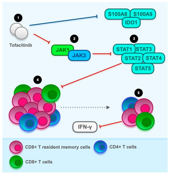
3.3. The Hyperactivation of T Cells as a Potential Therapeutic Target for JAK Inhibitors: General Overview with a Focus on CD8+ Resident Memory T Cells

ICI colitis presents a pathogenesis that, unfortunately, remains not entirely elucidated, thereby posing the challenge of formulating definitive therapies that target all aspects of the disease’s pathogenesis. This challenge is particularly pronounced in the context of ensuring the continuity of immunotherapy, an essential component of the patient’s cancer treatment, and, consequently, prioritizing oncological outcomes in the therapeutic management of the patient.
It is evident that one pivotal event in the pathogenesis of ICI colitis is the hyperactivation of effector T cells. This is underscored by the presence of a clear infiltrate of CD4+ and CD8+ T cells in the colonic microenvironment of patients treated with anti-CTLA4 and anti-PD1, suggesting a significant role for these cells in the inflammatory process’s pathogenesis [50]. However, it appears that the stimulus for such cellular clones is ICI-related. Consequently, patients treated with anti-CTLA4 seem to predominantly express CD4+ T-cells, whereas conversely, those treated with anti-PD1 undergo a preferential selection of CD8+ T-cells [50].
As anticipated, the JAK-TYK2-STAT pathway has the potential to downstream regulate an endless array of key molecular mediators in the regulation of inflammatory processes [26][27][28] (see Figure 2). Moreover, it seems that this population of memory cells may represent a potential therapeutic target for tofacitinib, acting as a pan-JAK inhibitor.
Figure 2. Cytokines and molecular mediators modulated by the Janus kinase (JAK) and tyrosine kinase (TYK2) system. The JAK/TYK2 system regulates the expression of numerous cytokines, growth factors, and hormones, resulting in a transcriptional crossroads of immune function and the inflammatory process. These kinases (i.e., JAK and TYK) interact with several transcription factors, namely those of the signal transducer and activator of transcription (STAT) family, which, through intranuclear translocation processes, regulate the gene expression of molecules that are part of the signalling pathway that combinations among all the mediators previously exposed determine. According to a simplified scheme (top of figure), activation of the membranous receptor complex consisting of the various combinations of JAK and TYK family members (bottom part of the figure) by binding of a ligand, which may be, for example, a cytokine or a transcription factor (1), which results in downstream activation, mainly by phosphorylation processes of STAT family members (2), which in turn are activated intranuclearly by acting as transcription factors and modulating gene expression of several genes coding for crucial molecules in the inflammatory process (3). Note: IL: interleukin; EPO: erythropoietin; GM-CSF: granulocyte–macrophage colony-stimulating factor; G-CSF: granulocyte-colony stimulating factor; TPO: thyroperoxidase; GH: growth hormone; LEP: leptin; OSM: oncostatin M; IFN: interferon.
Gene set enrichment analysis of bulk RNA sequencing data identified an enrichment of the interferon (IFN)-γ signalling pathway in ICI colitis. Data from the nano string RNAplex assay indicated a substantial down-regulation of JAK1, JAK3, STAT1, STAT2, STAT3, STAT4, and STAT5A upon tofacitinib treatment (Figure 3). The RNAplex assay conducted on colonic mucosal RNA before and after tofacitinib therapy demonstrated a down-regulation of crucial JAK-STAT signalling components downstream of IFN-γ signalling, aligning with the observed molecular alterations.
Figure 3. Hypothesis on the modulation of CD8+ CD103+ T resident memory cells by tofacitinib in immune checkpoint inhibitors (ICI) colitis. Drawing from a case involving a non-small cell lung cancer patient undergoing combination therapy (carboplatin/pemetrexed and pembrolizumab), this hypothesis suggests that tofacitinib, as a pan Janus kinase (JAK) inhibitor, may address the dysregulation of immunotherapy-induced CD8+ CD103+ T resident memory cell activation in the colonic microenvironment of ICI colitis patients. In the pre-therapy microenvironment with tofacitinib, CD8+ and CD4+ T cells exhibit extensive activation. Among CD8+ cells, the most active subset is CD8+ CD103+ T resident memory cells. Tofacitinib therapy leads to a substantial reduction in this activation, surpassing a fivefold decrease. At the genetic level, down-regulated molecules include JAK1, JAK3, signal transducer and activator of transcription (STAT) 1, STAT2, STAT3, and STAT4, potentially involved in interferon-γ (IFN-γ) signalling activation. This modulation of immunological processes may correspond to the clinical and endoscopic improvement observed in ICI colitis following tofacitinib therapy. In addition, the comprehensive transcriptional reaction to the administration of tofacitinib showcases a notable decrease in the expression of transcripts, specifically encompassing S100 calcium-binding protein (S100)A8, S100A9, and indoleamine 2,3-dioxygenase 1, also known as IDO1 (the blue line at the top of the figure).

4. Conclusions

ICI colitis unfortunately represents a common adverse event among patients undergoing cancer immunotherapy. Existing guidelines do not clearly advocate for the utilization of tofacitinib and other JAK inhibitors for managing this condition. Nevertheless, five case reports/series studies have demonstrated promising results in employing JAK inhibitors, specifically tofacitinib, to address ICI colitis in patients. The majority of these individuals were aged over 50, with metastatic cancer, and had experienced treatment failures across various conventional (e.g., antibiotics and steroids) and advanced (e.g., infliximab and vedolizumab) modalities.
While these studies are pioneering in nature, their significance in broader investigations necessitates validation to establish efficacy and safety rates. Given that tofacitinib and other JAK inhibitors exert moderate to severe immunosuppressive effects, careful consideration of their use is crucial in relation to the progression of the underlying malignancy.
In conclusion, JAK inhibitors emerge as genuine and potential therapeutic candidates for ICI colitis, thereby warranting evaluation in dedicated randomized controlled trials. The specific JAK or STAT implicated in ICI colitis remains unclear. Clarification on this matter will inform the precise utilization of targeted JAK-STAT inhibitors.

References

  1. Gong, Z.; Wang, Y. Immune Checkpoint Inhibitor-Mediated Diarrhea and Colitis: A Clinical Review. JCO Oncol. Prac. 2020, 16, 453–461.
  2. Singh, B.P.; Marshall, J.L.; He, A.R. Workup and Management of Immune-Mediated Colitis in Patients Treated with Immune Checkpoint Inhibitors. Oncologist 2020, 25, 197–202.
  3. Macovei Oprescu, A.; Tulin, R.; Slavu, I.; Venter, D.P.; Oprescu, C. Immune Checkpoint Inhibitor-Induced Gastrointestinal Toxicity: The Opinion of a Gastroenterologist. Cureus 2021, 13, e19945.
  4. König, D.; Läubli, H. Mechanisms of Immune-Related Complications in Cancer Patients Treated with Immune Checkpoint Inhibitors. Pharmacology 2021, 106, 123–136.
  5. Ribas, A.; Wolchok, J.D. Cancer Immunotherapy Using Checkpoint Blockade. Science 2018, 359, 1350–1355.
  6. Som, A.; Mandaliya, R.; Alsaadi, D.; Farshidpour, M.; Charabaty, A.; Malhotra, N.; Mattar, M.C. Immune Checkpoint Inhibitor-Induced Colitis: A Comprehensive Review. World J. Clin. Cases 2019, 7, 405–418.
  7. Abu-Sbeih, H.; Ali, F.S.; Luo, W.; Qiao, W.; Raju, G.S.; Wang, Y. Importance of Endoscopic and Histological Evaluation in the Management of Immune Checkpoint Inhibitor-Induced Colitis. J. Immunother. Cancer 2018, 6, 95.
  8. Hashash, J.G.; Francis, F.F.; Farraye, F.A. Diagnosis and Management of Immune Checkpoint Inhibitor Colitis. Gastroenterol. Hepatol. 2021, 17, 358–366.
  9. Shivaji, U.N.; Jeffery, L.; Gui, X.; Smith, S.C.L.; Ahmad, O.F.; Akbar, A.; Ghosh, S.; Iacucci, M. Immune Checkpoint Inhibitor-Associated Gastrointestinal and Hepatic Adverse Events and Their Management. Therap. Adv. Gastroenterol. 2019, 12, 1756284819884196.
  10. Abu-Sbeih, H.; Wang, Y. Management Considerations for Immune Checkpoint Inhibitor-Induced Enterocolitis Based on Management of Inflammatory Bowel Disease. Inflamm. Bowel Dis. 2020, 26, 662–668.
  11. Portenkirchner, C.; Kienle, P.; Horisberger, K. Checkpoint Inhibitor-Induced Colitis-A Clinical Overview of Incidence, Prognostic Implications and Extension of Current Treatment Options. Pharmaceuticals 2021, 14, 367.
  12. Smyth, M.J.; Teng, M.W. 2018 Nobel Prize in Physiology or Medicine. Clin. Transl. Immunol. 2018, 7, e1041.
  13. Ljunggren, H.; Jonsson, R.; Höglund, P. Seminal Immunologic Discoveries with Direct Clinical Implications: The 2018 Nobel Prize in Physiology or Medicine Honours Discoveries in Cancer Immunotherapy. Scand. J. Immunol. 2018, 88, e12731.
  14. Van Coillie, S.; Wiernicki, B.; Xu, J. Molecular and Cellular Functions of CTLA-4. Adv. Exp. Med. Biol. 2020, 1248, 7–32.
  15. Linsley, P.S.; Greene, J.L.; Brady, W.; Bajorath, J.; Ledbetter, J.A.; Peach, R. Human B7-1 (CD80) and B7-2 (CD86) Bind with Similar Avidities but Distinct Kinetics to CD28 and CTLA-4 Receptors. Immunity 1994, 1, 793–801.
  16. Linsley, P.S.; Brady, W.; Urnes, M.; Grosmaire, L.S.; Damle, N.K.; Ledbetter, J.A. CTLA-4 Is a Second Receptor for the B Cell Activation Antigen B7. J. Exp. Med. 1991, 174, 561–569.
  17. He, M.; Chai, Y.; Qi, J.; Zhang, C.W.H.; Tong, Z.; Shi, Y.; Yan, J.; Tan, S.; Gao, G.F. Remarkably Similar CTLA-4 Binding Properties of Therapeutic Ipilimumab and Tremelimumab Antibodies. Oncotarget 2017, 8, 67129–67139.
  18. Alsaab, H.O.; Sau, S.; Alzhrani, R.; Tatiparti, K.; Bhise, K.; Kashaw, S.K.; Iyer, A.K. PD-1 and PD-L1 Checkpoint Signaling Inhibition for Cancer Immunotherapy: Mechanism, Combinations, and Clinical Outcome. Front. Pharmacol. 2017, 8, 561.
  19. Finger, L.R.; Pu, J.; Wasserman, R.; Vibhakar, R.; Louie, E.; Hardy, R.R.; Burrows, P.D.; Billips, L.G. The Human PD-1 Gene: Complete cDNA, Genomic Organization, and Developmentally Regulated Expression in B Cell Progenitors. Gene 1997, 197, 177–187.
  20. Mallett, G.; Laurence, A.; Amarnath, S. Programmed Cell Death-1 Receptor (PD-1)-Mediated Regulation of Innate Lymphoid Cells. Int. J. Mol. Sci. 2019, 20, 2836.
  21. Ghosh, C.; Luong, G.; Sun, Y. A Snapshot of the PD-1/PD-L1 Pathway. J. Cancer 2021, 12, 2735–2746.
  22. Han, Y.; Liu, D.; Li, L. PD-1/PD-L1 Pathway: Current Researches in Cancer. Am. J. Cancer Res. 2020, 10, 727–742.
  23. Darnell, E.P.; Mooradian, M.J.; Baruch, E.N.; Yilmaz, M.; Reynolds, K.L. Immune-Related Adverse Events (irAEs): Diagnosis, Management, and Clinical Pearls. Curr. Oncol. Rep. 2020, 22, 39.
  24. Morad, G.; Helmink, B.A.; Sharma, P.; Wargo, J.A. Hallmarks of Response, Resistance, and Toxicity to Immune Checkpoint Blockade. Cell 2021, 184, 5309–5337.
  25. Khan, Z.; Di Nucci, F.; Kwan, A.; Hammer, C.; Mariathasan, S.; Rouilly, V.; Carroll, J.; Fontes, M.; Ley Acosta, S.; Guardino, E.; et al. Polygenic Risk for Skin Autoimmunity Impacts Immune Checkpoint Blockade in Bladder Cancer. Proc. Natl. Acad. Sci. USA 2020, 117, 12288–12294.
  26. Liu, C.; Kieltyka, J.; Fleischmann, R.; Gadina, M.; O’Shea, J.J. A Decade of JAK Inhibitors: What Have We Learned and What May Be the Future? Arthritis Rheumatol. 2021, 73, 2166–2178.
  27. McLornan, D.P.; Pope, J.E.; Gotlib, J.; Harrison, C.N. Current and Future Status of JAK Inhibitors. Lancet 2021, 398, 803–816.
  28. Damsky, W.; King, B.A. JAK Inhibitors in Dermatology: The Promise of a New Drug Class. J. Am. Acad. Dermatol. 2017, 76, 736–744.
  29. Kameda, H. JAK Inhibitors∼Overview∼. Immunol. Med. 2023, 46, 108–111.
  30. Benucci, M.; Damiani, A.; Infantino, M.; Manfredi, M.; Lari, B.; Grossi, V.; Gobbi, F.L.; Sarzi-Puttini, P. Cardiovascular Safety, Cancer and Jak-Inhibitors: Differences to Be Highlighted. Pharmacol. Res. 2022, 183, 106359.
  31. Kucharzik, T.; Ellul, P.; Greuter, T.; Rahier, J.F.; Verstockt, B.; Abreu, C.; Albuquerque, A.; Allocca, M.; Esteve, M.; Farraye, F.A.; et al. ECCO Guidelines on the Prevention, Diagnosis, and Management of Infections in Inflammatory Bowel Disease. J. Crohns Colitis 2021, 15, 879–913.
  32. Winthrop, K.L.; Cohen, S.B. Oral Surveillance and JAK Inhibitor Safety: The Theory of Relativity. Nat. Rev. Rheumatol. 2022, 18, 301–304.
  33. Chang, J.T. Pathophysiology of Inflammatory Bowel Diseases. N. Engl. J. Med. 2020, 383, 2652–2664.
  34. Terrin, M.; Migliorisi, G.; Dal Buono, A.; Gabbiadini, R.; Mastrorocco, E.; Quadarella, A.; Repici, A.; Santoro, A.; Armuzzi, A. Checkpoint Inhibitor-Induced Colitis: From Pathogenesis to Management. Int. J. Mol. Sci. 2023, 24, 11504.
  35. Sandborn, W.J.; Su, C.; Sands, B.E.; D’Haens, G.R.; Vermeire, S.; Schreiber, S.; Danese, S.; Feagan, B.G.; Reinisch, W.; Niezychowski, W.; et al. Tofacitinib as Induction and Maintenance Therapy for Ulcerative Colitis. N. Engl. J. Med. 2017, 376, 1723–1736.
  36. Raine, T.; Bonovas, S.; Burisch, J.; Kucharzik, T.; Adamina, M.; Annese, V.; Bachmann, O.; Bettenworth, D.; Chaparro, M.; Czuber-Dochan, W.; et al. ECCO Guidelines on Therapeutics in Ulcerative Colitis: Medical Treatment. J. Crohns Colitis 2022, 16, 2–17.
  37. Sandborn, W.J.; Ghosh, S.; Panes, J.; Schreiber, S.; D’Haens, G.; Tanida, S.; Siffledeen, J.; Enejosa, J.; Zhou, W.; Othman, A.A.; et al. Efficacy of Upadacitinib in a Randomized Trial of Patients with Active Ulcerative Colitis. Gastroenterology 2020, 158, 2139–2149.e14.
  38. Danese, S.; Vermeire, S.; Zhou, W.; Pangan, A.L.; Siffledeen, J.; Greenbloom, S.; Hébuterne, X.; D’Haens, G.; Nakase, H.; Panés, J.; et al. Upadacitinib as Induction and Maintenance Therapy for Moderately to Severely Active Ulcerative Colitis: Results from Three Phase 3, Multicentre, Double-Blind, Randomised Trials. Lancet 2022, 399, 2113–2128.
  39. Feagan, B.G.; Danese, S.; Loftus, E.V.; Vermeire, S.; Schreiber, S.; Ritter, T.; Fogel, R.; Mehta, R.; Nijhawan, S.; Kempiński, R.; et al. Filgotinib as Induction and Maintenance Therapy for Ulcerative Colitis (SELECTION): A Phase 2b/3 Double-Blind, Randomised, Placebo-Controlled Trial. Lancet 2021, 397, 2372–2384.
  40. Loftus, E.V.; Panés, J.; Lacerda, A.P.; Peyrin-Biroulet, L.; D’Haens, G.; Panaccione, R.; Reinisch, W.; Louis, E.; Chen, M.; Nakase, H.; et al. Upadacitinib Induction and Maintenance Therapy for Crohn’s Disease. N. Engl. J. Med. 2023, 388, 1966–1980.
  41. Sandborn, W.J.; Feagan, B.G.; Loftus, E.V.; Peyrin-Biroulet, L.; Van Assche, G.; D’Haens, G.; Schreiber, S.; Colombel, J.-F.; Lewis, J.D.; Ghosh, S.; et al. Efficacy and Safety of Upadacitinib in a Randomized Trial of Patients with Crohn’s Disease. Gastroenterology 2020, 158, 2123–2138.e8.
  42. Vermeire, S.; Schreiber, S.; Petryka, R.; Kuehbacher, T.; Hebuterne, X.; Roblin, X.; Klopocka, M.; Goldis, A.; Wisniewska-Jarosinska, M.; Baranovsky, A.; et al. Clinical Remission in Patients with Moderate-to-Severe Crohn’s Disease Treated with Filgotinib (the FITZROY Study): Results from a Phase 2, Double-Blind, Randomised, Placebo-Controlled Trial. Lancet 2017, 389, 266–275.
  43. Santos, S.; Gamelas, V.; Saraiva, R.; Simões, G.; Saiote, J.; Ramos, J. Tofacitinib: An Option for Acute Severe Ulcerative Colitis? GE Port. J. Gastroenterol. 2022, 29, 132–134.
  44. Steenholdt, C.; Dige Ovesen, P.; Brynskov, J.; Benedict Seidelin, J. Tofacitinib for Acute Severe Ulcerative Colitis: A Systematic Review. J. Crohns Colitis 2023, 17, 1354–1363.
  45. Gisbert, J.P.; García, M.J.; Chaparro, M. Rescue Therapies for Steroid-Refractory Acute Severe Ulcerative Colitis: A Review. J. Crohns Colitis 2023, 17, 972–994.
  46. Lamb, C.A.; Kennedy, N.A.; Raine, T.; Hendy, P.A.; Smith, P.J.; Limdi, J.K.; Hayee, B.; Lomer, M.C.E.; Parkes, G.C.; Selinger, C.; et al. British Society of Gastroenterology Consensus Guidelines on the Management of Inflammatory Bowel Disease in Adults. Gut 2019, 68, s1–s106.
  47. Sørensen, A.S.; Andersen, M.N.; Juul-Madsen, K.; Broksø, A.D.; Skejø, C.; Schmidt, H.; Vorup-Jensen, T.; Kragstrup, T.W. Tumor Necrosis Factor Alpha Neutralization Attenuates Immune Checkpoint Inhibitor-Induced Activation of Intermediate Monocytes in Synovial Fluid Mononuclear Cells from Patients with Inflammatory Arthritis. Arthritis Res. Ther. 2022, 24, 43.
  48. Haanen, J.; Obeid, M.; Spain, L.; Carbonnel, F.; Wang, Y.; Robert, C.; Lyon, A.R.; Wick, W.; Kostine, M.; Peters, S.; et al. Management of Toxicities from Immunotherapy: ESMO Clinical Practice Guideline for Diagnosis, Treatment and Follow-Up. Ann. Oncol. 2022, 33, 1217–1238.
  49. Abu-Sbeih, H.; Ali, F.S.; Alsaadi, D.; Jennings, J.; Luo, W.; Gong, Z.; Richards, D.M.; Charabaty, A.; Wang, Y. Outcomes of Vedolizumab Therapy in Patients with Immune Checkpoint Inhibitor-Induced Colitis: A Multi-Center Study. J. Immunother. Cancer 2018, 6, 142.
  50. Bellaguarda, E.; Hanauer, S. Checkpoint Inhibitor-Induced Colitis. Am. J. Gastroenterol. 2020, 115, 202–210.
More
Information
Contributors MDPI registered users' name will be linked to their SciProfiles pages. To register with us, please refer to https://encyclopedia.pub/register : , , , , , , , ,
View Times: 196
Revisions: 2 times (View History)
Update Date: 08 Feb 2024
1000/1000
Video Production Service