Submitted Successfully!
To reward your contribution, here is a gift for you: A free trial for our video production service.
Thank you for your contribution! You can also upload a video entry or images related to this topic.
Version Summary Created by Modification Content Size Created at Operation
1 -- 5013 2024-01-31 08:46:22 |
2 format correct Meta information modification 5013 2024-02-01 08:26:18 |

Video Upload Options

Do you have a full video?

Confirm

Are you sure to Delete?
Cite
If you have any further questions, please contact Encyclopedia Editorial Office.
Boo, Y.C. Rosmarinic Acid in Treatment of Fibrosis. Encyclopedia. Available online: https://encyclopedia.pub/entry/54563 (accessed on 19 May 2024).
Boo YC. Rosmarinic Acid in Treatment of Fibrosis. Encyclopedia. Available at: https://encyclopedia.pub/entry/54563. Accessed May 19, 2024.
Boo, Yong Chool. "Rosmarinic Acid in Treatment of Fibrosis" Encyclopedia, https://encyclopedia.pub/entry/54563 (accessed May 19, 2024).
Boo, Y.C. (2024, January 31). Rosmarinic Acid in Treatment of Fibrosis. In Encyclopedia. https://encyclopedia.pub/entry/54563
Boo, Yong Chool. "Rosmarinic Acid in Treatment of Fibrosis." Encyclopedia. Web. 31 January, 2024.
Rosmarinic Acid in Treatment of Fibrosis
Edit

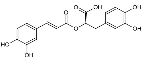
Fibrosis, which causes structural hardening and functional degeneration in various organs, is characterized by the excessive production and accumulation of connective tissue containing collagen, alpha-smooth muscle actin (α-SMA), etc. In traditional medicine, extracts of medicinal plants or herbal prescriptions have been used to treat various fibrotic diseases. RA, as well as the extracts of Glechoma hederacea, Melissa officinalis, Elsholtzia ciliata, Lycopus lucidus, Ocimum basilicum, Prunella vulgaris, Salvia rosmarinus (Rosmarinus officinalis), Salvia miltiorrhiza, and Perilla frutescens, have been shown to attenuate fibrosis of the liver, kidneys, heart, lungs, etc. in experimental animal models. Their antifibrotic effects were associated with the attenuation of oxidative stress, inflammation, cell activation, epithelial–mesenchymal transition, and fibrogenic gene expression. 

rosmarinic acid fibrosis Lamiaceae transforming growth factor β1 TGF-β1 Wnt peroxisomal proliferator-activated receptor γ PPARγ 5′ AMP-activated protein kinase AMPK

1. Introduction

In 2007, the mechanism of action of Wen-pi-tang-Hab-Wu-ling-san (WHW), which is an oriental herbal prescription currently used in Korea for the treatment of chronic renal failure, was investigated [1]. WHW extract inhibited the expression of alpha-smooth muscle actin (α-SMA) and the phosphorylation of small mothers against decapentaplegic (SMAD) 2 that were induced by transforming growth factor (TGF) β1 in Madin–Darby canine kidney cells (MDCKs), thus supporting its renal protective effects via the inhibition of the TGF-β1/SMAD2 pathway. In the same study, rosmarinic acid (RA, Figure 1) was proposed as an active compound responsible for the antifibrotic activity of WHW extract. At that time, there was little research regarding the antifibrotic actions of RA; however, since then, related research has substantially increased.
Figure 1. The chemical structure of rosmarinic acid (RA).
Fibrosis, which causes structural hardening and functional degeneration of major organs of the body, is characterized by the excessive production and accumulation of connective tissues containing collagen and α-SMA [2][3]. Fibrosis is caused by chronic exposure to physical, chemical, and biological stresses, and its progression is mediated by various factors, such as TGF-β1, platelet-derived growth factor (PDGF), connective tissue growth factor (CTGF), Wnt, heat shock proteins (HSPs), and reactive oxygen species (ROS) [4][5]. Because the disease mechanism of fibrosis for each organ has both common and different elements, research has been conducted to identify the therapeutic targets specific to individual organs as well as treatment strategies to recover them to a normal state [6][7][8][9][10].
In traditional medicine in the East and West, herbal prescriptions that are composed of several medicinal plants have been used to alleviate fibrotic diseases of the major organs, including the liver, kidneys, and lungs [11][12][13]. Interestingly, plant extracts with these effects contain several polyphenolic compounds in common, and studies on the pharmacological effects of these compounds are attracting attention [14][15][16]. A literature search by Alberti showed that plant extracts that contain hydroxycinnamic acids inhibited experimentally induced liver fibrosis [17]. Contents of hydroxycinnamic acids, such as caffeic acid, chlorogenic acid, and RA, were particularly high in Lamiaceae plants, and RA was found to be the most common main compound in these plants. The antioxidant and anti-inflammatory properties of RA underlie its therapeutic potential against various liver diseases, such as hepatitis, fibrosis, cirrhosis, and carcinoma [18].
RA is a secondary metabolite that is found in various plants, including the Lamiaceae, Boraginaceae, and Apiaceae families, and it is structurally an ester compound of caffeic acid and 3,4-dihydroxyphenyllactic acid [19]. As a polyphenol compound, RA is considered to function as a defensive phytoalexin in plants and has multiple biological activities in addition to its antioxidant and antimicrobial properties [20]. Because oxidative stress, which is increased by various internal and external factors, leads to excessive inflammatory responses and various chronic diseases, the medical application of RA for its antioxidant and anti-inflammatory properties is currently underway [21][22]. The value of RA as a nutraceutical, which promotes the overall health of the body, is high, and the medical application of RA is gradually and continuously expanding, as discussed in recent review papers [23][24][25].

2. Liver Fibrosis

HSCs are considered one of the most important therapeutic targets for liver fibrosis because they are activated, undergo myofibroblastic transdifferentiation, and participate in the progression of the disease [26]. RA inhibited cell proliferation and the expression of TGF-β, CTGF, and α-SMA in cultured HSCs in vitro [27]. It also conferred antifibrotic effects by ameliorating the biochemical and histopathological changes in the liver and the expression of TGF-β and CTGF in mice with liver fibrosis induced by CCl4 [27].
Peroxisomal proliferator-activated receptor (PPAR) γ functions normally in quiescent HSCs, and its epigenetic repression causes cell transdifferentiation to myofibroblastic cells that mediate liver fibrosis progression [27][28]. Restored expression of PPARγ can recover the normal phenotypes of HSCs. The Wnt signaling pathway is implicated in the epigenetic repression of PPARγ, which leads to the progression of fibrosis in different organs [29]. Impeding the canonical Wnt signaling pathway using the co-receptor antagonist DKK1 abolished the epigenetic repression of PPARγ and restored the gene expression and normal phenotypes of HSCs [28].
The herbal prescription Yang-Gan-Wan, whose main active polyphenolic constituents were identified as RA and baicalin, was shown to restore the normal phenotypes of HSCs through an epigenetic mechanism [30]. Yang-Gan-Wan reduced the expression and recruitment of the DNA methyl-CpG-binding protein MeCP2 to the PPARγ promoter, the expression of histone methyltransferase, the enhancer of zeste homolog 2 (EZH2), and the methylation of histone H3 lysine 27 (H3K27), with the subsequent induction of PPARγ. RA and baicalin restored the normal phenotypes of HSCs by means of epigenetic PPARγ induction. These compounds further suppressed the expression of Wnt family members, such as Wnt10b and Wnt3a, and the canonical Wnt signaling pathway. In addition, RA treatment inhibited HSC activation and the progression of liver fibrosis in mice with cholestatic liver fibrosis induced by BDL [30]. These results suggest that RA could alleviate liver fibrosis via the suppression of the canonical Wnt signaling pathway and epigenetic de-repression of PPARγ in HSCs.
RA was shown to inhibit cell proliferation and induce apoptosis in the activated hepatic stellate cell line (HSC-T6), which correlated with the reduced phosphorylation in STAT3 and the downregulation of cyclin D1 and B cell lymphoma/leukemia (Bcl) 2 [31]. Oxidative stress plays a role in liver fibrogenesis by activating the HSCs [32]. RA inhibited the activation of matrix metalloproteinase (MMP) 2 in a nuclear factor kappa B (NF-κB)-dependent mechanism in HSC-T6 cells [33]. In addition, RA suppressed the generation of ROS and lipid peroxidation while increasing the cellular glutathione (GSH) levels and the expression of catalytic subunits of glutamate cysteine ligase (GCLc) in a nuclear factor erythroid 2-related factor 2 (NRF2)/antioxidant response element (ARE)-dependent manner.
In Sprague Dawley (SD) rats with cholestatic liver injury induced by BDL, oral administration of the extract of Glechoma hederacea lowered the expression levels of TGF-β1, CTGF, SMAD2/3, and collagen and reduced the number of α-SMA-positive matrix-producing cells [34]. In addition, Glechoma hederacea extract attenuated the BDL-induced inflammatory cell infiltration and accumulation, the NF-κB and activator protein-1 (AP-1) activation, and the inflammatory cytokine production, whereas it inhibited the axis of the high mobility group box (HMGB) 1/Toll-like receptor (TLR) 4 intracellular signaling pathways. RA, which was identified as one of the most abundant polyphenolic compounds in Glechoma hederacea, also demonstrated protective effects against cholestasis-related liver injury via the attenuation of oxidative stress and the downregulation of HMGB1/TLR4, NF-κB, AP-1, and TGF-β1/SMAD signaling [35].
The Fuzheng Huayu recipe (FZHY) is an herbal prescription that is used in China to treat liver fibrosis [36][37]. Yang et al. identified more than eleven compounds and two metabolites in the serum from rats that were orally administered an extract of Danshen (Salvia miltiorrhiza), the main component of FZHY [36]. Of these compounds, salvianolic acid B (6 and 48 μM), caffeic acid (6 and 48 μM), and RA (48 μM) inhibited the proliferation of the HSC cell line (LX-2) and down-regulated the in vitro expression of α-SMA, thus supporting their contribution to the antihepatic fibrosis effects of FZHY. Plasma pharmacokinetics and tissue distribution profiles were analyzed following the oral administration of FZHY to normal and fibrotic rats induced by intraperitoneal injections of dimethylnitrosamine [37]. The results revealed that the serum levels of danshensu, salvianolic acid B, and RA were 1.69-, 2.37-, and 1.49-fold higher, respectively, in rats with liver fibrosis compared with the normal/nonfibrotic rats. It is suggested that the absorption and metabolism of active ingredients, including RA, in a mixed herbal prescription may vary depending on the disease state in the body and that they can exert therapeutic effects by modulating the relevant cells when administered at effective concentrations.
The hepatoprotective and antifibrotic effects of RA were compared with silymarin in thioacetamide-intoxicated SD rats [38]. RA at a daily oral dose of 10 mg kg−1 had similar hepatoprotective effects as silymarin at a dose of 50 mg kg−1 in that it lowered the serum levels of alanine aminotransferase (ALT) and aspartate aminotransferase (AST), reduced the tissue levels of malondialdehyde (MDA), and increased the tissue levels of GSH in the thioacetamide-intoxicated rats. Thioacetamide increased the tissue levels of the fibrotic markers, such as TIMP-1 and hydroxyproline, in the liver, and these changes were attenuated by RA (10 mg kg−1) and silymarin (50 mg kg−1) treatment to a similar extent. Both RA and silymarin decreased the expression of collagen and α-SMA in the livers of thioacetamide-intoxicated rats. In vitro experiments using HSC-T6 cells showed that RA decreased cell viability and increased both caspase-3-positive cells and α-SMA-positive cells, thus supporting the antiproliferative and proapoptotic effects of RA in this cell type.
When lemon balm (Melissa officinalis) extract and its constituent, RA, were administered orally, they were shown to alleviate nonalcoholic steatohepatitis and hepatic fibrosis in db/db mice fed a methionine- and choline-deficient (MCD) diet [39]. Oral treatments with the extract (200 mg kg−1) or RA (10 or 30 mg kg−1) alleviated liver damage that was monitored by the serum levels of ALT and AST. These treatments inhibited the accumulation of hepatic triglycerides and hydroxyproline as well as the expression of α-SMA and collagen (COL) 1A1 in the mouse model fed a MCD diet. In the associated in vitro experiments using HepG2 cells, palmitic acid caused the accumulation of lipids and triglycerides and the expression of lipogenic genes, such as sterol regulatory element-binding protein (SREBP) 1c, fatty acid synthase (FAS), and stearoyl-CoA desaturase (SCD) 1, whereas it suppressed the expression of lipolytic genes, such as carnitine palmitoyl transferase (CPT) 1L [39]. Treatment with either the extract or RA reversed these changes and restored the 5′ AMP-activated protein kinase (AMPK) phosphorylation and NRF2 activation as well as its downstream targets, including superoxide dismutase (SOD) 1 and heme oxygenase (HO) 1.
Lyu et al. demonstrated that advanced glycation end-products (AGEs) mediate high levels of cross-linking in the ECM of cirrhotic liver tissue [40]. AGE cross-linking in the collagen matrix could be effectively inhibited by RA in vitro. Furthermore, RA inhibited the AGE-mediated cross-linking in the liver ECM and alleviated late-stage liver fibrosis, as shown by the reduced levels of collagen in the scar tissue, hepatic hydroxyproline, and the proportion of α-SMA-positive cells in male C57BL/6 mice that were treated with CCl4 or fed the high-fat choline-deficient L-amino acid-defined (HFCDAA) diet, thus suggesting its potential to alleviate liver cirrhosis.

3. Kidney Fibrosis

EMT is a process of cell transdifferentiation that occurs during the development of many tissues [41]. Epithelial and mesenchymal cells have different morphologies, functions, and other phenotypes; epithelial cells express high levels of E-cadherin, whereas mesenchymal cells express high levels of N-cadherin, fibronectin, vimentin, collagen, and α-SMA. EMT can occur during the early stages of wound healing, organ fibrosis, and cancer metastasis; therefore, it is important in the pathogenesis of various diseases and a useful therapeutic target [42].
WHW, a herbal prescription composed of 14 medicinal plants, has been used in oriental traditional medicine for the treatment of chronic renal failure as well as for other purposes [1][43][44]. The previous study demonstrated that WHW extract (1 mg mL−1) attenuated the TGF-β1-induced morphological change of MDCKs undergoing EMT [1]. It inhibited the TGF-β1-stimulated expression of α-SMA and the phosphorylation of SMAD2. In the same study, activity-guided solvent fractionation and chromatographic separation of WHW extract identified RA as one of the main active compounds. It was further verified that RA (52–104 μM) suppressed the expression of α-SMA in MDCKs stimulated with TGF-β1 (unpublished data). In the study using cultured primary mouse macrophages and the RAW264.7 cell line, WHW extract (0.5 mg mL−1) suppressed the production of •NO as well as the mRNA expression and protein levels of nitric oxide synthase (NOS) 2, tumor necrosis factor (TNF) α, interleukin (IL) 1β, and IL-6 induced by lipopolysaccharide (LPS) [43]. WHW extract inhibited the phosphorylation of MAPK, such as ERK 1/2, p38, and c-Jun N-terminal kinase (JNK), and the nuclear translocation of NF-κB p65 protein in the LPS-stimulated RAW264.7 cells. These in vitro studies suggest anti-inflammatory and antifibrogenic properties of WHW extract that contains RA.
The effect of WHW on kidney ischemia/reperfusion injury was evaluated in a mouse model [45][46]. The mice were orally administered WHW extract (0.5–100 mg kg−1) daily for 14 days and then subjected to 30 min of bilateral renal ischemia. Then, oral administration of WHW continued for 2 days [45]. The ischemia/reperfusion resulted in the disruption of the kidney tubular epithelial cells and increased plasma creatinine levels in the model mice, but these changes were attenuated by the WHW treatment. WHW treatment also inhibited the post-ischemic increase of H2O2 and lipid peroxidation and the post-ischemic decrease in catalase, copper/zinc SOD, and manganese SOD activity in the kidney tissue. WHW treatment enhanced the post-ischemic increase in the expression levels of HSP27 and HSP72. In another study, WHW extract was administered orally to mice for 14 days, beginning 2 days after the ischemic procedure [46]. WHW treatment attenuated the progression of renal fibrosis and prevented the post-ischemic increase of H2O2 and lipid peroxidation, as well as the decrease of copper/zinc SOD and manganese SOD activity. In addition, WHW treatment reduced the phosphorylation of ERK1/2 and JNK1/2 and the activation of NF-κB in the kidneys induced by ischemia/reperfusion.
WHW treatment also inhibited the kidney fibrosis that was induced by unilateral ureteral obstruction (UUO) in mice [47]. Post-procedure WHW treatment (2, 10, or 50 mg kg−1) mitigated the UUO-induced kidney fibrosis, as determined by tubular atrophy and dilatation, collagen accumulation, interstitial space expansion, and leukocyte infiltration. WHW treatment prevented the increase in oxidative stress and decrease of catalase, copper/zinc SOD, and manganese SOD activity due to the UUO procedure. WHW reduced the expression of TGF-β1 and the phosphorylation of SMAD2/3 stimulated by the UUO procedure. Collectively, WHW extract can alleviate oxidative stress, inflammation, and fibrosis in the kidneys following an ischemia/reperfusion or UUO.
The effects of Elsholtzia ciliata extract, which contains luteolin and RA as its main phenolic compounds, on renal interstitial fibrosis were examined in male SD rats with UUO [48]. Orally administered extract (300 or 500 mg kg−1) reduced UUO-induced renal damage, histopathological alterations, and interstitial fibrosis. In addition, the renal expression levels of TNF-α, TGF-β1, and SMAD3 were reduced in the extract-treated rats compared to the untreated UUO model rats. The accompanying in vitro studies revealed that this extract (200 μg mL−l) ameliorated the LPS-induced overexpression of NF-κB, TNF-α, and IL-6, improved oxidative stress in RAW 264.7 cells, and suppressed the TGF-β-induced expression of α-SMA and MMP9 in human renal mesangial cells. Thus, Elsholtzia ciliata extract may impede the inflammatory and fibrogenic signaling pathways that cause renal fibrotic disease.
The inhibitory effects of RA on kidney fibrosis and EMT were examined using a UUO mouse model and an indoxyl sulfate-stimulated NRK-52E cell model [49]. C57BL/6 mice were orally administered RA (10 or 20 mg kg−1) before and after UUO surgery. UUO caused kidney damage, which was determined by the increase in serum creatine and blood urea nitrogen (BUN) levels, and the damage was reduced by RA treatment. The UUO-induced increase of α-SMA, collagen I, fibronectin, and vimentin and the decrease of E-cadherin were attenuated by RA treatment. NRK-52E cells were treated with 4 mM indoxyl sulfate with or without RA (20 or 40 μM). The indoxyl sulfate-induced proliferation and migration of the cells were inhibited by RA treatment. RA treatment further attenuated the increase of α-SMA, collagen I, fibronectin, vimentin, and TGF-β1 and the decrease of E-cadherin induced by indoxyl sulfate. RA treatment inhibited the phosphorylation of Akt in both the cell and animal models. Thus, RA was suggested to ameliorate renal interstitial fibrosis by inhibiting the Akt-mediated EMT.
RA was shown to attenuate the nephrotoxicity of cadmium both in vitro and in vivo [50]. RA prevented the apoptotic death of cultured mouse proximal tubular epithelial cells that were exposed to CdCl2. It attenuated the CdCl2-induced increased production of ROS/•NO, lipid peroxidation, protein carbonylation, the increased activity of NOX activity, and the decreased levels of coenzyme 9, coenzyme 10, and GSH, as well as the decreased activity of SOD, catalase, glucose-6-phosphate dehydrogenase (G6PDH), glutathione peroxidase (GPX), glutathione S-transferase (GST), and glutathione reductase (GR). CdCl2 stimulated apoptotic events that were mediated by Bcl-2, Bcl-2-associated death protein (Bad), apoptotic protease activating factor (Apaf) 1, cytochrome C, and caspases 3, 8, and 9, and the fibrogenic events that were accompanied by the upregulation of α-SMA, E-cadherin, and collagen IV in the cells, and all these events were attenuated by RA treatment. Swiss albino mice were treated with CdCl2 (4 mg kg−1) with or without RA treatment (50 mg kg−1), and the pathological changes were monitored by assessing the blood, urine, and tissues. The biochemical and histological data supported the pharmacological effects of RA attenuating oxidative stress, apoptosis, inflammation, and fibrosis in the kidneys caused by cadmium exposure.
The effects of Lycopus lucidus extract, which contains caffeic acid, luteolin-7-O-β-D-glucoside, and RA as the main phenolic compounds, on kidney fibrosis were examined in rhTGF-β1-stimulated SV40 MES13 cells and a streptozotocin-induced diabetic nephropathy rat model [51]. Treatment with the extract (50–400 μg mL−1) suppressed the activation of SMAD2 and ERK1/2 and downregulated TGF-βRI, TGF-βRII, SMAD4, and SMAD7 in TGF-β1-stimulated cells. This extract (3, 6, or 12 g kg−1) inhibited SMAD2 phosphorylation, reduced TGF-β1 expression, ameliorated the expansion of the mesangial area in glomerular tissue, reduced the levels of serum creatine and BUN, and reduced the total SOD activity in the streptozotocin-induced diabetic nephropathy rats. Thus, Lycopus lucidus extract is considered to be able to inhibit renal fibrosis by impeding the TGF-β signaling pathway and attenuating diabetic nephropathy.
Guanxinning injection, a herbal prescription composed of Radix et Rhizoma Salviae Miltiorrhizae (Salvia miltiorrhiza) and Chuanxiong Rhizoma (Ligusticum chuanxiong), was tested for its effects on renal fibrosis in mice with heart failure induced by transverse aortic constriction [52]. Guanxinning injection was administered via tail vein at doses of 3–12 mL kg−1, and it relieved the cardiac function indexes (ejection fraction, cardiac output, and left ventricle volume), kidney functional indexes (serum creatinine), and kidney fibrosis indexes (collagen volume fraction and CTGF) in the model mice. Guanxinning injection further increased catalase, GPX4, the x(c)(-)cysteine/glutamate antiporter SLC7A11, and ferritin heavy chain 1, while decreasing xanthine oxidase and NOS in the kidneys. Its major active ingredients include RA, caffeic acid, ferulic acid, etc. Thus, Guanxinning injection could help maintain cardiac function and alleviate the progression of fibrosis in the kidneys due to heart failure through the regulation of redox metabolism.
Patients with autosomal dominant polycystic kidney disease had higher expression levels of complement factor B and complement component 9 in the kidneys compared with control subjects or patients with other types of chronic kidney disease [53]. Oral administration of RA (300 mg kg−1, daily) for 4 weeks reduced serum creatine, BUN, kidney weight, cyst index, fibrosis, expression of complement factor B and C5b-9, and the number of Ki67-positive nuclei, as well as the inflammatory cells, without affecting the body weight in Pkd1−/− knockout mice. This supports the therapeutic potential of RA as an inhibitor of the complement pathway associated with autosomal dominant polycystic kidney disease progression [53].

4. Heart Fibrosis

The ethanolic extract of the aerial parts of basil (Ocimum basilicum), which contains RA as the principal phenolic compound, has been shown to improve cardiac function and inhibit the histopathological changes in response to isoproterenol-induced myocardial infarction (MI) in male Wistar albino rats [54]. Treatment with isoproterenol (100 mg kg−1) daily for 2 consecutive days caused an elevation in the ST segment in the electrocardiogram, indicative of a MI, a reduction in the left ventricular contractility, an increase in the left ventricular end-diastolic pressure, and an increase in myocardial necrosis and fibrosis, all of which were significantly attenuated by the subcutaneous injection of 10, 20, or 40 mg kg−1 of the extract twice per day. In addition, the isoproterenol-induced elevated MDA levels in the serum and myocardium were suppressed by the extract, thus suggesting that the cardioprotective effects of O. basilicum extract could be associated with its antioxidant activity.
The aqueous extract of Xia-Ku-Cao (Prunella vulgaris), as well as its caffeic acid, ursolic acid, and RA components, exhibited a cardioprotective effect in acute MI in male SD rats with left anterior descending coronary artery (LAD) ligation [55]. The extract (400 mg kg−1) administered by intragastric gavage after surgery improved cardiac function and reduced the infarct size, inflammation, fibrosis, oxidative damage, and apoptosis of cardiomyocytes. Phenolic compounds, such as caffeic acid (400 mg kg−1), ursolic acid (400 mg kg−1), and RA (400 mg kg−1), improved cardiac function and suppressed inflammatory aggregation and fibrosis in MI rat models. RNA-seq analysis and an additional in vitro study suggest that the therapeutic effect of P. vulgaris extract and its phenolic compounds might be related to the reduced expression of NOD-like receptor protein 3 (NLRP3), which is presumed to play a critical role in the inflammatory process during cardiac remodeling after a MI [56].
RA demonstrated antifibrotic effects in male SD rats with a MI induced by LAD ligation surgery [57]. The oral administration of RA (50, 100, or 200 mg kg−1) ameliorated the infarct size and MI-induced changes in the left ventricular systolic pressure, +dp/dtmax, and −dp/dtmax. RA attenuated the cardiac fibrosis, as determined by the collagen volume fraction and the expression of collagen I, collagen III, α-SMA, and hydroxyproline. RA treatment also decreased the expression of angiotensin-converting enzyme (ACE), angiotensin type 1 receptor (AT1R), and phospho-p38 MAPK while increasing the expression of ACE2, which may be associated with the cardioprotective effects of RA against cardiac dysfunction and fibrosis.
An injectable hydrogel encapsulating polydopamine-RA nanoparticles with dual responsiveness to pH and ROS was developed to achieve on-demand drug release in the MI microenvironment [58]. Oxidized xanthan gum grafted with 3-aminophenylboronic acid and dopamine-grafted gelatin were combined to prepare an “OGD” hydrogel. The polypyrrole-modified gelatin was combined with “OGD” to prepare an “OGDP” hydrogel. The polydopamine-RA nanoparticles were added to the “OGD” and “OGDP” hydrogels to prepare the “OGDR” and “OGDPR” hydrogels. Rats were subjected to LAD ligation to induce MI, and on day 2 after modeling, rats with a confirmed MI were subjected to multiple injections with the “OGD”, “OGDR”, “OGDP”, or “OGDPR” hydrogels in and around the infarct area. The OGDPR hydrogel showed the best results by exerting anti-inflammatory, antiapoptotic, and antifibrotic effects. The multifunctional hydrogel promoted the expression of heart-specific markers, thus restoring heart function after a MI.
RA also attenuated cardiac remodeling and fibrosis following a long-term pressure overload in male C57/B6 mice subjected to aortic banding to generate pressure overload, which led to cardiac dysfunction with reduced fractional shortening [59]. Orally administered RA (100 mg kg−1) attenuated the fibrotic response and cardiac dysfunction after pressure overload without affecting the hypertrophic response. Aortic banding increased the expression of collagen I and III, CTGF, fibronectin, TGF-β1, and α-SMA, and the phosphorylation of ERK, Akt, p38, AMPK, and acetyl-CoA carboxylase (ACC). RA attenuated the increased expression of collagen I and III, CTGF, fibronectin, TGF-β1, and α-SMA and enhanced the increased phosphorylation of AMPK and ACC without significantly affecting the phosphorylation of ERK, Akt, and p38. The cardioprotective effects of RA in reducing the expression of collagen I and III, the phosphorylation of SMAD, and the restored fractional shortening induced by aortic banding were abolished in AMPKα2 knockout mice.
RA has been demonstrated to alleviate the apoptosis of cardiomyocytes [60] and cardiac fibrosis induced by doxorubicin, an anticancer medicine, in male rats [61]. Doxorubicin (2 mg kg−1 per 48 h) increased the heart-to-body weight ratio, reduced the heart rate and blood pressure, and caused fibrosis and necrosis of the cardiac tissue [61]. These toxic effects of doxorubicin were attenuated by the administration of RA (10, 20, or 40 mg kg−1). RA administration also attenuated the doxorubicin-induced increase of MDA and decrease of GSH in the heart tissue, thereby implying that its antioxidant properties underlie the protective effects against doxorubicin-induced cardiotoxicity.

5. Lung Fibrosis

Rosemary (Rosmarinus officinalis) leaf extracts, which contain carnosic acid and RA, have been shown to inhibit bleomycin-induced pulmonary fibrosis [62]. Male Wistar rats were given a single dose of bleomycin (4 mg kg−1, intratracheal), and the extract (75 mg kg−1) was administered 3 days later and continued for 4 weeks (curative group), or administered 2 weeks before bleomycin administration and continued for 15 days thereafter (prophylactic group). The lung architecture was more retained in the curative and prophylactic groups compared to the bleomycin group, which was associated with lower fibrosis scores (2.33 and 1.8 vs. 3.7 of the bleomycin group, respectively), reduced MDA levels (141% and 108% vs. 258% of the normal value, respectively), high catalase levels (103% and 117% vs. 59% of the normal value, respectively), and enhanced GST activity (85% and 69% vs. 23% of the normal value, respectively). The antifibrotic and antioxidant effects of carnosic acid, RA, and their combination were verified in the bleomycin-induced fibrosis model in rats [63]. In addition, purified rosemary leaf extract with increased RA and carnosic acid contents and decreased essential oil content exhibited potent antifibrotic efficacy in an animal model [64].
RA attenuated the pulmonary fibrosis caused by irradiation. Treatments of male SD rats with RA (30, 60, or 120 mg kg−1) before 15 Gy of X-ray irradiation decreased the expression of inflammatory mediators, NF-κB phosphorylation, and the production of ROS [65]. RA inhibited Ras homolog family member A (RhoA)/Rho-associated protein kinase (Rock) signaling through the upregulation of the microRNA (miR) 19b-3p, which targets the myosin phosphatase target subunit 1 (MYPT1) and leads to the inhibition of TGF-β1 signaling and subsequent fibrosis [65].
Salvia miltiorrhiza extract, which contains salvianic acid A and B, caffeic acid, RA, and tanshinone IIA, attenuated ovalbumin-induced asthma in mice [66]. The ovalbumin challenge increased airway resistance, inflammatory cell infiltration, Th1/Th2 cytokine levels in the bronchoalveolar lavage fluid (BALF), cell hyperplasia, collagen deposition, and airway wall thickening. Daily treatment with the water extract of S. miltiorrhiza (156 mg kg−1) significantly alleviated these pathological changes. In the associated in vitro experiments, caffeic acid and RA potently reduced E-cadherin and vimentin in the TGF-β1-induced BEAS-2B cells and α-SMA and COL1A1 in the TGF-β1-induced MRC-5 cells. RA also attenuated the immunological and inflammatory responses and normalized the pathological features in the lungs of ovalbumin-induced asthmatic rats [67]. The asthmatic rats showed increased levels of IL-4, immunoglobulin (Ig) E, phospholipase A (PLA) 2, and total protein in the BALF and decreased levels of interferon (IFN) γ and the IFN-γ/IL-4 ratio compared to the control group. The RA treatment reduced the levels of IL-4, IgE, PLA2, and total protein and restored the IFN-γ/IL-4 ratio.

6. Post-Surgical Abdominal Adhesion

Peritoneal adhesion occurs when the peritoneum is damaged as a result of various injury events, including abdominal surgery [68]. Peritoneal bands connect the peritoneum to different visceral organs and cause abdominal discomfort, infertility in women, intestinal blockage, and other ailments [69]. RA (50 or 70 mg kg−1) inhibited the formation of postoperative peritoneal adhesion in male Wistar albino rats [70]. RA decreased the peritoneal adhesion score and was associated with reduced fibroblast proliferation, the expression of TGF-β1 (a fibrotic marker), TNF-α (an inflammation marker), and vascular endothelial growth factor (VEGF) (an angiogenesis marker), and reduced MDA levels (an oxidative stress marker). A polygalacturonic acid-conjugated cysteine hydrogel has been developed as a drug carrier for RA, and animal implant studies have shown that hydrogel films with and without RA reduce the incidence of post-surgical adhesion and early inflammatory reactions [71].

7. Fibrosis in the Salivary Glands

The three major types of salivary glands are the parotid, submaxillary, and sublingual glands [72]. The salivary glands have a high risk of exposure to oxidative damage after irradiation therapy for head and neck cancers [73]. The radioprotective effect of RA on the parotid gland was examined in SD rats irradiated with 15 Gy X-ray irradiation [74]. RA treatment (60 and 120 mg kg−1) attenuated the radiation-induced hyposalivation and oxidative stress in the parotid gland as effectively as amifostine (250 mg kg−1). The radiation increased NOX 4 and decreased PPARγ coactivator (PGC) 1α, which were normalized by RA treatment. RA reduced apoptosis by inhibiting p53/JNK activation and fibrosis by downregulating inflammatory factor levels. Therefore, RA has the therapeutic potential to treat radiation-induced parotid gland injuries.

8. Skin Wounds

The skin wound healing effects of RA were examined in Wistar albino rats with a 2 cm full-thickness skin wound [75]. The topical application of a 10% RA cream enhanced the wound size reduction more effectively compared to a 5% dexpanthenol cream application or nontreatment, which resulted in fewer scars. An oxidized dextran/amidated gelatin hydrogel, “ODex-AG”, and an RA-grafted hydrogel, “ODex-AG-RA”, were developed as dressings for skin injuries [76]. Both the “ODex-AG” and “ODex-AG-RA” hydrogels promoted skin wound healing in a rat model of a full-thickness skin wound. The “ODex-AG-RA” hydrogel effectively enhanced the collagen deposition and neovascularization (CD31) in the wounds and alleviated inflammation (TNF-α and CD163) and oxidative stress (MDA and H2O2) compared to the “ODex-AG” hydrogel. Overall, this study demonstrated the utility of RA-grafted hydrogels as a wound dressing with anti-inflammatory and antioxidant activities.

9. Pterygium in the Eyes

Pterygium occurs due to the uncontrolled excessive proliferation of epithelial tissue in the eyes, and the lesion can invade the cornea, potentially causing irregular corneal astigmatism and vision loss [77]. The antifibrotic effect of RA has been tested in pterygium epithelial cells [78]. RA treatment (100 µM) significantly decreased the cell viability and the protein expression of type I collagen, TGF-β1, TGF-βRII, phospho-SMAD1/5, phospho-SMAD2, phospho-SMAD, and SMAD4. Thus, RA is suggested to inhibit the TGF-β1/SMAD signaling pathway involved in pterygium epithelial cell-mediated fibrosis. Further studies are expected to evaluate the therapeutic potential of RA for the in vivo treatment of pterygium.

10. Fibrosis of Autologous Fat Grafts

After the autologous transplantation of inguinal fat pads to the parascapular area in SD rats, an ethanol solution of RA was intraperitoneally injected (20 mg kg−1) daily for a week and then weekly for 7 weeks [79]. The total oxidant status, MDA, and the expression levels of TNF-α and TGF-β1 were lower in the transplanted tissue of the RA-treated group compared to the control or vehicle (ethanol)-treated groups, which indicates that RA could reduce the necrosis, inflammation, and fibrosis of fat grafts [79].

References

  1. Lee, S.I.; Kim, H.J.; Baek, M.C.; Park, K.M.; Park, Y.; Yoon, C.H.; Boo, Y.C. Wen-pi-tang-Hab-Wu-ling-san, an oriental herbal prescription, attenuates epithelial-mesenchymal transdifferentiation stimulated by TGF-beta1 in kidney cells. Phytother. Res. 2007, 21, 548–553.
  2. Henderson, N.C.; Rieder, F.; Wynn, T.A. Fibrosis: From mechanisms to medicines. Nature 2020, 587, 555–566.
  3. Antar, S.A.; Ashour, N.A.; Marawan, M.E.; Al-Karmalawy, A.A. Fibrosis: Types, Effects, Markers, Mechanisms for Disease Progression, and Its Relation with Oxidative Stress, Immunity, and Inflammation. Int. J. Mol. Sci. 2023, 24, 4004.
  4. Lurje, I.; Gaisa, N.T.; Weiskirchen, R.; Tacke, F. Mechanisms of organ fibrosis: Emerging concepts and implications for novel treatment strategies. Mol. Asp. Med. 2023, 92, 101191.
  5. Gyorfi, A.H.; Matei, A.E.; Distler, J.H.W. Targeting TGF-beta signaling for the treatment of fibrosis. Matrix Biol. 2018, 68–69, 8–27.
  6. Distler, J.H.W.; Gyorfi, A.H.; Ramanujam, M.; Whitfield, M.L.; Konigshoff, M.; Lafyatis, R. Shared and distinct mechanisms of fibrosis. Nat. Rev. Rheumatol. 2019, 15, 705–730.
  7. Tan, Z.; Sun, H.; Xue, T.; Gan, C.; Liu, H.; Xie, Y.; Yao, Y.; Ye, T. Liver Fibrosis: Therapeutic Targets and Advances in Drug Therapy. Front. Cell Dev. Biol. 2021, 9, 730176.
  8. Sato, S.; Yanagihara, T.; Kolb, M.R.J. Therapeutic targets and early stage clinical trials for pulmonary fibrosis. Expert Opin. Investig. Drugs 2019, 28, 19–28.
  9. Rayego-Mateos, S.; Valdivielso, J.M. New therapeutic targets in chronic kidney disease progression and renal fibrosis. Expert Opin. Ther. Targets 2020, 24, 655–670.
  10. Park, S.; Nguyen, N.B.; Pezhouman, A.; Ardehali, R. Cardiac fibrosis: Potential therapeutic targets. Transl. Res. 2019, 209, 121–137.
  11. Latief, U.; Ahmad, R. Herbal remedies for liver fibrosis: A review on the mode of action of fifty herbs. J. Tradit. Complement. Med. 2018, 8, 352–360.
  12. Xu, Y.; Chen, J.; Wang, H.; Lu, Y. Research and application of herbal medicine in the treatment of chronic kidney disease since the 21st century: A visualized bibliometric analysis. Front. Pharmacol. 2022, 13, 971113.
  13. Wu, X.; Huang, J.; Wang, J.; Xu, Y.; Yang, X.; Sun, M.; Shi, J. Multi-Pharmaceutical Activities of Chinese Herbal Polysaccharides in the Treatment of Pulmonary Fibrosis: Concept and Future Prospects. Front. Pharmacol. 2021, 12, 707491.
  14. Wang, L.; Zhu, T.; Feng, D.; Li, R.; Zhang, C. Polyphenols from Chinese Herbal Medicine: Molecular Mechanisms and Therapeutic Targets in Pulmonary Fibrosis. Am. J. Chin. Med. 2022, 50, 1063–1094.
  15. Li, S.; Tan, H.Y.; Wang, N.; Cheung, F.; Hong, M.; Feng, Y. The Potential and Action Mechanism of Polyphenols in the Treatment of Liver Diseases. Oxid. Med. Cell Longev. 2018, 2018, 8394818.
  16. Zhou, Z.; Qiao, Y.; Zhao, Y.; Chen, X.; Li, J.; Zhang, H.; Lan, Q.; Yang, B. Natural products: Potential drugs for the treatment of renal fibrosis. Chin. Med. 2022, 17, 98.
  17. Alberti, A. Importance of dietary hydroxycinnamic acids in the therapy of liver fibrosis. Orv. Hetil. 2012, 153, 948–953.
  18. Elufioye, T.O.; Habtemariam, S. Hepatoprotective effects of rosmarinic acid: Insight into its mechanisms of action. Biomed. Pharmacother. 2019, 112, 108600.
  19. Guan, H.; Luo, W.; Bao, B.; Cao, Y.; Cheng, F.; Yu, S.; Fan, Q.; Zhang, L.; Wu, Q.; Shan, M. A Comprehensive Review of Rosmarinic Acid: From Phytochemistry to Pharmacology and Its New Insight. Molecules 2022, 27, 3292.
  20. Nieto, G.; Ros, G.; Castillo, J. Antioxidant and Antimicrobial Properties of Rosemary (Rosmarinus officinalis, L.): A Review. Medicines 2018, 5, 98.
  21. Dahchour, A. Anxiolytic and antidepressive potentials of rosmarinic acid: A review with a focus on antioxidant and anti-inflammatory effects. Pharmacol. Res. 2022, 184, 106421.
  22. Luo, C.; Zou, L.; Sun, H.; Peng, J.; Gao, C.; Bao, L.; Ji, R.; Jin, Y.; Sun, S. A Review of the Anti-Inflammatory Effects of Rosmarinic Acid on Inflammatory Diseases. Front. Pharmacol. 2020, 11, 153.
  23. Noor, S.; Mohammad, T.; Rub, M.A.; Raza, A.; Azum, N.; Yadav, D.K.; Hassan, M.I.; Asiri, A.M. Biomedical features and therapeutic potential of rosmarinic acid. Arch. Pharm. Res. 2022, 45, 205–228.
  24. Zhao, J.; Xu, L.; Jin, D.; Xin, Y.; Tian, L.; Wang, T.; Zhao, D.; Wang, Z.; Wang, J. Rosmarinic Acid and Related Dietary Supplements: Potential Applications in the Prevention and Treatment of Cancer. Biomolecules 2022, 12, 1410.
  25. Azhar, M.K.; Anwar, S.; Hasan, G.M.; Shamsi, A.; Islam, A.; Parvez, S.; Hassan, M.I. Comprehensive Insights into Biological Roles of Rosmarinic Acid: Implications in Diabetes, Cancer and Neurodegenerative Diseases. Nutrients 2023, 15, 4297.
  26. Bansal, R.; Poelstra, K. Hepatic Stellate Cell Targeting Using Peptide-Modified Biologicals. Methods Mol. Biol. 2023, 2669, 269–284.
  27. Li, G.S.; Jiang, W.L.; Tian, J.W.; Qu, G.W.; Zhu, H.B.; Fu, F.H. In vitro and in vivo antifibrotic effects of rosmarinic acid on experimental liver fibrosis. Phytomedicine 2010, 17, 282–288.
  28. Miao, C.G.; Yang, Y.Y.; He, X.; Huang, C.; Huang, Y.; Zhang, L.; Lv, X.W.; Jin, Y.; Li, J. Wnt signaling in liver fibrosis: Progress, challenges and potential directions. Biochimie 2013, 95, 2326–2335.
  29. Xu, C.Y.; Wang, J.; Zhu, T.J.; Shen, Y.; Tang, X.S.; Fang, L.; Xu, Y.Z. Cross-Talking Between PPAR and WNT Signaling and its Regulation in Mesenchymal Stem Cell Differentiation. Curr. Stem Cell Res. Ther. 2016, 11, 247–254.
  30. Yang, M.D.; Chiang, Y.M.; Higashiyama, R.; Asahina, K.; Mann, D.A.; Mann, J.; Wang, C.C.; Tsukamoto, H. Rosmarinic acid and baicalin epigenetically derepress peroxisomal proliferator-activated receptor gamma in hepatic stellate cells for their antifibrotic effect. Hepatology 2012, 55, 1271–1281.
  31. Zhang, J.J.; Wang, Y.L.; Feng, X.B.; Song, X.D.; Liu, W.B. Rosmarinic acid inhibits proliferation and induces apoptosis of hepatic stellate cells. Biol. Pharm. Bull. 2011, 34, 343–348.
  32. De Smet, V.; Eysackers, N.; Merens, V.; Kazemzadeh Dastjerd, M.; Halder, G.; Verhulst, S.; Mannaerts, I.; van Grunsven, L.A. Initiation of hepatic stellate cell activation extends into chronic liver disease. Cell Death Dis. 2021, 12, 1110.
  33. Lu, C.; Zou, Y.; Liu, Y.; Niu, Y. Rosmarinic acid counteracts activation of hepatic stellate cells via inhibiting the ROS-dependent MMP-2 activity: Involvement of Nrf2 antioxidant system. Toxicol. Appl. Pharmacol. 2017, 318, 69–78.
  34. Wang, Y.Y.; Lin, S.Y.; Chen, W.Y.; Liao, S.L.; Wu, C.C.; Pan, P.H.; Chou, S.T.; Chen, C.J. Glechoma hederacea extracts attenuate cholestatic liver injury in a bile duct-ligated rat model. J. Ethnopharmacol. 2017, 204, 58–66.
  35. Lin, S.Y.; Wang, Y.Y.; Chen, W.Y.; Liao, S.L.; Chou, S.T.; Yang, C.P.; Chen, C.J. Hepatoprotective activities of rosmarinic acid against extrahepatic cholestasis in rats. Food Chem. Toxicol. 2017, 108, 214–223.
  36. Yang, T.; Shen, D.P.; Wang, Q.L.; Tao, Y.Y.; Liu, C.H. Investigation of the absorbed and metabolized components of Danshen from Fuzheng Huayu recipe and study on the anti-hepatic fibrosis effects of these components. J. Ethnopharmacol. 2013, 148, 691–700.
  37. Yang, T.; Liu, S.; Wang, C.H.; Tao, Y.Y.; Zhou, H.; Liu, C.H. Comparative pharmacokinetic and tissue distribution profiles of four major bioactive components in normal and hepatic fibrosis rats after oral administration of Fuzheng Huayu recipe. J. Pharm. Biomed. Anal. 2015, 114, 152–158.
  38. El-Lakkany, N.M.; El-Maadawy, W.H.; Seif El-Din, S.H.; Hammam, O.A.; Mohamed, S.H.; Ezzat, S.M.; Safar, M.M.; Saleh, S. Rosmarinic acid attenuates hepatic fibrogenesis via suppression of hepatic stellate cell activation/proliferation and induction of apoptosis. Asian Pac. J. Trop. Med. 2017, 10, 444–453.
  39. Kim, M.; Yoo, G.; Randy, A.; Son, Y.J.; Hong, C.R.; Kim, S.M.; Nho, C.W. Lemon Balm and Its Constituent, Rosmarinic Acid, Alleviate Liver Damage in an Animal Model of Nonalcoholic Steatohepatitis. Nutrients 2020, 12, 1166.
  40. Lyu, C.; Kong, W.; Liu, Z.; Wang, S.; Zhao, P.; Liang, K.; Niu, Y.; Yang, W.; Xiang, C.; Hu, X.; et al. Advanced glycation end-products as mediators of the aberrant crosslinking of extracellular matrix in scarred liver tissue. Nat. Biomed. Eng. 2023, 7, 1437–1454.
  41. Yang, J.; Antin, P.; Berx, G.; Blanpain, C.; Brabletz, T.; Bronner, M.; Campbell, K.; Cano, A.; Casanova, J.; Christofori, G.; et al. Guidelines and definitions for research on epithelial-mesenchymal transition. Nat. Rev. Mol. Cell Biol. 2020, 21, 341–352.
  42. Dongre, A.; Weinberg, R.A. New insights into the mechanisms of epithelial-mesenchymal transition and implications for cancer. Nat. Rev. Mol. Cell Biol. 2019, 20, 69–84.
  43. Jung, H.W.; Yoon, C.H.; Kim, Y.H.; Boo, Y.C.; Park, K.M.; Park, Y.K. Wen-Pi-Tang-Hab-Wu-Ling-San extract inhibits the release of inflammatory mediators from LPS-stimulated mouse macrophages. J. Ethnopharmacol. 2007, 114, 439–445.
  44. Jung, H.W.; Jung, J.K.; Ramalingam, M.; Yoon, C.H.; Bae, H.S.; Park, Y.K. Anti-diabetic effect of Wen-pi-tang-Hab-Wu-ling-san extract in streptozotocin-induced diabetic rats. Indian J. Pharmacol. 2012, 44, 97–102.
  45. Seok, Y.M.; Kim, J.; Choi, K.C.; Yoon, C.H.; Boo, Y.C.; Park, Y.; Park, K.M. Wen-pi-tang-Hab-Wu-ling-san attenuates kidney ischemia/reperfusion injury in mice A role for antioxidant enzymes and heat-shock proteins. J. Ethnopharmacol. 2007, 111, 333.
  46. Seok, Y.M.; Kim, J.; Park, M.J.; Boo, Y.C.; Park, Y.K.; Park, K.M. Wen-pi-tang-Hab-Wu-ling-san attenuates kidney fibrosis induced by ischemia/reperfusion in mice. Phytother. Res. 2008, 22, 1057.
  47. Jung, K.J.; Kim, J.; Park, Y.K.; Yoon, Y.R.; Park, K.M. Wen-pi-tang-Hab-Wu-ling-san reduces ureteral obstructive renal fibrosis by the reduction of oxidative stress, inflammation, and TGF-beta/Smad2/3 signaling. Food Chem. Toxicol. 2010, 48, 522–529.
  48. Kim, T.W.; Kim, Y.J.; Seo, C.S.; Kim, H.T.; Park, S.R.; Lee, M.Y.; Jung, J.Y. Elsholtzia ciliata (Thunb.) Hylander attenuates renal inflammation and interstitial fibrosis via regulation of TGF-ss and Smad3 expression on unilateral ureteral obstruction rat model. Phytomedicine 2016, 23, 331–339.
  49. Hsieh, Y.H.; Tsai, J.P.; Ting, Y.H.; Hung, T.W.; Chao, W.W. Rosmarinic acid ameliorates renal interstitial fibrosis by inhibiting the phosphorylated-AKT mediated epithelial-mesenchymal transition in vitro and in vivo. Food Funct. 2022, 13, 4641–4652.
  50. Joardar, S.; Dewanjee, S.; Bhowmick, S.; Dua, T.K.; Das, S.; Saha, A.; De Feo, V. Rosmarinic Acid Attenuates Cadmium-Induced Nephrotoxicity via Inhibition of Oxidative Stress, Apoptosis, Inflammation and Fibrosis. Int. J. Mol. Sci. 2019, 20, 2027.
  51. Yao, Y.; Yang, J.; Wang, D.; Zhou, F.; Cai, X.; Lu, W.; Hu, C.; Gu, Z.; Qian, S.; Guan, X.; et al. The aqueous extract of Lycopus lucidus Turcz ameliorates streptozotocin-induced diabetic renal damage via inhibiting TGF-beta1 signaling pathway. Phytomedicine 2013, 20, 1160–1167.
  52. Wang, C.; Zhou, J.; Wang, S.; Liu, Y.; Long, K.; Sun, T.; Zhi, W.; Yang, Y.; Zhang, H.; Zhao, Y.; et al. Guanxining injection alleviates fibrosis in heart failure mice and regulates SLC7A11/GPX4 axis. J. Ethnopharmacol. 2023, 310, 116367.
  53. Su, Z.; Wang, X.; Gao, X.; Liu, Y.; Pan, C.; Hu, H.; Beyer, R.P.; Shi, M.; Zhou, J.; Zhang, J.; et al. Excessive activation of the alternative complement pathway in autosomal dominant polycystic kidney disease. J. Intern. Med. 2014, 276, 470–485.
  54. Fathiazad, F.; Matlobi, A.; Khorrami, A.; Hamedeyazdan, S.; Soraya, H.; Hammami, M.; Maleki-Dizaji, N.; Garjani, A. Phytochemical screening and evaluation of cardioprotective activity of ethanolic extract of Ocimum basilicum L. (basil) against isoproterenol induced myocardial infarction in rats. Daru 2012, 20, 87.
  55. Wei, J.; Leng, L.; Sui, Y.; Song, S.; Owusu, F.B.; Li, X.; Cao, Y.; Li, P.; Wang, H.; Li, R.; et al. Phenolic acids from Prunella vulgaris alleviate cardiac remodeling following myocardial infarction partially by suppressing NLRP3 activation. Phytother. Res. 2024, 38, 384–399.
  56. Fei, Q.; Ma, H.; Zou, J.; Wang, W.; Zhu, L.; Deng, H.; Meng, M.; Tan, S.; Zhang, H.; Xiao, X.; et al. Metformin protects against ischaemic myocardial injury by alleviating autophagy-ROS-NLRP3-mediated inflammatory response in macrophages. J. Mol. Cell Cardiol. 2020, 145, 1–13.
  57. Liu, Q.; Tian, J.; Xu, Y.; Li, C.; Meng, X.; Fu, F. Protective Effect of RA on Myocardial Infarction-Induced Cardiac Fibrosis via AT1R/p38 MAPK Pathway Signaling and Modulation of the ACE2/ACE Ratio. J. Agric. Food Chem. 2016, 64, 6716–6722.
  58. Zhang, L.; Bei, Z.; Li, T.; Qian, Z. An injectable conductive hydrogel with dual responsive release of rosmarinic acid improves cardiac function and promotes repair after myocardial infarction. Bioact. Mater. 2023, 29, 132–150.
  59. Zhang, X.; Ma, Z.G.; Yuan, Y.P.; Xu, S.C.; Wei, W.Y.; Song, P.; Kong, C.Y.; Deng, W.; Tang, Q.Z. Rosmarinic acid attenuates cardiac fibrosis following long-term pressure overload via AMPKalpha/Smad3 signaling. Cell Death Dis. 2018, 9, 102.
  60. Zhang, X.; Zhu, J.X.; Ma, Z.G.; Wu, H.M.; Xu, S.C.; Song, P.; Kong, C.Y.; Yuan, Y.P.; Deng, W.; Tang, Q.Z. Rosmarinic acid alleviates cardiomyocyte apoptosis via cardiac fibroblast in doxorubicin-induced cardiotoxicity. Int. J. Biol. Sci. 2019, 15, 556–567.
  61. Rahbardar, M.G.; Eisvand, F.; Rameshrad, M.; Razavi, B.M.; Hosseinzadeh, H. In Vivo and In Vitro Protective Effects of Rosmarinic Acid against Doxorubicin-Induced Cardiotoxicity. Nutr. Cancer 2022, 74, 747–760.
  62. Bahri, S.; Ben Ali, R.; Gasmi, K.; Mlika, M.; Fazaa, S.; Ksouri, R.; Serairi, R.; Jameleddine, S.; Shlyonsky, V. Prophylactic and curative effect of rosemary leaves extract in a bleomycin model of pulmonary fibrosis. Pharm. Biol. 2017, 55, 462–471.
  63. Bahri, S.; Mies, F.; Ben Ali, R.; Mlika, M.; Jameleddine, S.; Mc Entee, K.; Shlyonsky, V. Rosmarinic acid potentiates carnosic acid induced apoptosis in lung fibroblasts. PLoS ONE 2017, 12, e0184368.
  64. Bahri, S.; Ali, R.B.; Abdennabi, R.; Nahdi, A.; Mlika, M.; Jameleddine, S. Industrial Elimination of Essential Oils from Rosmarinus officinalis: In Support of the Synergic Antifibrotic Effect of Rosmarinic and Carnosic Acids in Bleomycin Model of Lung Fibrosis. Nutr. Cancer 2021, 73, 2376–2387.
  65. Zhang, T.; Ma, S.; Liu, C.; Hu, K.; Xu, M.; Wang, R. Rosmarinic Acid Prevents Radiation-Induced Pulmonary Fibrosis through Attenuation of ROS/MYPT1/TGFbeta1 Signaling Via miR-19b-3p. Dose Response 2020, 18, 1559325820968413.
  66. Luo, J.; Zhang, L.; Zhang, X.; Long, Y.; Zou, F.; Yan, C.; Zou, W. Protective effects and active ingredients of Salvia miltiorrhiza Bunge extracts on airway responsiveness, inflammation and remodeling in mice with ovalbumin-induced allergic asthma. Phytomedicine 2019, 52, 168–177.
  67. Shakeri, F.; Eftekhar, N.; Roshan, N.M.; Rezaee, R.; Moghimi, A.; Boskabady, M.H. Rosmarinic acid affects immunological and inflammatory mediator levels and restores lung pathological features in asthmatic rats. Allergol. Immunopathol. 2019, 47, 16–23.
  68. Tang, J.; Xiang, Z.; Bernards, M.T.; Chen, S. Peritoneal adhesions: Occurrence, prevention and experimental models. Acta Biomater. 2020, 116, 84–104.
  69. Oelhafen, K.; Shayota, B.J.; Muhleman, M.; Klaassen, Z.; Shoja, M.M.; Tubbs, R.S.; Loukas, M. Peritoneal bands: A review of anatomical distribution and clinical implications. Am. Surg. 2012, 78, 377–384.
  70. Kakanezhadi, A.; Rezaei, M.; Raisi, A.; Dezfoulian, O.; Davoodi, F.; Ahmadvand, H. Rosmarinic acid prevents post-operative abdominal adhesions in a rat model. Sci. Rep. 2022, 12, 18593.
  71. Peng, H.H.; Chen, Y.M.; Lee, C.I.; Lee, M.W. Synthesis of a disulfide cross-linked polygalacturonic acid hydrogel for biomedical applications. J. Mater. Sci. Mater. Med. 2013, 24, 1375–1382.
  72. de Paula, F.; Teshima, T.H.N.; Hsieh, R.; Souza, M.M.; Nico, M.M.S.; Lourenco, S.V. Overview of Human Salivary Glands: Highlights of Morphology and Developing Processes. Anat. Rec. 2017, 300, 1180–1188.
  73. Castelli, J.; Simon, A.; Louvel, G.; Henry, O.; Chajon, E.; Nassef, M.; Haigron, P.; Cazoulat, G.; Ospina, J.D.; Jegoux, F.; et al. Impact of head and neck cancer adaptive radiotherapy to spare the parotid glands and decrease the risk of xerostomia. Radiat. Oncol. 2015, 10, 6.
  74. Zhang, T.; Liu, C.; Ma, S.; Gao, Y.; Wang, R. Protective Effect and Mechanism of Action of Rosmarinic Acid on Radiation-Induced Parotid Gland Injury in Rats. Dose Response 2020, 18, 1559325820907782.
  75. Kuba, M.C.; Turkoglu, A.; Oguz, A.; Tuncer, M.C.; Kaya, S.; Basol, O.; Bilge, H.; Tatli, F. Comparison of local rosmarinic acid and topical dexpanthenol applications on wound healing in a rat experimental wound model. Folia Morphol. 2021, 80, 618–624.
  76. Yin, Y.; Xu, Q.; Wei, X.; Ma, Q.; Li, D.; Zhao, J. Rosmarinic Acid-Grafted Dextran/Gelatin Hydrogel as a Wound Dressing with Improved Properties: Strong Tissue Adhesion, Antibacterial, Antioxidant and Anti-Inflammatory. Molecules 2023, 28, 4034.
  77. Shahraki, T.; Arabi, A.; Feizi, S. Pterygium: An update on pathophysiology, clinical features, and management. Ther. Adv. Ophthalmol. 2021, 13, 25158414211020152.
  78. Chen, Y.Y.; Tsai, C.F.; Tsai, M.C.; Chen, W.K.; Hsu, Y.W.; Lu, F.J. Anti-fibrotic effect of rosmarinic acid on inhibition of pterygium epithelial cells. Int. J. Ophthalmol. 2018, 11, 189–195.
  79. Cin, B.; Ciloglu, N.S.; Omar, S.; Kaya Terzi, N. Effect of Rosmarinic Acid and Alcohol on Fat Graft Survival in Rat Model. Aesthetic Plast. Surg. 2020, 44, 177–185.
More
Information
Contributor MDPI registered users' name will be linked to their SciProfiles pages. To register with us, please refer to https://encyclopedia.pub/register :
View Times: 61
Revisions: 2 times (View History)
Update Date: 20 Feb 2024
1000/1000