Submitted Successfully!
To reward your contribution, here is a gift for you: A free trial for our video production service.
Thank you for your contribution! You can also upload a video entry or images related to this topic.
Version Summary Created by Modification Content Size Created at Operation
1 -- 3208 2024-01-31 08:01:43 |
2 layout & references Meta information modification 3208 2024-02-01 01:37:32 |

Video Upload Options

Do you have a full video?

Confirm

Are you sure to Delete?
Cite
If you have any further questions, please contact Encyclopedia Editorial Office.
Abbas, R.; Chakkour, M.; Zein El Dine, H.; Obaseki, E.F.; Obeid, S.T.; Jezzini, A.; Ghssein, G.; Ezzeddine, Z. General Overview of Klebsiella pneumonia. Encyclopedia. Available online: https://encyclopedia.pub/entry/54560 (accessed on 19 May 2024).
Abbas R, Chakkour M, Zein El Dine H, Obaseki EF, Obeid ST, Jezzini A, et al. General Overview of Klebsiella pneumonia. Encyclopedia. Available at: https://encyclopedia.pub/entry/54560. Accessed May 19, 2024.
Abbas, Rim, Mohamed Chakkour, Hiba Zein El Dine, Eseiwi Folorunsho Obaseki, Soumaya T. Obeid, Aya Jezzini, Ghassan Ghssein, Zeinab Ezzeddine. "General Overview of Klebsiella pneumonia" Encyclopedia, https://encyclopedia.pub/entry/54560 (accessed May 19, 2024).
Abbas, R., Chakkour, M., Zein El Dine, H., Obaseki, E.F., Obeid, S.T., Jezzini, A., Ghssein, G., & Ezzeddine, Z. (2024, January 31). General Overview of Klebsiella pneumonia. In Encyclopedia. https://encyclopedia.pub/entry/54560
Abbas, Rim, et al. "General Overview of Klebsiella pneumonia." Encyclopedia. Web. 31 January, 2024.
General Overview of Klebsiella pneumonia
Edit

The opportunistic pathogen Klebsiella pneumoniae (K. pneumoniae) can colonize mucosal surfaces and spread from mucosae to other tissues, causing fatal infections. Medical equipment and the healthcare setting can become colonized by Klebsiella species, which are widely distributed in nature and can be found in water, soil, and animals. Moreover, a substantial number of community-acquired illnesses are also caused by this organism worldwide. These infections are characterized by a high rate of morbidity and mortality as well as the capacity to spread metastatically. Hypervirulent Klebsiella strains are thought to be connected to these infections. Four components are critical to this bacterium’s pathogenicity—the capsule, lipopolysaccharide, fimbriae, and siderophores. 

Klebsiella pneumoniae pathogenesis epidemiology Metallophores

1. Introduction

Klebsiella pneumoniae (K. pneumoniae), a Gram-negative bacterium, is recognized as an opportunistic pathogen capable of causing a wide range of infections in humans. Traditionally, K. pneumoniae has been linked to bacteremia, pneumonia, and urinary tract infections (UTIs) in immunocompromised or often hospitalized individuals [1]. In addition, K. pneumoniae isolates that belong to the carbapenem-resistant Enterobacteriaceae (CRE) family are characterized by their resistance to carbapenems, a class of commonly used wide-spectrum antibiotics. These isolates have been identified by the World Health Organization (WHO) as a “critical concern”, meaning that there is a serious need for the research and development of new antibiotics to target this significant threat to human health [2]. In fact, K. pneumoniae is the major cause of nosocomial pneumonia and one of the few Gram-negative rods that can cause primary pneumonia. Classical K. pneumonia (cKP) is characterized by nosocomial infections, which are most common in elderly or immunodeficient patients [3]. Over the recent decades, a unique strain of K. pneumoniae, known as hypervirulent K. pneumoniae (hvKp), has emerged, causing infections among both healthy and immunocompromised individuals. In contrast to cKP, the hypervirulent strain frequently infects healthy people [4] and is mostly linked to high pathogenicity and mortality rates [5]. Liver abscess in healthy individuals is the primary clinical manifestation of hvKp infection, in addition to several infections, including pneumonia, meningitis, and endophthalmitis [6]. The ability of hvKp to spread metastatically from the site of infection in an immunocompetent host sets it apart from cKP and other Enterobacteriaceae members [7]. Most hvKp strains have proven to be quite responsive to antibiotics thus far. However, it seems that the hvKp phenotypes and the antibiotic-resistant cKP phenotypes have already started to merge. According to previous findings, certain hvKp strains have developed the ability to produce carbapenemases and extended-spectrum β-lactamases (ESBLs), allowing hvKp to become an antibiotic-resistant strain [8][9].

2. Bacteriology of K. pneumoniae

K. pneumoniae belongs to the Gram-negative Enterobacteriaceae family, along with several common pathogens, such as Escherichia coli, Salmonella, and Shigella [10]. It is a rod-shaped, non-motile, and non-spore-forming bacterium, ranging from 0.3 to 2.0 μm in width and 0.6 to 6.0 μm in length, with a distinctive mucoid appearance on agar plates. As a facultative anaerobe, K. pneumoniae is adapted to thrive in both aerobic (presence of oxygen) and anaerobic (absence of oxygen) environments. Biochemically, it is lactose-fermenting, catalase-positive, and cytochrome oxidase-negative.
A hallmark feature of K. pneumoniae is its encapsulated morphology. The bacterium produces a pronounced extracellular polysaccharide layer, a capsule, which encloses the cell structure and acts as a shield against host innate immunity [11]. Capsules are known to promote immune evasion by hindering host clearance through phagocytosis [12], enhancing bacterial resistance to intracellular killing [13], and antigenically mimicking host glycans. The presence of a capsule increases bacterial survival, and its loss makes K. pneumoniae less or nonvirulent [14]. Capsule production is controlled by a single capsule polysaccharide (cps) locus, which harbors several genes [15]. To date, K. pneumoniae is known to produce at least 79 types of capsules [15], which differ from one another by the structure and components of the repeating polysaccharide unit in the capsular polysaccharide [11]. Unlike cKP, hvKp strains can produce hypercapsules through several specific virulence genes, such as c-rmpA, c-rmpA2, p-rmpA, p-rmpA2, and wzy-K1 [16]. This phenotype is notably found in K1 and K2 serotypes and is linked to hypervirulence [17].
As a Gram-negative bacterium, both cKp and hvKp have lipopolysaccharides (LPS) in their outer membrane, also termed endotoxin. LPS consists of lipid A, a core oligosaccharide, and O-antigens that serve as a protective layer against complement-mediated killing [18]. To date, there are at least 8 O-antigen serotypes in K. pneumoniae, with the O1 antigen being the most common among clinical strains.
Moreover, two types of fimbriae are widespread in K. pneumoniae strains [19]: Type 1 and 3 fimbriae, which are encoded by the fim and mrkABCD operons, respectively, and aid in bacterial adhesion and invasion of host cells and biofilm formation.

3. Pathophysiology of K. pneumoniae

The common opportunistic K. pneumoniae greatly affects individuals with compromised immunity or whose immunity is weakened by other infections. However, the very invasive hypervirulent K. pneumoniae is capable of infecting healthy individuals, leading to community-acquired infections, including pyogenic liver abscess, meningitis, necrotizing fasciitis, endophthalmitis, and severe pneumonia [6]. The development of hospital-acquired infection is usually preceded by gastrointestinal (GI) tract colonization by K. pneumoniae. Additionally, this colonization can further extend to the urinary tract, respiratory tract, and bloodstream [20]. Another infectious aspect of K. pneumoniae is its biofilm, which can develop on medical equipment (catheters and endotracheal tubes), causing a substantial infection source in catheterized patients [21]. Furthermore, K. pneumoniae nosocomial infections tend to be chronic due to two main factors: The development of immune-evading biofilms in vivo and the manufacturing of enzymes that may inactivate a certain antibiotic, making the organism resistant to this antibiotic, such as the extended-spectrum β-lactamases or carbapenemases. The production of antibiotic-resistance enzymes makes it nearly impossible for physicians to find an effective antibiotic to treat infected patients [22].
After entering the human body, K. pneumoniae can colonize the respiratory tract, infect the lungs, and cause severe pneumonia. To establish a prominent infection, one of the major strategies of K. pneumoniae is to evade the host’s immunity, particularly via conferring resistance to phagocytosis by immune cells [12]. Several virulent factors contributing to the pathophysiology of K. pneumoniae have been studied extensively. These factors include the production of capsular polysaccharides (CPSs), lipopolysaccharides (LPSs), fimbriae, outer membrane proteins (OMPs), and iron-binding siderophores [23]. The CPS is considered the primary virulence factor of K. pneumonia; it is an acid polysaccharide composed of three to six repeating units of sugar. Its synthesis involves the Wzy-dependent polymerization pathway and is controlled by the cps gene cluster [24][25]. Glycosyltransferases catalyze the assembly of single sugar-repeat units, initiating CPS synthesis, which is then transported to the inner cell membrane where it undergoes Wzy-dependent polymerization and export to the cell surface [26]. After crossing the epithelial barrier, pathogens become prone to phagocytosis by macrophages, neutrophils, or dendritic cells (DCs). However, the presence of a thick capsule on the surface of K. pneumoniae blocks its binding to and internalization by immune cells. In fact, the K1-CPS in the hypervirulent K. pneumoniae significantly limits the interaction between the bacteria and macrophages compared with non-hypervirulent strains [27]. In addition, CPS plays an anti-inflammatory role by inhibiting the secretion of IL-8 by respiratory pathway epithelial cells, thus, cutting off any downstream immune response to IL-8. Respiratory tract epithelia express Toll-like receptors (TLRs), which recognize cell surface molecules carried by pathogens and trigger downstream signaling pathways, leading to IL-8 secretion. CPS has the capability to bind TLR2 and TLR4 on pulmonary epithelial cells and block their downstream signaling [26]. Another response to infection by airway epithelial cells is the secretion of several antimicrobial polypeptides to weaken and kill the pathogen. CPS attached to the surface of K. pneumoniae forms a shield that protects the bacteria from these antimicrobial molecules, while free CPSs released by the bacteria capture these molecules before reaching the bacterial surface. CPS also inhibits antimicrobial substance secretion by epithelial cells by blocking the TLR-mediated responses [26]. Moreover, K. pneumoniae CPS can prevent DC maturation, reducing the number of natural killer cells and blocking their migration to the site of infection [26]. Another role for CPSs in evading immune response is their ability to block the assembly of the complement system components and prevent the subsequent membrane attack complex pores from breaching the bacterial outer membrane [28].
Another virulent factor is the lipopolysaccharide (LPS). LPSs in hypervirulent K. pneumoniae are directly linked to increased virulence. Studies showed that LPSs, and particularly the O-antigen, block the complement system components from accessing the bacteria, thus, protecting it from immune complement-mediated destruction [29]. After the onset of bacteremia, O-antigen also stimulates bacterial spread to and colonization of internal organs [29]. Comparatively, lipid A and core polysaccharides provide the bacteria with resistance to antibacterial molecules secreted by the host cells [30] and phagocytosis by macrophages [31]. Furthermore, LPS, and particularly its lipid A component, can induce a strong immune response by activating Toll-like receptor 4 (TLR4), leading to the expression of different cytokines and chemokines and the recruitment of neutrophils and macrophages. However, it is still not clearly established whether the LPS produced by hvKp plays a distinctive role in their enhanced virulence.
In addition to CPS and LPS, K. pneumoniae has thin surface appendages called fimbriae on their outer membranes. At least four types of experimentally verified fimbriae exist: fimbriae type 1, fimbriae type 3, Kpc fimbriae, and KPF-28 fimbriae [26]. Fimbriae are thin, stiff, adhesive, thread-like projections located on the surface of the bacterial outer membrane. These structures stretch beyond the capsule and facilitate bacterial bonds to mannose-rich structures on host cells or extracellular matrices. Thus, fimbriae play a key role in facilitating bacterial adhesion to and infection spread within internal tissues. Type 1 fimbriae are required by K. pneumoniae to establish infection in the urinary tract [32], while type 3 fimbriae mediate the adhesion of K. pneumoniae to epithelial cells in kidney and lung tissues [33]. On the other hand, type 3 fimbriae and kpc fimbriae majorly contribute to K. pneumoniae biofilm formation [34][35]. It is worth mentioning that biofilm formation is considered a critical virulence trait for many microorganisms, including K. pneumoniae. In fact, bacterial biofilms account for 65–80% of bacterial infections, where biofilms provide elevated resistance to host immunity, antimicrobial factors, and exogenic stressors [36]. These short pili structures have been shown to bind to extracellular matrix proteins (such as type IV and V collagens) as well as to abiotic surfaces (such as urinary catheters), revealing their remarkable contribution to biofilm formation [18][21][37]. Experimental evidence shows that KPF-28 fimbriae mediate K. pneumoniae attachment to human colon carcinoma cell lines, suggesting the involvement of KFP-28 fimbriae in bacterial colonization in the intestinal tissue [38].
Finally, all the above factors are essential contributors to K. pneumoniae virulence, yet there are still more. The bacterial outer membrane proteins (OMPs) are the missing part that completes the virulence puzzle created by all other virulent factors. For instance, OmpA, one of the major outer membrane proteins in K. pneumoniae, can attenuate the inflammatory response triggered by airway epithelial cells. This inhibitory role of OmpA is independent of CPS and has an additive effect; thus, CPS alone is not enough to trigger full inhibition of epithelial-inflammatory reaction [39]. OmpA can also prevent phagocytosis by macrophages located in alveolar sacs [31] and resist the cytotoxicity of the host’s antimicrobial molecules [40]. The outer membrane porins are additional essential OMPs. In addition to their role in evading phagocytosis, these porins facilitate the diffusion of nutrients, carbohydrates, and hydrophilic molecules essential for bacterial growth into the cell [41]. Efflux pumps, such as the AcrAB pump in K. pneumoniae, form another group of outer membrane proteins. They play critical roles in pumping antibiotics, antimicrobial peptides, and other harmful substances to the outside of the bacteria [42].
In summary, the pathophysiology of bacteria involves mechanisms of survival, development, and virulent infection within a host organism. In other words, the pathophysiology of K. pneumoniae is directly linked to its ability to evade the host’s immune system via its CPS and LPS, efficiently attach to and colonize its surroundings with the help of fimbriae and acquire nutrients from its surroundings through OMPs. Nevertheless, iron-binding siderophores (discussed in detail below) contribute hugely to K. pneumoniae pathophysiology through their role in iron uptake, which is an indispensable element for bacterial growth and replication.

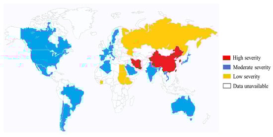
4. Epidemiology of K. pneumoniae (Mainly Hypervirulent K. pneumoniae in Eastern Asia)

Several factors can result in the colonization of K. pneumoniae in a particular region. Risk factors such as regional healthcare practices, use and misuse of antibiotics, infection control measures, nutrition, gender, and age could predispose a community to K. pneumoniae infection. As such, the colonization rate varies from country to country. K. pneumoniae may exist as part of the normal microbiota in both animal and human hosts. In humans, K. pneumoniae coexists with the microbial flora in the intestinal tract and nasopharynx. K. pneumoniae in stool samples ranges from 5% to 38%, while the detection rate in nasopharyngeal samples ranges from 1% to 6% [20]. However, hospitalized patients tend to have significantly higher carrier levels, with up to 77% in the stool and 19% in the pharynx [20]. Apart from the contributory factor of the hospital environment, antibiotic misuse practices have also led to a high rate of this colonization [43]. Healthy individuals may harbor cKP colonization, yet infection is uncommon without some degree of host compromise. On the other hand, healthy individuals carrying hvKp are significantly more susceptible to infection. The remainder of this section will focus on hvKp.
Case studies in the 1980s originating from Taiwan were the first to document instances of community-acquired liver abscesses induced by hvKp in individuals without underlying health issues, alongside severe concurrent complications, such as meningitis and endophthalmitis [44][45]. Since then, other cases of hvKp have increasingly been reported across the globe, possibly due to increasing population migration [46][47][48][49][50][51]. However, the frequency of hvKp infection in Asia remains high. For example, in a Chinese study conducted from January 2013 to October 2015, a total of 369 K. pneumoniae isolates were consecutively isolated from the specimens of patients with various invasive infections at the First Affiliated Hospital of Wenzhou Medical University located in Wenzhou, east China. The K. pneumoniae isolates from the specimens of the respiratory tract, urinary tract, and intestinal tract were excluded in this investigation because it is difficult to discriminate invasive isolates from colonizing isolates. This study identified 22.89% of isolated K. pneumoniae strains to be of hvKp [52]. This is significantly higher than a report from Spain (conducted in a teaching hospital) between 2007 and 2013 (5.4%, 53/878) and a study in Alberta, Canada (8.2%) [53][54]. A predilection of the hvKp infection in Asians has been observed even among Asians residing in Western countries, although the reason for this remains unclear. However, it is evident that there is a growing occurrence of infections caused by hvKp in other ethnicities and climes. As such, there is a need for heightened awareness of this condition, which may be further complicated by other infections [6]. Figure 1 summarizes the geographic distribution of the hvKp infection [55][56][57][58][59].
Figure 1. Global distribution of hvKp. The severity of hvKp is differentiated by color. Regions with high severity have endemic spread of hvKp. This manifests as severe clinical outcomes in regions. Moderate severity regions have moderate case studies of hvKp infections. Likewise, the low-severity region has few hospital-reported cases of hvKp infection [55][56][57][58][59].
Current research is unsure of the exact mechanisms through which hvKp spreads through communities; however, based on data from the cKP bacteria, probable vectors may include contaminated water or food, direct transmission between close personal contacts, or animal-to-human transmission [55]. In support of these routes of acquisition, an investigation of ready-to-eat vegetables identified the hvKp strain, positive for the “string test”, isolated from cucumber [60]. Additionally, hvKp strains have been isolated from public water environments in Brazil, implicating contaminated water as another source of acquisition of these bacteria [61]. The gastrointestinal carriage is regarded as a major source of infection, especially in intensive care patients [62]. Investigation of fecal bacteria samples from healthy and K. pneumoniae liver abscess (KLA) patients showed similar bacterial serotypes and genotypes [63]. Interestingly, a case report from Japan showed that the stool cultures from two healthy relatives of KLA patients were hvKp-positive, rendering the relatives carriers [64]. These two reports suggest that the gut is also a reservoir for hvKp, and as proposed by Zhu J. et al. [65], this is plausible as this could be the passageway through which hvKp spreads from the intestinal barrier to the liver, causing KLA.
Several factors stand behind the higher incidence of infections caused by K. pneumoniae compared to other Gram-negative opportunistic pathogens, including bacteria’s ability to tolerate starvation [66], inherent antibiotic resistance [67], outcompete other bacteria [68], voluntarily exchange DNA with other human microbiome bacteria [69], and acquired antibiotic resistance and virulence-enhancing genes [70].
Antibiotics are diverse chemical substances that play a crucial role in treating and controlling the spread of infectious diseases. Two mechanisms exist through which antimicrobial drugs act on bacteria, either via a bacteriostatic or bactericidal mode of action, which hinders bacterial replication or kills the bacteria, respectively [19]. One of the recent and major challenges to public health is antimicrobial resistance (AMR). AMR is defined as the microorganisms’ ability to live and thrive in the presence of antimicrobial agents, a phenomenon associated with increased morbidity and mortality rates [71]. Bacterial resistance can be divided into three patterns: MDR (multidrug-resistant), where bacteria are resistant to more than one antimicrobial agent; XDR (extensively drug-resistant), refers to bacteria that remain susceptible to only one to two antimicrobial categories; PDR (pan-drug resistance) which includes bacteria that are resistant to all agents in all antimicrobial categories [72]. Classical K. pneumonia (cKP) has a natural resistance against certain antibiotics, such as ampicillin, carbenicillin, and ticarcillin, due to the production of an enzyme known as chromosomal penicillinase, sulfhydryl variable (SHV-1) [73]. However, third- and fourth-generation cephalosporins, quinolones, or carbapenems are effective antimicrobial drugs for treating K. pneumonia [74], making cKP a non-concerning pathogen. Two major types of antibiotic resistance were frequently found in K. pneumoniae infections. The first mechanism involves the production of the extended-spectrum beta-lactamase (ESBL) enzyme, which acts on beta-lactam antibiotics, including penicillin, cephalosporins, and monobactams. Thus, ESBL-producing K. pneumoniae is rendered resistant to such antibiotics [23]. Today, MDR ESBL-producing cKP are among the pathogens most associated with nosocomial infections [73]. Carbapenem drugs such as imipenem and meropenem are still the “gold standard” therapy for treating serious and invasive ESBL infections and are linked to better outcomes in patients with severe infections. In addition to carbapenems, β-lactam/β-lactamase inhibitor (BLBLI) combinations such as piperacillin–tazobactam (PTZ) are also found to be effective in treating ESBL-producing K. pneumonia [75]. The second mechanism is more concerning, through which K. pneumonia will develop resistance to almost all available beta-lactams, including carbapenems. This resistance is obtained by the help of carbapenemase enzymes that are capable of hydrolyzing carbapenems. These types of bacteria are referred to as CRKP, which is short for carbapenem-resistant Klebsiella pneumonia [23]. There are indications that, in certain conditions, combination therapy is advantageous. In accordance with some studies, colistin is best taken in combination with another antibiotic, although the effectiveness of this treatment is still debatable [76].
A Kp isolate was considered hvKp when at least three or more of the following virulence genes were detected with >99% full-length gene coverage: iucA, iroB, peg-344, rmpA, and rmpA2 [16]. Moreover, both cKP and hvKp possess type 1 (mannose-sensitive) and type 3 (mannose-resistant) fimbriae. In cKP strains, these fimbriae have been shown to adhere to host epithelial cells from the respiratory and urinary tracts and add to infection. Although little work has been conducted on hvKp, a recent study examined the regulation of type 3 fimbriae in the hvKp strain CG43. This study confirmed observations that type 3 fimbriae contribute to biofilm formation and demonstrated that expression positively correlated with iron concentration [6].

References

  1. Choby, J.E.; Howard-Anderson, J.; Weiss, D.S. Hypervirulent Klebsiella pneumoniae-clinical and molecular perspectives. J. Intern. Med. 2020, 287, 283–300.
  2. WHO. Global Priority List of Antibiotic-Resistant Bacteria to Guide Research, Discovery, and Development of New Antibiotics; World Health Organization: Geneva, Switzerland, 2017.
  3. Magill, S.S.; O’Leary, E.; Janelle, S.J.; Thompson, D.L.; Dumyati, G.; Nadle, J.; Wilson, L.E.; Kainer, M.A.; Lynfield, R.; Greissman, S.; et al. Changes in Prevalence of Health Care-Associated Infections in U.S. Hospitals. N. Engl. J. Med. 2018, 379, 1732–1744.
  4. Pomakova, D.K.; Hsiao, C.-B.; Beanan, J.M.; Olson, R.; MacDonald, U.; Keynan, Y.; Russo, T.A. Clinical and phenotypic differences between classic and hypervirulent Klebsiella pneumonia: An emerging and under-recognized pathogenic variant. Eur. J. Clin. Microbiol. Infect. Dis. 2012, 31, 981–989.
  5. Lan, P.; Shi, Q.; Zhang, P.; Chen, Y.; Yan, R.; Hua, X.; Jiang, Y.; Zhou, J.; Yu, Y. Core genome allelic profiles of clinical Klebsiella pneumoniae strains using a random forest algorithm based on multilocus sequence typing scheme for hypervirulence analysis. J. Infect. Dis. 2020, 221, 263–271.
  6. Shon, A.S.; Bajwa, R.P.; Russo, T.A. Hypervirulent (hypermucoviscous) Klebsiella pneumoniae: A new and dangerous breed. Virulence 2013, 4, 107–118.
  7. Lee, H.C.; Chuang, Y.C.; Yu, W.L.; Lee, N.Y.; Chang, C.M.; Ko, N.Y.; Wang, L.R.; Ko, W.C. Clinical implications of hypermucoviscosity phenotype in Klebsiella pneumoniae isolates: Association with invasive syndrome in patients with community-acquired bacteraemia. J. Intern. Med. 2006, 259, 606–614.
  8. Li, W.; Sun, G.; Yu, Y.; Li, N.; Chen, M.; Jin, R.; Jiao, Y.; Wu, H. Increasing occurrence of antimicrobial-resistant hypervirulent (hypermucoviscous) Klebsiella pneumoniae isolates in China. Clin. Infect. Dis. 2014, 58, 225–232.
  9. Liu, Y.; Li, X.Y.; Wan, L.G.; Jiang, W.Y.; Yang, J.H.; Li, F.Q. Virulence and transferability of resistance determinants in a novel Klebsiella pneumonia sequence type 1137 in China. Microb. Drug Resist. 2014, 20, 150–155.
  10. Bergey, D.H.; David, R.; Boone, G.M.; Garrity, R.W.C.; Bergey, D.H. Bergey’s Manual of Systematic Bacteriology; Springer: New York, NY, USA; London, UK, 2001.
  11. Wen, Z.; Zhang, J.-R. Chapter 3—Bacterial Capsules. In Molecular Medical Microbiology, 2nd ed.; Elsevier Ltd.: Amsterdam, The Netherlands, 2015.
  12. Domenico, P.; Salo, R.J.; Cross, A.S.; Cunha, B.A. Polysaccharide capsule-mediated resistance to opsonophagocytosis in Klebsiella pneumonia. Infect. Immun. 1994, 62, 4495–4499.
  13. Brissac, T.; Martínez, E.; Kruckow, K.L.; Riegler, A.N.; Ganaie, F.; Im, H.; Bakshi, S.; Arroyo-Diaz, N.M.; Spencer, B.L.; Saad, J.S.; et al. Capsule Promotes Intracellular Survival and Vascular Endothelial Cell Translocation during Invasive Pneumococcal Disease. mBio 2021, 12, e0251621.
  14. Lawlor, M.S.; Handley, S.A.; Miller, V.L. Comparison of the host responses to wild-type and cpsB mutant Klebsiella pneumoniae infections. Infect. Immun. 2006, 74, 5402–5407.
  15. Pan, Y.J.; Lin, T.L.; Chen, C.T.; Chen, Y.Y.; Hsieh, P.F.; Hsu, C.R.; Wu, M.C.; Wang, J.T. Genetic analysis of capsular polysaccharide synthesis gene clusters in 79 capsular types of Klebsiella spp. Sci. Rep. 2015, 5, 15573.
  16. Russo, T.A.; Olson, R.; Fang, C.T.; Stoesser, N.; Miller, M.; MacDonald, U.; Hutson, A.; Barker, J.H.; La Hoz, R.M.; Johnson, J.R. Identification of biomarkers for the differentiation of hypervirulent Klebsiella pneumonia from classical K. pneumoniae. J. Clin. Microbiol. 2018, 56, e00776-18.
  17. Wang, L.; Shen, D.; Wu, H.; Ma, Y. Resistance of hypervirulent Klebsiella pneumoniae to both intracellular and extracellular killing of neutrophils. PLoS ONE 2017, 12, e0173638.
  18. Shankar-Sinha, S.; Valencia, G.A.; Janes, B.K.; Rosenberg, J.K.; Whitfield, C.; Bender, R.A.; Standiford, T.J.; Younger, J.G. The Klebsiella pneumoniae O antigen contributes to bacteremia and lethality during murine pneumonia. Infect. Immun. 2004, 72, 1423–1430.
  19. Abushaheen, M.A.; Muzaheed; Fatani, A.J.; Alosaimi, M.; Mansy, W.; George, M.; Jhugroo, P. Antimicrobial Resistance, Mechanisms and Its Clinical Significance. Dis. Mon. 2020, 66, 100971.
  20. Podschun, R.; Ullmann, U. Klebsiella spp. as nosocomial pathogens: Epidemiology, taxonomy, typing methods, and pathogenicity factors. Clin. Microbiol. Rev. 1998, 11, 589–603.
  21. Schroll, C.; Barken, K.B.; Krogfelt, K.A.; Struve, C. Role of type 1 and type 3 fimbriae in Klebsiella pneumoniae biofilm formation. BMC Microbiol. 2010, 10, 179.
  22. Munoz-Price, L.S.; Poirel, L.; Bonomo, R.A.; Schwaber, M.J.; Daikos, G.L.; Cormican, M.; Cornaglia, G.; Garau, J.; Gniadkowski, M.; Hayden, M.K.; et al. Clinical Epidemiology of the Global Expansion of Klebsiella pneumoniae Carbapenemases. Lancet Infect. Dis. 2013, 13, 785–796.
  23. Paczosa, M.K.; Mecsas, J. Klebsiella pneumoniae: Going on the Offense with a Strong Defense. Microbiol. Mol. Biol. Rev. 2016, 80, 629–661.
  24. Hsu, C.R.; Lin, T.L.; Pan, Y.J.; Hsieh, P.F.; Wang, J.T. Isolation of a Bacteriophage specific for a new capsular type of Klebsiella pneumoniae and Characterization of its Polysaccharide Depolymerase. PLoS ONE 2013, 8, e70092.
  25. Ramos, P.I.; Picão, R.C.; Vespero, E.C.; Pelisson, M.; Zuleta, L.F.G.; Almeida, L.G.P.; Gerber, A.L.; Vasconcelos, A.T.R.; Gales, A.C.; Nicolás, M.F. Pyrosequencing-Based Analysis Reveals a Novel Capsular Gene Cluster in a KPC-Producing Klebsiella pneumoniae Clinical Isolate Identified in Brazil. BMC Microbiol. 2012, 12, 173.
  26. Li, B.; Zhao, Y.; Liu, C.; Chen, Z.; Zhou, D. Molecular Pathogenesis of Klebsiella pneumoniae. Future Microbiol. 2014, 9, 1071–1081.
  27. Yang, F.L.; Yang, Y.L.; Liao, P.C.; Chou, J.C.; Tsai, K.C.; Yang, A.S.; Sheu, F.; Lin, T.L.; Hsieh, P.F.; Wang, J.T.; et al. Structure and Immunological Characterization of the Capsular Polysaccharide of a Pyrogenic Liver Abscess caused by Klebsiella pneumoniae: Activation of Macrophages through Toll-like Receptor 4. J. Biol. Chem. 2011, 286, 21041–21051.
  28. Gonzalez-Ferrer, S.; Peñaloza, H.F.; Budnick, J.A.; Bain, W.G.; Nordstrom, H.R.; Lee, J.S.; Van Tyne, D. Finding Order in the Chaos: Outstanding Questions in Klebsiella pneumoniae Pathogenesis. Infect. Immun. 2021, 89, e00693-20.
  29. Merino, S.; Altarriba, M.; Izquierdo, L.; Nogueras, M.M.; Regue, M.; Tomas, J.M. Cloning and Sequencing of the Klebsiella pneumoniae O5 wb Gene Cluster and its Role in Pathogenesis. Infect. Immun. 2000, 68, 2435–2440.
  30. Clements, A.; Tull, D.; Jenney, A.W.; Farn, J.L.; Kim, S.H.; Bishop, R.E.; McPhee, J.B.; Hancock, R.E.; Hartland, E.L.; Pearse, M.J.; et al. Secondary Acylation of Klebsiella pneumoniae Lipopolysaccharide Contributes to Sensitivity to Antibacterial Peptides. J. Biol. Chem. 2007, 282, 15569–15577.
  31. March, C.; Cano, V.; Moranta, D.; Llobet, E.; Pérez-Gutiérrez, C.; Tomás, J.M.; Suárez, T.; Garmendia, J.; Bengoechea, J.A. Role of Bacterial Surface Structures on the Interaction of Klebsiella pneumoniae with Phagocytes. PLoS ONE 2013, 8, e56847.
  32. Struve, C.; Bojer, M.; Krogfelt, K.A. Characterization of Klebsiella pneumoniae Type 1 Fimbriae by Detection of Phase Variation during Colonization and Infection and Impact on Virulence. Infect. Immun. 2008, 76, 4055–4065.
  33. Stahlhut, S.G.; Struve, C.; Krogfelt, K.A. Klebsiella pneumoniae Type 3 Fimbriae Agglutinate Yeast in a Mannose-Resistant Manner. J. Med. Microbiol. 2012, 61 Pt 3, 317–322.
  34. Struve, C.; Bojer, M.; Krogfelt, K.A. Identification of a Conserved Chromosomal Region Encoding Klebsiella pneumoniae Type 1 and Type 3 Fimbriae and Assessment of the Role of Fimbriae in Pathogenicity. Infect. Immun. 2009, 77, 5016–5024.
  35. Wu, C.C.; Huang, Y.J.; Fung, C.P.; Peng, H.L. Regulation of the Klebsiella pneumoniae Kpc Fimbriae by the Site-Specific Recombinase KpcI. Microbiology 2010, 156 Pt 7, 1983–1992.
  36. Guerra, M.E.S.; Destro, G.; Vieira, B.; Lima, A.S.; Ferraz, L.F.C.; Hakansson, A.P.; Darrieux, M.; Converso, T.R. Klebsiella pneumoniae Biofilms and Their Role in Disease Pathogenesis. Front. Cell. Infect. Microbiol. 2022, 12, 877995.
  37. Sebghati, T.A.; Korhonen, T.K.; Hornick, D.B.; Clegg, S. Characterization of the type 3 fimbrial adhesins of Klebsiella strains. Infect. Immun. 1998, 66, 2887–2894.
  38. Di Martino, P.; Livrelli, V.; Sirot, D.; Joly, B.; Darfeuille-Michaud, A. A New Fimbrial Antigen Harbored by CAZ-5/ SHV-4-Producing Klebsiella pneumoniae Strains Involved in Nosocomial Infections. Infect. Immun. 1996, 64, 2266–2273.
  39. Regueiro, V.; Moranta, D.; Frank, C.G.; Larrarte, E.; Margareto, J.; March, C.; Garmendia, J.; Bengoechea, J.A. Klebsiella pneumoniae Subverts the Activation of Inflammatory Responses in a NOD1-Dependent Manner. Cell Microbiol. 2011, 13, 135–153.
  40. Llobet, E.; March, C.; Gimenez, P.; Bengoechea, J.A. Klebsiella pneumoniae OmpA Confers Resistance to Antimicrobial Peptides. Antimicrob. Agents Chemother. 2009, 53, 298–302.
  41. Tsai, Y.K.; Fung, C.P.; Lin, J.C.; Chen, J.-H.; Chang, F.-Y.; Chen, T.-L.; Siu, L.K. Klebsiella pneumoniae Outer Membrane Porins OmpK35 and OmpK36 Play Roles in Both Antimicrobial Resistance and Virulence. Antimicrob. Agents Chemother. 2011, 55, 1485–1493.
  42. Padilla, E.; Llobet, E.; Domenech-Sanchez, A.; Martinez-Martinez, L.; Bengoechea, J.A.; Alberti, S. Klebsiella pneumoniae AcrAB Efflux Pump Contributes to Antimicrobial Resistance and Virulence. Antimicrob. Agents Chemother. 2010, 54, 177–183.
  43. Pollack, M.; Nieman, R.; Reinhardt, J.; Charache, P.; Jett, M.; Hardy, P., Jr. Factors influencing colonisation and antibiotic-resistance patterns of gram-negative bacteria in hospital patients. Lancet 1972, 300, 668–671.
  44. Chiu, C.T.; Lin, D.Y.; Liaw, Y.F. Metastatic Septic Endophthalmitis in Pyogenic Liver Abscess. J. Clin. Gastroenterol. 1988, 10, 524–527.
  45. Liu, Y.C. Klebsiella pneumoniae Liver Abscess Associated With Septic Endophthalmitis. Arch. Intern. Med. 1986, 146, 1913.
  46. Victor, L.Y.; Hansen, D.S.; Ko, W.C.; Sagnimeni, A.; Klugman, K.P.; Von Gottberg, A.; Goossens, H.; Wagener, M.M.; Benedi, V.J.; International Klebsiella Study Group. Virulence Characteristics of Klebsiella and Clinical Manifestations of K. pneumoniae Bloodstream Infections. Emerg. Infect. Dis. 2007, 13, 986–993.
  47. Coutinho, R.L.; Visconde, M.F.; Descio, F.J.; Nicoletti, A.G.; Pinto, F.C.; da Silva, A.C.R.; Rodrigues-Costa, F.; Gales, A.C.; Furtado, G.H. Community-acquired invasive liver abscess syndrome caused by a K1 serotype Klebsiella pneumoniae isolate in Brazil: A case report of hypervirulent ST23. Mem. Inst. Oswaldo Cruz 2014, 109, 970–971.
  48. Fazili, T.; Sharngoe, C.; Endy, T.; Kiska, D.; Javaid, W.; Polhemus, M. Klebsiella pneumoniae Liver Abscess: An Emerging Disease. Am. J. Med. Sci. 2016, 351, 297–304.
  49. Sturm, E.; Tai, A.; Lin, B.; Kwong, J.; Athan, E.; Howden, B.P.; Angliss, R.D.; Asaid, R.; Pollard, J. Bilateral osteomyelitis and liver abscess caused by hypervirulent Klebsiella pneumoniae—A rare clinical manifestation (case report). BMC Infect. Dis. 2018, 18, 1–5.
  50. Sánchez-López, J.; García-Caballero, A.; Navarro-San Francisco, C.; Quereda, C.; Ruiz-Garbajosa, P.; Navas, E.; Dronda, F.; Morosini, M.I.; Cantón, R.; Diez-Aguilar, M. Hypermucoviscous Klebsiella pneumoniae: A challenge in community acquired infection. ID Cases 2019, 17, e00547.
  51. Chung, D.R.; Lee, S.S.; Lee, H.R.; Kim, H.B.; Choi, H.J.; Eom, J.S.; Kim, J.S.; Choi, Y.H.; Lee, J.S.; Chung, M.H.; et al. Emerging invasive liver abscess caused by K1 serotype Klebsiella pneumoniae in Korea. J. Infect. 2007, 54, 578–583.
  52. Guo, Y.; Wang, S.; Zhan, L.; Jin, Y.; Duan, J.; Hao, Z.; Lv, J.; Qi, X.; Chen, L.; Kreiswirth, B.N.; et al. Microbiological and Clinical Characteristics of Hypermucoviscous Klebsiella pneumoniae Isolates Associated with Invasive Infections in China. Front. Cell Infect. Microbiol. 2017, 7, 24.
  53. Cubero, M.; Grau, I.; Tubau, F.; Pallarés, R.; Dominguez, M.; Liñares, J.; Ardanuy, C. Hypervirulent Klebsiella pneumoniae clones causing bacteraemia in adults in a teaching hospital in Barcelona, Spain (2007–2013). Clin. Microbiol. Infect. 2016, 22, 154–160.
  54. Peirano, G.; Pitout, J.D.; Laupland, K.B.; Meatherall, B.; Gregson, D.B. Population-Based Surveillance for Hypermucoviscosity Klebsiella pneumoniae Causing Community-Acquired Bacteremia in Calgary, Alberta. Can. J. Infect. Dis. Med. Microbiol. 2013, 24, 61–64.
  55. Russo, T.A.; Marr, C.M. Hypervirulent Klebsiella pneumonia. Clin. Microbiol. Rev. 2019, 32, 10–1128.
  56. Albasha, A.M.; Osman, E.H.; Abd-Alhalim, S.; Alshaib, E.F.; Al-Hassan, L.; Altayb, H.N. Detection of several carbapenems resistant and virulence genes in classical and hyper-virulent strains of Klebsiella pneumoniae isolated from hospitalized neonates and adults in Khartoum. BMC Res. Notes 2020, 13, 312.
  57. Vasilyev, I.Y.; Nikolaeva, I.V.; Siniagina, M.N.; Kharchenko, A.M.; Shaikhieva, G.S. Multidrug-Resistant Hypervirulent Klebsiella pneumoniae Found Persisting Silently in Infant Gut Microbiota. Int. J. Microbiol. 2020, 2020, 4054393.
  58. Sewunet, T.; Asrat, D.; Woldeamanuel, Y.; Ny, S.; Westerlund, F.; Aseffa, A.; Giske, C.G. High prevalence of blaCTX-M-15 and nosocomial transmission of hypervirulent epidemic clones of Klebsiella pneumoniae at a tertiary hospital in Ethiopia. JAC Antimicrob. Resist. 2021, 3, dlab001.
  59. Emam, S.M.; Abdelrahman, S.; Hasan, A.A.; EL-Melouk, M.S. Hypervirulent Klebsiella pneumoniae at Benha University Hospitals. Egypt. J. Hosp. Med. 2023, 90, 3592–3597.
  60. Liu, B.T.; Zhang, X.Y.; Wan, W.S.; Hao, J.J.; De Jiang, R.; Song, F.J. Characteristics of carbapenem-resistant Enterobacteriaceae in ready-to-eat vegetables in China. Front. Microbiol. 2018, 9, 359969.
  61. Furlan, J.P.R.; Savazzi, E.A.; Stehling, E.G. Genomic insights into multidrug-resistant and hypervirulent Klebsiella pneumoniae co-harboring metal resistance genes in aquatic environments. Ecotoxicol. Environ. Saf. 2020, 201, 110782.
  62. Gorrie, C.L.; Mirčeta, M.; Wick, R.R.; Edwards, D.J.; Thomson, N.R.; Strugnell, R.A.; Pratt, N.F.; Garlick, J.S.; Watson, K.M.; Pilcher, D.V.; et al. Gastrointestinal Carriage Is a Major Reservoir of Klebsiella pneumoniae Infection in Intensive Care Patients. Clin. Infect. Dis. 2017, 65, 208–215.
  63. Fung, C.P.; Lin, Y.T.; Lin, J.C.; Chen, T.L.; Yeh, K.M.; Chang, F.Y.; Chuang, H.C.; Wu, H.S.; Tseng, C.P.; Siu, L.K. Klebsiella pneumoniae in Gastrointestinal Tract and Pyogenic Liver Abscess. Emerg. Infect. Dis. 2012, 18, 1322.
  64. Harada, S.; Tateda, K.; Mitsui, H.; Hattori, Y.; Okubo, M.; Kimura, S.; Sekigawa, K.; Kobayashi, K.; Hashimoto, N.; Itoyama, S.; et al. Familial Spread of a Virulent Clone of Klebsiella pneumoniae Causing Primary Liver Abscess. J. Clin. Microbiol. 2011, 49, 2354–2356.
  65. Zhu, J.; Wang, T.; Chen, L.; Du, H. Virulence Factors in Hypervirulent Klebsiella pneumoniae. Front. Microbiol. 2021, 12, 642484.
  66. Baker, J.L.; Hendrickson, E.L.; Tang, X.; Lux, R.; He, X.; Edlund, A.; McLean, J.S.; Shi, W. Klebsiella and Providencia Emerge as Lone Survivors Following Long-Term Starvation of Oral Microbiota. Proc. Natl. Acad. Sci. USA 2019, 116, 8499–8504.
  67. Holt, K.E.; Wertheim, H.; Zadoks, R.N.; Baker, S.; Whitehouse, C.A.; Dance, D.; Jenney, A.; Connor, T.R.; Hsu, L.Y.; Severin, J.; et al. Genomic Analysis of Diversity, Population Structure, Virulence, and Antimicrobial Resistance in Klebsiella pneumoniae, an Urgent Threat to Public Health. Proc. Natl. Acad. Sci. USA 2015, 112, 3574–3581.
  68. Hsieh, P.F.; Lu, Y.R.; Lin, T.L.; Lai, L.Y.; Wang, J.T. Klebsiella pneumoniae Type VI Secretion System Contributes to Bacterial Competition, Cell Invasion, Type-1 Fimbriae Expression, and in vivo Colonization. J. Infect. Dis. 2019, 219, 637–647.
  69. Dunn, S.J.; Connor, C.; McNally, A. The Evolution and Transmission of Multi-Drug Resistant Escherichia coli and Klebsiella pneumoniae: The Complexity of Clones and Plasmids. Curr. Opin. Microbiol. 2019, 51, 51–56.
  70. Wyres, K.L.; Wick, R.R.; Judd, L.M.; Froumine, R.; Tokolyi, A.; Gorrie, C.L.; Lam, M.M.C.; Duchêne, S.; Jenney, A.; Holt, K.E. Distinct Evolutionary Dynamics of Horizontal Gene Transfer in Drug Resistant and Virulent Clones of Klebsiella pneumoniae. PLoS Genet. 2019, 15, 1008114.
  71. Frieri, M.; Kumar, K.; Boutin, A. Antibiotic Resistance. J. Infect. Public Health 2017, 10, 369–378.
  72. Magiorakos, A.P.; Srinivasan, A.; Carey, R.B.; Carmeli, Y.; Falagas, M.E.; Giske, C.G.; Monnet, D.L. Multidrug-Resistant, Extensively Drug-Resistant and Pandrug-Resistant Bacteria: An International Expert Proposal for Interim Standard Definitions for Acquired Resistance. Clin. Microbiol. Infect. 2012, 18, 268–281.
  73. Stojowska-Swędrzyńska, K.; Łupkowska, A.; Kuczyńska-Wiśnik, D.; Laskowska, E. Antibiotic Heteroresistance in Klebsiella pneumoniae. Int. J. Mol. Sci. 2021, 23, 449.
  74. Prince, S.E.; Dominger, K.A.; Cunha, B.A.; Klein, N.C. Klebsiella pneumoniae Pneumonia. Heart Lung 1997, 26, 413–417.
  75. Pana, Z.D.; Zaoutis, T. Treatment of Extended-Spectrum β-Lactamase-Producing Enterobacteriaceae (ESBLs) Infections: What Have We Learned Until Now? F1000Research 2018, 7. F1000 Faculty Rev-1347.
  76. Fritzenwanker, M.; Imirzalioglu, C.; Herold, S.; Wagenlehner, F.M.; Zimmer, K.-P.; Chakraborty, T. Treatment Options for Carbapenem-Resistant Gram-Negative Infections. Dtsch. Arztebl. Int. 2018, 115, 345–352.
More
Information
Subjects: Microbiology
Contributors MDPI registered users' name will be linked to their SciProfiles pages. To register with us, please refer to https://encyclopedia.pub/register : , , , , , , ,
View Times: 123
Revisions: 2 times (View History)
Update Date: 01 Feb 2024
1000/1000