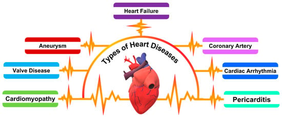
NT-ProBNP, a prominent cardiac biomarker, has gained significant recognition for its role in the diagnosis and prognosis of HF. This biomarker is synthesized as a response to ventricular volume expansion and pressure overload, conditions often indicative of cardiac stress and dysfunction. NT-ProBNP, a cleavage byproduct of proBNP, is released primarily by cardiac myocytes. Its elevated levels in the bloodstream are not only diagnostic but also correlate proportionally with the severity of heart disease, offering a nuanced understanding of cardiac function. Distinguished by its superior specificity and sensitivity, NT-ProBNP is particularly valuable in diagnosing HF, especially in cases with unclear clinical manifestations. Beyond its diagnostic capabilities, NT-ProBNP serves as a crucial tool for patient risk stratification, guiding therapeutic decision-making, and monitoring the response to treatment in both acute and chronic HF scenarios. Its comprehensive utility in various clinical settings solidifies NT-ProBNP’s position as a vital biomarker in the landscape of cardiovascular healthcare and patient management
[5][6][7]. A recent report by Hall et al. explores BNP, initially isolated from porcine brains and later identified as a cardiac hormone. The study explains that BNP and atrial natriuretic peptide together form a critical system in the heart. The synthesis and secretion of proBNP, mainly triggered by myocyte stretch, result in active BNP and NT-proBNP. The research highlights that heart failure leads to increased BNP secretion due to factors like wall stretch and neurohormonal activation. The research also outlines BNP’s physiological effects, including diuresis and vasodilation, and details the clearance mechanisms for BNP and NT-proBNP, noting the latter’s longer half-life and higher plasma concentration
[8].
The emergence of biosensor technology, characterized by the fusion of biological components with physicochemical detectors, represents a transformative advancement in the realm of biomedical diagnostics. These sophisticated devices are engineered to convert biological interactions into quantifiable electronic signals, thus facilitating rapid, sensitive, and specific detection of a diverse array of analytes. Among these, biomarkers such as NT-ProBNP, pivotal in cardiovascular disease diagnostics, are of particular interest. The incorporation of nanomaterials into biosensor frameworks has catalyzed a paradigm shift in biosensing capabilities
[10]. Nanomaterials, endowed with distinct physicochemical attributes like an extensive surface area-to-volume ratio, augmented reactivity, and pronounced quantum mechanical effects, have significantly enhanced the analytical performance of biosensors
[11][12][13][14][15]. They contribute to elevated sensitivity and selectivity, expanding the detection limits and improving the fidelity of biosensor readings
[16][17][18][19][20].
The detection of NT-ProBNP, a crucial biomarker for cardiovascular diseases, has been significantly enhanced by the integration of nanomaterials into biosensor designs. These advancements leverage the unique properties of nanomaterials to achieve high sensitivity and specificity in NT-ProBNP detection. In a recent article by Qian et al., a highly sensitive and selective immunosensor is designed for NT-pro BNP detection, employing flower-like Bi
2WO
6/Ag
2S nanoparticles (F-Bi
2WO
6/Ag
2S) as the photoelectrochemical matrix. This matrix is coupled with a graphene oxide and polydopamine (GO/PDA) composite for signal amplification. The sensor capitalizes on the efficient photocurrent conversion provided by the cascade-like band-edge level between F-Bi
2WO
6 and Ag
2S, further enhanced by the GO/PDA conjugates. These conjugates aid in sweeping the holes and preventing the recombination of photogenerated electron-hole pairs, with graphene oxide’s excellent conductivity facilitating rapid electron transfer. The resulting photocurrent correlates directly with NT-pro BNP concentration, with the sensor demonstrating a detection range from 0.1 pg/mL to 100 ng/mL and a low detection limit of 0.03 pg/mL
[21].
In a study by Li et al., a novel electrochemical immunoassay is developed for NT-pro BNP detection. This sensor features an amorphous bimetallic sulfide of CoSnS
x-Pd as a signal amplifier and Fe
3O
4@PPy-Au as a magnetic substrate. The CoSnSx, synthesized via a stoichiometric co-precipitation method, exhibits excellent electrochemical behavior due to the synergistic effect of SnS
2 and cobalt, enhanced by Pd nanoparticles’ electrocatalytic activity towards H
2O
2 oxidation. The Fe
3O
4@PPy-Au substrate, immobilized on the magnetic glassy carbon electrode, enhances conductivity and stability. This design achieves a wide detection range from 0.1 pg/mL to 50 ng/mL and a low detection limit of 31.5 fg/mL for NT-pro BNP, showing potential for broad biomarker detection in medical diagnostics
[22].
2. An Overview of Various Nanomaterial Types, Their Distinct Properties, and Applications in the Detection of NT-ProBNP
2.1. Metal Nanoparticles
Gold (AuNPs) and silver (AgNPs) are renowned for their localized surface plasmon resonance (LSPR) properties, which are highly sensitive to changes in the refractive index near the nanoparticle surface. This sensitivity is leveraged in colorimetric assays where shifts in the LSPR spectrum serve as indicators of molecular interactions, such as the binding of NT-ProBNP. The colorimetric response resulting from these shifts can be easily observed, making AuNPs and AgNPs ideal for rapid and straightforward detection methods. The intensity and peak wavelength of the LSPR are influenced by factors including particle size, shape, and the dielectric environment, allowing for finely-tuned biosensor designs.
Platinum (Pt), palladium (Pd), and copper (Cu) nanoparticles offer distinct electrocatalytic properties and electrical conductance, which are advantageous in the development of electrochemical sensors for NT-ProBNP detection. Platinum nanoparticles, for instance, provide excellent catalytic activity towards various biochemical reactions, enhancing the electron transfer processes necessary for sensitive electrochemical detection. Palladium nanoparticles also exhibit similar catalytic properties, with a unique affinity for hydrogen absorption, which can be utilized in biosensing applications. Copper nanoparticles contribute to improved electrical conductance and have been employed in electrochemical sensors for their cost-effectiveness and efficient catalytic behavior. These metals can be utilized to fabricate highly sensitive and specific electrochemical biosensors for NT-ProBNP, capitalizing on their unique properties to detect minute changes in electrochemical signals that correspond to the presence of the biomarker
[18][22].
2.2. Carbon-Based Nanomaterials
Carbon nanotubes (CNTs) are recognized for their exceptional electrical conductivity, substantial surface area, and distinct electron transport characteristics. These properties make CNTs highly effective in electrochemical sensor applications. In the context of NT-ProBNP detection, CNTs are employed to enhance signal transduction. When NT-ProBNP antibodies are bound to the surface of CNTs, their unique electron transport properties facilitate efficient signal relay, amplifying the detection capabilities of the sensor. Additionally, the large surface area of CNTs offers ample sites for antibody attachment, thereby increasing the sensor’s sensitivity and allowing for the detection of low concentrations of NT-ProBNP.
Graphene and graphene oxide, with their high surface area and excellent electrical conductivity, are versatile materials in biosensor technology. Their planar structure allows for substantial functionalization with various biomolecules, including antibodies specific to NT-ProBNP. This functionalization is crucial for creating selective and sensitive biosensing interfaces. Graphene-based materials are particularly useful in field-effect transistor (FET)-based sensors, where the conductive properties of graphene can be modulated by the binding of target biomolecules, leading to measurable changes in electrical signals. Additionally, their use in fluorescence-quenching sensors for NT-ProBNP detection is noteworthy. In these applications, graphene or graphene oxide can quench the fluorescence of labeled antibodies or NT-ProBNP, with the quenching effect being reversed upon the binding of the target molecule. This property is exploited to create highly sensitive sensors that can detect minute changes in fluorescence, correlating with the concentration of NT-ProBNP in a sample
[20][23].
2.3. Semiconductor Nanoparticles
Quantum Quantum Dots (QDs) are semiconductor nanoparticles renowned for their quantum confinement effects, which allow for size-tunable fluorescence emission, providing a unique advantage in biosensor design. This size dependency of their optical properties enables precise control over the emission wavelength, making QDs highly customizable for various applications. Their high photostability further adds to their appeal, ensuring consistent fluorescence over extended periods. In NT-ProBNP detection, QDs are employed as fluorescent probes within biosensors. These biosensors leverage changes in the fluorescence properties of the QDs, induced by the binding of NT-ProBNP to the sensor, to signal the presence of this biomarker. The sensitivity of QDs to even minor changes in fluorescence makes them particularly effective in detecting low concentrations of NT-ProBNP, offering a powerful tool for early and accurate heart failure diagnosis
[19].
2.4. Magnetic Nanoparticles
Magnetic nanoparticles, particularly those based on iron oxide, are crucial in biosensing due to their superparamagnetic properties. These properties allow for easy manipulation and concentration of the nanoparticles using external magnetic fields, a feature immensely beneficial in assays for NT-ProBNP detection. In such applications, these nanoparticles are functionalized with specific biomolecules that bind to NT-ProBNP. Once bound, they can be magnetically concentrated, significantly enhancing the precision and sensitivity of the assay. This concentration is particularly advantageous in subsequent steps of electrochemical or optical detection, where the gathered nanoparticles facilitate amplified signals or observable changes in optical properties. This method not only improves the detection sensitivity but also allows for more efficient and accurate quantification of NT-ProBNP levels, making iron oxide-based magnetic nanoparticles a vital component in advanced biosensor systems for heart failure diagnostics
[24].
2.5. Silicon Nanoparticles
Silicon nanoparticles stand out in biosensing applications due to their exceptional photoluminescent properties combined with notable biocompatibility, making them ideal candidates for bioconjugation with molecules for fluorescence-based detection. Their quantum confinement-induced luminescence is particularly advantageous for optical biosensing techniques. When used in NT-ProBNP detection, these nanoparticles can be conjugated with specific biomolecules, such as antibodies or aptamers, targeting NT-ProBNP. Upon binding with NT-ProBNP in a sample, these nanoparticles exhibit changes in fluorescence intensity or energy transfer, providing a quantifiable signal. This change in fluorescence is directly correlated with the concentration of NT-ProBNP, enabling the precise and sensitive detection of this biomarker. The biocompatibility of silicon nanoparticles further enhances their suitability for use in complex biological environments, making them a valuable tool in the development of advanced optical biosensors for heart failure diagnostics
[25].
2.6. Lipid-Based Nanomaterials
Lipid-based nanomaterials, particularly liposomes and lipid bilayers, are increasingly utilized in biosensor technology due to their ability to mimic biological membranes, offering a biocompatible interface that closely resembles the natural cellular environment. This unique characteristic makes them especially suitable for biosensors that require membrane-like structures or are designed to interact directly with biological systems. Liposomes, which are spherical vesicles composed of lipid bilayers, can encapsulate various substances, including enzymes, drugs, or signaling molecules, making them versatile carriers for targeted delivery in biosensing applications. In the context of NT-ProBNP detection, these lipid-based nanomaterials can provide a conducive environment for the biomarker, facilitating more natural interactions and binding events. The integration of liposomes and lipid bilayers in biosensors enhances the bio-relevance of the sensing environment, thereby improving the sensor’s sensitivity and specificity towards NT-ProBNP. Their compatibility with a wide range of detection methods, including optical and electrochemical techniques, further underscores their utility in developing advanced biosensors for heart failure diagnostics
[26].
2.7. Polymeric Nanoparticles
Polymeric nanoparticles, crafted from biocompatible polymers, offer significant versatility and functionality in the realm of biosensing. Their composition allows for customization in terms of encapsulation and controlled release properties, making them highly adaptable for various diagnostic applications. In the detection of NT-ProBNP, a critical biomarker for HF, these nanoparticles can be specifically engineered to carry and protect biomolecules or drugs that are relevant to the sensing mechanism. This capability not only ensures the stability and bioavailability of these agents but also enhances the specificity and sensitivity of the sensors. By encapsulating recognition elements or enzymes that react with NT-ProBNP, polymeric nanoparticles can provide a focused and efficient means of detecting the biomarker. Furthermore, their ability to release these agents in a controlled manner can lead to more consistent and reliable sensor responses. This controlled release mechanism also opens possibilities for multiphasic sensing strategies, where different stages of detection can be orchestrated for improved accuracy. The inherent biocompatibility and tailorability of polymeric nanoparticles make them invaluable tools in the design of advanced biosensors, particularly for applications requiring high specificity and sensitivity, such as the detection of NT-ProBNP in heart failure diagnostics
[27].
2.8. Nanowires and Nanorods
Nanowires and nanorods, particularly those made of metals like gold or silver, are highly valued in biosensor technology due to their high aspect ratio, which confers an extensive surface area relative to their volume. This extensive surface area is crucial for the immobilization of biomolecules, such as antibodies or aptamers specific to NT-ProBNP, facilitating a higher density of functionalization. This property significantly enhances the sensitivity and efficiency of biosensors, as it allows for more interactions between the target biomarker and the sensor’s surface. Additionally, the conductive properties of metallic nanowires and nanorods are advantageous in signal transduction, particularly in electrochemical and optical biosensors. In electrochemical sensors, their conductive nature can lead to improved electron transfer, enhancing the signal-to-noise ratio. In optical biosensors, these nanostructures can interact with light through mechanisms like surface plasmon resonance, providing a basis for sensitive detection methods based on changes in optical properties. The incorporation of nanowires and nanorods into biosensor designs, therefore, offers a means to significantly amplify the detection capabilities of NT-ProBNP, contributing to more accurate and sensitive diagnostics for HF
[28].
2.9. Nanodiamonds
Nanodiamonds, emerging as a novel class of nanomaterials in the biosensing field, are distinguished by their stable photoluminescent properties and high biocompatibility, making them well-suited for medical and biological applications. These unique characteristics stem from their inert carbon-based structure and surface defects, which can be engineered to emit stable fluorescence. This fluorescence stability is a significant advantage in biosensing as it ensures consistent signal quality over time, which is essential for reliable diagnostics. Nanodiamonds can be functionalized with specific biomolecules, such as antibodies or aptamers targeting NT-ProBNP, to facilitate selective biomolecular interactions. In fluorescence-based detection systems for NT-ProBNP, the binding events between these functionalized nanodiamonds and the target biomarker result in modulation of their fluorescence properties. This change in fluorescence, either in intensity, wavelength, or lifetime, provides a quantifiable means of detecting NT-ProBNP presence and concentration. The ability to tailor the surface chemistry of nanodiamonds for specific interactions, combined with their photostability and biocompatibility, positions them as a promising material in the development of advanced, fluorescence-based biosensors for accurate and sensitive detection of HF biomarkers like NT-ProBNP
[29].
2.10. Plasmonic Nanostructures
Plasmonic nanostructures, such as nanostars or nanocages, represent an advanced class of materials in the field of optical biosensing, primarily due to their enhanced localized surface plasmon resonance (LSPR) effects. These complex nanostructures, with their unique geometries, offer a greater surface area and more hotspots for plasmonic activity compared to simpler nanoparticles. The intricate shapes of nanostars or the hollow interiors of nanocages lead to more pronounced and tunable LSPR properties, making them highly effective for specific optical sensing applications. In the context of NT-ProBNP detection, these plasmonic nanostructures are utilized to capitalize on their sensitive optical response to changes in the local refractive index, which occurs upon biomolecular binding events. When functionalized with recognition elements for NT-ProBNP, the interaction with the biomarker induces shifts in the LSPR spectrum, which can be detected as changes in absorption, scattering, or fluorescence. This sensitivity to minute refractive index changes allows for the detection of low concentrations of NT-ProBNP with high precision. The use of these plasmonic nanostructures in optical biosensors therefore offers a path to highly sensitive and specific NT-ProBNP detection, crucial for the early and accurate diagnosis of heart failure
[30].
2.11. Hybrid Nanocomposites
Hybrid nanocomposites, formed by integrating different nanomaterials, capitalize on the synergistic properties emerging from their combination, leading to enhanced performance in biosensing applications. By merging materials like magnetic nanoparticles, conductive polymers, or quantum dots, these composites achieve functionalities that surpass those of their individual components. For instance, the catalytic activity can be amplified, the electrical conductivity can be optimized, and the optical characteristics can be finely tuned. In NT-ProBNP detection, these hybrid nanocomposites are particularly valuable for implementing multifaceted detection strategies. They can combine magnetic properties for the concentration and separation of target biomarkers with optical properties for signal detection, such as fluorescence or plasmonic effects. This dual functionality enables more efficient capture and sensitive detection of NT-ProBNP, enhancing both the specificity and sensitivity of the assay. The versatility of hybrid nanocomposites allows for the development of advanced biosensing platforms that can effectively integrate various detection mechanisms, offering a powerful approach for comprehensive and accurate NT-ProBNP analysis in heart failure diagnostics
[31].
In the study by Li et al., a new electrochemiluminescence (ECL) strategy for NT-proBNP detection is introduced, utilizing CuS grown on reduced graphene oxide (CuS-rGO) to quench a luminol/H
2O
2 system. The system involves luminol grafted onto Au@Fe
3O
4-Cu
3(PO
4)
2 nanoflowers, enhancing ECL intensity through the catalytic reduction of H
2O
2. The quenching mechanism by CuS-rGO is based on ECL resonance energy transfer (RET), confirmed by spectral overlaps. The sensor shows a remarkable decrease in ECL signal post-immunoreaction and exhibits a broad detection range from 0.5 pg/mL to 20 ng/mL, with a low detection limit of 0.12 pg/mL. This approach combines high sensitivity, stability, and specificity, marking a significant advancement in NT-proBNP biosensing
[32].

