Submitted Successfully!
To reward your contribution, here is a gift for you: A free trial for our video production service.
Thank you for your contribution! You can also upload a video entry or images related to this topic.
Version Summary Created by Modification Content Size Created at Operation
1 -- 4295 2024-01-26 23:10:10 |
2 format Meta information modification 4295 2024-01-29 02:47:06 |

Video Upload Options

Do you have a full video?

Confirm

Are you sure to Delete?
Cite
If you have any further questions, please contact Encyclopedia Editorial Office.
Lopez-Ayuso, C.A.; Aranda-Herrera, B.; Guzman-Rocha, D.; Chávez, P.A.; Garcia-Contreras, R. Synthetic Proteins in Dental Applications. Encyclopedia. Available online: https://encyclopedia.pub/entry/54428 (accessed on 27 April 2024).
Lopez-Ayuso CA, Aranda-Herrera B, Guzman-Rocha D, Chávez PA, Garcia-Contreras R. Synthetic Proteins in Dental Applications. Encyclopedia. Available at: https://encyclopedia.pub/entry/54428. Accessed April 27, 2024.
Lopez-Ayuso, Christian Andrea, Benjamin Aranda-Herrera, Dulce Guzman-Rocha, Patricia Alejandra Chávez, Rene Garcia-Contreras. "Synthetic Proteins in Dental Applications" Encyclopedia, https://encyclopedia.pub/entry/54428 (accessed April 27, 2024).
Lopez-Ayuso, C.A., Aranda-Herrera, B., Guzman-Rocha, D., Chávez, P.A., & Garcia-Contreras, R. (2024, January 26). Synthetic Proteins in Dental Applications. In Encyclopedia. https://encyclopedia.pub/entry/54428
Lopez-Ayuso, Christian Andrea, et al. "Synthetic Proteins in Dental Applications." Encyclopedia. Web. 26 January, 2024.
Synthetic Proteins in Dental Applications
Edit

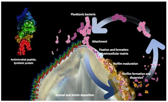
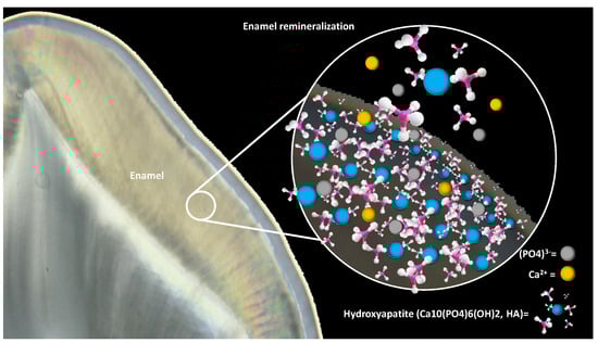
Biotechnology and artificial intelligence have sparked a revolution in dentistry, with a focus on restoring natural tissue functions. This transformation has given rise to bioactive materials, inspired by biomimetics, aimed at replicating the processes found in nature. As synthetic biology advances, there is a heightened focus on signaling systems crucial for bio-based diagnostics and therapeutics. Dentistry now harnesses synthetic proteins for tissue regeneration and dental material enhancement. A current research priority is bacterial biofilm inhibition, vital for dental health. Given the role of Streptococcus mutans in dental caries, the development of synthetic antimicrobial peptides targeting this bacterium is underway. The balance of dental enamel between demineralization and remineralization impacts caries formation. Factors such as the presence of hydroxyapatite and salivary peptides influence enamel health. Recent studies have spotlighted salivary protein-inspired peptides for enhanced remineralization.

protein peptides bacteria demineralization

1. Introduction

The development of biotechnology and artificial intelligence within the dental area has evolved in recent years [1]; many of the new materials currently being developed are primarily aimed at restoring the biological functions of tissues, and with this, the formulation of bioactive materials based on biomimetics [2][3]. The purpose is to mimic the biological approaches and strategies of nature [4] and this has been booming. More recently with the knowledge and advances in synthetic biology [5], the construction of signaling systems has ranged from proteins, pathways, networks, and organisms, landing in diagnostics and therapeutics, and focusing on the behavior of biological processes [6]. One of the sources of inspiration for these engineering processes involves proteins that trigger molecular and cellular functions [7].
With this, synthetic proteins arise in different fields of medicine [8] and in dentistry [9]; their development is mainly related to tissue regeneration and improvement of dental materials [10][11][12]. Synthetic proteins are created and modified with biotechnology and chemical synthesis techniques, by means of which synthetic amino acids can be produced, or the modification of existing proteins can be performed to carry out specific functions according to a need [13]. Their design has specific purposes; in bone and tissue regeneration, proteins related to growth factors stimulate, promote, and accelerate tissue regeneration, and healing processes, as well as stimulating cell differentiation to specific oral tissues [14][15]. Within dental materials, they are used to improve adhesion and biocompatibility with the surrounding tissues [16][17][18][19][20].

2. Bacterial Biofilm Inhibition

Dental caries remains a global public health concern [21]. The beginning of its evolution is related to the accumulation of organized biofilm and the presence of sugars from the diet, which are metabolized by bacteria producing acids, which decrease the pH of the oral cavity to a critical point (below 5.5 pH) which causes a loss of ions in the mineralized tissues of the teeth [22]. To contrast this effect, it is important, among other preventive actions, to have mineralized dental structures and adequate control of bacterial biofilms [23].
The oral cavity harbors a great variety of microorganisms; it is estimated that more than 700 different species of bacteria can coexist, although this figure may vary according to each individual and his or her oral health condition [24]. Among the most numerous species are S treptococcus mutans (S. mutans), Streptococcus salivarius (S. salivarius), Porphyromonas gingivalis (P. gingivalis), among others [25], according to their quantity, these bacteria can be found in a balance within the oral microbiota or can develop a dysbiosis [26].
Biofilms are microbial communities organized three-dimensionally in an extracellular matrix adhered to surfaces [27]. In their development, S. mutans are among the first colonizers and the main microorganisms responsible for the dental caries process, derived from the production of extracellular polysaccharides, such as glucan, which favors the colonization of various species of bacteria that contribute to the formation of highly structured bacterial groups [28].
Given this role played by S. mutans, recently, researchers have been analyzing the use of both natural and synthetic antimicrobial peptides, which provide a potential effect against this microorganism and thus in decreasing bacterial adhesion [23]. The proposal of synthetic peptides focuses on mimicry of natural peptides [29][30], fusion of functional sequences [31], modifications, and new designs.
Antimicrobial peptides (AMPs) are a vital component of the body’s innate immune response, safeguarding the host against microbial infections. They are typically short proteins, usually consisting of fewer than 100 amino acids (aa). They are characterized by a positive charge and a significant proportion of hydrophobic amino acids [32]. According to their three-dimensional structure, which is essential to determine their biological function, selective interaction with other molecules, and catalysis in chemical reactions as well as molecular recognition (antigen-antibody), there are four families of AMPs: Alpha family (α)—α-helix structure, length of 30 to 40 aa, amphipathic, rich in lysine and arginine; Beta family (β)—beta-sheet structure, disulfide-rich peptides; Alpha-beta family (αβ)—structure with α-helices and beta-sheets; Non-alpha-beta family (non-αβ)—structure containing neither helices nor sheets, present abundance of proline, glycine, histidine, arginine, and tryptophan. The three-dimensional structure is essential in the design of pharmaceuticals, to interact in a specific way.
More than 2000 AMPs with immunomodulatory, antimicrobial, and antibiofilm activities have been identified, within them peptide 1018 (peptide 478963: VRLIVAVRIWRR) has demonstrated antimicrobial activity and reduction in biofilm formation on hydroxyapatite structures [33]. Polyfemusin I (PI) peptide grafting a diphosphoserine (Ser(p)-Ser(p)-tooth-binding domain to create Ser(p)-Ser(p)-polyfemusin I peptide (DPS-PI) demonstrated in in vitro study a binding to the tooth surface that prevented S. mutans adhesion and inhibited the development of oral biofilms in vivo [34].
The structure of cationic, short, amphipathic, α-helical AMPs suggests that they are more potent against cariogenic bacteria. Some peptides designed following this structure, such as GH12, GLLWHLHLLHHLLLH-NH2, caused cell lysis and pore formation in cell membranes, suggesting that it probably works through the mechanism of microbial activity of amphipathic alpha-helical peptides, where peptides accumulate in the bacterial membrane by electrostatic interactions of negatively charged groups on the cell surface, so that subsequently, the hydrophobic parts are inserted into lipid bilayers causing pores and membrane disruption. Thus, it can be considered as a potential antimicrobial agent in the inhibition of biofilm of species such as S. mutans [35].
Currently, there is a growing interest in the inhibition of biofilm formation using peptide therapeutics due to their high specificity. The construction of the bioactive molecule phosphoserine-histatin 5 (Sp-H5) creates electrostatic attractive forces between positively charged amino acids (Lys12, Arg13, Lys14, Lys18, and Arg23) and the negatively charged phosphate anion of hydroxyapatite. These forces inhibit the adhesion of S. mutans and promote enamel remineralization. Specifically, Arg13, Lys14, Lys18, and Arg23 interact with the negatively charged phosphate anion of hydroxyapatite to inhibit S. mutans adhesion and facilitate enamel remineralization [33] (Figure 1).
Figure 1. Enamel biofilm formation and antimicrobial synthetic protein bioactivity. The right side shows the development by which planktonic bacteria adhere to the tooth surface and begin to form an extracellular matrix, subsequently forming communities co-aggregated with other bacteria giving rise to a more complex and dense structure called bacterial biofilm, which matures and disperses. On the left is a representative image of synthetic proteins. Their highly specific amphipathic α-helical structure interacts with hydroxyapatite through electrostatic forces, which inhibit bacterial adhesion and biofilm formation. This, in turn, promotes the exchange of mineralized ions, facilitating the remineralization process.

3. Enamel Remineralization

Dental enamel is composed of 96% inorganic material, 3% water, and 1% organic matrix [36], and is constituted of basic units currently referred to as enamel rods [37] arranged in interprismatic zones [38]. Mineralization is carried out by ameloblasts through the elaboration and secretion of protein matrix [38] throughout life. This mineralized tissue is exposed to a dynamic process of demineralization and remineralization. When teeth are exposed to acidic or sugary foods and beverages, these acids can erode tooth enamel and cause demineralization, because in this condition, the pH decreases below 5.5 (which is considered the critical pH) and consequently hydroxyapatite (HA) begins to dissolve. Therefore, demineralization of the inorganic enamel matrix takes place when this process occurs in a prolonged and repeated manner [21]. On the other hand, the buffering capacity of the saliva counteracts this condition by depositing mineral ions in the molecular structure of the enamel with the help of specific proteins such as amelogenins and HA interaction, but when this balance is lost, the demineralization process predominates. In this imbalance, together with constant salivary pH fluctuations, the presence of cariogenic bacterial biofilms, and the consumption of fermentable carbohydrates, the pathophysiology of dental caries is established [39][40].
One of the key components of tooth enamel strength that aids the remineralization process is HA, a chemical compound with a crystalline form of calcium phosphate, found naturally in teeth and bones, but is nowadays also available synthetically [41][42][43]. In teeth, 96% of the enamel is HA, with the remaining 3% consisting of water, collagen, and other proteins. The presence of inorganic ions in human saliva indicates a state of supersaturation with respect to HA crystals; inorganic ions in high concentrations in human saliva indicate that HA crystals are supersaturated and that salivary proteins regulate remineralization [41][42][44][45].
In recent years research has been developed to make more efficient remineralization of enamel using synthetic peptides in physiological conditions. When applied to the teeth, HA binds to the enamel and releases crucial calcium and phosphate ions, which are integral for the creation of apatite crystals [42]. This helps to restore the mineral structure of the enamel and strengthen the enamel [38]. In general, self-assembly peptide is recognized as a good template for HA initiation of nucleation or growth.
Various research studies have demonstrated that salivary proteins, such as histatin and statherin, exhibit a significant attraction to HA [43]. The adsorption of specific salivary proteins and peptides onto dental surfaces precedes the formation of dental biofilm, resulting in the creation of a film known as the acquired enamel pellicle (AEP). The composition of the AEP plays a vital role in determining its functions and understanding this can be valuable in designing strategies to combat dental caries. Marin et al., investigated how engineered peptides DR9-DR9 and DR-RR14 within the AEP control dental caries. They employed a validated in vitro biofilm model of S. mutans and found that the DR9-RR14 peptide significantly reduced enamel demineralization (p < 0.0001) in both experimental conditions. These findings suggest that DR9-RR14 may primarily control caries development through a physicochemical mechanism [46].
Synthetic peptides offer the advantage of being programmable for assembly into specific conformations and nanostructural patterns, making them highly applicable in biomimetic bone and tooth mineralization. Cheng Li et al., conducted a study to design and synthesize self-assembling β-sheet peptides ID4 and ID8, optimizing their short sequence patterns. They investigated the self-assembly and gelation properties of these peptides and assessed their stability under neutral physiological conditions. Furthermore, the study explored their effects on HAP nucleation and their potential for in vitro remineralization of initial caries lesions. They obtained very interesting results, in which ID8 showed better efficacy than ID4 [47].
Currently, new remineralization proposals are based on proteins such as amelogenin, dentin phosphoprotein, or synthetic peptides with physiological properties like salivary proteins; their interaction is specific in demineralized areas promoting a supersaturated state of calcium and phosphate ions [48]. Amelogenin is a protein of the extracellular matrix, of which 90% presents three domains, an amelogenin that has a tyrosine peptide (TRAP), a central dominium proline, and a terminal glutamine. Yaur Li et al., studied the effect of TRAP. They synthesized this peptide in vitro to study its remineralization effect of dental grinding in bovine; results showed decreased lesion depth and production of HA crystals. The mechanism of acting as a calcium ion transporter enhances the efficacy of the recombinant amelogenin peptide TRAP in the regeneration of enamel tissue [45].
Another important finding related to amelogenin, is that it is a leucine-rich enamel matrix protein that promotes mineralization [49]. In this sense, some research has been conducted on the Gln-Pro-X gene sequence synthesizing the amelogenin-derived QP5 peptide (QPYQPVQPHQPMQQQQPQTKREEVD) composed of five Gln-Pro-X repeats (QPYQPVQPHQQPMQPQ) and the C-terminal end of amelogenin (TKREEVD). The results of the studies identify the ability of the QP5 peptide to bind to demineralized enamel and HA, increasing the surface microhardness of dental enamel and favoring a lower loss of minerals [50]. Another leucine-rich amelogenin peptide (LRAP) has been proposed for remineralization in vitro of the deeper layers of the enamel. It was found that the concentration of 120 μg mL−1 LRAP (commercial porcine LRAP) microhardness and mineral distribution were improved by promoting the remineralization pattern [51].
The continuous research on amelogenin’s physiological significance has driven the development of peptides derived from it. It was discovered that certain structural elements such as polyproline, N- and C-terminal domains, and C-terminal orientation play crucial roles in promoting biomimetic remineralization processes [52][53].
Amelogenins assemble into nanospheres, globular aggregates, and nanochains during enamel formation, which favors the formation of HA crystals. In recent years different studies have been conducted to characterize these primary structures of native amelogenin from different species, such as human, bovine, porcine, and murine [52], highlighting that the N-terminal domain is a unique site with the ability to bind calcium. The highly charged C-terminal domain adopts a favorable structure to promote remineralization of the HA crystals, porcine and murine [52], highlighting that the N-terminal domain is a single site with the ability to bind calcium. The highly charged C-terminal domain adopts a favorable structure to promote remineralization; in the development of peptides, eleven or more polyproline repeats are suggested to allow the formation of HA crystals [53][54]. It has also been reported that peptides with eight repetitive aspartate–serine–serine (8DSS) sequences exhibit biostimulatory effects in the mineralization of murine enamel [55].
Efforts to maintain an adequate balance in the de-remineralization processes and new proposals with the modification or creation of peptides are focused on promoting an increase in the microhardness of the enamel to avoid the gradual loss of mineral ions and the formation of microscopic pores that can cause the penetration of bacteria into the dentin-pulp complex (Figure 2).
Figure 2. Understanding the significance of hydroxyapatite in ion exchange of calcium and phosphate for biomimetic remineralization. Its structure is mainly composed of repeating units of calcium phosphate; the presence of hydroxyl ions gives it unique biochemical properties, which makes it biocompatible and bioactive. The release of calcium and phosphate ions favors remineralization of tooth enamel, which is why many synthesized peptides are based on mimicking the function of this compound.

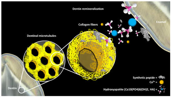
4. Stimulation of the Dentin-Pulp Complex

One of the current challenges in dentistry is the development of biomaterials that present adequate adhesion to collagen fibers and in turn promote the proliferation and differentiation of the cells of the dentin-pulp complex [56]. Collagen is a fibrous protein that constitutes a major part of the extracellular matrix of dentinogenesis providing strength, stability, and protection to this highly structured complex [57][58]. Collagen is a protein found in various tissues of the human body, including skin, bones, tendons, and teeth.
Regarding the function of collagen as a dental remineralizer, it is important to understand its role in tooth structure and dental mineralization in general. Collagen is pivotal in maintaining structural integrity and creating a vital biological setting for cellular interactions. It contributes to the formation and architecture of connective tissues and is a component of the organic matrix in tooth enamel. Although collagen itself is not a dental remineralizer, it is an essential component of the organic matrix of enamel that provides a supporting structure for hydroxyapatite crystals [59][60][61].
The proposed synthetic β-sheet peptide ID8 (Ile-Asp-Ile-Asp-Ile-Asp-Ile-Asp-Ile-Asp) promotes a calcium response and self-assembles to induce hydroxyapatite nucleation and intrafibrillar mineralization of collagen, contributing to calcium retention within collagen increasing its hydrophilicity. These peptides have similar regulatory effects or other macromolecules, so it is expected that they can be used as potential candidates in tissue mineralization [62].
Intrafibrillar mineralization of collagen in the “hybrid layer” is a key point in addressing collagen demineralization itself, which interferes with improving the adhesion of current restorations [62][63]. Cloyd et al., proposed a multifunctional peptide that combines a hydroxyapatite-binding peptide (HABP1) with a collagen-binding peptide (TKKLTLRT), evaluated its use for potential direct mineralization within the hybrid layer in dentin and identified intermolecular interactions that enhance the nanomechanical properties and intrafibrillar remineralization in collagen, which could promote sealing of the restorative material–tooth structure interface [63].
Peptides derived from the sequence of dentin phosphoprotein, containing multiple repetitions of the tripeptide aspartate–serine–serine (DSS), exhibit strong binding to calcium phosphate compounds. When immobilized, these peptides can effectively attract calcium phosphate to polystyrene beads modified with the peptide or to human dentin surfaces that have been demineralized. The affinity on the hydroxyapatite surface is defined by the following sequence: (DSS)n repeats, and although similar repeated sequences-(NTT)n, (DTT)n, (ETT)n, (NSS)n, (ESS)n, (DAA)n, (ASS)n, and (NAA)n [44].
Cell-based tissue engineering promotes microenvironments that facilitate odontogenic differentiation for the regeneration of the dentin-pulp complex. Some proposals in the literature focus on simulating the extracellular matrix or creating peptides that promote improvements in adhesion and rapid vascularization of the pulp tissue [60][61].
Biomaterials for dental pulp regeneration include both natural and synthetic materials. However, synthetic materials must closely mimic natural tissues to achieve biomimetic modifications, ensuring precise control over the temporal and spatial aspects of pulp-dentin complex regeneration. To achieve this objective, two primary strategies are utilized: stem cell transplantation and stem cell homing [57]. Notably, the stem cell homing strategy eliminates the need for in vitro stem cell isolation and cultivation, rendering it a promising clinical approach.
The continuous work on the biological characteristics of dental pulp stem cells offers a broad perspective in regenerating the pulp-dentin complex, maintaining histological characteristics similar to native tissue, physiological functions focused on pulp innervation, and immunity [57]. Utilizing synthetic proteins for dental pulp regeneration involves a strategic use of signaling molecules and the activation of vital signal transduction pathways, such as Wnt/β-catenin and BMP/Smad. These pathways enhance various stem cell functions, such as migration, proliferation, odontoblastic differentiation, and the promotion of nerve and blood vessel regeneration [58] (Figure 3).
Figure 3. Dentin-pulp complex biomimetic remineralization by synthetic peptide. The yellow diagrams presented in the graph represent the dentinal tissue. Intrafibrillar collagen mineralization plays a crucial role in improving the adhesion of current dental restorations. To achieve this, the use of synthetic proteins that are incorporated into collagen was investigated. These synthetic proteins promote a calcium response and, upon self-assembly, induce the formation of hydroxyapatite, which favors the remineralization of the affected tissue, increasing the protection of the dentin-pulp complex.

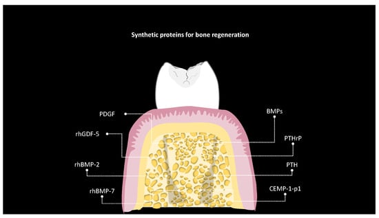
5. Bone Regeneration

Bone regeneration in the field of dentistry has experienced significant advances thanks to the application of synthetic proteins [64]. These innovations have become an essential pillar that has enabled success in dental procedures that require the restoration and repair of bone tissue. In the context of bone regeneration in dentistry, various peptides have been investigated for their potential to promote bone tissue formation or accelerate healing in dental procedures [65].
Currently, various growth factors (GFs) have been assessed, such as bone morphogenetic protein (BMP), platelet-derived growth factor (PDGF), and parathyroid hormone (PTH) peptides, to promote local bone regeneration [66]. However, within the most relevant and extensively studied proteins to date, the role of BMP stands out in the field of medicine. Such proteins have received approval from the United States Food and Drug Administration (FDA) for their use in specific clinical applications [67], with the approval for maxillofacial and oral surgery in 2007 and commercially available as Infuse Bone Graft (Medtronic, Parkway, Minneapolis, MN, USA). These osteoinductive proteins constitute a set of molecules derived from a non-mineralized bone matrix. They have the potential to induce differentiation of pluripotent mesenchymal cells into osteoprogenitor cells [66]. A small number of this group of proteins have osteoinductive properties. Schwarz et al., evaluated in vitro the influence of recombinant human growth and differentiation factor 5 (rhGDF-5) and natural bone mineral (NBM) coated with recombinant human bone morphogenetic protein 2 (rhBMP-2) as potential candidates for guided tissue regeneration, concluding that both proteins exhibited potential for use in this procedure [68]. Dupoirieux et al. reported on the effect of dimeric rhGDF-5 and its monomeric form rhGDF-5C465A on the bone healing of cranial defects in rats [69]. Furthermore, an evaluation was carried out to examine the impact of rhGDF-5 on bone regeneration in the membrane surrounding titanium dental implants placed in the mandibles of beagle dogs [70]. Lately, in the realm of periodontology, an investigation was conducted to evaluate maxillary sinus augmentation using bio-implants containing bone morphogenetic protein and autogenous bone in a rabbit model [71]. In terms of viable proteins, research has also emphasized the application of composite pedicled muscle flaps and rhBMP-7 for mandibular reconstruction, resulting in positive results [72]. It is crucial to highlight that science is exploring not only proteins but also small peptides because they are easy to synthesize and manipulate while exhibiting low immunogenic activity. In both in vivo and in vitro studies, the peptide derived from cementum protein 1 (CEMP-1-p1) was examined. It has shown a significant capacity to promote the proliferation and differentiation of human periodontal ligament cells, encouraging a phenotype resembling mineralization.
The importance of maintaining an adequate balance of minerals such as calcium and phosphorus in the regeneration process is attributed to the critical role of the parathyroid hormone. This hormone plays an essential function in regulating homeostasis, which has a direct impact on bone health [65]. PTH is a natural protein consisting of 84 amino acids, from which a 34 amino acid peptide has been evaluated, showing activity like the full protein. PTH 1-34 stimulates the proliferation and differentiation of osteoblasts and prevents their apoptosis [73]. Aside from the PTH peptide (1-34), related proteins such as PTHrP (1-36) and the recently developed abaloparatide peptide may exhibit distinct anabolic effects in individuals with osteoporosis, making them relevant candidates for application. This is particularly relevant due to the significant correlation between osteoporosis and tooth loss [74].
One of the most recent studies in dentistry, conducted by Hsu et al., focused on evaluating three peptides: intermittent PTH (1-34), PTHrP (1-36), and abaloparatide. These peptides influenced the expression of genes related to bone formation in cementoblast cells, including Bsp, Col1a1, Opg, RANKL, and Mmp13. PTH (1-34) demonstrating an immediate effect by increasing COL1A1 protein levels after treatment, while abaloparatide showed a delayed increase in COL1A1, observed 18 h after treatment. In contrast, PTHrP had no significant impact on the expression of COL1A1 in these cells. Moreover, microcomputed tomography revealed that PTH (1-34) injections led to heightened mineral density in the molar roots of mice. In contrast, abaloparatide injections increased mineral density in both incisors and molar roots in mice [75].
These discoveries indicate the potential for the development of treatments focused on enhancing both dental and bone health. Such advancements could carry substantial implications for dental practitioners and their patients (Figure 4).
Figure 4. Synthetic proteins for bone regeneration. This illustrative diagram showcases a collection of synthetic proteins and peptides that hold notable importance in the realm of bone regeneration within the field of dentistry—synthetic proteins for bone regeneration. Among these compounds, Bone Morphogenetic Proteins (BMPs), Abaloparatide, PTHrP (Parathyroid Hormone-Related Protein), PTH (Parathyroid Hormone), the peptide CEMP-1-p1 (Cementum-Related Peptide), rhBMP-7 (Recombinant Human Bone Morphogenetic Protein-7), rhBMP-2 (Recombinant Human Bone Morphogenetic Protein-2), and rhGDF-5 (Recombinant Human Growth/Differentiation Factor-5) are featured. Additionally, Platelet-Derived Growth Factor (PDGF) is included. Collectively, these substances represent pivotal elements in the promotion and enhancement of bone regeneration within the field of dentistry.

6. Challenges and Limitations

Synthetic biology holds great promise for addressing current clinical needs. Its focus on improving biological functions and gene expressions in diverse cellular and molecular areas has contributed to the engineering of new peptides; however, most advances within this field are still controlled in vitro and in vivo settings [76].
Each clinical area still has limitations identified by researchers within these controlled settings, with most experts finding that further studies are required to assess cytotoxic resistance before an effective commercial product can be developed [29]. To know the appropriate concentrations of peptides designed to counteract antimicrobial actions is necessary, as it has been identified that bacteria can exhibit tolerance in the production of extracellular polymeric matrix as a defense mechanism [30], suggesting the importance of monitoring bacterial resistance testing to avoid these mechanisms.
It has been recognized that the interaction of the constructed peptides and the oral environment to which they are subjected is highly complex, due to the self-regulatory mechanisms of each organism, which is why it is suggested that toxicity tests be carried out using various animal models before initiating controlled clinical trials [43].
Another limitation identified is that most in vitro models work with monospecies biofilm, and when a highly communicated and structured multispecies biofilm has been identified in the organism, the design of experiments based on this organization could suggest the effect within a more biomimic environment [35].
Although refinements of biological responses using bioengineering concepts continue to be made, there are also controversies surrounding the processing of these synthetic proteins, including waste management, alteration of the natural gene pool, and the possibility that microorganisms designed for specific functions may have unwanted side effects in the environment [77].

7. Ethical Considerations and Regulatory Aspects

The development of synthetic biology is still in an emerging form; however, its rapid development favors a more accessible and less expensive technology, which leads to the need to reinforce bioethical standards among researchers. The promotion of a continuous and renewed dialogue between the scientific community and social experts is suggested to strengthen the code of ethics. To encourage participation and accountability in this area, there are several scientific committees in Europe, the USA and Singapore; however, the implementation of global governance would allow for greater ethical commitment in the various nations developing these technologies.
The International Genetically Engineered Machine (iGEM) competition has included debate and reflection upon the safety and security needs for applications of synthetic biology. These efforts indicate a willingness by the community to engage with the social sciences; yet the next step requires more formal, permanent, and recurring collaboration between stakeholders in synthetic biology’s physical and social sciences [77]. Currently, no risks related to synthetic biology have been identified; most ethical concerns are based on the need to implement regulations governing these techniques to reduce the potential abuses associated with them in the future.

References

  1. Patil, S.; Albogami, S.; Hosmani, J.; Mujoo, S.; Kamil, M.A.; Mansour, M.A.; Abdul, H.N.; Bhandi, S.; Ahmed, S.S.S.J. Artificial Intelligence in the Diagnosis of Oral Diseases: Applications and Pitfalls. Diagnostics 2022, 12, 1029.
  2. Singer, L.; Fouda, A.; Bourauel, C. Biomimetic approaches and materials in restorative and regenerative dentistry: Review article. BMC Oral. Health 2023, 23, 105.
  3. Kumar, N.; Maher, N.; Amin, F.; Ghabbani, H.; Zafar, M.S.; Rodríguez-Lozano, F.J.; Oñate-Sánchez, R.E. Biomimetic Approaches in Clinical Endodontics. Biomimetics 2022, 7, 229.
  4. Jafari, N.; Habashi, M.S.; Hashemi, A.; Shirazi, R.; Tanideh, N.; Tamadon, A. Application of bioactive glasses in various dental fields. Biomater. Res. 2022, 26, 31.
  5. Hanczyc, M.M. Engineering Life: A Review of Synthetic Biology. Artif. Life 2020, 26, 260–273.
  6. Stein, V.; Alexandrov, K. Synthetic protein switches: Design principles and applications. Trends Biotechnol. 2015, 33, 101–110.
  7. Khoury, G.A.; Smadbeck, J.; Kieslich, C.A.; Floudas, C.A. Protein folding and de novo protein design for biotechnological applications. Trends Biotechnol. 2014, 32, 99–109.
  8. Zheng, X.; Wu, Y.; Bi, J.; Huang, Y.; Cheng, Y.; Li, Y.; Wu, Y.; Cao, G.; Tian, Z. The use of supercytokines, immunocytokines, engager cytokines, and other synthetic cytokines in immunotherapy. Cell. Mol. Immunol. 2022, 19, 192–209.
  9. Samiei, M.; Alipour, M.; Khezri, K.; Saadat, Y.R.; Forouhandeh, H.; Abdolahinia, E.D.; Vahed, S.Z.; Sharifi, S.; Dizaj, S.M. Application of collagen and mesenchymal stem cells in regenerative dentistry. Curr. Stem. Cell Res. Ther. 2022, 17, 606–620.
  10. Tavelli, L.; Chen, C.Y.; Barootchi, S.; Kim, D.M. Efficacy of biologics for the treatment of periodontal infrabony defects: An American Academy of Periodontology best evidence systematic review and network meta-analysis. J. Periodontol. 2022, 93, 1803–1826.
  11. Firipis, K.; Nisbet, D.R.; Franks, S.J.; Kapsa, R.M.I.; Pirogova, E.; Williams, R.J.; Quigley, A. Enhancing Peptide Biomaterials for Biofabrication. Polymers 2021, 13, 2590.
  12. Wang, Y.; Katyal, P.; Montclare, J.K. Protein-engineered functional materials. Adv. Healthc. Mater. 2019, 8, 1801374.
  13. Takahara, M.; Kamiya, N. Synthetic strategies for artificial lipidation of functional proteins. Chemistry 2020, 26, 4645–4655.
  14. Ahn, W.; Lee, J.H.; Kim, S.R.; Lee, J.; Lee, E.J. Designed protein-and peptide-based hydrogels for biomedical sciences. J. Mater. Chem. B 2021, 9, 1919–1940.
  15. Wise, S.G.; Mithieux, S.M.; Weiss, A.S. Engineered tropoelastin and elastin-based biomaterials. Adv. Protein Chem. Struct. Biol. 2009, 78, 1–24.
  16. Fang, D.; Yuran, S.; Reches, M.; Catunda, R.; Levin, L.; Febbraio, M. A peptide coating preventing the attachment of Porphyromonas gingivalis on the surfaces of dental implants. J. Periodontal. Res. 2020, 55, 503–510.
  17. Babaji, P.; Melkundi, M.; Bhagwat, P.; Mehta, V. An in vitro Evaluation of remineralizing capacity of self-assembling peptide (SAP) P11-4 and casein phosphopeptides-amorphous calcium phosphate (CPP-ACP) on artificial enamel. Pesqui. Bras. Odontopediatria Clínica Integr. 2019, 19.
  18. Chen, X.; Zhou, X.C.; Liu, S.; Wu, R.F.; Aparicio, C.; Wu, J.Y. In vivo osseointegration of dental implants with an antimicrobial peptide coating. J. Mater. Sci. Mater. Med. 2017, 28, 76.
  19. Bagno, A.; Piovan, A.; Dettin, M.; Chiarion, A.; Brun, P.; Gambaretto, R.; Fontana, G.; Di Bello, C.; Palù, G.; Castagliuolo, I. Human osteoblast-like cell adhesion on titanium substrates covalently functionalized with synthetic peptides. Bone 2007, 40, 693–699.
  20. Dettin, M.; Conconi, M.T.; Gambaretto, R.; Pasquato, A.; Folin, M.; Di Bello, C.; Parnigotto, P.P. Novel osteoblast-adhesive peptides for dental/orthopedic biomaterials. J. Biomed. Mater. Res. 2002, 60, 466–471.
  21. Pitts, N.B.; Twetman, S.; Fisher, J.; Marsh, P.D. Understanding dental caries as a non-communicable disease. Br. Dent. J. 2021, 231, 749–753.
  22. Mathur, V.P.; Dhillon, J.K. Dental caries: A disease which needs attention. Indian J. Pediatr. 2018, 85, 202–206.
  23. Niu, J.Y.; Yin, I.X.; Wu, W.K.K.; Li, Q.L.; Mei, M.L.; Chu, C.H. Antimicrobial peptides for the prevention and treatment of dental caries: A concise review. Arch. Oral Biol. 2021, 122, 105022.
  24. Dewhirst, F.E.; Chen, T.; Izard, J.; Paster, B.J.; Tanner, A.C.; Yu, W.H.; Lakshmana, A.; Wade, W.G. The human oral microbiome. J. Bacteriol. 2010, 192, 5002–5017.
  25. Gross, E.L.; Beall, C.J.; Kutsch, S.R.; Firestone, N.D.; Leys, E.J.; Griffen, A.L. Beyond Streptococcus mutans: Dental caries onset linked to multiple species by 16S rRNA community analysis. PLoS ONE 2012, 7, e47722.
  26. Lamont, R.J.; Koo, H.; Hajishengallis, G. The oral microbiota: Dynamic communities and host interactions. Nat. Rev. Microbiol. 2018, 16, 745–759.
  27. Yang, Y.; Xia, L.; Haapasalo, M.; Wei, W.; Zhang, D.; Ma, J.; Shen, Y. A novel hydroxyapatite-binding antimicrobial peptide against oral biofilms. Clin. Oral Investig. 2019, 23, 2705–2712.
  28. Jakubovics, N.S.; Goodman, S.D.; Mashburn-Warren, L.; Stafford, G.P.; Cieplik, F. The dental plaque biofilm matrix. Periodontology 2000 2021, 86, 32–56.
  29. Da Silva, B.R.; Conrado, A.J.S.; Pereira, A.L.; Evaristo, F.F.V.; Arruda, F.V.S.; Vasconcelos, M.A.; Lorenzón, E.N.; Cilli, E.M.; Teixeira, E.H. Antibacterial activity of a novel antimicrobial peptide KR12-KAEK derived from KR-12 against Streptococcus mutans planktonic cells and biofilms. Biofouling 2017, 33, 835–846.
  30. Da Silva, B.R.; De Freitas, V.A.A.; Carneiro, V.A.; Arruda, F.V.S.; Lorenzón, E.N.; De Aguiar, A.S.W.; Cilli, E.M.; Cavada, E.H.; Teixeira, E.H. Antimicrobial activity of the synthetic peptide Lys-a1 against oral streptococci. Peptides 2013, 42, 78–83.
  31. He, J.; Yarbrough, D.K.; Kreth, J.; Anderson, M.H.; Shi, W.; Eckert, R. Systematic approach to optimizing specifically targeted antimicrobial peptides against Streptococcus mutans. Antimicrob. Agents Chemother. 2010, 54, 2143–2151.
  32. Izadi, N.; Keikha, M.; Ghazvini, K.; Karbalaei, M. Oral antimicrobial peptides and new therapeutic strategies for plaque-mediated diseases. Gene Rep. 2020, 21, 100811.
  33. Zhou, L.; Wong, H.M.; Zhang, Y.Y.; Li, Q.L. Constructing an antibiofouling and mineralizing bioactive tooth surface to protect against decay and promote self-healing. ACS Appl. Mater. Interfaces 2019, 12, 3021–3031.
  34. Zhang, L.Y.; Fang, Z.H.; Li, Q.L.; Cao, C.Y. A tooth-binding antimicrobial peptide to prevent the formation of dental biofilm. J. Mater. Sci. Mater. Med. 2019, 30, 1–9.
  35. Wang, Y.; Fan, Y.; Zhou, Z.; Tu, H.; Ren, Q.; Wang, X.; Ding, L.; Zhou, X.; Zhang, L. De novo synthetic short antimicrobial peptides against cariogenic bacteria. Arch. Oral Biol. 2017, 80, 41–50.
  36. Xu, C.; Yao, X.; Walker, M.P.; Wang, Y. Chemical/molecular structure of the dentin-enamel junction is dependent on the intratooth location. Calcif. Tissue Int. 2009, 84, 221–228.
  37. Bartlett, J.D. Dental enamel development: Proteinases and their enamel matrix substrates. Int. Sch. Res. Not. 2013, 2013, 684607.
  38. Farooq, I.; Bugshan, A. The role of salivary contents and modern technologies in the remineralization of dental enamel: A narrative review. F1000Research 2020, 9, 171.
  39. Philip, N. State of the art enamel remineralization systems: The next frontier in caries management. Caries Res. 2019, 53, 284–295.
  40. Kastenbom, L.; Falsen, A.; Larsson, P.; Sunnegårdh-Grönberg, K.; Davidson, T. Costs and health-related quality of life in relation to caries. BMC Oral Health 2019, 19, 187.
  41. Anand, S.; Rejula, F.; Sam, J.V.G.; Christaline, R.; Nair, M.G.; Dinakaran, S. Comparative evaluation of effect of nanohydroxyapatite and 8% arginine containing toothpastes in managing dentin hypersensitivity: Double blind randomized clinical trial. Acta. Med. 2017, 60, 114–119.
  42. Kassebaum, N.J.; Bernabé, E.; Dahiya, M.; Bhandari, B.; Murray, C.J.; Marcenes, W. Global burden of untreated caries: A systematic review and metaregression. J. Dent. Res. 2015, 94, 650–658.
  43. Valente, M.T.; Moffa, E.B.; Crosara, K.T.B.; Xiao, Y.; de Oliveira, T.M.; Machado, M.A.D.A.M.; Siqueira, W.L. Acquired enamel pellicle engineered peptides: Effects on hydroxyapatite crystal growth. Sci. Rep. 2018, 8, 3766.
  44. Yarbrough, D.K.; Hagerman, E.; Eckert, R.; He, J.; Choi, H.; Cao, N.; Le, K.; Hedger, J.; Qi, F.; Anderson, M.; et al. Specific binding and mineralization of calcified surfaces by small peptides. Calcif. Tissue Int. 2010, 86, 58–66.
  45. Li, Y.; Li, Y.; Bai, Q.; Wen, M.; Ma, D.; Lin, Y.; Chu, J. Recombinant amelogenin peptide TRAP promoting remineralization of early enamel caries: An in vitro study. Front. Physiol. 2023, 14, 1076265.
  46. Marin, L.M.; Xiao, Y.; Cury, J.A.; Siqueira, W.L. Engineered salivary peptides reduce enamel demineralization provoked by cariogenic S. Mutans biofilm. Microorganisms 2022, 10, 742.
  47. Li, Z.C.; Xi, Q.I.N.; Qian, R.E.N.; Die, H.U.; Tian, T.I.A.N.; Ting, H.E.; Li, W.; Zhang, L.L. Rational Design of β-sheet Peptides with Self-Assembly into Nanofibres on Remineralisation of Initial Caries Lesions. Chin. J. Dent. Res. 2020, 23, 131–141.
  48. Marin, L.M.; Xiao, Y.; Cury, J.A.; Siqueira, W.L. Modulation of Streptococcus mutans Adherence to Hydroxyapatite by Engineered Salivary Peptides. Microorganisms 2022, 10, 223.
  49. Kwak, S.Y.; Litman, A.; Margolis, H.C.; Yamakoshi, Y.; Simmer, J.P. Biomimetic enamel regeneration mediated by leucine-rich amelogenin peptide. J. Dent. Res. 2017, 96, 524–530.
  50. Ding, L.; Han, S.; Wang, K.; Zheng, S.; Zheng, W.; Peng, X.; Niu, Y.; Li, W.; Zhang, L. Remineralization of enamel caries by an amelogenin-derived peptide and fluoride in vitro. Regen. Biomater. 2020, 7, 283–292.
  51. Hossein, B.G.; Sadr, A.; Espigares, J.; Hariri, I.; Nakashima, S.; Hamba, H.; Shafiei, F.; Moztarzadeh, F.; Tagami, J. Study on the influence of leucine-rich amelogenin peptide (LRAP) on the remineralization of enamel defects via micro-focus x-ray computed tomography and nanoindentation. Biomed. Mater. 2015, 10, 035007.
  52. Dissanayake, S.S.; Ekambaram, M.; Li, K.C.; Harris, P.W.; Brimble, M.A. Identification of key functional motifs of native amelogenin protein for dental enamel remineralisation. Molecules 2020, 25, 4214.
  53. Wang, D.; Deng, J.; Deng, X.; Fang, C.; Zhang, X.; Yang, P. Controlling enamel remineralization by amyloid-like amelogenin mimics. Adv. Mater. 2020, 32, e2002080.
  54. Chu, J.; Feng, X.; Guo, H.; Zhang, T.; Zhao, H.; Zhang, Q. Remineralization efficacy of an amelogenin-based synthetic peptide on carious lesions. Front. Physiol. 2018, 9, 842.
  55. Zheng, W.; Ding, L.; Wang, Y.; Han, S.; Zheng, S.; Guo, Q.; Li, W.; Zhou, X.; Zhang, L. The effects of 8DSS peptide on remineralization in a rat model of enamel caries evaluated by two nondestructive techniques. J. Appl. Biomater. Funct. Mater. 2019, 17, 2280800019827798.
  56. Han, S.; Peng, X.; Ding, L.; Lu, J.; Liu, Z.; Wang, K.; Zhang, L. TVH-19, a synthetic peptide, induces mineralization of dental pulp cells in vitro and formation of tertiary dentin in vivo. Biochem. Biophys. Res. Commun. 2021, 534, 837–842.
  57. Machla, F.; Angelopoulos, I.; Epple, M.; Chatzinikolaidou, M.; Bakopoulou, A. Biomolecule-Mediated Therapeutics of the Dentin-Pulp Complex: A Systematic Review. Biomolecules 2022, 12, 285.
  58. Zou, J.; Mao, J.; Shi, X. Influencing factors of pulp-dentin complex regeneration and related biological strategies. Zhejiang Da Xue Xue Bao Yi Xue Ban 2022, 51, 350–361.
  59. McGuire, J.D.; Walker, M.P.; Mousa, A.; Wang, Y.; Gorski, J.P. Type VII collagen is enriched in the enamel organic matrix associated with the dentin-enamel junction of mature human teeth. Bone 2014, 63, 29–35.
  60. Xia, K.; Chen, Z.; Chen, J.; Xu, H.; Xu, Y.; Yang, T.; Zhang, Q. RGD- and VEGF-Mimetic Peptide Epitope-Functionalized Self-Assembling Peptide Hydrogels Promote Dentin-Pulp Complex Regeneration. Int. J. Nanomed. 2020, 15, 6631–6647.
  61. Chattopadhyay, S.; Raines, R.T. Review collagen-based biomaterials for wound healing. Biopolymers 2014, 101, 821–833.
  62. Li, Z.; Ren, Q.; Han, S.; Ding, L.; Qin, X.; Hu, D.; He, T.; Tian, T.; Lu, Z.; Zhang, L. Promoting effect of a calcium-responsive self-assembly β-sheet peptide on collagen intrafibrillar mineralization. Regen Biomater. 2022, 9, rbac059.
  63. Cloyd, A.K.; Boone, K.; Ye, Q.; Snead, M.L.; Spencer, P.; Tamerler, C. Engineered Peptides Enable Biomimetic Route for Collagen Intrafibrillar Mineralization. Int. J. Mol. Sci. 2023, 24, 6355.
  64. Thalakiriyawa, D.S.; Dissanayaka, W.L. Advances in Regenerative Dentistry Approaches: An Update. Int. Dent. J. 2023.
  65. Pountos, I.; Panteli, M.; Lampropoulos, A.; Jones, E.; Calori, G.M.; Giannoudis, P.V. The role of peptides in bone healing and regeneration: A systematic review. BMC Med. 2016, 14, 103.
  66. Roberto-Rodrigues, M.; Fernandes, R.M.P.; Senos, R.; Scoralick, A.C.D.; Bastos, A.L.; Santos, T.M.P.; Viana, L.P.; Lima, I.; Guzman-Silva, M.A.; Kfoury-Júnior, J.R. Novel rat model of nonunion fracture with vascular deficit. Injury 2015, 46, 649–654.
  67. Díaz-Sánchez, R.M.; Yáñez-Vico, R.M.; Fernández-Olavarría, A.; Mosquera-Pérez, R.; Iglesias-Linares, A.; Torres-Lagares, D. Current approaches of bone morphogenetic proteins in dentistry. J. Oral Implantol. 2015, 41, 337–342.
  68. Schwarz, F.; Ferrari, D.; Sager, M.; Herten, M.; Hartig, B.; Becker, J. Guided bone regeneration using rhGDF-5-and rhBMP-2-coated natural bone mineral in rat calvarial defects. Clin. Oral Implants Res. 2009, 20, 1219–1230.
  69. Dupoirieux, L.; Pohl, J.; Hanke, M.; Pourquier, D. A preliminary report on the effect of dimeric rhGDF-5 and its monomeric form rhGDF-5C465A on bone healing of rat cranial defects. J. Craniomaxillofac. Surg. 2009, 37, 30–35.
  70. Lee, J.S.; Wikesjö, U.M.E.; Jung, U.W.; Choi, S.H.; Pippig, S.; Siedler, M.; Kim, C.K. Periodontal wound healing/regeneration following implantation of recombinant human growth/differentiation factor-5 in a β-tricalcium phosphate carrier into one-wall intrabony defects in dogs. J. Clin. Periodontol. 2010, 37, 382–389.
  71. Ho, S.K.C.; Frcd, C.; Peel, S.A.F.; Hu, Z.M. Augmentation of the maxillary sinus: Comparison of bioimplants containing bone morphogenetic protein and autogenous bone in a rabbit model. J. Can. Dent. Assoc. 2010, 76, a108.
  72. Ayoub, A.; Challa, S.R.R.; Abu-Serriah, M.; McMahon, J.; Moos, K.; Creanor, S.; Odell, E. Use of a composite pedicled muscle flap and rhBMP-7 for mandibular reconstruction. Int. J. Oral Maxillofac. Surg. 2007, 36, 1183–1192.
  73. Whitfield, J.F.; Morley, P.; Willick, G.E. Parathyroid hormone, its fragments and their analogs for the treatment of osteoporosis. Treat. Endocrinol. 2002, 1, 175–190.
  74. Qin, L.; Raggatt, L.J.; Partridge, N.C. Parathyroid hormone: A double-edged sword for bone metabolism. Trends Endocrinol. Metab. 2004, 15, 60–65.
  75. Hsu, C.; He, Z.; Le Henaff, C.; Partridge, N.C. Differential effects of parathyroid hormone, parathyroid hormone-related protein, and abaloparatide on collagen 1 expression by mouse cementoblasts and mouse tooth root density. Am. J. Orthod. Dentofac. Orthop. 2023, 163, 378–388.e1.
  76. Safaei, M.; Mobini, G.R.; Abiri, A.; Shojaeian, A. Synthetic biology in various cellular and molecular fields: Applications, limitations, and perspective. Mol. Biol. Rep. 2020, 47, 6207–6216.
  77. Trump, B.D.; Cegan, J.C.; Wells, E.; Keisler, J.; Linkov, I. A critical juncture for synthetic biology: Lessons from nanotechnology could inform public discourse and further development of synthetic biology. EMBO Rep. 2018, 19, e46153.
More
Information
Contributors MDPI registered users' name will be linked to their SciProfiles pages. To register with us, please refer to https://encyclopedia.pub/register : , , , ,
View Times: 118
Revisions: 2 times (View History)
Update Date: 29 Jan 2024
1000/1000