Submitted Successfully!
To reward your contribution, here is a gift for you: A free trial for our video production service.
Thank you for your contribution! You can also upload a video entry or images related to this topic.
Version Summary Created by Modification Content Size Created at Operation
1 -- 4491 2024-01-23 09:34:54 |
2 layout -1 word(s) 4490 2024-01-24 02:19:15 |

Video Upload Options

Do you have a full video?

Confirm

Are you sure to Delete?
Cite
If you have any further questions, please contact Encyclopedia Editorial Office.
Lee, S.J.; Park, J. Targeted Drug-Delivery Systems with Aptamers. Encyclopedia. Available online: https://encyclopedia.pub/entry/54232 (accessed on 08 May 2024).
Lee SJ, Park J. Targeted Drug-Delivery Systems with Aptamers. Encyclopedia. Available at: https://encyclopedia.pub/entry/54232. Accessed May 08, 2024.
Lee, Su Jin, Jee-Woong Park. "Targeted Drug-Delivery Systems with Aptamers" Encyclopedia, https://encyclopedia.pub/entry/54232 (accessed May 08, 2024).
Lee, S.J., & Park, J. (2024, January 23). Targeted Drug-Delivery Systems with Aptamers. In Encyclopedia. https://encyclopedia.pub/entry/54232
Lee, Su Jin and Jee-Woong Park. "Targeted Drug-Delivery Systems with Aptamers." Encyclopedia. Web. 23 January, 2024.
Targeted Drug-Delivery Systems with Aptamers
Edit

The field of drug delivery has witnessed remarkable progress, driven by the quest for more effective and precise therapeutic interventions. Among the myriad strategies employed, the integration of aptamers as targeting moieties and stimuli-responsive systems has emerged as a promising avenue, particularly in the context of anticancer therapy. The conventional chemotherapy paradigm often suffers from systemic toxicity, as potent cytotoxic agents are indiscriminately delivered throughout the body, causing adverse effects on healthy tissues. To address this limitation, the integration of smart targeting mechanisms has gained prominence. Within this paradigm, aptamers, short nucleic acid sequences with a unique ability to bind specifically to target molecules, have emerged as valuable targeting ligands. Aptamers share similarities with antibodies as they exhibit a high affinity for specific targets, making them a focus of research in disease-targeted therapy owing to their remarkable selectivity. Regarded as promising therapeutic agents, aptamers possess attributes such as non-immunogenicity, high specificity, and stability.

aptamer targeted drug delivery stimuli-responsive drug delivery

1. Aptamers

In 1990, a groundbreaking development marked the emergence of aptamers as synthetic ligands endowed with the capacity for molecular recognition [1][2]. Aptamers, in essence, represent sophisticated analogs of antibodies, consisting of concise single-stranded oligonucleotides capable of selectively and strongly binding to proteins, peptides, metal ions, and cells. This exceptional selectivity and high affinity stem from their distinctive three-dimensional conformation, which is achieved through intramolecular interactions. Furthermore, aptamers are garnering increasing attention in the realm of drug targeting due to their advantages such as low toxicity, minimal or absent immunogenicity, and more economical production, particularly when compared to antibodies [3].
Aptamers have been synthesized using the Systematic Evolution of Ligands by Exponential Enrichment (SELEX) technique [4][5]. This method enables the generation of aptamers capable of specifically targeting proteins that are overexpressed on the surface of particular cells, analogous to a donor–receptor system involving cancer cells. The SELEX technique can enhance the aptamers’ ability to bind to known or unidentified receptors located on the cell membrane [6]. The binding affinity of an aptamer is typically characterized by dissociation rate constants (kd), which are associated with dissociation and association rate constants (koff/kon). Moreover, aptamers can be functionalized with a variety of materials on the intended sites. Furthermore, aptamers with small molecular weights can be rapidly eliminated through renal filtration in vivo [7]. Aptamer sequences readily accommodate the loading of antitumor drugs targeted specifically to cancer cells.
Hence, researchers have employed biological, biocompatible, and biodegradable carrier materials like lipids, lactic acid, or chitosan, as well as various approved non-toxic polymers (owing to their ease of production), or synthetic materials such as titanium dioxide (TiO2) or cadmium (Cd) as a means to enhance the loading of anticancer agents [8].
Zeng et al. formed aptamer–drug conjugates with doxorubicin (DOX) and an aptamer for targeted cancer therapy in vitro and in vivo [9]. Synthesizing tripartite newkome-type monomers (TNMs) of DOX with pH-sensitive hydrazone bonds improves therapeutic potential. Apt–TNM-DOX, created via a self-loading process, stably carries 15 DOX molecules per aptamer. The pH-dependent DOX release at a pH level of 5.0 targets lymphoma cells selectively, minimizing off-target effects. Apt–TNM-DOX, a non-toxic approach, shows promise for aptamer-based targeted therapeutics, potentially reducing chemotherapy’s non-specific side effects.
It is worth highlighting that the use of PEGylated-functionalized materials can reduce systemic clearance and enhance the stability of nanoparticles, which is crucial for the passive targeting of tumors by nanocarriers [10]. Furthermore, the incorporation of different types of aptamer-based modifications on the surface of polymeric nanocarriers can result in heightened selectivity and sensitivity when targeting specific organs, tissues, and other sites [11].

2. Aptamer-Modified Nanomaterials

Because of their distinctive physicochemical attributes, exceedingly small size, expansive surface area, and exceptional loading capacity, nanomaterials surmount numerous constraints associated with conventional therapeutic and diagnostic approaches [12]. The essence of advancing nanomedicine lies in enhancing the specific recognition capability for pathological tissues. The conjugation of aptamers with nanomaterials signifies a noteworthy advancement in targeted drug delivery (Figure 1) [13]. In this section, various representative aptamer-based nanomaterials employed for drug delivery will be discussed.
Figure 1. A variety of aptamer conjugates for targeted drug-delivery system.

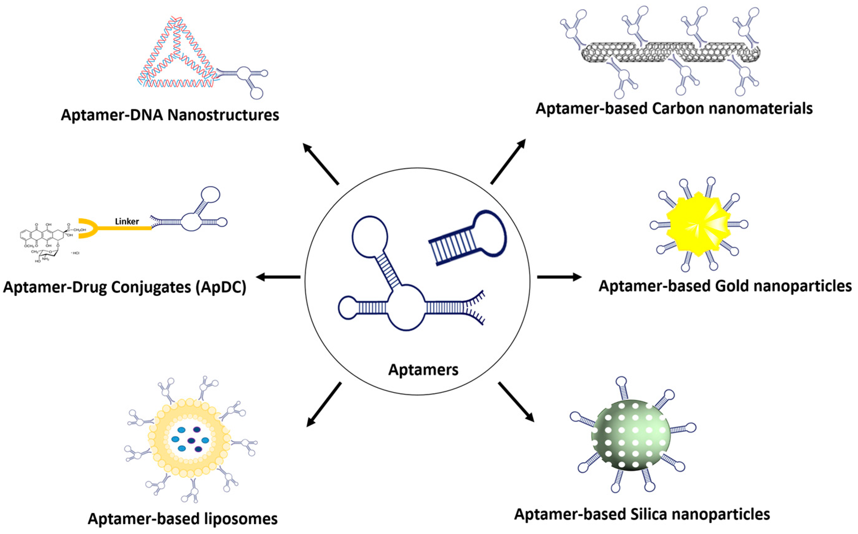
2.1. Aptamer–Gold Conjugation

Being a significant nanomaterial, gold nanoparticles have garnered substantial attention in the field of biomedicine owing to their elevated surface-to-volume ratio, low toxicity, outstanding stability, and biological compatibility [14][15]. Aptamer-conjugated gold nanomaterials (Apt-AuNPs), combining the distinctive advantageous features of aptamers and gold nanoparticles, have found extensive application in cancer diagnosis and therapy [16].
Since the investigation by the Mirkin group utilized DNA molecules to construct a polymeric network of nanoparticles [17], numerous studies have emerged, including the development of enzyme-responsive Apt-AuNPs for the detection of mucin 1 protein (MUC1) [18], the utilization of Apt-AuNPs in conjunction with graphene oxide for the photothermal therapy of breast cancer [19], and the design of an aptamer-functionalized AuNPs-Fe3O4-GS capture probe for monitoring circulating tumor cells in whole blood [20].
In a recent study, Khorshid et al. investigated the utilization of gold nanoparticles functionalized with an anti-HER-2 aptamer for the precise delivery of dasatinib (DSB) to breast cancer cells [21]. The loading efficiency of the activated drug on both plain and porous gold nanoparticles was notably elevated (52% and 68%, respectively) compared to that of free DSB within gold nanoparticles (1 to 2.5%). Porous gold nanoparticles functionalized with the aptamer and loaded with activated dasatinib demonstrated heightened cytotoxicity and enhanced cellular uptake in comparison to nanoparticles containing modified DSB or unactivated DSB.
Specifically targeted gold nanobipyramids (GNBs) demonstrate potential as photothermal therapeutic agents and have diverse applications such as contrast agents, biosensors, and drug-delivery vehicles. Navyatha et al. explored the efficiency of targeting moieties (aptamers and antibodies) in specifically targeting the MUC1 protein and their impact on cytotoxicity [22]. The results indicate that aptamer-conjugated GNBs exhibit reduced cytotoxicity compared to antibody-conjugated ones. Aptamer-conjugated GNBs are more effective in photoablating MCF7 cell lines than HCT116 cell lines, highlighting their potential for targeted photothermal therapy.

2.2. Aptamer–Silica Conjugation

Silica nanoparticles have emerged as feasible carriers in drug-delivery systems [23]. These particles have effectively enabled controlled drug release both in vivo and in vitro, achieved through pH and temperature variations, photochemical reactions, and specific redox reactions [24]. Integrated with aptamers, the silica nanoparticles have demonstrated the ability to amplify the therapeutic effects against cancer with a reduced dosage of the drug [25].
Recently, Heydari et al. studied surface-modified mesoporous silica nanoparticles (MSNs) for the targeted delivery of anticancer agents (daunorubicin and cytarabine) to K562 leukemia cancer cells [26]. The MSNs were further enhanced with the KK1B10 aptamer (Apt) to improve uptake by K562 cells through ligand–receptor interactions. MSNs coated with CS and conjugated with the aptamer exhibited a significantly lower IC50 value of 2.34 µg/mL compared to MSNs without the aptamer conjugation (IC50 value of 12.27 µg/mL). The aptamer-modified MSNs exhibited lower IC50 values against cancer cell lines and demonstrated enhanced anticancer activity in animal models, highlighting their potential as effective targeted anticancer agents with controlled drug release properties.
Kianpour et al. studied the oncoprotein cell migration-inducing hyaluronidase 2 (CEMIP2) in colorectal cancer (CRC) and developed an aptamer-based silica nanoparticle for targeted therapy [27]. The cell-SELEX technique identified aptCEMIP2(101), which specifically interacts with full-length CEMIP2. Treatment with aptCEMIP2(101) reduced CEMIP2-mediated tumorigenesis in vitro. Mesoporous silica nanoparticles (MSN) carrying aptCEMIP2(101) and Dox significantly suppressed tumorigenesis, with Dox@MSN-aptCEMIP2(101) showing higher efficacy compared to Dox@MSN and MSN-aptCEMIP2(101) in CRC-derived cells. This research revealed CEMIP2 as a novel oncogene and introduced an effective aptamer-based targeted therapeutic strategy.
Xie et al. focused on enhancing the targeted delivery of doxorubicin (DOX) to colon cancer cells using aptamer-modified mesoporous silica nanoparticles (Ap-MSN-DOX) [28]. The nanoparticles were characterized for various properties, and results demonstrated increased binding to EpCAM-overexpressing SW620 colon cancer cells. This led to enhanced cellular uptake and cytotoxicity compared to non-aptamer-modified nanoparticles (MSN-DOX). Ap-MSN-DOX also exhibited the significant inhibition of EpCAM expression on SW620 cells, indicating its potential for targeted delivery to improve therapeutic efficacy while minimizing side effects.

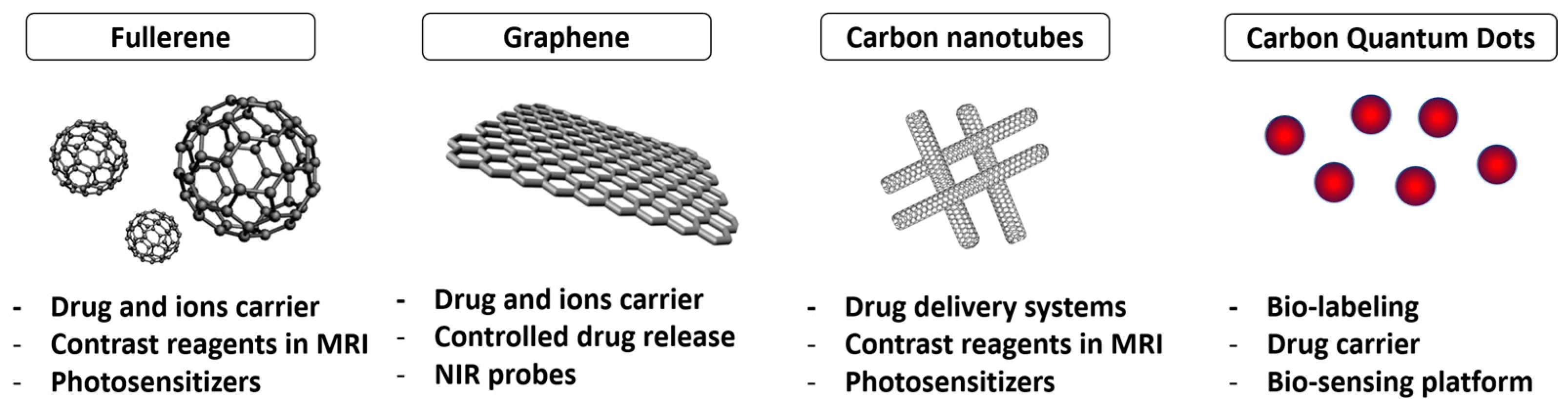
2.3. Aptamer–Carbon Conjugation

Carbon nanomaterials such as graphene and carbon nanotubes are highly valued for their mechanical, optical, and thermal properties for various biomedical applications [29] (Figure 2).
Figure 2. Carbon nanomaterials (CNMs) for theranostics.
The conjugation of aptamers with carbon nanomaterials (CNMs) has given rise to theranostic agents [30], revolutionizing personalized cancer medicine [31], target-specific imaging, and the label-free diagnosis of diverse cancers [32]. Aptamer-functionalized CNMs serve as nanovesicles for the precise delivery of anticancer agents, such as doxorubicin [33] and 5-fluorouracil [34], directly to tumor sites. This innovative approach holds promise for advanced, targeted therapeutic strategies with reduced side effects in medical treatments.
Zavareh et al. developed a targeted drug-delivery system, the 5-fluorouracil-chitosan-carbon quantum dot-aptamer (5-FU-CS-CQD-Apt) nanoparticle, using a water-in-oil emulsification method for breast cancer treatment [34]. Characterized by high drug-loading and entrapment efficiency, the nanoparticle exhibited an average size of 122.7 nm and a zeta potential of +31.2 mV. In vitro studies demonstrated controlled drug release, and functional assays indicated the biocompatibility of the blank nanoparticle and the efficient tumor cell-killing capabilities of 5-FU-CS-CQD-Apt, making it a potential carbon nanocarrier for breast cancer treatment.
Zhao et al. introduced aptamer-functionalized Fe3O4@carbon@doxorubicin nanoparticles (Apt-Fe3O4@C@DOX) for synergistic chemo–photothermal cancer therapy [33]. The nanoparticles exhibit high photothermal conversion efficiency and pH/heat-induced drug release. In vitro experiments demonstrate enhanced toxicity towards lung adenocarcinoma cells (A549) with combined chemo–photothermal therapy compared to individual treatments. Furthermore, the nanoparticles exhibit decreasing contrast enhancement in magnetic resonance imaging, suggesting potential applications as contrast agents for the T2-weighted imaging of tumor tissues. Apt-Fe3O4@C@DOX nanoparticles hold significant promise for cancer therapy.
Fullerenes composed of 60 and 70 carbon atoms are spherical carbon allotropes commonly utilized in drug-delivery systems like carbon nanotubes (CNTs) and graphene. With a size around 1 nm, their geometry and surface area make them suitable for drug release applications [35]. Fullerenes, featuring a stable ellipsoidal structure with apolar properties, allow for modifications with various drugs or biomolecules, including radioactive atoms for diagnostic purposes. Notably, C60 exhibits a high stability due to the delocalization of π electrons in benzene rings, showing minimal toxicity in in vitro and in vivo cytotoxicity studies.
Fullerene-based photosensitizers, particularly trimalonic acid-modified C70 fullerene (TF70), show significant potential in photodynamic therapy (PDT) [36]. An aptamer-guided TF70 photosensitizer demonstrates enhanced PDT efficiency against A549 lung cancer cells, even in the presence of serum. The conjugation of the aptamer (R13) improves the lysosomal localization of TF70-R13, leading to the increased production of intracellular reactive oxygen species (ROS) under light irradiation, effectively killing cells. TF70-R13’s enhanced photodynamic efficiency and good biocompatibility position it as a highly promising tumor-specific photosensitizer for PDT.
Carbon nanotubes (CNTs) have garnered considerable interest as potential nanocarriers for drug delivery [37]. Their distinctive properties, such as an ultrahigh length-to-diameter ratio and efficient cellular uptake, make them promising nanocarriers in this field. The unique conjugated structure of CNTs is well-suited for π–π stacking interactions with various drugs and therapeutic molecules that possess aromatic rings, including anthracyclines. This interaction enhances the potential for the effective loading and delivery of such drugs, contributing to the attractiveness of CNTs as nanocarriers in drug-delivery applications.
Chen et al. developed an aptamer-siRNA chimera (Chim), polyethyleneimine (PEI), 5-fluorouracil (5-FU), carbon nanotube (CNT), and collagen membrane, which demonstrated sustained 5-FU release for over two weeks [38]. The aptamer-siRNA chimera enabled specific binding to gastric cancer cells, facilitating the targeted delivery of 5-FU and silencing drug-resistant genes. In vitro experiments revealed that Chim/PEI/5-FU/CNT nanoparticles induced apoptosis in 5-FU-resistant gastric cancer cells, inhibiting their invasion and proliferation. Animal studies showed the significant inhibition of mitogen-activated protein kinase (MAPK) expression and effective treatment of peritoneal dissemination of 5-FU-resistant gastric cancer.
Graphene oxides (GOs) have emerged as promising drug carriers for targeted delivery systems due to their good endocytosis, biocompatibility, and ample surface area for drug loading [39]. GO’s dispersing capability in water and physiological environments, attributed to its abundant functional groups like epoxide, hydroxyl, and carboxyl groups, enhances its appeal. The interaction between these groups and drug functional groups, including hydrogen bonding, π−π stackings, and hydrophobic interactions, facilitates efficient drug loading onto the nanocarrier.
Shahidi et al. engineered drug-delivery cargo by decorating carboxylated graphene oxide (cGO) with an aptamer, HB5, for the simultaneous delivery of DOX and silibinin (Sili) in a combination therapy against MCF-7 and SK-BR-3 breast cancer cells [40]. Apt-cGO exhibited a sheet-like nanostructure with a high entrapment efficiency for both Sili (70.42%) and DOX (84.22%). The nanocomposites, selectively taken up by breast cancer cells, released both drugs upon the cleavage of the cGO–drug interaction. Apt-cGO-DOX-Sili nanocomposites demonstrated higher in vitro cytotoxicity than free drugs, suppressing cancer cell survival signals and inducing apoptosis, suggesting a promising drug-delivery approach for breast chemotherapy.
Ganoderenic acid D (GAD) from Ganoderma lucidum, which is known to show anticancer activity [41], was loaded onto a graphene oxide-polyethylene glycol-anti-epidermal growth factor receptor (GO-PEG-EGFR) carrier to create a targeting antitumor nanocomposite (GO-PEG@GAD) [42]. The carrier, modified with anti-EGFR aptamer, achieved a high loading content (77.3%) and encapsulation efficiency (89.1%). Targeting to HeLa cells was confirmed in vitro and in vivo. The subcutaneously implanted tumor mass significantly decreased by 27.27% after GO-PEG@GAD treatment. The nanocomposite’s in vivo anti-cervical carcinoma activity was attributed to the activation of the intrinsic mitochondrial pathway.
A pH-sensitive nano-graphene oxide (nGO)-based system was developed for delivering Curcumin (Cur) to MCF cancer cells [43]. Cur is loaded onto nGO, decorated with bovine serum albumin (BSA) for improved stability and protection, and functionalized with the AS1411 aptamer. The system exhibits 8.9% drug loading, 78.9% loading efficiency, and preferential release in acidic conditions. MTT tests show growth inhibition, with the AS1411 aptamer enhancing efficiency toward MCF7 cells due to its significant affinity for highly expressed nucleolin on MCF7 plasma membranes.

3. Aptamer–Drug Conjugate

Aptamers, known for high specificity, show promise in targeted therapy. They excel in delivering therapeutic agents against toxins or hypoimmunogenic agents, surpassing current antibody techniques. Aptamers serve as carriers for aptamer–drug conjugates (ApDCs), offering advantages in targeted drug delivery [44][45]. Similar to antibodies in recognition, aptamers allow for the design of various ApDCs, typically comprising an aptamer, linker, and drug. Aptamers guide therapeutic delivery to disease sites, modulating target biomarker functions. Their chemical stability, simplicity of modification, and molecular engineering enable versatile conjugation with therapeutics. ApDCs, effectively inhibiting tumor growth in vitro and in vivo, present a compelling avenue for targeted therapy.
In pursuit of an enhanced drug-targeting approach, Gray et al. developed a novel class of targeted anticancer therapeutics—aptamers linked to potent chemotherapeutics [46]. The E3 aptamer, selected for its specificity to prostate cancer cells, was successfully conjugated to monomethyl auristatin E (MMAE) and monomethyl auristatin F (MMAF). This resulting cytotoxic agent effectively killed prostate cancer cells in vitro while sparing normal cells. In vivo, the E3 aptamer targeted tumors, and the MMAF–E3 conjugate significantly curtailed prostate cancer growth in mice. Additionally, antidotes were introduced to counteract unintended cytotoxicity, serving as a safety switch in vivo.
The Sgc8-c aptamer, binding to PTK7, enables the recognition of haemato-oncological malignancies. Aptamer–drug conjugates, specifically Sgc8-c-carb-da, were developed by hybridizing Sgc8-c with dasatinib for lymphoma chemotherapy [47]. This conjugate demonstrated the targeted inhibition of lymphocyte growth, inducing cell death, proliferation arrest, and affecting mitochondrial potential. In an in vitro assay mimicking in vivo conditions, Sgc8-c-carb-da exhibited 2.5-fold higher cytotoxic effects than dasatinib, offering a promising therapeutic concept for lymphoma and highlighting opportunities for novel targeted biotherapeutics through chemical synthesis.
The human transferrin receptor-targeted DNA aptamer (HG1-9)-fluorophore conjugates were utilized for visualizing their internalization and intracellular transport [48]. Unlike transferrin, these aptameric conjugates demonstrated prolonged cellular retention, escaping degradation in late endosomes or lysosomes. About 90% of internalized HG1-9 was retained in cellular vesicles at pH levels between 6.0 and 6.8, facilitating efficient drug release. These results highlight HG1-9 as a versatile tool for the specific and effective delivery of diverse therapeutics with accurate release.
Henri et al. investigated a DNA aptamer targeting the cancer biomarker EpCAM for delivering chemotherapy [49]. Findings suggest EpCAM aptamers effectively bind to epithelial ovarian cancer, providing a tunable ligand alternative with specificity and sensitivity. Aptamers demonstrated cytotoxicity in monolayer, tumorsphere, and tumor-enriching assays, highlighting their potential for cancer therapeutics. The study supports aptamers’ adaptability through post-SELEX engineering and proposes their role in developing targeted drug delivery for novel cancer treatments.
A multivalent nanomedicine, HApt-tFNA@Dxd, was developed by combining the anti-HER2 aptamer (HApt), tetrahedral framework nucleic acid (tFNA), and deruxtecan (Dxd) [50]. HApt-tFNA@Dxd exhibited enhanced structural stability, targeted cytotoxicity to HER2-positive gastric cancer, and improved tissue aggregation in tumors compared to free Dxd and tFNA@Dxd. The study represents a significant advance in developing DNA-based nanomaterials for HER2-positive cancer therapy, showcasing HApt-tFNA@Dxd as a promising chemotherapeutic medicine.
Jo et al. developed a tumor-specific bifunctional G-Quadruplex aptamer(BGA) with a dual function, inhibiting topoisomerase 1 (TOP1) and targeting nucleolin (NCL)-positive MCF-7 cells [51]. The BGA–DM1 conjugate demonstrated a 20-fold stronger anticancer effect than free DM1 and was even 10-fold stronger than AS1411 (NCL aptamer)-DM1. The research suggests that biased libraries can yield aptamers with effector functions for developing potent aptamer–drug conjugates, offering a distinct approach to targeted cancer therapy with synergistic effects compared to traditional antibody–drug conjugates.
Liu et al. introduced a CD71/CD44 dual-aptamer-gemcitabine (CD71-CD44-GEMs) conjugate for treating bladder cancer by co-targeting cancer cells and cancer stem cells (CSCs) [52]. Evaluations demonstrated CD71-CD44-GEMs’ selective binding and significant inhibitory effects on bladder cancer in vitro and in vivo. The conjugate outperformed single-target GEM conjugates (CD71-GEMs or CD44-GEMs) in terms of binding affinity and inhibitory efficacy, making CD71-CD44-GEMs a promising approach for treating bladder cancer by effectively targeting both cancer cells and CSCs.
Lysosome-targeting chimeras (LYTACs) offer a promising avenue for targeted protein degradation, extending to extracellular targets. However, the conventional method involving the antibody-trivalent N-acetylgalactosamine (tri-GalNAc) conjugation is complex and time-consuming. Addressing these challenges, Wu et al. introduced aptamer-based LYTACs (Apt-LYTACs), enabling the efficient and rapid degradation of the extracellular protein PDGF and membrane protein PTK7 in liver cells [53]. This innovative approach leverages the advantages of aptamer synthesis, overcoming issues associated with conventional LYTACs.
The exploration of proteolysis-targeting chimeras (PROTACs) is becoming a promising tools for achieving targeted protein degradation. Nevertheless, the development of drugs utilizing heterobifunctional PROTAC molecules is commonly hindered by challenges such as inadequate membrane permeability, limited in vivo effectiveness, and non-specific distribution. S. He et al. introduces a novel approach to enhance targeted protein degradation using aptamer-proteolysis-targeting chimeras (APCs). The first designed aptamer–PROTAC conjugate (APC) combines a BET-targeting PROTAC with the nucleic acid aptamer AS1411 [54]. This strategy improves tumor-specific targeting, resulting in enhanced in vivo BET degradation and antitumor potency, along with reduced toxicity in an MCF-7 xenograft model. The findings suggest that the aptamer–PROTAC conjugation approach holds promise for developing tumor-specific targeting PROTACs, expanding applications in PROTAC-based drug development.

4. Aptamer Conjugation with Organic Material

4.1. Aptamer–Liposome Conjugation

Liposomes stand out as highly successful drug-delivery systems, with several FDA-approved liposome-based systems for treating diseases in clinical settings [55]. These lipid-based structures have demonstrated the ability to extend the presence of aptamers in the bloodstream. Passive targeting mechanisms, based on the enhanced permeation and retention (EPR) effect for liposomal drug delivery, showed undesirable systemic side effects and suboptimal antitumor effectiveness [55]. The potential enhancement is conceivable through the utilization of delivery vehicles such as aptamers possessing active tumor-targeting capabilities [56][57].
Iman et al. compared the efficacy of nucleolin-targeted PEGylated liposomal doxorubicin (PLD) with PLD in delivering doxorubicin to tumors [58]. Using AS1411 aptamer-coupled liposomes (AS-PLD), the research evaluated cytotoxicity, competition, and cellular uptake in vitro, as well as biodistribution, pharmacokinetics, and therapeutic efficacy in C26 tumor models in mice. The results showed that AS-PLD was more potent in killing cancer cells, exhibited specificity in targeting C26 cells, and demonstrated increased accumulation in tumors after 72 h. The study concludes that AS-PLD is more therapeutically efficient than PLD, making it a suitable active-targeted formulation for cancer treatment.
Han et al. sought to transport miRNA into hepatocellular carcinoma (HCC) cells utilizing liposomes [59]. To boost specificity for HCC cells, the liposomes were altered with the liver cancer-tropic aptamer TLS11a [60]. In mice, liposomes with the aptamer selectively accumulated in the liver area, contrasting with aptamer-free liposomes, which dispersed throughout the body. In this system, the aptamer with liposomes exhibited the utmost delivery efficiency. Khodarahmi et al. studied the clinical use of 5-Fluorouracil (5-FU) for colon cancer, and targeted liposomes were created using an optimized thin film method [61]. The anti-nucleolin aptamer (AS1411) served as a ligand for specific colon targeting. Liposomes were coated with alginate and chitosan to form nanocapsules. Characterization via FT-IR, DLS, zeta potential, and FESEM revealed spherical liposomes (120 nm) and nanocapsules (170 nm). In vitro MTT cytotoxicity studies on the HT-29 colon cancer cell line demonstrated that aptamer-conjugated liposomes induced higher cell death than aptamer-free liposomes and the free drug. Simulated release experiments confirmed the nanocapsules’ efficiency in releasing cargo specifically under colonic conditions. In another study, AS1411 aptamer-conjugated liposomes were employed for the targeted delivery of siRNA against the COL1A1 gene in colorectal cancer (CRC) cells [62]. Cationic liposomes were synthesized, and the confirmation of siRNA loading and aptamer conjugation was achieved through the gel shift assay and spectrophotometry. Cellular studies demonstrated that the liposomal delivery of COL1A1 siRNA into HCT116 and HEK293 cells significantly reduced gene expression, lowered cell viability, increased chemotherapy sensitivity, and induced apoptosis. Aptamer conjugation enhanced these effects in HCT116 cells. The study suggests that the AS1411-targeted liposomal delivery of COL1A1 siRNA is a promising therapeutic strategy for overcoming treatment resistance in CRC.

4.2. Aptamer–Micelle Conjugation

Micelles, composed of amphiphilic molecules self-assembled in aqueous solutions, offer significant advantages in drug delivery [63]. The core–shell structure allows for the encapsulation of hydrophobic drugs within the inner core, shielding them from the aqueous environment, while the outer shell provides water solubility. This unique architecture enhances drug stability and bioavailability. Micelles can exploit the enhanced permeability and retention (EPR) effect [64], selectively accumulating in tumor tissues for targeted drug delivery. Additionally, the nanoscale size of micelles facilitates passive targeting and improved cellular uptake. Furthermore, their biocompatibility and ability to incorporate various therapeutic agents make micelles versatile carriers, allowing for the co-delivery of multiple drugs [65] or imaging agents [66], enhancing therapeutic efficacy [63], and personalized medicine strategies [67]. Hence, the assembly of aptamer–micelle conjugates demonstrates significant promise in recognizing cancer cells and has potential applications for in vivo drug delivery [68].
Tian et al. investigated the targeting efficiency and therapeutic efficacy of aptamer-modified polymeric micelles as a drug carrier encapsulating DOX [69]. In vitro cytotoxicity studies revealed enhanced targeting and cytotoxic efficacy against human pancreatic cancer cells (Panc-1 cells) compared to free DOX and DOX-loaded micelles. The aptamer-decorated system exhibits superior tumor penetration into Panc-1 cell spheroids and successful DOX release, suggesting the potential effectiveness of aptamer-modified polymeric micelles for targeted delivery in pancreatic cancer treatment.
The micelle incorporates a targeting aptamer, with the aim of minimizing the therapeutic dosage and reducing off-target effects [68]. The dual redox/pH-sensitive poly (β-amino ester) copolymeric micelles are coupled with the CSRLSLPGSSSKpalmSSS peptide and TA1 aptamer as dual-targeting ligands for synergistic targeting in the 4T1 breast cancer model. A physicochemical characterization confirmed the transformative nature of these stealth nanoparticles (NPs). The micelles, altered into ligand-capped (SRL-2 and TA1) NPs after exposure to the tumor microenvironment, exhibited reduced protein corona formation in Raw 264.7 cells. Notably, the dual-targeted micelles showed a significantly higher accumulation in the 4T1 tumor microenvironment, deep penetration 24 h post-intraperitoneal injection, and remarkable tumor growth inhibition in vivo with a 10% lower therapeutic dose of salinomycin.
The AS1411 aptamer was studied to develop nucleolar-targeted theranostic pluronic F127-TPGS micelles for brain cancer therapy [70]. Docetaxel (DTX) and upconversion nanoparticles (UCNP) were loaded into micelles, and the TPGS-AS1411 aptamer conjugate was added for brain cancer cell targeting. Micelles were 90–165 nm with a uniform distribution. The DTX and UCNP encapsulation efficiencies were 74–88% and 38–40%, respectively, with sustained DTX release for 72 h. DUTP-AS1411 aptamer micelles were biocompatible and showed a higher effectiveness than Taxotere® in cytotoxicity studies. Brain distribution studies revealed improved efficacy compared to Taxotere®, and histopathology studies demonstrated reduced toxicity. The study suggests DUTP-AS1411 aptamer micelles as a promising therapeutic approach for brain cancer with improved efficacy and reduced toxicity.

4.3. Aptamer–siRNA Chimeras

RNA interference (RNAi), first observed in Caenorhabditis elegans, involves 21- to 23-nucleotide RNA duplexes triggering mRNA degradation [71]. The potential of RNAi for therapeutics was evident when exogenous small interfering RNAs (siRNAs) silenced gene expression in mammalian cells [71]. RNAi’s appealing therapeutic properties include stringent target specificity, low immunogenicity, and simple design. However, a major challenge is delivering siRNAs across cell membranes in vivo. To optimize siRNAs for in vivo use, targeting therapeutic siRNAs to specific cell types is crucial for minimizing side effects and reducing treatment costs. Aptamer–siRNA chimeras offer numerous advantages for in vivo applications [72]. Both aptamers and siRNAs exhibit low immunogenicity, allowing for safe use. They can be produced in large quantities at a cost-effective rate and are adaptable to various chemical modifications, enhancing resistance to degradation and improving in vivo pharmacokinetics. The smaller size of aptamers, in comparison to antibodies, facilitates effective in vivo delivery by enhancing tissue penetration.
A novel RNA therapeutic strategy targeting Osteopontin (OPN) in the tumor microenvironment has been developed by Wei et al. [73]. Recognizing OPN’s role in tumor progression and immune suppression, OPN siRNA was linked to the nucleolin aptamer (Ncl-OPN siRNA) for cancer targeting and connected to the TLR9-binding CpG oligodeoxynucleotide (CpG ODN-OPN siRNA) for myeloid targeting. Treating various cell lines revealed 70–90% OPN inhibition compared to scramble controls, demonstrating therapeutic efficacy in lung and breast cancer cell models. These aptamer–siRNA conjugates show promise as therapeutics with lower toxicity than traditional cytotoxic therapies.
The aptamer–siRNA chimera is used in the development of effective anticancer drugs targeting cancer-associated fibroblasts (CAFs). An aptamer-based conjugate was designed, incorporating a signal transducer and activator of transcription-3 (STAT3) siRNA linked to an aptamer inhibiting platelet-derived growth factor receptor (PDGFRβ). The conjugate effectively delivered STAT3 siRNA to non-small-cell lung cancer (NSCLC) cells, inhibiting CAF-induced cancer cell growth and migration, and reducing spheroid dimension. The conjugate also altered the CAF phenotype, acting as a double agent by inhibiting the entire tumor bulk. This proof-of-principle suggests innovative horizons in NSCLC therapy through aptamer-based siRNA drugs targeting CAF pro-tumor functions.
Utilizing the AS1411 aptamer conjugated with SMG1 RNAi, aptamer-linked siRNA chimeras (AsiCs) were developed to enhance the response to immune-checkpoint blockade (ICB) therapy [74]. AS1411 demonstrated binding to various tumor cell lines and induced cytotoxicity. AS1411-SMG1 AsiCs exhibited a robust antitumor response in local and systemic treatments across different tumor types, improving ICB response. Notably, AS1411-SMG1 AsiCs were well-tolerated with no detected side effects, offering a promising platform to increase the effectiveness of ICB therapy for a broader range of cancer patients.
The development of albumin-binding aptamer–siRNA chimeras was explored to enhance siRNA bioavailability [74]. By fusing RNA-binding aptamers directly to siRNA, these chimeras demonstrated stability in serum, retaining potent gene knockdown capabilities in vitro. In vivo, the best-performing chimera exhibited a 1.6-fold increase in absolute circulation half-life compared to controls. These aptamer–siRNA chimeras effectively improved bioavailability without compromising biological activity, suggesting a promising strategy for drug-delivery applications, particularly for biologic drugs with poor bioavailability.

References

  1. Ellington, A.D.; Szostak, J.W. In vitro selection of RNA molecules that bind specific ligands. Nature 1990, 346, 818–822.
  2. Tuerk, C.; Gold, L. Systematic evolution of ligands by exponential enrichment: RNA ligands to bacteriophage T4 DNA polymerase. Science 1990, 249, 505–510.
  3. Alshaer, W.; Hillaireau, H.; Fattal, E. Aptamer-guided nanomedicines for anticancer drug delivery. Adv. Drug Deliv. Rev. 2018, 134, 122–137.
  4. Choi, J.W.; Seo, M.; Kim, K.; Kim, A.R.; Lee, H.; Kim, H.S.; Park, C.G.; Cho, S.W.; Kang, J.H.; Joo, J.; et al. Aptamer Nanoconstructs Crossing Human Blood-Brain Barrier Discovered via Microphysiological System-Based SELEX Technology. ACS Nano 2023, 17, 8153–8166.
  5. Nguyen, T.T.Q.; Kim, E.R.; Gu, M.B. A new cognate aptamer pair-based sandwich-type electrochemical biosensor for sensitive detection of Staphylococcus aureus. Biosens. Bioelectron. 2022, 198, 113835.
  6. Wan, L.Y.; Yuan, W.F.; Ai, W.B.; Ai, Y.W.; Wang, J.J.; Chu, L.Y.; Zhang, Y.Q.; Wu, J.F. An exploration of aptamer internalization mechanisms and their applications in drug delivery. Expert Opin. Drug Deliv. 2019, 16, 207–218.
  7. Röthlisberger, P.; Hollenstein, M. Aptamer chemistry. Adv. Drug Deliv. Rev. 2018, 134, 3–21.
  8. Fu, Z.; Xiang, J. Aptamers, the nucleic acid antibodies, in cancer therapy. Int. J. Mol. Sci. 2020, 21, 2793.
  9. Zeng, Z.; Qi, J.; Wan, Q.; Zu, Y. Aptamers with Self-Loading Drug Payload and pH-Controlled Drug Release for Targeted Chemotherapy. Pharmaceutics 2021, 13, 1221.
  10. Zhang, Y.; Zhang, H.; Chan, D.W.H.; Ma, Y.; Lu, A.; Yu, S.; Zhang, B.; Zhang, G. Strategies for developing long-lasting therapeutic nucleic acid aptamer targeting circulating protein: The present and the future. Front. Cell Dev. Biol. 2022, 10, 1048148.
  11. Vazquez-Gonzalez, M.; Willner, I. Aptamer-Functionalized Micro- And Nanocarriers for Controlled Release. ACS Appl. Mater. Interfaces 2021, 13, 9520–9541.
  12. Li, S.L.; Jiang, P.; Jiang, F.L.; Liu, Y. Recent Advances in Nanomaterial-Based Nanoplatforms for Chemodynamic Cancer Therapy. Adv. Funct. Mater. 2021, 31, 2100243.
  13. Mahmoudpour, M.; Ding, S.; Lyu, Z.; Ebrahimi, G.; Du, D.; Ezzati Nazhad Dolatabadi, J.; Torbati, M.; Lin, Y. Aptamer functionalized nanomaterials for biomedical applications: Recent advances and new horizons. Nano Today 2021, 39, 101177.
  14. Shiao, Y.S.; Chiu, H.H.; Wu, P.H.; Huang, Y.F. Aptamer-functionalized gold nanoparticles as photoresponsive nanoplatform for Co-drug delivery. ACS Appl. Mater. Interfaces 2014, 6, 21832–21841.
  15. Yang, X.; Yang, M.; Pang, B.; Vara, M.; Xia, Y. Gold Nanomaterials at Work in Biomedicine. Chem. Rev. 2015, 115, 10410–10488.
  16. Lv, Q.-Y.; Cui, H.-F.; Song, X. Aptamer-based technology for gastric cancer theranostics. Anal. Methods 2023, 15, 2142–2153.
  17. Elghanian, R.; Storhoff, J.J.; Mucic, R.C.; Letsinger, R.L.; Mirkin, C.A. Selective Colorimetric Detection of Polynucleotides Based on the Distance-Dependent Optical Properties of Gold Nanoparticles. Science 1997, 277, 1078–1081.
  18. Hu, R.; Wen, W.; Wang, Q.; Xiong, H.; Zhang, X.; Gu, H.; Wang, S. Novel electrochemical aptamer biosensor based on an enzyme–gold nanoparticle dual label for the ultrasensitive detection of epithelial tumour marker MUC1. Biosens. Bioelectron. 2014, 53, 384–389.
  19. Yang, L.; Tseng, Y.-T.; Suo, G.; Chen, L.; Yu, J.; Chiu, W.-J.; Huang, C.-C.; Lin, C.-H. Photothermal Therapeutic Response of Cancer Cells to Aptamer–Gold Nanoparticle-Hybridized Graphene Oxide under NIR Illumination. ACS Appl. Mater. Interfaces 2015, 7, 5097–5106.
  20. Dou, B.; Xu, L.; Jiang, B.; Yuan, R.; Xiang, Y. Aptamer-Functionalized and Gold Nanoparticle Array-Decorated Magnetic Graphene Nanosheets Enable Multiplexed and Sensitive Electrochemical Detection of Rare Circulating Tumor Cells in Whole Blood. Anal. Chem. 2019, 91, 10792–10799.
  21. Khorshid, M.; Varshosaz, J.; Rostami, M.; Haghiralsadat, F.; Akbari, V.; Khorshid, P. Anti HER-2 aptamer functionalized gold nanoparticles of dasatinib for targeted chemo-radiotherapy in breast cancer cells. Biomater. Adv. 2023, 154, 213591.
  22. Navyatha, B.; Nara, S. Aptamer- and antibody-conjugated gold nanobipyramids—A study on cytotoxicity towards breast cancer cell lines. J. Nanoparticle Res. 2023, 25, 165.
  23. Manzano, M.; Vallet-Regí, M. Mesoporous Silica Nanoparticles for Drug Delivery. Adv. Funct. Mater. 2020, 30, 1902634.
  24. Fu, Z.; Xiang, J. Aptamer-Functionalized Nanoparticles in Targeted Delivery and Cancer Therapy. Int. J. Mol. Sci. 2020, 21, 9123.
  25. Vandghanooni, S.; Barar, J.; Eskandani, M.; Omidi, Y. Aptamer-conjugated mesoporous silica nanoparticles for simultaneous imaging and therapy of cancer. TrAC Trends Anal. Chem. 2020, 123, 115759.
  26. Heydari, S.R.; Ghahremani, M.H.; Atyabi, F.; Bafkary, R.; Jaafari, M.R.; Dinarvand, R. Aptamer-modified chitosan-capped mesoporous silica nanoparticles for co-delivery of cytarabine and daunorubicin in leukemia. Int. J. Pharm. 2023, 646, 123495.
  27. Kianpour, M.; Huang, C.-W.; Vejvisithsakul, P.P.; Wang, J.-Y.; Li, C.-F.; Shiao, M.-S.; Pan, C.-T.; Shiue, Y.-L. Aptamer/doxorubicin-conjugated nanoparticles target membranous CEMIP2 in colorectal cancer. Int. J. Biol. Macromol. 2023, 245, 125510.
  28. Xie, X.; Li, F.; Zhang, H.; Lu, Y.; Lian, S.; Lin, H.; Gao, Y.; Jia, L. EpCAM aptamer-functionalized mesoporous silica nanoparticles for efficient colon cancer cell-targeted drug delivery. Eur. J. Pharm. Sci. 2016, 83, 28–35.
  29. Cui, X.; Xu, S.; Wang, X.; Chen, C. The nano-bio interaction and biomedical applications of carbon nanomaterials. Carbon 2018, 138, 436–450.
  30. Kościk, I.; Jankowski, D.; Jagusiak, A. Carbon Nanomaterials for Theranostic Use. C 2021, 8, 3.
  31. Yaghoubi, F.; Naghib, S.M.; Motlagh, N.S.H.; Haghiralsadat, F.; Jaliani, H.Z.; Tofighi, D.; Moradi, A. Multiresponsive carboxylated graphene oxide-grafted aptamer as a multifunctional nanocarrier for targeted delivery of chemotherapeutics and bioactive compounds in cancer therapy. Nanotechnol. Rev. 2021, 10, 1838–1852.
  32. Viraka Nellore, B.P.; Pramanik, A.; Chavva, S.R.; Sinha, S.S.; Robinson, C.; Fan, Z.; Kanchanapally, R.; Grennell, J.; Weaver, I.; Hamme, A.T.; et al. Aptamer-conjugated theranostic hybrid graphene oxide with highly selective biosensing and combined therapy capability. Faraday Discuss. 2014, 175, 257–271.
  33. Zhao, C.; Song, X.; Jin, W.; Wu, F.; Zhang, Q.; Zhang, M.; Zhou, N.; Shen, J. Image-guided cancer therapy using aptamer-functionalized cross-linked magnetic-responsive Fe3O4@carbon nanoparticles. Anal. Chim. Acta 2019, 1056, 108–116.
  34. Zavareh, H.S.; Pourmadadi, M.; Moradi, A.; Yazdian, F.; Omidi, M. Chitosan/carbon quantum dot/aptamer complex as a potential anticancer drug delivery system towards the release of 5-fluorouracil. Int. J. Biol. Macromol. 2020, 165, 1422–1430.
  35. Biju, V. Chemical modifications and bioconjugate reactions of nanomaterials for sensing, imaging, drug delivery and therapy. Chem. Soc. Rev. 2014, 43, 744–764.
  36. Liu, Q.; Xu, L.; Zhang, X.; Li, N.; Zheng, J.; Guan, M.; Fang, X.; Wang, C.; Shu, C. Enhanced Photodynamic Efficiency of an Aptamer-Guided Fullerene Photosensitizer toward Tumor Cells. Chem. Asian J. 2013, 8, 2370–2376.
  37. Jha, R.; Singh, A.; Sharma, P.K.; Fuloria, N.K. Smart carbon nanotubes for drug delivery system: A comprehensive study. J. Drug Deliv. Sci. Technol. 2020, 58, 101811.
  38. Chen, W.; Yang, S.; Wei, X.; Yang, Z.; Liu, D.; Pu, X.; He, S.; Zhang, Y. Construction of Aptamer-siRNA Chimera/PEI/5-FU/Carbon Nanotube/Collagen Membranes for the Treatment of Peritoneal Dissemination of Drug-Resistant Gastric Cancer. Adv. Healthc. Mater. 2020, 9, 2001153.
  39. Liu, J.; Cui, L.; Losic, D. Graphene and graphene oxide as new nanocarriers for drug delivery applications. Acta Biomater. 2013, 9, 9243–9257.
  40. Shahidi, M.; Haghiralsadat, B.F.; Abazari, O.; Hemati, M.; Dayati, P.; Jaliani, H.Z.; Motlagh, N.S.H.; Naghib, S.M.; Moradi, A. HB5 aptamer-tagged graphene oxide for co-delivery of doxorubicin and silibinin, and highly effective combination therapy in breast cancer. Cancer Nanotechnol. 2023, 14, 59.
  41. Liu, Z.; Robinson, J.T.; Sun, X.; Dai, H. PEGylated Nanographene Oxide for Delivery of Water-Insoluble Cancer Drugs. J. Am. Chem. Soc. 2008, 130, 10876–10877.
  42. Lu, J.; Zhang, A.; Zhang, F.; Linhardt, R.J.; Zhu, Z.; Yang, Y.; Zhang, T.; Lin, Z.; Zhang, S.; Zhao, H.; et al. Ganoderenic acid D-loaded functionalized graphene oxide-based carrier for active targeting therapy of cervical carcinoma. Biomed. Pharmacother. 2023, 164, 114947.
  43. Baneshi, M.; Dadfarnia, S.; Haji Shabani, A.M.; Sabbagh, S.K.; Bardania, H. AS1411 aptamer-functionalized graphene oxide-based nano-carrier for active-target and pH-sensitive delivery of curcumin. J. Iran. Chem. Soc. 2022, 19, 2367–2376.
  44. He, J.; Duan, Q.; Ran, C.; Fu, T.; Liu, Y.; Tan, W. Recent progress of aptamer–drug conjugates in cancer therapy. Acta Pharm. Sin. B 2023, 13, 1358–1370.
  45. Xuan, W.; Peng, Y.; Deng, Z.; Peng, T.; Kuai, H.; Li, Y.; He, J.; Jin, C.; Liu, Y.; Wang, R.; et al. A basic insight into aptamer-drug conjugates (ApDCs). Biomaterials 2018, 182, 216–226.
  46. Powell Gray, B.; Kelly, L.; Ahrens, D.P.; Barry, A.P.; Kratschmer, C.; Levy, M.; Sullenger, B.A. Tunable cytotoxic aptamer–drug conjugates for the treatment of prostate cancer. Proc. Natl. Acad. Sci. USA 2018, 115, 4761–4766.
  47. Sicco, E.; Cerecetto, H.; Calzada, V.; Moreno, M. Targeted-Lymphoma Drug Delivery System Based on the Sgc8-c Aptamer. Cancers 2023, 15, 922.
  48. Zhang, N.; Wang, J.; Bing, T.; Liu, X.; Shangguan, D. Transferrin receptor-mediated internalization and intracellular fate of conjugates of a DNA aptamer. Mol. Ther. Nucleic Acids 2022, 27, 1249–1259.
  49. Henri, J.L.; Nakhjavani, M.; McCoombe, S.; Shigdar, S. Cytotoxic effects of aptamer-doxorubicin conjugates in an ovarian cancer cell line. Biochimie 2023, 204, 108–117.
  50. Ma, W.; Yang, Y.; Liu, Z.; Zhao, R.; Wan, Q.; Chen, X.; Tang, B.; Zhou, Y.; Lin, Y. Self-Assembled Multivalent Aptamer Drug Conjugates: Enhanced Targeting and Cytotoxicity for HER2-Positive Gastric Cancer. ACS Appl. Mater. Interfaces 2023, 15, 43359–43373.
  51. Jo, J.; Bae, S.; Jeon, J.; Youn, H.; Lee, G.; Ban, C. Bifunctional G-Quadruplex Aptamer Targeting Nucleolin and Topoisomerase 1: Antiproliferative Activity and Synergistic Effect of Conjugated Drugs. Bioconjug. Chem. 2023, 34, 238–247.
  52. Liu, B.; Wang, J.; Peng, Y.; Zeng, H.; Zhang, Q.; Deng, M.; Xiang, W.; Liu, J.; Hu, X.; Liu, X.; et al. CD71/CD44 dual-aptamer-gemcitabine conjugate for tumor co-targeting treatment of bladder cancer. Chem. Eng. J. 2023, 464, 142597.
  53. Wu, Y.; Lin, B.; Lu, Y.; Li, L.; Deng, K.; Zhang, S.; Zhang, H.; Yang, C.; Zhu, Z. Aptamer-LYTACs for Targeted Degradation of Extracellular and Membrane Proteins. Angew. Chemie Int. Ed. 2023, 62, e202218106.
  54. He, S.; Gao, F.; Ma, J.; Ma, H.; Dong, G.; Sheng, C. Aptamer-PROTAC Conjugates (APCs) for Tumor-Specific Targeting in Breast Cancer. Angew. Chemie Int. Ed. 2021, 60, 23299–23305.
  55. Woo, J.; Chiu, G.N.C.; Karlsson, G.; Wasan, E.; Ickenstein, L.; Edwards, K.; Bally, M.B. Use of a passive equilibration methodology to encapsulate cisplatin into preformed thermosensitive liposomes. Int. J. Pharm. 2008, 349, 38–46.
  56. Jain Singhai, N.; Maheshwari, R.; Khatri, K. New insights in aptamer-targeted nanoliposomes for the treatment of breast cancer. J. Drug Deliv. Sci. Technol. 2023, 87, 104880.
  57. Moosavian, S.A.; Kesharwani, P.; Singh, V.; Sahebkar, A. 6-Aptamer-functionalized liposomes for targeted cancer therapy. In Woodhead Publishing Series in Biomaterials; Woodhead Publishing: Cambridge, UK, 2023; pp. 141–172. ISBN 978-0-323-85881-6.
  58. Iman, M.; Moosavian, S.A.; Zamani, P.; Jaafari, M.R. Preparation of AS1411 aptamer-modified PEGylated liposomal doxorubicin and evaluation of its anti-cancer effects in vitro and in vivo. J. Drug Deliv. Sci. Technol. 2023, 81, 104255.
  59. Han, S.; Xue, L.; Wei, Y.; Yong, T.; Jia, W.; Qi, Y.; Luo, Y.; Liang, J.; Wen, J.; Bie, N.; et al. Bone Lesion-Derived Extracellular Vesicles Fuel Prometastatic Cascades in Hepatocellular Carcinoma by Transferring ALKBH5-Targeting miR-3190-5p. Adv. Sci. 2023, 10, 2207080.
  60. Hu, Z.; He, J.; Gong, W.; Zhou, N.; Zhou, S.; Lai, Z.; Zheng, R.; Wang, Y.; Yang, X.; Yang, W.; et al. TLS11a Aptamer/CD3 Antibody Anti-Tumor System for Liver Cancer. J. Biomed. Nanotechnol. 2018, 14, 1645–1653.
  61. Khodarahmi, M.; Abbasi, H.; Kouchak, M.; Mahdavinia, M.; Handali, S.; Rahbar, N. Nanoencapsulation of aptamer-functionalized 5-Fluorouracil liposomes using alginate/chitosan complex as a novel targeting strategy for colon-specific drug delivery. J. Drug Deliv. Sci. Technol. 2022, 71, 103299.
  62. Manoochehri, H.; Jalali, A.; Tanzadehpanah, H.; Taherkhani, A.; Najafi, R. Aptamer-conjugated nanoliposomes containing COL1A1 siRNA sensitize CRC cells to conventional chemotherapeutic drugs. Colloids Surf. B Biointerfaces 2022, 218, 112714.
  63. Alven, S.; Aderibigbe, B.A. The Therapeutic Efficacy of Dendrimer and Micelle Formulations for Breast Cancer Treatment. Pharmaceutics 2020, 12, 1212.
  64. Biancacci, I.; De Lorenzi, F.; Theek, B.; Bai, X.; May, J.-N.; Consolino, L.; Baues, M.; Moeckel, D.; Gremse, F.; von Stillfried, S.; et al. Monitoring EPR Effect Dynamics during Nanotaxane Treatment with Theranostic Polymeric Micelles. Adv. Sci. 2022, 9, e2103745.
  65. Cho, H.; Lai, T.C.; Tomoda, K.; Kwon, G.S. Polymeric micelles for multi-drug delivery in cancer. AAPS PharmSciTech 2015, 16, 10–20.
  66. Vorobiev, V.; Adriouach, S.; Crowe, L.A.; Lenglet, S.; Thomas, A.; Chauvin, A.-S.; Allémann, E. Vascular-targeted micelles as a specific MRI contrast agent for molecular imaging of fibrin clots and cancer cells. Eur. J. Pharm. Biopharm. 2021, 158, 347–358.
  67. Prencipe, F.; Diaferia, C.; Rossi, F.; Ronga, L.; Tesauro, D. Forward Precision Medicine: Micelles for Active Targeting Driven by Peptides. Molecules 2021, 26, 4049.
  68. Salahpour-Anarjan, F.; Zare, F.; Hosseini, F.; Ahranjani, S.D.; Alipour, M.; Gozali, E. 7-Aptamer-functionalized micelles for targeted cancer therapy. In Woodhead Publishing Series in Biomaterials; Woodhead Publishing: Cambridge, UK, 2023; pp. 173–189. ISBN 978-0-323-85881-6.
  69. Tian, L.; Pei, R.; Zhong, L.; Ji, Y.; Zhou, D.; Zhou, S. Enhanced targeting of 3D pancreatic cancer spheroids by aptamer-conjugated polymeric micelles with deep tumor penetration. Eur. J. Pharmacol. 2021, 894, 173814.
  70. Chauhan, M.; Singh, R.P.; Yadav, B.; Shekhar, S.; Kumar, A.; Mehata, A.K.; Nayak, A.K.; Dutt, R.; Garg, V.; Kailashiya, V.; et al. Development and characterization of micelles for nucleolin-targeted co-delivery of docetaxel and upconversion nanoparticles for theranostic applications in brain cancer therapy. J. Drug Deliv. Sci. Technol. 2023, 87, 104808.
  71. Cao, Z.; Tong, R.; Mishra, A.; Xu, W.; Wong, G.C.L.; Cheng, J.; Lu, Y. Reversible Cell-Specific Drug Delivery with Aptamer-Functionalized. Angew. Chem. Int. Ed. 2009, 48, 6494–6498.
  72. McNamara, J.O.; Andrechek, E.R.; Wang, Y.; Viles, K.D.; Rempel, R.E.; Gilboa, E.; Sullenger, B.A.; Giangrande, P.H. Cell type–specific delivery of siRNAs with aptamer-siRNA chimeras. Nat. Biotechnol. 2006, 24, 1005–1015.
  73. Wei, J.; Song, R.; Sabbagh, A.; Marisetty, A.; Shukla, N.; Fang, D.; Najem, H.; Ott, M.; Long, J.; Zhai, L.; et al. Cell-directed aptamer therapeutic targeting for cancers including those within the central nervous system. Oncoimmunology 2022, 11, 2062827.
  74. Zhang, L.; Mu, C.; Zhang, T.; Wang, Y.; Wang, Y.; Fan, L.; Liu, C.; Chen, H.; Shen, J.; Wei, K.; et al. Systemic Delivery of Aptamer-Conjugated XBP1 siRNA Nanoparticles for Efficient Suppression of HER2+ Breast Cancer. ACS Appl. Mater. Interfaces 2020, 12, 32360–32371.
More
Information
Contributors MDPI registered users' name will be linked to their SciProfiles pages. To register with us, please refer to https://encyclopedia.pub/register : ,
View Times: 166
Revisions: 2 times (View History)
Update Date: 24 Jan 2024
1000/1000