Submitted Successfully!
To reward your contribution, here is a gift for you: A free trial for our video production service.
Thank you for your contribution! You can also upload a video entry or images related to this topic.
Version Summary Created by Modification Content Size Created at Operation
1 -- 4633 2024-01-19 17:36:21 |
2 layout & references Meta information modification 4633 2024-01-22 02:24:48 |

Video Upload Options

Do you have a full video?

Confirm

Are you sure to Delete?
Cite
If you have any further questions, please contact Encyclopedia Editorial Office.
Avdonin, P.P.; Blinova, M.S.; Generalova, G.A.; Emirova, K.M.; Avdonin, P.V. The Role of the Complement System in HUS. Encyclopedia. Available online: https://encyclopedia.pub/entry/54133 (accessed on 18 May 2024).
Avdonin PP, Blinova MS, Generalova GA, Emirova KM, Avdonin PV. The Role of the Complement System in HUS. Encyclopedia. Available at: https://encyclopedia.pub/entry/54133. Accessed May 18, 2024.
Avdonin, Piotr P., Maria S. Blinova, Galina A. Generalova, Khadizha M. Emirova, Pavel V. Avdonin. "The Role of the Complement System in HUS" Encyclopedia, https://encyclopedia.pub/entry/54133 (accessed May 18, 2024).
Avdonin, P.P., Blinova, M.S., Generalova, G.A., Emirova, K.M., & Avdonin, P.V. (2024, January 19). The Role of the Complement System in HUS. In Encyclopedia. https://encyclopedia.pub/entry/54133
Avdonin, Piotr P., et al. "The Role of the Complement System in HUS." Encyclopedia. Web. 19 January, 2024.
The Role of the Complement System in HUS
Edit

Hemolytic uremic syndrome (HUS) is an acute disease and the most common cause of childhood acute renal failure. HUS is characterized by a triad of symptoms: microangiopathic hemolytic anemia, thrombocytopenia, and acute kidney injury. In most of the cases, HUS occurs as a result of infection caused by Shiga toxin-producing microbes: hemorrhagic Escherichia coli and Shigella dysenteriae type 1. They account for up to 90% of all cases of HUS. The remaining 10% of cases grouped under the general term atypical HUS represent a heterogeneous group of diseases with similar clinical signs. Emerging evidence suggests that in addition to E. coli and S. dysenteriae type 1, a variety of bacterial and viral infections can cause the development of HUS. In particular, infectious diseases act as the main cause of aHUS recurrence. The pathogenesis of most cases of atypical HUS is based on congenital or acquired defects of complement system. 

hemolytic uremic syndrome complement system thrombotic microangiopathy

1. Introduction

Hemolytic uremic syndrome (HUS) is a form of thrombotic microangiopathy (TMA), which is characterized by the presence of three pronounced symptoms: thrombocytopenia, acute renal failure, and microangiopathic hemolytic anemia. This syndrome is one of the most common causes of renal failure in children. The development of HUS is based on a whole range of different causes that determine the course of the disease, treatment approaches, and outcome. These may be infectious diseases, cobalamin C defects, mutations in the gene encoding diacylglycerol kinase ε (DGKE), genes of complement system factors, antibodies to complement factor H, organ and tissue transplantation, tumor, autoimmune diseases, etc. The etiology of HUS lay down the basis of its classification. Initially, it was accepted to divide all cases of HUS into two main groups: typical and atypical HUS. Typical HUS usually includes all cases caused by infection with hemorrhagic strains of Escherichia coli and Shigella dysenteriae. In this case, the key pathogenic factor causing the development of HUS are the toxins produced by pathogenic bacteria. At the same time, all cases not associated with infection with strains of E. coli and S. dysenteriae were traditionally classified as aHUS. As data on the causes and mechanisms of HUS development have accumulated, the classification of HUS has changed [1][2][3]. In 2016, an international expert group of clinicians and basic scientists studying HUS proposed a classification dividing all cases of HUS into seven groups [4]:
  • HUS caused by hemorrhagic Shiga toxin-producing E. coli (STEC-HUS);
  • Secondary HUS (due to cancer, organ and tissue transplantation, medications, autoimmune disorders, malignant hypertension, and HIV infection);
  • HUS associated with infections caused by the H1N1 influenza virus and S. pneumoniae;
  • HUS associated with cobalamin C defect;
  • HUS associated with mutations in the DGKE gene;
  • HUS caused by dysregulation of the alternative complement pathway (mutations in complement genes and antibodies to factor H);
  • HUS of unknown etiology.
This classification is based on etiological features, but a deeper understanding of the pathogenetic mechanisms of HUS is also necessary to develop rational treatment methods. The results of recent studies indicate that the complement system is involved in the pathogenesis of HUS in these infections. In some cases, it has been established that the development of pathogen-induced HUS is accompanied by activation of the alternative pathway of the complement system. On the other hand, in the case of HUS that is associated with disturbances in the regulation of the alternative complement pathway, the question remains open as to what events serve as a trigger for its development in these cases. The presence of mutations in the genes of complement factors does not in itself trigger the pathological process. Since the complement system is part of the human immune system, it is logical to assume that infectious diseases can act as a trigger leading to a disruption of its functioning. This is confirmed by statistical data according to which in 79% of cases, a relapse of aHUS develops against the background of infectious diseases, mainly viral [4]. It is important to note that the range of infectious diseases that can provoke the development of HUS is expanding.

2. Complement System

The role of dysregulation of the alternative complement pathway in endothelial cell damage and the development of TMA was first considered in 1998, when abnormalities in the CFH gene, encoding complement factor H, were discovered in patients with HUS [5]. Normally, the complement system plays an important role in the body’s humoral defenses, enabling pathogen detection and elimination [6]. The complement system consists of more than 40 proteins, including regulatory proteins and complement receptors [7]. Complement factors are predominantly synthesized by hepatocytes and are present in the blood plasma in an inactive form. Synthesis of complement system components can also occur in neutrophils (C7) [8] and adipose tissue (factor D) and, to a lesser extent, in macrophages/monocytes, endothelial cells, keratinocytes, and renal epithelial cells [9]. Data also appeared on the existence of a local complement system and the presence of proteins and complement receptors inside immune and non-immune cells. This system was called the complosome [10]. The functions of the complement system are extensive and are not limited to protecting the body from pathogens, as originally thought.
Complement factors are involved in the opsonization and lysis of pathogens [11][12], recruitment of phagocytes for their destruction [13], modulation of smooth muscle contraction and vascular permeability, removal of immune complexes and cellular debris [14], angiogenesis, tissue regeneration and wound healing, provision of proliferative signals for adaptive immune cells [15], initiation and enhancement of the adaptive immune response [16], and neuroprotection [17][18].
Complement activation occurs through one or several pathways, which are called the classical, lectin, or alternative pathway (Figure 1).
Figure 1. Complement activation pathways. The CP and LP are activated via antibody–antigen complexes or by sugar moieties on the surfaces of bacteria, respectively, whereupon C4b is surface deposited in a complex with C2b, forming the LP/CP C3 convertase (C4bC2b). The AP is constitutively activated by spontaneous thioester hydrolysis. Either the LP/CP or AP C3 convertase (C3bBb) may result in deposition of surface C3b [B] and generation of respective C5 convertases. C5b production triggers the assembly of the lytic membrane attack complex [A] by the addition of C6, C7, C8, and multiple C9 molecules. C3a and C5a, the smaller fragments, are referred to as anaphylatoxins. They mediate chemotaxis, inflammation and do not contribute to further downstream complement activation [C]. Under physiological conditions, complement activation is tightly controlled by the regulators of complement activation (FI, FH/FHL-1, CR1, CD59, C4BP, CD55, CI-INH).
The end result of complement activation is the formation of the membrane attack complex, which creates pores in the cell membranes of some pathogens and infected cells, which can lead to their death.
All components of the classical complement pathway and membrane attack complex are designated by the letter C followed by a number. Native components have a simple numerical designation, for example, C1 and C2. The numbering does not reflect their place in the chain of molecular interactions during complement activation, but the order of discovery of the complement factor. In this regard, the sequence of reactions C1, C4, C2, C3, C5, C6, C7, C8, C9 does not look entirely logical. Activation of the complement system is accompanied by the cleavage of native factors with the formation of complexes with specific activities. The products of cleavage reactions are indicated by adding lowercase letters. The larger fragment is designated by the letter b, and the smaller fragment by a. The exception is C2; the larger active cleavage fragment has long been designated C2a. Instead of being numbered, the components of the alternative pathway are designated by different capital letters, for example, factor B and factor D. As with the classical pathway, their cleavage products are designated by adding lowercase letters a and b; thus the large fragment B is called Bb, and the small fragment Ba. Finally, in the lectin mannose binding pathway, the first enzymes activated are the mannan-binding lectin-a-associated serine proteases MASP-1 and MASP-2.
The classical pathway plays a role in both innate and adaptive immunity. It is initiated upon recognition of antibody–antigen complexes or surface-bound pentraxins by complement fragment C1q. C1q is part of the C1 complex, which consists of one molecule of C1q associated with two molecules of each of the zymogens C1r and C1s. Its binding to the target causes a conformational change in the C1r2–C1s2 complex, which leads to the activation of autocatalytic enzymatic activity in C1r; the active form of C1r then cleaves the associated C1s to form an active serine protease. Once activated, C1s cleaves C4 and C2 to produce two large fragments, C4b and C2b, which together form the classical pathway C3 convertase (C4b2b), and two small fragments, C4a and C2a, whose functions are not completely clear. In turn, C3 convertase, remaining on the surface of the pathogen, cleaves a large amount of C3 to form fragments C3a and C3b [19]. Fragment C3a is an anaphylatoxin with proinflammatory activity. The C3b fragment either covalently binds to neighboring molecules on the surface of the pathogen, allowing recognition and phagocytosis by phagocytes, or binds to the C3 convertase to form the C5 convertase C4b2b3b. Unbound C3b is inactivated by hydrolysis.
The lectin pathway is triggered by the recognition of microbial glycans by pattern recognition receptors (PRRs). These include (1) mannose binding lectin (MBL) of the collectin family and (2) ficolins. MBL-associated serine proteases (MASP-1 and MASP-2) are evolutionarily related to C1r and C1s and function in a similar manner [20]. When interacting with MBL, MASP proenzyme molecules are activated and acquire the ability to cleave complement components C4 and C2, similarly to the C1 complex. Further reactions of the lectin and classical pathways coincide.
The alternative pathway is associated with constant spontaneous hydrolysis of the thioester bond in the C3 molecule. The resulting C3(H2O) molecule interacts with factor B. The binding of factor B to C3(H2O) allows a plasma protease, called factor D, to break down factor B into Ba and Bb. The Bb fragment remains associated with C3(H2O) to form the C3(H2O)Bb complex. This complex is a liquid phase C3 convertase, and although it is only produced in small quantities, it can cleave many C3 molecules into C3a and C3b. The amplification phase begins, as a result of which the cleavage of factors B and C3 increases. The accumulating fragment of C3b, having contacted the surface of the host cell or pathogen, is able to bind factor B, ensuring its cleavage by factor D with the formation of the alternative pathway C3 convertase, C3bBb. C3bBb activity is stabilized by factor P or properdin, which is found on cell surfaces with reduced sialic acid content (e.g., foreign cell membranes). In turn, the binding of C3b to C3bBb results in the formation of the C5 convertase C3bBbC3b [19].

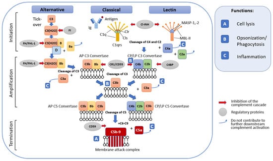
2.1. General Terminal Stage of Complement Activation

The terminal stage of the complement activation cascade is triggered by the formation of C5 convertase. C5 convertases of the alternative and classical pathways act in a similar way. They split C5 into C5b and C5a. In this case, C5a plays the role of a chemotactic and anaphylactogenic molecule, while C5b, having contacted other complement components fixed on the cell membrane, participates in the formation of the lytic membrane attack complex (MAC) [21]. In addition to pore formation and cell lysis (especially Gram-negative bacteria), the functions of MAC also include stimulatory activity in the polarization of T-helper cells and the role of soluble MAC in platelet activation [22][23].

2.2. Complement Regulatory Mechanisms

Excessive activation and dysregulation of complement and misrecognition of cellular debris or grafts can lead to various pathological conditions [21]. That is why restraining regulatory mechanisms acting at different stages of the cascade reaction are necessary. Regulatory factors can be present both in the liquid phase and on the cell surface. The major regulators of fluid phase complement include serum C1INH, C4BP, protein S, factor H, factor I, and anaphylatoxin inhibitor AI. Cell-associated regulators include CD55 (DAF), CD59 (MAC-IP or Protectin), MCP (or CD46), CR1 (or CD35), and CRIg [19]. Complement regulators act in different ways (Figure 1). For example, C1INH inactivates the C1r and C1s proteases in the C1 complex of the classical complement pathway; CD55(DAF) causes degradation of C4b2b and thus interrupts the formation of classical and lectin pathway C3 convertase, and CD59 prevents final assembly of the membrane attack complex [6][19].
A major role in the inactivation of C3bBb is played by complement factor H (CFH), a plasma glycoprotein consisting of 20 short consensus repeats (SCRs). Factor H is able to bind C3b, thereby preventing the cleavage of C5 and factor B on cell surfaces and inhibiting the formation of C3 and C5 convertases. In addition, factor H is a cofactor for factor I, a regulator that mediates the proteolytic cleavage of C3b [19]. Factor H acts both in the fluid phase and on cell surfaces by recognizing host cells directly through specific glycosaminoglycans and sialic acid or indirectly, for example through C-reactive protein (CRP) [24].
The proteins Vitronectin and Clusterin also take part in the regulation of terminal MAC assembly. Vitronectin, also known as protein S, preferentially binds to C5b-7 and interacts with C9, inhibiting its polymerization, thereby preventing the formation of a lytic pore in the membrane of the attacked cell [25]. Clusterin specifically binds to C7, the beta subunit of C8 and C9, also inhibiting the polymerization of C9 [26].
The complement system is also regulated by other systems, including the blood coagulation system, which will be discussed below.

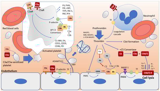
3. Interactions of the Complement System with the Blood Coagulation System

The complement system and the blood coagulation system have a common evolutionary origin, which led to the presence of common activators and inhibitors and synergy in their work, which is extremely important for the body’s fight against pathogens [27]. The function of the blood coagulation system is not only to ensure the integrity of the cardiovascular system, but also to localize the source of infection if it enters the body and stimulate inflammation mediated by the complement system. In turn, the complement system engages the blood coagulation system in the fight against pathogens. A striking example of such interaction is the syndrome of disseminated intravascular coagulation in sepsis. Thus, the functional relationship between the complement system and the blood coagulation system provides protection for the body. Dysregulation of the activity of these systems and the pathways of interaction between them can lead to the development of severe complications, including complement-associated thrombotic microangiopathies.

3.1. Blood Coagulation System

The blood coagulation system is a series of sequential proteolytic reactions in response to damage to a vessel or some other stimulus, which results in the conversion of inactive proteins—blood clotting factors—into their corresponding proteases, which ultimately leads to the formation of a fibrin clot. It is important to note that all protease complexes of the blood coagulation system depend on Ca2+ ions and anionic phospholipids (aPL). One of the key sources of aPL are platelets. In response to vascular damage, platelet activation occurs. They bind to proteins at the site of damage and are activated and then aggregate. In this case, a regulated transfer of aPL occurs from the inner layer of the bilayer cell membrane to the outer one [28][29] and the release of the contents of the granules [30], which promotes coagulation. Platelets release important hemostatic components into the circulation including platelet activating factor, platelet factor 4 (PF4), P-selectin, adenosine diphosphate, and polyphosphate. They produce local cell-stimulating effects, recruit and activate neutrophils and monocytes, and may promote further availability of aPL, an important cofactor for the assembly of all coagulation cofactor/enzyme protein complexes. There are two ways to activate the formation of thrombin, a key factor in the blood coagulation system that catalyzes the formation of a fibrin clot. These are the extrinsic tissue factor TF pathway and the intrinsic contact pathway (Figure 2).
Figure 2. The coagulation cascade and its regulators. Coagulation is initiated via the extrinsic or intrinsic pathway. The extrinsic pathway initiates by exposure of tissue factor (FIII) and assembly of the extrinsic tenase, leading to prothrombinase and ultimate thrombin (IIa) production. Thrombin (IIa) is responsible for direct fibrin clot formation, further stabilized by FXIIIa. The intrinsic pathway is initiated by FXII interacting with negatively charged surfaces, autoactivation, and via kallikrein. Activated FXIIa activates FXI (FXIa), which activates FIX (FIXa) that binds FVIIIa, forming the tenase complex, where the intrinsic pathway converges with the extrinsic pathway. There are many interactions between components within this complex system. For example, thrombin can activate FXIII, FV, and FVII. Activation of the coagulation system is finely balanced and controlled through specific regulatory mechanisms, including activity of proteins such as antithrombin (ATIII), activated protein C (APC), heparin cofactor II (HCII), and tissue factor pathway inhibitor (TFPI).

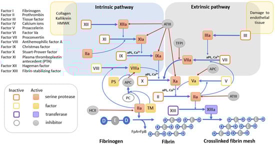
3.1.1. Tissue Factor Pathway

Tissue factor (TF) is a transmembrane glycoprotein that is constitutively expressed by subendothelial cells and serves as a high-affinity receptor cofactor for FVII. When the vessel is damaged, TF becomes available for binding to FVII. This connection ensures autoproteolytic activation of FVII with the formation of the TF/FVIIa complex, called external tenase [31]. As part of this complex, TF accelerates the FVIIa-mediated conversion of the FX factor into its active form FXa by approximately 100,000 times [32]. Extrinsic tenase TF/FVIIa can also initiate activation of FIX [33] and FVIII [34]. FIXa and FVIIIa are a protease and its cofactor, respectively, that form the FIXa/FVIIIa complex called intrinsic tenase. Like extrinsic tenase, intrinsic tenase catalyzes the activation of FX. Once the concentration of FXa exceeds the threshold required to overcome the effects of circulating endogenous anticoagulants such as tissue factor pathway inhibitor (TFPI) [35] and antithrombin (AT), FXa activates its cofactor FV [36][37]. Assembly of the Ca2+-dependent prothrombinase complex FVa/FXa on a membrane containing aPL results in the cleavage of prothrombin to produce the potent serine protease thrombin [38]. Thrombin, in turn, triggers the polymerization of soluble fibrinogen by proteolytic conversion into cross-linked fibrin by thrombin-activated FXIII to produce a stable clot.

3.1.2. The Internal Contact Activation Pathway

The internal contact activation pathway is a critical link within the thromboinflammatory network, which is closely related to complement and coagulation. The intrinsic pathway is mediated by circulating factor FXII. Factor FXII constitutively exhibits low levels of activity [39]. Upon contact with a negatively charged surface, it catalyzes its own activation and that of plasma prekallekrein to form the plasma serine protease plasma kallikrein (Pka). To do this, it recruits the high molecular weight kininogen HK as a cofactor, which significantly accelerates the process [40]. Negatively charged surfaces for FXII may include damaged blood vessels, pathogens, DNA, RNA [41], neutrophil extracellular traps (NETs) [42], anionic polysaccharides, polyphosphates, activated endothelial cells, and platelets [43][44]. When there is enough FXIIa, FXI is activated. FXIa activates the formation of the intrinsic tenase FIXa/FVIIIa and ultimately the formation of fibrin by thrombin.
Thrombin recognizes several protein substrates at once, which contribute to its own generation and, as a consequence, amplification of the coagulation reaction [45]. It activates FV and FVIII and converts FXI to FXIa, the latter promoting the generation of its own tenase activity by further activating FIX. Combined with high levels of active Fva, increased FVIIIa/FIXa tenase activity significantly increases subsequent prothrombinase Xa/Va assembly and thrombin generation. At the same time, thrombin, in combination with its cofactor thrombomodulin, activates protein C. Activated protein C, associated with protein S, cleaves factors Fva and FVIIIa, thereby preventing excessive thrombin formation. Plasma kallikrein Pka, as part of the HK/Pka/FXIIa complex, cleaves HK to produce the proinflammatory bradykinin (BK) [46]. One of the functions of bradykinin is to activate the release of tissue plasminogen activator tPA from endothelial cells [47]. The formation of a complex of tPA with plasminogen (Pg) directly on the fibrin clot causes proteolysis of plasminogen to form the corresponding serine protease, plasmin. Plasmin breaks down fibrin, restoring blood flow.

3.2. Synergism in the Functioning of the Complement System and the Blood Coagulation System as a Key Factor in Thrombus Formation in HUS

As already mentioned, the complement system and the blood coagulation system have common activators and inhibitors that coordinate their relationships and activities (Figure 3) [27]. Thus, anaphylotoxins C3a and C5a, through their receptors, activate platelets sensitized to C3a and C5a, changing their adhesive properties and stimulating aggregation. As a result of platelet activation, factors contained in α-granules are released that modulate inflammation and coagulation, including blood coagulation factors FV, FVIII and FXI, fibrinogen, vWF, P-selectin, plasminogen Pg, TFPI, PAI-1, PAF, PF4, regulatory complement factors C1-INH, FH, CD55, CD59, CD46, FD, etc. [27]. During platelet activation, P-selectin, chondroitin sulfate A, and gC1q-R receptors are exposed on their surface. C1q, through the gC1q-R receptor, on the one hand, can trigger the classical pathway of the complement system [48]; on the other hand, it causes conformational changes in the GpIIbIIIa integrin, which supports platelet adhesion and aggregation [49]. Exposed on the surface of activated platelets, P-selectin binds to its ligand C3b to ensure the assembly of alternative pathway C3 convertase, which can be enhanced by properdin [50][51].
Figure 3. Complement and coagulation crosstalk. The complement and coagulation systems have common evolutionary origins. They exhibit several interactions that can affect activation, amplification and regulatory functions in both systems. Anaphylatoxins C3a and C5a, through their receptors, activate platelets sensitized to C3a and C5a, changing their adhesive properties and stimulating aggregation. Activated platelets secret FV, FVIII and FXI, fibrinogen, vWF, P-selectin, Pg, TFPI, PAI-1, PAF, PF4, as well as regulatory complement factors C1-INH, FH, CD55, CD59, CD46, FD, etc. Exposed P-selectin binds to its ligand C3b to ensure the assembly of alternative pathway C3 convertase. C5a triggers surface expression of TF by endothelial cells, monocytes, and neutrophils. Activated by C3a and C5a, endothelial cells express vWF, which can cause platelets aggregates formation. MASP2 protease, either alone or as part of the activated MBL-MASP2 and L-FCN-MASP2 complexes, is capable of stimulating fibrinogen metabolism and fibrin clot formation by cleaving prothrombin to form thrombin. Combined activity of thrombin and C5 convertase yielded C5a and C5b(T). C5b(T) forms the C5b(T)-9 complex with significantly higher lytic activity compared to C5b-9. Thrombin may also be able to enhance the C3 convertase assembly via activation of FD or, on the other hand, induce PAR1-mediated expression of complement decay accelerating factor (DAF), a membrane complement inhibitor.
Endothelial cells also express C3a and C5a receptors [52][53][54], the interaction of which causes activation of leukocyte adhesion molecules, P-selectin, VWF, and TF [55], suppression of thrombomodulin [56], and damage to the glycocalyx [57]. In addition to endothelial cells, C5a also triggers the expression of TF on the surface of monocytes and neutrophils and the expression of the plasminogen activator inhibitor PAI-1 in mast cells. Normally, these cells express tissue plasminogen activator t-PA. An increased level of PAI-1 expression compared to t-PA leads to a change in the regulatory activity of mast cells from profibrinolytic to prothrombotic [58].
MASP2 protease, either alone or as part of the activated MBL-MASP2 and L-FCN-MASP2 complexes, is capable of stimulating fibrinogen metabolism and fibrin clot formation by cleaving prothrombin to form thrombin [59]. The MASP1 protease, although significantly lower in comparison with thrombin, has very similar activity. It cleaves the factor XIII A chain and the fibrinogen beta chain at sites identical to thrombin, but differs from thrombin in cleaving the fibrinogen alpha chain [60]. In addition, activated MASP1 can stimulate endothelial cells through the PAR4 receptor, which leads to the exposure of TF and P-selectin.
Finally, platelets, endothelial cells, and leukocytes are particularly sensitive to sublytic concentrations of C5b-9 (sC5b-9). The sC5b-9 complex induces the transbilayer flip of aPL, which is required for the activation of coagulation through the assembly of the corresponding tenases for the terminal generation of thrombin [61][62]. In turn, blood coagulation factors are also capable of activating the complement system at various stages. Thrombin, especially at high concentrations, cleaves C5 to form a fragment corresponding to anaphylatoxin C5a [63][64][65]. The combined activity of thrombin and C5 convertase leads to the formation of cleavage products C5a and C5b(T). In this case, C5b(T) forms the C5b(T)-9 complex with significantly higher lytic activity compared to C5b-9 [65], thereby enhancing the thromboinflammatory response to damage.
Thrombin can also enhance complement by acting through its own protease-activated receptors (PARs) on the plasma membrane. Thus, exposure of platelets to thrombin induces deposition of C3 and MAC [66][67]. Thrombin may also be able to indirectly enhance the assembly of C3 convertase through activation of FD [68][69]. On the other hand, thrombin induces PAR1-mediated expression of the complement accelerating factor DAF, a membrane inhibitor of the complement system [70]. Plasmin can also act as a regulator of the complement system.
Plasmin cleaves C3 and C5 to form anaphylatoxins C3a and C5a, but this does not lead to the formation of convertases, which may be due to the proteolytic activity of plasmin towards C3b and C5b [63][71][72]. Factors IXa, Xa, XIa, and PKa have been reported to cleave C5 bypassing true convertases in a C3-independent manner [63][64][73][74]. In turn, PKa can cleave FH and FB [75][76]. Thus, both systems are capable of amplifying each other’s activity and, after activation, require control by appropriate inhibitory mechanisms.
One of the key regulators of coagulation is antithrombin, the primary inhibitor of thrombin, FXa, and FIXa [77][78]. In the complement system, it inhibits MASP1 and MASP2 of the lectin pathway [79]. In turn, the complement system inhibitor C1-INH, which blocks several proteases, including C1r and C1s [80], MASP1 and MASP2 [81], and is also able to directly bind C3b, blocking the formation of C3 convertases, control the activation of PK in the hemostatic system and neutralizes PKa and FXIIa activity [82], and inhibit plasmin [83].
Tissue factor pathway inhibitor (TFPI), while an endogenous extrinsic tenase inhibitor [35][84][85], also inhibits the lectin pathway by preventing the MASP2 protease from cleaving factors C4 and C2 [86].
There is an evidence suggesting a potential coregulatory relationship between FH and FXIa [87]. FXIa degrades FH [88], reducing FH binding to endothelial cells, its cofactor activity in FI-mediated C3b inactivation, and its C3b/Bb degradation function. In turn, FH inhibits the activation of FXI by thrombin or FXIIa. A complex of FH with FXIIa was detected in plasma [89]. Evidence suggests that FH may promote ADAMTS13-mediated proteolysis of ULVWF to form monomers and dimers [90][91][92][93]. In turn, smaller forms of VWF are not only less amenable to C3b binding, but they may also act as a cofactor for C3b inactivation by FI [94].
Another regulator of the coagulation system, thrombomodulin, stimulates the production of CPB2 [95], which inactivates the proinflammatory mediators bradykinin, osteopontin, and the critical anaphylatoxins C3a and C5a [96]. Thrombomodulin also enhances FI-mediated inactivation of C3b in the presence of FH or C4b-binding protein [97][98][99].
Summarizing the above data, it can be said that the biochemical pathways of the complement system and the blood coagulation system intersect and influence each other, have common cellular targets, and common pathways of activation and regulation. The activity of both systems is coordinated through complex feedback mechanisms, and the disruption of these mechanisms can lead to various severe complications, including the development of complement-associated thrombotic microangiopathies. Thus, in 5% of patients with aHUS, various heterozygous missense mutations in the thrombomodulin gene THBD were identified [97]. These mutations were found to be associated with a reduced ability to inactivate C3b and activate CPB2. Plasminogen deficiency variants are also associated with aHUS [100]. There are proposals to expand the panel of analyzed genes in patients with aHUS to include genes encoding factors of the blood coagulation system [100].

References

  1. Aigner, C.; Schmidt, A.; Gaggl, M.; Sunder-Plassmann, G. An updated classification of thrombotic microangiopathies and treatment of complement gene variant-mediated thrombotic microangiopathy. Clin. Kidney J. 2019, 12, 333–337.
  2. Brocklebank, V.; Wood, K.M.; Kavanagh, D. Thrombotic microangiopathy and the kidney. Clin. J. Am. Soc. Nephrol. CJASN 2018, 13, 300–317.
  3. Fakhouri, F.; Fila, M.; Hummel, A.; Ribes, D.; Sellier-Leclerc, A.L.; Ville, S.; Pouteil-Noble, C.; Coindre, J.P.; Le Quintrec, M.; Rondeau, E.; et al. Eculizumab discontinuation in children and adults with atypical hemolytic-uremic syndrome: A prospective multicenter study. Blood 2021, 137, 2438–2449.
  4. Loirat, C.; Fakhouri, F.; Ariceta, G.; Besbas, N.; Bitzan, M.; Bjerre, A.; Coppo, R.; Emma, F.; Johnson, S.; Karpman, D.; et al. An international consensus approach to the management of atypical hemolytic uremic syndrome in children. Pediatr. Nephrol. 2016, 31, 15–39.
  5. Warwicker, P.; Goodship, T.H.; Donne, R.L.; Pirson, Y.; Nicholls, A.; Ward, R.M.; Turnpenny, P.; Goodship, J.A. Genetic studies into inherited and sporadic hemolytic uremic syndrome. Kidney Int. 1998, 53, 836–844.
  6. Zipfel, P.F.; Skerka, C. Complement regulators and inhibitory proteins. Nat. Rev. Immunol. 2009, 9, 729–740.
  7. Cserhalmi, M.; Papp, A.; Brandus, B.; Uzonyi, B.; Jozsi, M. Regulation of regulators: Role of the complement factor h-related proteins. Semin. Immunol. 2019, 45, 101341.
  8. Wurzner, R.; Joysey, V.C.; Lachmann, P.J. Complement component c7. Assessment of in vivo synthesis after liver transplantation reveals that hepatocytes do not synthesize the majority of human c7. J. Immunol. 1994, 152, 4624–4629.
  9. Carroll, M.C. The role of complement and complement receptors in induction and regulation of immunity. Annu. Rev. Immunol. 1998, 16, 545–568.
  10. Arbore, G.; Kemper, C.; Kolev, M. Intracellular complement—The complosome—In immune cell regulation. Mol. Immunol. 2017, 89, 2–9.
  11. Bhakdi, S.; Tranum-Jensen, J. C5b-9 assembly: Average binding of one c9 molecule to c5b-8 without poly-c9 formation generates a stable transmembrane pore. J. Immunol. 1986, 136, 2999–3005.
  12. Podack, E.R.; Tschoop, J.; Muller-Eberhard, H.J. Molecular organization of c9 within the membrane attack complex of complement. Induction of circular c9 polymerization by the c5b-8 assembly. J. Exp. Med. 1982, 156, 268–282.
  13. Ward, P.A.; Newman, L.J. A neutrophil chemotactic factor from human c′5. J. Immunol. 1969, 102, 93–99.
  14. Ogden, C.A.; Elkon, K.B. Role of complement and other innate immune mechanisms in the removal of apoptotic cells. Curr. Dir. Autoimmun. 2006, 9, 120–142.
  15. Anliker-Ort, M.; Dingemanse, J.; van den Anker, J.; Kaufmann, P. Treatment of rare inflammatory kidney diseases: Drugs targeting the terminal complement pathway. Front. Immunol. 2020, 11, 599417.
  16. Dempsey, P.W.; Allison, M.E.; Akkaraju, S.; Goodnow, C.C.; Fearon, D.T. C3d of complement as a molecular adjuvant: Bridging innate and acquired immunity. Science 1996, 271, 348–350.
  17. Chen, J.Y.; Cortes, C.; Ferreira, V.P. Properdin: A multifaceted molecule involved in inflammation and diseases. Mol. Immunol. 2018, 102, 58–72.
  18. Mukherjee, P.; Thomas, S.; Pasinetti, G.M. Complement anaphylatoxin c5a neuroprotects through regulation of glutamate receptor subunit 2 in vitro and in vivo. J. Neuroinflamm. 2008, 5, 5.
  19. Ling, M.; Murali, M. Analysis of the complement system in the clinical immunology laboratory. Clin. Lab. Med. 2019, 39, 579–590.
  20. Garred, P.; Genster, N.; Pilely, K.; Bayarri-Olmos, R.; Rosbjerg, A.; Ma, Y.J.; Skjoedt, M.O. A journey through the lectin pathway of complement-mbl and beyond. Immunol. Rev. 2016, 274, 74–97.
  21. Pouw, R.B.; Ricklin, D. Tipping the balance: Intricate roles of the complement system in disease and therapy. Semin. Immunopathol. 2021, 43, 757–771.
  22. Bossi, F.; Fischetti, F.; Pellis, V.; Bulla, R.; Ferrero, E.; Mollnes, T.E.; Regoli, D.; Tedesco, F. Platelet-activating factor and kinin-dependent vascular leakage as a novel functional activity of the soluble terminal complement complex. J. Immunol. 2004, 173, 6921–6927.
  23. Chen, Y.; Yang, C.; Jin, N.; Xie, Z.; Tang, Y.; Fei, L.; Jia, Z.; Wu, Y. Terminal complement complex c5b-9-treated human monocyte-derived dendritic cells undergo maturation and induce th1 polarization. Eur. J. Immunol. 2007, 37, 167–176.
  24. Jozsi, M.; Barlow, P.N.; Meri, S. Editorial: Function and dysfunction of complement factor h. Front. Immunol. 2021, 12, 831044.
  25. Johnson, E.; Berge, V.; Hogasen, K. Formation of the terminal complement complex on agarose beads: Further evidence that vitronectin (complement s-protein) inhibits c9 polymerization. Scand. J. Immunol. 1994, 39, 281–285.
  26. Tschopp, J.; Chonn, A.; Hertig, S.; French, L.E. Clusterin, the human apolipoprotein and complement inhibitor, binds to complement c7, c8 beta, and the b domain of c9. J. Immunol. 1993, 151, 2159–2165.
  27. Pryzdial, E.L.G.; Leatherdale, A.; Conway, E.M. Coagulation and complement: Key innate defense participants in a seamless web. Front. Immunol. 2022, 13, 918775.
  28. Lenoir, G.; D’Ambrosio, J.M.; Dieudonne, T.; Copic, A. Transport pathways that contribute to the cellular distribution of phosphatidylserine. Front. Cell Dev. Biol. 2021, 9, 737907.
  29. Protty, M.B.; Jenkins, P.V.; Collins, P.W.; O’Donnell, V.B. The role of procoagulant phospholipids on the surface of circulating blood cells in thrombosis and haemostasis. Open Biol. 2022, 12, 210318.
  30. Heijnen, H.; van der Sluijs, P. Platelet secretory behaviour: As diverse as the granules … Or not? J. Thromb. Haemost. 2015, 13, 2141–2151.
  31. Komiyama, Y.; Pedersen, A.H.; Kisiel, W. Proteolytic activation of human factors ix and x by recombinant human factor viia: Effects of calcium, phospholipids, and tissue factor. Biochemistry 1990, 29, 9418–9425.
  32. Pryzdial, E.L.G. Maestro tissue factor reaches new height. Blood 2017, 130, 1604–1605.
  33. Lu, G.; Broze, G.J., Jr.; Krishnaswamy, S. Formation of factors ixa and xa by the extrinsic pathway: Differential regulation by tissue factor pathway inhibitor and antithrombin iii. J. Biol. Chem. 2004, 279, 17241–17249.
  34. Kamikubo, Y.; Mendolicchio, G.L.; Zampolli, A.; Marchese, P.; Rothmeier, A.S.; Orje, J.N.; Gale, A.J.; Krishnaswamy, S.; Gruber, A.; Ostergaard, H.; et al. Selective factor viii activation by the tissue factor-factor viia-factor xa complex. Blood 2017, 130, 1661–1670.
  35. Mast, A.E.; Ruf, W. Regulation of coagulation by tissue factor pathway inhibitor: Implications for hemophilia therapy. J. Thromb. Haemost. JTH 2022, 20, 1290–1300.
  36. Olson, S.T.; Richard, B.; Izaguirre, G.; Schedin-Weiss, S.; Gettins, P.G. Molecular mechanisms of antithrombin-heparin regulation of blood clotting proteinases. A paradigm for understanding proteinase regulation by serpin family protein proteinase inhibitors. Biochimie 2010, 92, 1587–1596.
  37. Schuijt, T.J.; Bakhtiari, K.; Daffre, S.; Deponte, K.; Wielders, S.J.; Marquart, J.A.; Hovius, J.W.; van der Poll, T.; Fikrig, E.; Bunce, M.W.; et al. Factor xa activation of factor v is of paramount importance in initiating the coagulation system: Lessons from a tick salivary protein. Circulation 2013, 128, 254–266.
  38. Krishnaswamy, S.; Nesheim, M.E.; Pryzdial, E.L.; Mann, K.G. Assembly of prothrombinase complex. Methods Enzymol. 1993, 222, 260–280.
  39. Shamanaev, A.; Emsley, J.; Gailani, D. Proteolytic activity of contact factor zymogens. J. Thromb. Haemost. JTH 2021, 19, 330–341.
  40. Mailer, R.K.; Rangaswamy, C.; Konrath, S.; Emsley, J.; Renne, T. An update on factor xii-driven vascular inflammation. Biochim. Biophys. Acta Mol. Cell Res. 2022, 1869, 119166.
  41. Kannemeier, C.; Shibamiya, A.; Nakazawa, F.; Trusheim, H.; Ruppert, C.; Markart, P.; Song, Y.; Tzima, E.; Kennerknecht, E.; Niepmann, M.; et al. Extracellular rna constitutes a natural procoagulant cofactor in blood coagulation. Proc. Natl. Acad. Sci. USA 2007, 104, 6388–6393.
  42. von Bruhl, M.L.; Stark, K.; Steinhart, A.; Chandraratne, S.; Konrad, I.; Lorenz, M.; Khandoga, A.; Tirniceriu, A.; Coletti, R.; Kollnberger, M.; et al. Monocytes, neutrophils, and platelets cooperate to initiate and propagate venous thrombosis in mice in vivo. J. Exp. Med. 2012, 209, 819–835.
  43. Muller, F.; Mutch, N.J.; Schenk, W.A.; Smith, S.A.; Esterl, L.; Spronk, H.M.; Schmidbauer, S.; Gahl, W.A.; Morrissey, J.H.; Renne, T. Platelet polyphosphates are proinflammatory and procoagulant mediators in vivo. Cell 2009, 139, 1143–1156.
  44. Renne, T. The procoagulant and proinflammatory plasma contact system. Semin. Immunopathol. 2012, 34, 31–41.
  45. Pryzdial, E.L.G.; Lee, F.M.H.; Lin, B.H.; Carter, R.L.R.; Tegegn, T.Z.; Belletrutti, M.J. Blood coagulation dissected. Transfus. Apher. Sci. 2018, 57, 449–457.
  46. Maas, C.; Renne, T. Coagulation factor xii in thrombosis and inflammation. Blood 2018, 131, 1903–1909.
  47. Brown, N.J.; Gainer, J.V.; Stein, C.M.; Vaughan, D.E. Bradykinin stimulates tissue plasminogen activator release in human vasculature. Hypertension 1999, 33, 1431–1435.
  48. Yin, W.; Ghebrehiwet, B.; Weksler, B.; Peerschke, E.I. Classical pathway complement activation on human endothelial cells. Mol. Immunol. 2007, 44, 2228–2234.
  49. Nayak, A.; Ferluga, J.; Tsolaki, A.G.; Kishore, U. The non-classical functions of the classical complement pathway recognition subcomponent c1q. Immunol. Lett. 2010, 131, 139–150.
  50. Hamad, O.A.; Nilsson, P.H.; Wouters, D.; Lambris, J.D.; Ekdahl, K.N.; Nilsson, B. Complement component c3 binds to activated normal platelets without preceding proteolytic activation and promotes binding to complement receptor 1. J. Immunol. 2010, 184, 2686–2692.
  51. Saggu, G.; Cortes, C.; Emch, H.N.; Ramirez, G.; Worth, R.G.; Ferreira, V.P. Identification of a novel mode of complement activation on stimulated platelets mediated by properdin and c3(h2o). J. Immunol. 2013, 190, 6457–6467.
  52. Monsinjon, T.; Gasque, P.; Chan, P.; Ischenko, A.; Brady, J.J.; Fontaine, M.C. Regulation by complement c3a and c5a anaphylatoxins of cytokine production in human umbilical vein endothelial cells. FASEB J. 2003, 17, 1003–1014.
  53. Propson, N.E.; Roy, E.R.; Litvinchuk, A.; Kohl, J.; Zheng, H. Endothelial c3a receptor mediates vascular inflammation and blood-brain barrier permeability during aging. J. Clin. Investig. 2021, 131, e140966.
  54. Shivshankar, P.; Li, Y.D.; Mueller-Ortiz, S.L.; Wetsel, R.A. In response to complement anaphylatoxin peptides c3a and c5a, human vascular endothelial cells migrate and mediate the activation of b-cells and polarization of t-cells. FASEB J. 2020, 34, 7540–7560.
  55. Foreman, K.E.; Vaporciyan, A.A.; Bonish, B.K.; Jones, M.L.; Johnson, K.J.; Glovsky, M.M.; Eddy, S.M.; Ward, P.A. C5a-induced expression of p-selectin in endothelial cells. J. Clin. Investig. 1994, 94, 1147–1155.
  56. Fang, W.; Guo, Z.H.; Zhang, B.Q.; Wu, X.F.; Li, P.; Lv, F.L.; Su, L. . Zhongguo Wei Zhong Bing Ji Jiu Yi Xue Chin. Crit. Care Med. Zhongguo Weizhongbing Jijiuyixue 2009, 21, 168–171.
  57. Bongoni, A.K.; Lu, B.; McRae, J.L.; Salvaris, E.J.; Toonen, E.J.M.; Vikstrom, I.; Baz Morelli, A.; Pearse, M.J.; Cowan, P.J. Complement-mediated damage to the glycocalyx plays a role in renal ischemia-reperfusion injury in mice. Transplant. Direct 2019, 5, e341.
  58. Wojta, J.; Huber, K.; Valent, P. New aspects in thrombotic research: Complement induced switch in mast cells from a profibrinolytic to a prothrombotic phenotype. Pathophysiol. Haemost. Thromb. 2003, 33, 438–441.
  59. Gulla, K.C.; Gupta, K.; Krarup, A.; Gal, P.; Schwaeble, W.J.; Sim, R.B.; O’Connor, C.D.; Hajela, K. Activation of mannan-binding lectin-associated serine proteases leads to generation of a fibrin clot. Immunology 2010, 129, 482–495.
  60. Krarup, A.; Gulla, K.C.; Gal, P.; Hajela, K.; Sim, R.B. The action of mbl-associated serine protease 1 (masp1) on factor xiii and fibrinogen. Biochim. et Biophys. Acta 2008, 1784, 1294–1300.
  61. Hamilton, K.K.; Hattori, R.; Esmon, C.T.; Sims, P.J. Complement proteins c5b-9 induce vesiculation of the endothelial plasma membrane and expose catalytic surface for assembly of the prothrombinase enzyme complex. J. Biol. Chem. 1990, 265, 3809–3814.
  62. Wiedmer, T.; Esmon, C.T.; Sims, P.J. Complement proteins c5b-9 stimulate procoagulant activity through platelet prothrombinase. Blood 1986, 68, 875–880.
  63. Amara, U.; Flierl, M.A.; Rittirsch, D.; Klos, A.; Chen, H.; Acker, B.; Bruckner, U.B.; Nilsson, B.; Gebhard, F.; Lambris, J.D.; et al. Molecular intercommunication between the complement and coagulation systems. J. Immunol. 2010, 185, 5628–5636.
  64. Huber-Lang, M.; Sarma, J.V.; Zetoune, F.S.; Rittirsch, D.; Neff, T.A.; McGuire, S.R.; Lambris, J.D.; Warner, R.L.; Flierl, M.A.; Hoesel, L.M.; et al. Generation of c5a in the absence of c3: A new complement activation pathway. Nat. Med. 2006, 12, 682–687.
  65. Krisinger, M.J.; Goebeler, V.; Lu, Z.; Meixner, S.C.; Myles, T.; Pryzdial, E.L.; Conway, E.M. Thrombin generates previously unidentified c5 products that support the terminal complement activation pathway. Blood 2012, 120, 1717–1725.
  66. Polley, M.J.; Nachman, R. The human complement system in thrombin-mediated platelet function. J. Exp. Med. 1978, 147, 1713–1726.
  67. Polley, M.J.; Nachman, R.L. Human complement in thrombin-mediated platelet function: Uptake of the c5b-9 complex. J. Exp. Med. 1979, 150, 633–645.
  68. Dobo, J.; Szakacs, D.; Oroszlan, G.; Kortvely, E.; Kiss, B.; Boros, E.; Szasz, R.; Zavodszky, P.; Gal, P.; Pal, G. Masp-3 is the exclusive pro-factor d activator in resting blood: The lectin and the alternative complement pathways are fundamentally linked. Sci. Rep. 2016, 6, 31877.
  69. Oroszlan, G.; Kortvely, E.; Szakacs, D.; Kocsis, A.; Dammeier, S.; Zeck, A.; Ueffing, M.; Zavodszky, P.; Pal, G.; Gal, P.; et al. Masp-1 and masp-2 do not activate pro-factor d in resting human blood, whereas masp-3 is a potential activator: Kinetic analysis involving specific masp-1 and masp-2 inhibitors. J. Immunol. 2016, 196, 857–865.
  70. Lidington, E.A.; Haskard, D.O.; Mason, J.C. Induction of decay-accelerating factor by thrombin through a protease-activated receptor 1 and protein kinase c-dependent pathway protects vascular endothelial cells from complement-mediated injury. Blood 2000, 96, 2784–2792.
  71. Foley, J.H.; Walton, B.L.; Aleman, M.M.; O’Byrne, A.M.; Lei, V.; Harrasser, M.; Foley, K.A.; Wolberg, A.S.; Conway, E.M. Complement activation in arterial and venous thrombosis is mediated by plasmin. eBioMedicine 2016, 5, 175–182.
  72. Ward, P.A. A plasmin-split fragment of c′3 as a new chemotactic factor. J. Exp. Med. 1967, 126, 189–206.
  73. Mannes, M.; Dopler, A.; Zolk, O.; Lang, S.J.; Halbgebauer, R.; Hochsmann, B.; Skerra, A.; Braun, C.K.; Huber-Lang, M.; Schrezenmeier, H.; et al. Complement inhibition at the level of c3 or c5: Mechanistic reasons for ongoing terminal pathway activity. Blood 2021, 137, 443–455.
  74. Wetsel, R.A.; Kolb, W.P. Expression of c5a-like biological activities by the fifth component of human complement (c5) upon limited digestion with noncomplement enzymes without release of polypeptide fragments. J. Exp. Med. 1983, 157, 2029–2048.
  75. DiScipio, R.G. The activation of the alternative pathway c3 convertase by human plasma kallikrein. Immunology 1982, 45, 587–595.
  76. Saito, A. Plasma kallikrein is activated on dermatan sulfate and cleaves factor h. Biochem. Biophys. Res. Commun. 2008, 370, 646–650.
  77. Ellis, V.; Scully, M.; MacGregor, I.; Kakkar, V. Inhibition of human factor xa by various plasma protease inhibitors. Biochim. Biophys. Acta 1982, 701, 24–31.
  78. Osterud, B.; Miller-Andersson, M.; Abildgaard, U.; Prydz, H. The effect of antithrombin iii on the activity of the coagulation factors vii, ix and x. Thromb. Haemost. 1976, 35, 295–304.
  79. Parej, K.; Dobo, J.; Zavodszky, P.; Gal, P. The control of the complement lectin pathway activation revisited: Both c1-inhibitor and antithrombin are likely physiological inhibitors, while alpha2-macroglobulin is not. Mol. Immunol. 2013, 54, 415–422.
  80. Ziccardi, R.J. Activation of the early components of the classical complement pathway under physiologic conditions. J. Immunol. 1981, 126, 1769–1773.
  81. Rossi, V.; Cseh, S.; Bally, I.; Thielens, N.M.; Jensenius, J.C.; Arlaud, G.J. Substrate specificities of recombinant mannan-binding lectin-associated serine proteases-1 and -2. J. Biol. Chem. 2001, 276, 40880–40887.
  82. Kerr, F.K.; Thomas, A.R.; Wijeyewickrema, L.C.; Whisstock, J.C.; Boyd, S.E.; Kaiserman, D.; Matthews, A.Y.; Bird, P.I.; Thielens, N.M.; Rossi, V.; et al. Elucidation of the substrate specificity of the masp-2 protease of the lectin complement pathway and identification of the enzyme as a major physiological target of the serpin, c1-inhibitor. Mol. Immunol. 2008, 45, 670–677.
  83. Ratnoff, O.D. Some relationships among hemostasis, fibrinolytic phenomena, immunity, and the inflammatory response. Adv. Immunol. 1969, 10, 145–227.
  84. Maroney, S.A.; Ellery, P.E.; Mast, A.E. Alternatively spliced isoforms of tissue factor pathway inhibitor. Thromb. Res. 2010, 125 (Suppl. 1), S52–S56.
  85. Mast, A.E. Tissue factor pathway inhibitor: Multiple anticoagulant activities for a single protein. Arterioscler. Thromb. Vasc. Biol. 2016, 36, 9–14.
  86. Keizer, M.P.; Pouw, R.B.; Kamp, A.M.; Patiwael, S.; Marsman, G.; Hart, M.H.; Zeerleder, S.; Kuijpers, T.W.; Wouters, D. Tfpi inhibits lectin pathway of complement activation by direct interaction with masp-2. Eur. J. Immunol. 2015, 45, 544–550.
  87. Puy, C.; Pang, J.; Reitsma, S.E.; Lorentz, C.U.; Tucker, E.I.; Gailani, D.; Gruber, A.; Lupu, F.; McCarty, O.J.T. Cross-talk between the complement pathway and the contact activation system of coagulation: Activated factor xi neutralizes complement factor h. J. Immunol. 2021, 206, 1784–1792.
  88. Chen, L.J.; Liu, D.T.; Tam, P.O.; Chan, W.M.; Liu, K.; Chong, K.K.; Lam, D.S.; Pang, C.P. Association of complement factor h polymorphisms with exudative age-related macular degeneration. Mol. Vis. 2006, 12, 1536–1542.
  89. Thangaraj, S.S.; Christiansen, S.H.; Graversen, J.H.; Sidelmann, J.J.; Hansen, S.W.K.; Bygum, A.; Gram, J.B.; Palarasah, Y. Contact activation-induced complex formation between complement factor h and coagulation factor xiia. J. Thromb. Haemost. JTH 2020, 18, 876–884.
  90. Feng, S.; Liang, X.; Cruz, M.A.; Vu, H.; Zhou, Z.; Pemmaraju, N.; Dong, J.F.; Kroll, M.H.; Afshar-Kharghan, V. The interaction between factor h and von willebrand factor. PLoS ONE 2013, 8, e73715.
  91. Nolasco, L.; Nolasco, J.; Feng, S.; Afshar-Kharghan, V.; Moake, J. Human complement factor h is a reductase for large soluble von willebrand factor multimers—Brief report. Arterioscler. Thromb. Vasc. Biol. 2013, 33, 2524–2528.
  92. Rayes, J.; Roumenina, L.T.; Dimitrov, J.D.; Repesse, Y.; Ing, M.; Christophe, O.; Jokiranta, T.S.; Halbwachs-Mecarelli, L.; Borel-Derlon, A.; Kaveri, S.V.; et al. The interaction between factor h and vwf increases factor h cofactor activity and regulates vwf prothrombotic status. Blood 2014, 123, 121–125.
  93. Turner, N.; Nolasco, L.; Nolasco, J.; Sartain, S.; Moake, J. Thrombotic microangiopathies and the linkage between von willebrand factor and the alternative complement pathway. Semin. Thromb. Hemost. 2014, 40, 544–550.
  94. Feng, S.; Liang, X.; Kroll, M.H.; Chung, D.W.; Afshar-Kharghan, V. Von willebrand factor is a cofactor in complement regulation. Blood 2015, 125, 1034–1037.
  95. Bajzar, L.; Manuel, R.; Nesheim, M.E. Purification and characterization of tafi, a thrombin-activable fibrinolysis inhibitor. J. Biol. Chem. 1995, 270, 14477–14484.
  96. Campbell, W.D.; Lazoura, E.; Okada, N.; Okada, H. Inactivation of c3a and c5a octapeptides by carboxypeptidase r and carboxypeptidase n. Microbiol. Immunol. 2002, 46, 131–134.
  97. Delvaeye, M.; Noris, M.; De Vriese, A.; Esmon, C.T.; Esmon, N.L.; Ferrell, G.; Del-Favero, J.; Plaisance, S.; Claes, B.; Lambrechts, D.; et al. Thrombomodulin mutations in atypical hemolytic-uremic syndrome. N. Engl. J. Med. 2009, 361, 345–357.
  98. Heurich, M.; Preston, R.J.; O’Donnell, V.B.; Morgan, B.P.; Collins, P.W. Thrombomodulin enhances complement regulation through strong affinity interactions with factor h and c3b-factor h complex. Thromb. Res. 2016, 145, 84–92.
  99. Tateishi, K.; Imaoka, M.; Matsushita, M. Dual modulating functions of thrombomodulin in the alternative complement pathway. Biosci. Trends 2016, 10, 231–234.
  100. Bu, F.; Maga, T.; Meyer, N.C.; Wang, K.; Thomas, C.P.; Nester, C.M.; Smith, R.J. Comprehensive genetic analysis of complement and coagulation genes in atypical hemolytic uremic syndrome. J. Am. Soc. Nephrol. JASN 2014, 25, 55–64.
More
Information
Contributors MDPI registered users' name will be linked to their SciProfiles pages. To register with us, please refer to https://encyclopedia.pub/register : , , , ,
View Times: 86
Revisions: 2 times (View History)
Update Date: 22 Jan 2024
1000/1000