Your browser does not fully support modern features. Please upgrade for a smoother experience.
Submitted Successfully!
 +1 credit
Thank you for your contribution! You can also upload a video entry or images related to this topic.
For video creation, please contact our Academic Video Service.
| Version | Summary | Created by | Modification | Content Size | Created at | Operation |
|---|---|---|---|---|---|---|
| 1 | Wojciech Kazimierczak | -- | 1669 | 2024-01-17 09:04:45 | | | |
| 2 | Wendy Huang | Meta information modification | 1669 | 2024-01-17 11:46:44 | | |
Video Upload Options
We provide professional Academic Video Service to translate complex research into visually appealing presentations. Would you like to try it?
Cite
If you have any further questions, please contact Encyclopedia Editorial Office.
Kazimierczak, N.; Kazimierczak, W.; Serafin, Z.; Nowicki, P.; Nożewski, J.; Janiszewska-Olszowska, J. AI Applications in Dental Diagnostics and Cephalometric Analysis. Encyclopedia. Available online: https://encyclopedia.pub/entry/53950 (accessed on 06 June 2026).
Kazimierczak N, Kazimierczak W, Serafin Z, Nowicki P, Nożewski J, Janiszewska-Olszowska J. AI Applications in Dental Diagnostics and Cephalometric Analysis. Encyclopedia. Available at: https://encyclopedia.pub/entry/53950. Accessed June 06, 2026.
Kazimierczak, Natalia, Wojciech Kazimierczak, Zbigniew Serafin, Paweł Nowicki, Jakub Nożewski, Joanna Janiszewska-Olszowska. "AI Applications in Dental Diagnostics and Cephalometric Analysis" Encyclopedia, https://encyclopedia.pub/entry/53950 (accessed June 06, 2026).
Kazimierczak, N., Kazimierczak, W., Serafin, Z., Nowicki, P., Nożewski, J., & Janiszewska-Olszowska, J. (2024, January 17). AI Applications in Dental Diagnostics and Cephalometric Analysis. In Encyclopedia. https://encyclopedia.pub/entry/53950
Kazimierczak, Natalia, et al. "AI Applications in Dental Diagnostics and Cephalometric Analysis." Encyclopedia. Web. 17 January, 2024.
Copy Citation
Artificial intelligence (AI) describes the ability of machines to perform tasks that are classified as intelligent. AI can be classified into two main categories: symbolic AI and machine learning (ML). Symbolic AI involves structuring an algorithm in a way that is easily understandable to humans. Machine learning (ML) is the predominant paradigm in the field of AI. The advent of artificial intelligence (AI) in medicine has transformed various medical specialties, including orthodontics. AI has shown promising results in enhancing the accuracy of diagnoses, treatment planning, and predicting treatment outcomes. Its usage in orthodontic practices worldwide has increased with the availability of various AI applications and tools.
AI
cephalometric analysis
orthodontic
dental diagnostics
CBCT
1. Introduction
Artificial intelligence (AI), a term first introduced in 1955 by John McCarthy, describes the ability of machines to perform tasks that are classified as intelligent [1]. During these 70 years, there have been cycles of significant optimism associated with the development of AI, alternating with periods of failure, reductions in research funding, and pessimism [2]. The 2015 victory of AlphaGo, a Google-developed AI application, over the “GO” world champion represented a breakthrough [2]. This AI success over a human player sparked further development and interest, which was raised by the introduction of the Chat-GPT in 2022. These events served as precursors to the remarkable growth of AI applications in various fields, including everyday life and medicine [2].
AI algorithms have already proven effective in various medical specialties, surpassing the capabilities of experienced clinicians [3][4][5][6][7]. These algorithms enable the analysis, organization, visualization, and classification of healthcare data. The development of AI algorithms in medicine has gained momentum in recent years, particularly in radiology, where medical imaging accounts for approximately 85% of FDA-approved AI programs (data for 2023) [8].
In the field of diagnostic imaging, AI can be categorized into three main domains: operational AI, which enhances healthcare delivery; diagnostic AI, which aids in the interpretation of clinical images; and predictive AI, which forecasts future outcomes [9]. Currently, the primary goals of AI in diagnostic imaging are to detect and segment structures and classify pathologies [10]. AI tools can analyze images obtained from various imaging modalities, ranging from X-ray to MRI [11][12][13][14][15].
Orthodontics, with its emphasis on cephalometric analysis and pretreatment imaging, is particularly well suited for the implementation of AI. However, AI is also being utilized in orthodontics for applications beyond cephalometric analysis. The literature on the use of AI in orthodontics can be divided into five main areas: diagnosis and treatment planning, automated landmark detection and cephalometric analysis, assessment of growth and development, treatment outcome evaluation, and miscellaneous applications [16].
The number of AI companies in the healthcare industry has experienced a remarkable increase, indicating significant growth in commercial prospects for AI [9]. AI tools are no longer limited to researchers and scientists involved in research and development projects. They are now accessible through commercially available web-based products as well. In orthodontics, the adoption of AI has led to the creation of various AI-based programs, such as WeDoCeph (Audax, Ljubljana, Slovenia), WebCeph (Assemble Circle, Seoul, Republic of Korea), and CephX (ORCA Dental AI, Las Vegas, NV, USA). These systems can automatically identify cephalometric landmarks, compute angles and distances, and generate cephalometric reports with significant findings. AI programs are now easily accessible on mobile devices, making AI tools widely available and promoting equal access for all interested users. As a result, orthodontic practices and scientific researchers utilizing AI applications have notably increased. However, this accessibility has also sparked concerns about patient safety, especially when AI is used for diagnosis and treatment.
2. Dental Diagnostics
The use of medical imaging methods is essential in dental patient care because they aid in the clinical diagnosis of pathologies related to teeth and their surrounding structures [17][18][19]. Radiological methods, such as orthopantomograms (OPGs) and cone-beam computed tomography (CBCT), play crucial roles in orthodontic diagnosis, treatment planning, and monitoring [20][21][22]. However, with the increasing number of radiological examinations being performed [23], there is a need for a comprehensive tool to support the process of radiological diagnosis. In response to this demand, multimodular diagnostic systems based on AI have emerged.
One such AI-based system, developed by Diagnocat Ltd. (San Francisco, CA, USA), utilizes CNNs and provides precise and comprehensive dental diagnostics. The system enables tooth segmentation and enumeration, oral pathology diagnosis (including periapical lesions and caries), and volumetric assessment. Several scientific papers have validated the diagnostic performance of this program, demonstrating its high efficacy and accuracy [24][25][26][27][28]. A study by Orhan et al. [24] reported that the AI system achieved 92.8% accuracy in the detection of periapical lesions in CBCT images and showed no statistically significant difference in volumetric measurements compared to manual methods. Similarly, a study evaluating the diagnostic accuracy of the program for periapical lesion detection on periapical radiographs (PRs) yielded comparable results [25]. However, conflicting results have also been reported, particularly regarding the accuracy of AI in the assessment of periapical lesions in OPGs [29].
In a recent study by Ezhov (2021) [30], the overall diagnostic performance of two groups, one aided by AI and the other unaided, was compared in oral CBCT evaluation. The AI system used in this research included modules for tooth and jaw segmentation, tooth localization and enumeration, periodontitis, caries, and periapical lesion detection. The results showed that the AI system significantly improved the diagnostic capabilities of dentists, with higher sensitivity and specificity values observed in the AI-aided group than in the unaided group (sensitivity: 0.8537 vs. 0.7672; specificity: 0.9672 vs. 0.9616).
Several systematic reviews and meta-analyses have been conducted on the utilization of AI for identifying caries and periapical lucencies [31][32][33][34][35][36][37][38][39][40][41][42]. In a recent comprehensive study by Rahimi [41], the accuracy of classification models for caries detection was evaluated across 48 studies. The reported diagnostic accuracy varied significantly based on the imaging modality, ranging from 68% to 99.2%. The diagnostic odds ratio, which indicates the effectiveness of the test, also varied greatly from 2.27 to 32,767 across studies. The study concluded that deep learning models show promise for caries detection and may aid clinical workflows. One of the earliest meta-analyses conducted in 2019 on the computer-aided detection of radiolucent lesions in the maxillofacial region [33] yielded a pooled accuracy estimate of 88.75% (95% CI = 85.19–92.30); however, only four studies were included. A more recent meta-analysis by Sadr [39] included 18 studies and revealed that the pooled sensitivity and specificity were 0.925 (95% CI, 0.862–0.960) and 0.852 (95% CI, 0.810–0.885), respectively. The researchers concluded that deep learning showed highly accurate results in detecting periapical radiolucent lesions in dental radiographs. These findings suggest that multimodal AI programs may serve as first-line diagnostic aids and decision support systems, improving patient care at multiple levels. Figure 1 shows a sample of the Diagnocat report.
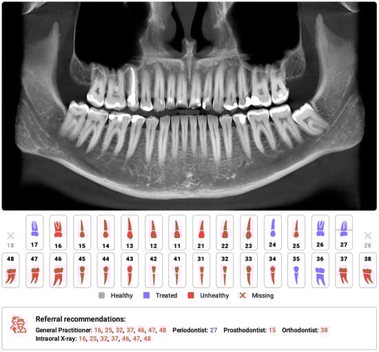
Figure 1. Part of the automatic diagnostic report from a CBCT scan was obtained prior to orthodontic treatment on a 24-year-old male. The software automatically identified the absence of teeth 18 and 28, as well as changes in the remaining teeth, primarily consisting of attrition and the presence of dental fillings. The program has recommended further consultations as necessary.
3. Cephalometric Analysis
Cephalometric analysis (CA) is an important diagnostic tool in orthodontics that has been in use since 1931 [43]. Over the years, advancements in technology have revolutionized CA by replacing manual assessments with digital software. This approach simplifies the measurement process and provides an automatic display of the analysis results. Automated CA has been shown to be more stable and repeatable than manual analyses, which rely heavily on operator-dependent landmark identification and often exhibit significant variability [44][45][46][47]. Accurate and repeatable landmark identification is crucial for reliable CA outcomes. Several studies have demonstrated the effectiveness of AI in identifying cephalometric landmarks. Although lateral radiography remains the most commonly used method in CA, recent AI advancements have sparked renewed interest in the use of cone-beam computed tomography (CBCT) [48].
The effectiveness of AI in identifying cephalometric landmarks has been studied since 1998 [49]. Numerous studies have used various automated methods and have consistently achieved high accuracy in landmark identification [46][47][50][51][52][53][54][55][56][57][58][59]. A recent study by Hwang et al. (2020) [47] concluded that automated cephalometric landmark identification can be as reliable as an experienced human reader. Similarly, Kim et al. [52], Lee et al. [58], and Dobratulin et al. [50] achieved landmark definition accuracies between 88% and 92% using AI. These researchers also found that, compared with manual methods, AI methods demonstrated greater accuracy in landmark identification and reduced the time and human labor required. In other studies conducted by Hwang et al. [46] and Yu et al. [57], the researchers found no statistically significant differences between the results of automated cephalometric analysis and those calculated via manually identified landmarks. Additionally, AI has been shown to significantly improve the workflow of practices, reducing analysis time by up to 80 times compared to manual analysis [59]. Figure 2 shows the definitions of the sampled cephalometric landmarks.
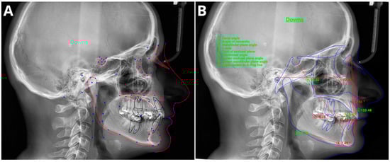
Figure 2. Sample of automatic cephalometric landmark tracings performed using CephX (A) and WebCeph (B) on an 18-year-old male. The results of Downs cephalometric analysis superimposed on tracings (B). Measurements outside the standard range marked in red and with asterix *.
The utilization of CBCT in CA was first reported in the 2000s [60], but its use has remained limited due to inefficiency and time constraints. However, recent advancements in AI have revived interest in CBCT-based CA. Several studies [61][62][63][64][65][66][67][68] have shown that AI techniques are accurate and efficient for automatically identifying and analyzing landmarks, surpassing manual approaches. Kim et al. [67] found that the repeatability of artificial neural networks was higher than achieved by human reades, while Muraev et al. [68] reported that artificial neural networks (ANNs) performed as well as or better than inexperienced readers in identifying landmarks. However, Bao et al. (2023) [69] recently revealed that manual tracing is still necessary to increase the accuracy of automated AI analysis, indicating the importance of manual supervision.
Meta-analyses have generally shown high accuracy in identifying cephalometric landmarks [70][71][72][73][74][75][76]. However, the results are strongly dependent on predefined thresholds, with lower accuracies reported at a 2 mm threshold [70][72][75]. Serafin et al. [76] conducted a study in 2023 and reported a mean difference of 2.44 mm between three-dimensional (3D) automated and manual landmarking. A meta-regression analysis indicated a significant association between publication year and mean error, suggesting that recent advances in deep learning (DL) algorithms have significantly improved landmark annotation accuracy. Overall, AI tools have shown promising results in automated cephalometric analyses, but caution is advised due to potential biases in evaluated studies [70][71][72][74].
References
- McCarthy, J.; Minsky, M.L.; Rochester, N.; Shannon, C.E. A Proposal for the Dartmouth Summer Research Project on Artificial Intelligence. AI Mag. 2006, 27, 12.
- Haenlein, M.; Kaplan, A. A Brief History of Artificial Intelligence: On the Past, Present, and Future of Artificial Intelligence. Calif. Manag. Rev. 2019, 61, 5–14.
- Schwendicke, F.; Golla, T.; Dreher, M.; Krois, J. Convolutional Neural Networks for Dental Image Diagnostics: A Scoping Review. J. Dent. 2019, 91, 103226.
- Esteva, A.; Kuprel, B.; Novoa, R.A.; Ko, J.; Swetter, S.M.; Blau, H.M.; Thrun, S. Dermatologist-Level Classification of Skin Cancer with Deep Neural Networks. Nature 2017, 542, 686.
- Gulshan, V.; Peng, L.; Coram, M.; Stumpe, M.C.; Wu, D.; Narayanaswamy, A.; Venugopalan, S.; Widner, K.; Madams, T.; Cuadros, J.; et al. Development and Validation of a Deep Learning Algorithm for Detection of Diabetic Retinopathy in Retinal Fundus Photographs. JAMA 2016, 316, 2402–2410.
- Mazurowski, M.A.; Buda, M.; Saha, A.; Bashir, M.R. Deep Learning in Radiology: An Overview of the Concepts and a Survey of the State of the Art with Focus on MRI. J. Magn. Reson. Imaging 2019, 49, 939–954.
- Saida, T.; Mori, K.; Hoshiai, S.; Sakai, M.; Urushibara, A.; Ishiguro, T.; Satoh, T.; Nakajima, T. Differentiation of Carcinosarcoma from Endometrial Carcinoma on Magnetic Resonance Imaging Using Deep Learning. Pol. J. Radiol. 2022, 87, 521–529.
- McNabb, N.K.; Christensen, E.W.; Rula, E.Y.; Coombs, L.; Dreyer, K.; Wald, C.; Treml, C. Projected Growth in FDA-Approved Artificial Intelligence Products Given Venture Capital Funding. J. Am. Coll. Radiol. 2023.
- Pianykh, O.S.; Langs, G.; Dewey, M.; Enzmann, D.R.; Herold, C.J.; Schoenberg, S.O.; Brink, J.A. Continuous Learning AI in Radiology: Implementation Principles and Early Applications. Radiology 2020, 297, 6–14.
- Milam, M.E.; Koo, C.W. The Current Status and Future of FDA-Approved Artificial Intelligence Tools in Chest Radiology in the United States. Clin. Radiol. 2023, 78, 115–122.
- Giełczyk, A.; Marciniak, A.; Tarczewska, M.; Kloska, S.M.; Harmoza, A.; Serafin, Z.; Woźniak, M. A Novel Lightweight Approach to COVID-19 Diagnostics Based on Chest X-Ray Images. J. Clin. Med. 2022, 11, 5501.
- Kloska, A.; Tarczewska, M.; Giełczyk, A.; Kloska, S.M.; Michalski, A.; Serafin, Z.; Woźniak, M. Influence of Augmentation on the Performance of the Double ResNet-Based Model for Chest X-Ray Classification. Pol. J. Radiol. 2023, 88, 244–250.
- Fujima, N.; Kamagata, K.; Ueda, D.; Fujita, S.; Fushimi, Y.; Yanagawa, M.; Ito, R.; Tsuboyama, T.; Kawamura, M.; Nakaura, T.; et al. Current State of Artificial Intelligence in Clinical Applications for Head and Neck MR Imaging. Magn. Reson. Med. Sci. 2023, 22, 401–414.
- Matsubara, K.; Ibaraki, M.; Nemoto, M.; Watabe, H.; Kimura, Y. A Review on AI in PET Imaging. Ann. Nucl. Med. 2022, 36, 133–143.
- Wang, B.; Jin, S.; Yan, Q.; Xu, H.; Luo, C.; Wei, L.; Zhao, W.; Hou, X.; Ma, W.; Xu, Z.; et al. AI-Assisted CT Imaging Analysis for COVID-19 Screening: Building and Deploying a Medical AI System. Appl. Soft Comput. 2021, 98, 106897.
- Bichu, Y.M.; Hansa, I.; Bichu, A.Y.; Premjani, P.; Flores-Mir, C.; Vaid, N.R. Applications of Artificial Intelligence and Machine Learning in Orthodontics: A Scoping Review. Prog. Orthod. 2021, 22, 18.
- Vandenberghe, B.; Jacobs, R.; Bosmans, H. Modern Dental Imaging: A Review of the Current Technology and Clinical Applications in Dental Practice. Eur. Radiol. 2010, 20, 2637–2655.
- Drage, N. Cone Beam Computed Tomography (CBCT) in General Dental Practice. Prim. Dent. J. 2018, 7, 26–30.
- Gallichan, N.; Albadri, S.; Dixon, C.; Jorgenson, K. Trends in CBCT Current Practice within Three UK Paediatric Dental Departments. Eur. Arch. Paediatr. Dent. 2020, 21, 537–542.
- Kapetanović, A.; Oosterkamp, B.C.M.; Lamberts, A.A.; Schols, J.G.J.H. Orthodontic Radiology: Development of a Clinical Practice Guideline. Radiol. Medica 2021, 126, 72–82.
- de Grauwe, A.; Ayaz, I.; Shujaat, S.; Dimitrov, S.; Gbadegbegnon, L.; Vande Vannet, B.; Jacobs, R. CBCT in Orthodontics: A Systematic Review on Justification of CBCT in a Paediatric Population Prior to Orthodontic Treatment. Eur. J. Orthod. 2019, 41, 381–389.
- Garlapati, K.; Gandhi Babu, D.B.; Chaitanya, N.C.S.K.; Guduru, H.; Rembers, A.; Soni, P. Evaluation of Preference and Purpose of Utilisation of Cone Beam Computed Tomography (CBCT) Compared to Orthopantomogram (OPG) by Dental Practitioners—A Cross-Sectional Study. Pol. J. Radiol. 2017, 82, 248–251.
- Hajem, S.; Brogårdh-Roth, S.; Nilsson, M.; Hellén-Halme, K. CBCT of Swedish Children and Adolescents at an Oral and Maxillofacial Radiology Department. A Survey of Requests and Indications. Acta Odontol. Scand. 2020, 78, 38–44.
- Orhan, K.; Bayrakdar, I.S.; Ezhov, M.; Kravtsov, A.; Özyürek, T. Evaluation of Artificial Intelligence for Detecting Periapical Pathosis on Cone-Beam Computed Tomography Scans. Int. Endod. J. 2020, 53, 680–689.
- Issa, J.; Jaber, M.; Rifai, I.; Mozdziak, P.; Kempisty, B.; Dyszkiewicz-Konwińska, M. Diagnostic Test Accuracy of Artificial Intelligence in Detecting Periapical Periodontitis on Two-Dimensional Radiographs: A Retrospective Study and Literature Review. Medicina 2023, 59, 768.
- Orhan, K.; Shamshiev, M.; Ezhov, M.; Plaksin, A.; Kurbanova, A.; Ünsal, G.; Gusarev, M.; Golitsyna, M.; Aksoy, S.; Mısırlı, M.; et al. AI-Based Automatic Segmentation of Craniomaxillofacial Anatomy from CBCT Scans for Automatic Detection of Pharyngeal Airway Evaluations in OSA Patients. Sci. Rep. 2022, 12, 11863.
- Vujanovic, T.; Jagtap, R. Evaluation of Artificial Intelligence for Automatic Tooth and Periapical Pathosis Detection on Panoramic Radiography. Oral. Surg. Oral. Med. Oral. Pathol. Oral. Radiol. 2023, 135, e51.
- Brignardello-Petersen, R. Artificial Intelligence System Seems to Be Able to Detect a High Proportion of Periapical Lesions in Cone-Beam Computed Tomographic Images. J. Am. Dent. Assoc. 2020, 151, e83.
- Zadrożny, Ł.; Regulski, P.; Brus-Sawczuk, K.; Czajkowska, M.; Parkanyi, L.; Ganz, S.; Mijiritsky, E. Artificial Intelligence Application in Assessment of Panoramic Radiographs. Diagnostics 2022, 12, 224.
- Ezhov, M.; Gusarev, M.; Golitsyna, M.; Yates, J.M.; Kushnerev, E.; Tamimi, D.; Aksoy, S.; Shumilov, E.; Sanders, A.; Orhan, K. Clinically Applicable Artificial Intelligence System for Dental Diagnosis with CBCT. Sci. Rep. 2021, 11, 15006.
- Li, S.; Liu, J.; Zhou, Z.; Zhou, Z.; Wu, X.; Li, Y.; Wang, S.; Liao, W.; Ying, S.; Zhao, Z. Artificial Intelligence for Caries and Periapical Periodontitis Detection. J. Dent. 2022, 122, 104107.
- Ramezanzade, S.; Laurentiu, T.; Bakhshandah, A.; Ibragimov, B.; Kvist, T.; Bjørndal, L.; Bjørndal, L.; Dawson, V.S.; Fransson, H.; Frisk, F.; et al. The Efficiency of Artificial Intelligence Methods for Finding Radiographic Features in Different Endodontic Treatments—A Systematic Review. Acta Odontol. Scand. 2023, 81, 422–435.
- Silva, V.K.S.; Vieira, W.A.; Bernardino, Í.M.; Travençolo, B.A.N.; Bittencourt, M.A.V.; Blumenberg, C.; Paranhos, L.R.; Galvão, H.C. Accuracy of Computer-Assisted Image Analysis in the Diagnosis of Maxillofacial Radiolucent Lesions: A Systematic Review and Meta-Analysis. Dentomaxillofacial Radiol. 2020, 49, 20190204.
- Setzer, F.C.; Shi, K.J.; Zhang, Z.; Yan, H.; Yoon, H.; Mupparapu, M.; Li, J. Artificial Intelligence for the Computer-Aided Detection of Periapical Lesions in Cone-Beam Computed Tomographic Images. J. Endod. 2020, 46, 987–993.
- Prados-Privado, M.; Villalón, J.G.; Martínez-Martínez, C.H.; Ivorra, C.; Prados-Frutos, J.C. Dental Caries Diagnosis and Detection Using Neural Networks: A Systematic Review. J. Clin. Med. 2020, 9, 3579.
- Reyes, L.T.; Knorst, J.K.; Ortiz, F.R.; Ardenghi, T.M.H. Machine Learning in the Diagnosis and Prognostic Prediction of Dental Caries: A Systematic Review. Caries Res. 2022, 56, 161–170.
- Badr, F.F.; Jadu, F.M. Performance of Artificial Intelligence Using Oral and Maxillofacial CBCT Images: A Systematic Review and Meta-Analysis. Niger. J. Clin. Pract. 2022, 25, 1918–1927.
- Khanagar, S.B.; Alfouzan, K.; Awawdeh, M.; Alkadi, L.; Albalawi, F.; Alfadley, A. Application and Performance of Artificial Intelligence Technology in Detection, Diagnosis and Prediction of Dental Caries (DC)—A Systematic Review. Diagnostics 2022, 12, 1083.
- Sadr, S.; Mohammad-Rahimi, H.; Motamedian, S.R.; Zahedrozegar, S.; Motie, P.; Vinayahalingam, S.; Dianat, O.; Nosrat, A. Deep Learning for Detection of Periapical Radiolucent Lesions: A Systematic Review and Meta-Analysis of Diagnostic Test Accuracy. J. Endod. 2023, 49, 248–261.e3.
- Abesi, F.; Maleki, M.; Zamani, M. Diagnostic Performance of Artificial Intelligence Using Cone-Beam Computed Tomography Imaging of the Oral and Maxillofacial Region: A Scoping Review and Meta-Analysis. Imaging Sci. Dent. 2023, 53, 101–108.
- Mohammad-Rahimi, H.; Motamedian, S.R.; Rohban, M.H.; Krois, J.; Uribe, S.E.; Mahmoudinia, E.; Rokhshad, R.; Nadimi, M.; Schwendicke, F. Deep Learning for Caries Detection: A Systematic Review. J. Dent. 2022, 122, 104115.
- Abesi, F.; Jamali, A.S.; Zamani, M. Accuracy of Artificial Intelligence in the Detection and Segmentation of Oral and Maxillofacial Structures Using Cone-Beam Computed Tomography Images: A Systematic Review and Meta-Analysis. Pol. J. Radiol. 2023, 88, 256–263.
- Leonardi, R.; Giordano, D.; Maiorana, F.; Spampinato, C. Automatic Cephalometric Analysis: A Systematic Review. Angle Orthod. 2008, 78, 145–151.
- Chen, Y.J.; Chen, S.K.; Yao, J.C.C.; Chang, H.F. The Effects of Differences in Landmark Identification on the Cephalometric Measurements in Traditional versus Digitized Cephalometry. Angle Orthod. 2004, 74, 155–161.
- Dias Da Silveira, H.L.; Dias Silveira, H.E. Reproducibility of Cephalometric Measurements Made by Three Radiology Clinics. Angle Orthod. 2006, 76, 394–399.
- Hwang, H.-W.; Moon, J.-H.; Kim, M.-G.; Donatelli, R.E.; Lee, S.-J. Evaluation of Automated Cephalometric Analysis Based on the Latest Deep Learning Method. Angle Orthod. 2021, 91, 329–335.
- Hwang, H.W.; Park, J.H.; Moon, J.H.; Yu, Y.; Kim, H.; Her, S.B.; Srinivasan, G.; Aljanabi, M.N.A.; Donatelli, R.E.; Lee, S.J. Automated Identification of Cephalometric Landmarks: Part 2-Might It Be Better than Human? Angle Orthod. 2020, 90, 69–76.
- Chung, E.J.; Yang, B.E.; Park, I.Y.; Yi, S.; On, S.W.; Kim, Y.H.; Kang, S.H.; Byun, S.H. Effectiveness of Cone-Beam Computed Tomography-Generated Cephalograms Using Artificial Intelligence Cephalometric Analysis. Sci. Rep. 2022, 12, 20585.
- Rudolph, D.J.; Sinclair, P.M.; Coggins, J.M. Automatic Computerized Radiographic Identification of Cephalometric Landmarks. Am. J. Orthod. Dentofac. Orthop. 1998, 113, 173–179.
- Dobratulin, K.; Gaidel, A.; Kapishnikov, A.; Ivleva, A.; Aupova, I.; Zelter, P. The Efficiency of Deep Learning Algorithms for Detecting Anatomical Reference Points on Radiological Images of the Head Profile. In Proceedings of the ITNT 2020–6th IEEE International Conference on Information Technology and Nanotechnology, Samara, Russia, 26–29 May 2020.
- Park, J.H.; Hwang, H.W.; Moon, J.H.; Yu, Y.; Kim, H.; Her, S.B.; Srinivasan, G.; Aljanabi, M.N.A.; Donatelli, R.E.; Lee, S.J. Automated Identification of Cephalometric Landmarks: Part 1—Comparisons between the Latest Deep-Learning Methods YOLOV3 and SSD. Angle Orthod. 2019, 89, 903–909.
- Kim, H.; Shim, E.; Park, J.; Kim, Y.J.; Lee, U.; Kim, Y. Web-Based Fully Automated Cephalometric Analysis by Deep Learning. Comput. Methods Programs Biomed. 2020, 194, 105513.
- Grau, V.; Alcañiz, M.; Juan, M.C.; Monserrat, C.; Knoll, C. Automatic Localization of Cephalometric Landmarks. J. Biomed. Inf. 2001, 34, 146–156.
- Yao, J.; Zeng, W.; He, T.; Zhou, S.; Zhang, Y.; Guo, J.; Tang, W. Automatic Localization of Cephalometric Landmarks Based on Convolutional Neural Network. Am. J. Orthod. Dentofac. Orthop. 2022, 161, e250–e259.
- Nishimoto, S.; Sotsuka, Y.; Kawai, K.; Ishise, H.; Kakibuchi, M. Personal Computer-Based Cephalometric Landmark Detection with Deep Learning, Using Cephalograms on the Internet. J. Craniofacial Surg. 2019, 30, 91–95.
- Kunz, F.; Stellzig-Eisenhauer, A.; Zeman, F.; Boldt, J. Artificial Intelligence in Orthodontics: Evaluation of a Fully Automated Cephalometric Analysis Using a Customized Convolutional Neural Network. J. Orofac. Orthop. 2020, 81, 52–68.
- Yu, H.J.; Cho, S.R.; Kim, M.J.; Kim, W.H.; Kim, J.W.; Choi, J. Automated Skeletal Classification with Lateral Cephalometry Based on Artificial Intelligence. J. Dent. Res. 2020, 99, 249–256.
- Lee, J.H.; Yu, H.J.; Kim, M.J.; Kim, J.W.; Choi, J. Automated Cephalometric Landmark Detection with Confidence Regions Using Bayesian Convolutional Neural Networks. BMC Oral Health 2020, 20, 270.
- Nishimoto, S. Locating Cephalometric Landmarks with Multi-Phase Deep Learning. J. Dent. Health Oral. Res. 2023, 4, 1–13.
- Palomo, J.M.; Yang, C.Y.; Hans, M.G. Clinical Application of Three-Dimensional Craniofacial Imaging in Orthodontics. J. Med. Sci. 2005, 25, 269.
- Kazimierczak, N.; Kazimierczak, W.; Serafin, Z.; Nowicki, P.; Lemanowicz, A.; Nadolska, K.; Janiszewska-Olszowska, J. Correlation Analysis of Nasal Septum Deviation and Results of AI-Driven Automated 3D Cephalometric Analysis. J. Clin. Med. 2023, 12, 6621.
- Ed-Dhahraouy, M.; Riri, H.; Ezzahmouly, M.; Bourzgui, F.; El Moutaoukkil, A. A New Methodology for Automatic Detection of Reference Points in 3D Cephalometry: A Pilot Study. Int. Orthod. 2018, 16, 328–337.
- Gupta, A.; Kharbanda, O.P.; Sardana, V.; Balachandran, R.; Sardana, H.K. A Knowledge-Based Algorithm for Automatic Detection of Cephalometric Landmarks on CBCT Images. Int. J. Comput. Assist. Radiol. Surg. 2015, 10, 1737–1752.
- Ma, Q.; Kobayashi, E.; Fan, B.; Nakagawa, K.; Sakuma, I.; Masamune, K.; Suenaga, H. Automatic 3D Landmarking Model Using Patch-Based Deep Neural Networks for CT Image of Oral and Maxillofacial Surgery. Int. J. Med. Robot. Comput. Assist. Surg. 2020, 16, e2093.
- Montúfar, J.; Romero, M.; Scougall-Vilchis, R.J. Hybrid Approach for Automatic Cephalometric Landmark Annotation on Cone-Beam Computed Tomography Volumes. Am. J. Orthod. Dentofac. Orthop. 2018, 154, 140–150.
- Gupta, A.; Kharbanda, O.P.; Sardana, V.; Balachandran, R.; Sardana, H.K. Accuracy of 3D Cephalometric Measurements Based on an Automatic Knowledge-Based Landmark Detection Algorithm. Int. J. Comput. Assist. Radiol. Surg. 2016, 11, 1297–1309.
- Kim, M.-J.; Liu, Y.; Oh, S.H.; Ahn, H.-W.; Kim, S.-H.; Nelson, G. Evaluation of a Multi-Stage Convolutional Neural Network-Based Fully Automated Landmark Identification System Using Cone-Beam Computed Tomographysynthesized Posteroanterior Cephalometric Images. Korean J. Orthod. 2021, 51, 77–85.
- Muraev, A.A.; Tsai, P.; Kibardin, I.; Oborotistov, N.; Shirayeva, T.; Ivanov, S.; Ivanov, S.; Guseynov, N.; Aleshina, O.; Bosykh, Y.; et al. Frontal Cephalometric Landmarking: Humans vs Artificial Neural Networks. Int. J. Comput. Dent. 2020, 23, 139–148.
- Bao, H.; Zhang, K.; Yu, C.; Li, H.; Cao, D.; Shu, H.; Liu, L.; Yan, B. Evaluating the Accuracy of Automated Cephalometric Analysis Based on Artificial Intelligence. BMC Oral. Health 2023, 23, 191.
- de Queiroz Tavares Borges Mesquita, G.; Vieira, W.A.; Vidigal, M.T.C.; Travençolo, B.A.N.; Beaini, T.L.; Spin-Neto, R.; Paranhos, L.R.; de Brito Júnior, R.B. Artificial Intelligence for Detecting Cephalometric Landmarks: A Systematic Review and Meta-Analysis. J. Digit. Imaging 2023, 36, 1158–1179.
- Schwendicke, F.; Chaurasia, A.; Arsiwala, L.; Lee, J.H.; Elhennawy, K.; Jost-Brinkmann, P.G.; Demarco, F.; Krois, J. Deep Learning for Cephalometric Landmark Detection: Systematic Review and Meta-Analysis. Clin. Oral. Investig. 2021, 25, 4299–4309.
- Londono, J.; Ghasemi, S.; Hussain Shah, A.; Fahimipour, A.; Ghadimi, N.; Hashemi, S.; Khurshid, Z.; Dashti, M. Evaluation of Deep Learning and Convolutional Neural Network Algorithms Accuracy for Detecting and Predicting Anatomical Landmarks on 2D Lateral Cephalometric Images: A Systematic Review and Meta-Analysis. Saudi Dent. J. 2023, 35, 487–497.
- Jihed, M.; Dallel, I.; Tobji, S.; Amor, A. Ben The Impact of Artificial Intelligence on Contemporary Orthodontic Treatment Planning—A Systematic Review and Meta-Analysis. Sch. J. Dent. Sci. 2022, 9, 70–87.
- Junaid, N.; Khan, N.; Ahmed, N.; Abbasi, M.S.; Das, G.; Maqsood, A.; Ahmed, A.R.; Marya, A.; Alam, M.K.; Heboyan, A. Development, Application, and Performance of Artificial Intelligence in Cephalometric Landmark Identification and Diagnosis: A Systematic Review. Healthcare 2022, 10, 2454.
- Rauniyar, S.; Jena, S.; Sahoo, N.; Mohanty, P.; Dash, B.P. Artificial Intelligence and Machine Learning for Automated Cephalometric Landmark Identification: A Meta-Analysis Previewed by a Systematic Review. Cureus 2023, 15, e40934.
- Serafin, M.; Baldini, B.; Cabitza, F.; Carrafiello, G.; Baselli, G.; Del Fabbro, M.; Sforza, C.; Caprioglio, A.; Tartaglia, G.M. Accuracy of Automated 3D Cephalometric Landmarks by Deep Learning Algorithms: Systematic Review and Meta-Analysis. Radiol. Medica 2023, 128, 544–555.
More
Information
Subjects:
Dentistry, Oral Surgery & Medicine
Contributors
MDPI registered users' name will be linked to their SciProfiles pages. To register with us, please refer to https://encyclopedia.pub/register
:
View Times:
1.3K
Revisions:
2 times
(View History)
Update Date:
17 Jan 2024
Table of Contents


Notice
You are not a member of the advisory board for this topic. If you want to update advisory board member profile, please contact office@encyclopedia.pub.
OK
Confirm
Only members of the Encyclopedia advisory board for this topic are allowed to note entries. Would you like to become an advisory board member of the Encyclopedia?
Yes
No
${ textCharacter }/${ maxCharacter }
Submit
Cancel
Back
Comments
${ item }
|
${ item.createdUser.fullName }
${ item.createdAt }
${ item.vote }
${ item.reply }
Delete
${ reply.createdUser.fullName }
${ reply.createdAt }
${ reply.vote }
Delete
There is no reply to this comment~
${ item.replyTextCharacter }/${ item.replyMaxCharacter }
Submit
Cancel
More
No more~
There is no comment~
${ textCharacter }/${ maxCharacter }
Submit
Cancel
${ selectedItem.replyTextCharacter }/${ selectedItem.replyMaxCharacter }
Submit
Cancel
Confirm
Are you sure to Delete?
Yes
No