Submitted Successfully!
To reward your contribution, here is a gift for you: A free trial for our video production service.
Thank you for your contribution! You can also upload a video entry or images related to this topic.
Version Summary Created by Modification Content Size Created at Operation
1 -- 3755 2024-01-17 02:23:55 |
2 Reference format revised. + 3 word(s) 3758 2024-01-17 03:02:20 |

Video Upload Options

Do you have a full video?

Confirm

Are you sure to Delete?
Cite
If you have any further questions, please contact Encyclopedia Editorial Office.
Malla, R.; Kundrapu, D.B.; Bhamidipati, P.; Nagaraju, G.P.; Muniraj, N. Role of Yes-Associated Protein in Breast Cancer. Encyclopedia. Available online: https://encyclopedia.pub/entry/53928 (accessed on 07 July 2024).
Malla R, Kundrapu DB, Bhamidipati P, Nagaraju GP, Muniraj N. Role of Yes-Associated Protein in Breast Cancer. Encyclopedia. Available at: https://encyclopedia.pub/entry/53928. Accessed July 07, 2024.
Malla, Ramarao, Durga Bhavani Kundrapu, Priyamvada Bhamidipati, Ganji Purnachandra Nagaraju, Nethaji Muniraj. "Role of Yes-Associated Protein in Breast Cancer" Encyclopedia, https://encyclopedia.pub/entry/53928 (accessed July 07, 2024).
Malla, R., Kundrapu, D.B., Bhamidipati, P., Nagaraju, G.P., & Muniraj, N. (2024, January 17). Role of Yes-Associated Protein in Breast Cancer. In Encyclopedia. https://encyclopedia.pub/entry/53928
Malla, Ramarao, et al. "Role of Yes-Associated Protein in Breast Cancer." Encyclopedia. Web. 17 January, 2024.
Role of Yes-Associated Protein in Breast Cancer
Edit

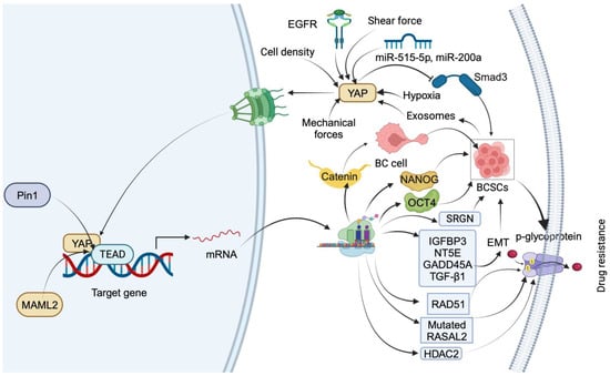
The hippo/yes-associated protein (YAP) protein is a critical oncogenic mediator within the Hippo signaling pathway and has been implicated in various cancer types. In breast cancer, it frequently becomes activated, thereby contributing to developing drug-resistance mechanisms. Studies have underscored the intricate interplay between YAP and ferroptosis within the breast tumor microenvironment. YAP exerts a negative regulatory effect on ferroptosis, promoting cancer cell survival and drug resistance.

breast cancer drug resistance ferroptosis Hippo-YAP pathway natural compounds

1. Introduction

Breast cancer (BC) represents a complex and multifaceted challenge in the realm of clinical oncology. It is characterized by intrinsic heterogeneity, encompassing diverse molecular subtypes such as luminal (luminal A and B) and non-luminal (human epidermal growth factor receptor 2 (HER2)-enriched and basal-like) BCs [1]. The basal-like subtype is characterized by its aggressive features, including a high histological grade and mutations in the TP53 gene. Due to the absence of ER, PR expression, and lack of HER2 enrichment, it is often referred to as triple-negative breast cancer (TNBC). This subtype is strongly associated with the inhibition of BRCA1 function and largely affects younger women. Also, it frequently exhibits overexpression of the EGFR gene [2]. Patients with TNBC exhibit poor survival and an enhanced probability of metastases, particularly to the distant lymph nodes, lungs, and brain [3]. Studies describe the complexity and multifaceted nature of TNBC, underscoring the imperative for a comprehensive understanding of the underlying mechanisms of resistance to established treatment modalities [4]. In recent years, considerable research has been devoted to unraveling the mechanisms underlying chemoresistance in TNBC.
The emergence of TNBC chemoresistance is increasingly recognized as a complex process that hinges on intricate interactions within the TME. These interactions involve drug efflux mechanisms in bulk tumor cells and cancer stem cells (CSCs), all of which are orchestrated by alterations in multiple signaling pathways [5]. Accumulating evidence underscores that drug resistance in cancer relies on intricate bidirectional interactions between tumor cells and the TME. This interplay involves various components that form a complex three-dimensional network, with a central role attributed to the Hippo-Yes-associated protein (YAP) pathway [6][7]. Furthermore, the TME contributes to drug resistance by modulating ferroptosis, a regulated cell-death mechanism in TNBC [8]. Consequently, comprehending the drug resistance mediated by the Hippo-YAP pathway within the TME, particularly through its impact on ferroptosis, could provide valuable insights for advancing the treatment of drug-resistant TNBC.

2. Hippo Signaling Pathway

The Hippo pathway, a key regulator of tissue growth and organ size, was originally reported in Drosophila melanogaster. It is conserved evolutionarily from Protista to eukaryotes. Its core consists of both tumor-suppressor and oncogenic proteins. The mammalian Hippo pathway is constituted mainly of tumor suppressor core kinases such as microtubule-associated serine/threonine-protein kinase 1/2 (MST1/2) and large tumor suppressor kinase 1/2 (LATS1/2) along with oncogenic downstream mediators YAP as well as TAZ. In addition, Salvador 1 or WW45, TEA domain family members (TEAD1-4), and Vestigial-like family member 4 are also core components of the Hippo pathway.
The components of the Hippo pathway are associated with diverse functions, including regulation of transcriptional programs of cell survival and migration as well as self-renewal and differentiation mechanisms. The Hippo pathway is controlled by various intrinsic as well as extrinsic cues like cell–cell contact, stress, cell polarity, mechanical forces, and hormonal factors [9]. Several upstream regulators of core kinases control the components of the Hippo pathway. When the Hippo pathway is activated by upstream activators, MST1/2 is activated and phosphorylates LATS1/2 by complexing with SAV1. Subsequently, phosphorylated LATS1/2 inactivates YAP by phosphorylating serine residue at 127 and TAZ at 66, 89, 117, and 311 and their subsequent binding to TEADs. This phosphorylation promotes the sequestration of YAP/TAZ in the cytosol by enhancing the binding with the 14-3-3 protein. A proteosome-dependent mechanism degrades the phosphorylated YAP/TAZ by interacting with β-TrCP. For example, Merlin (Neurofibromain 2), a tumor suppressor [10]; G-protein coupled receptor [11]; miRNAs [12]; and adherens, tight junctions, and signaling pathways (MAPK, Notch, and Wnt/β-catenin) [13] inhibit the activity of YAP/TAZ by promoting phosphorylation by activating the kinases.

3. YAP Protein and Its Impact on Breast Cancer

YAP expression was reported in different subtypes of BC. An immunohistochemical assessment of YAP expression in BC tissues, along with its correlation with clinicopathological parameters and patient survival, indicates predominant localization within tumor cell nuclei. Notably, YAP expression correlates with PR status and the luminal A subtype. Kaplan–Meier (KM) analyses demonstrate a favorable association of YAP expression with disease-free survival (DFS) and overall survival (OS) in luminal A BC patients. Additionally, a positive association with favorable DFS is observed in patients with invasive ductal carcinoma, luminal B (HER2−), and luminal B (HER2+) BCs [14]. These findings underscore the potential prognostic value of YAP expression in BC, particularly within specific subtypes. In an independent histochemical investigation, higher levels of cytoplasmic YAP and pYAP were observed in HER-2-type BC. Notably, stromal expressions of YAP and pYAP were elevated in luminal B and HER-2-type BCs. Univariate analysis revealed an association between nuclear YAP expression in tumor cells and a shortened OS [15].
Human protein is encoded by the YAP gene, which is found on chromosome 11q22 [16]. It was initially reported in chickens as a copartner of yes protein tyrosine kinase. The human form of YAP mediates oncogenesis via interaction with regulators and mediators of oncogenesis through different domains. The SH3 domain of the yes protein tyrosine kinase interacts with YAP in the proline-rich region (PVKQPPPLAP) [17]. A conserved module, namely, the WW domain with two well-preserved and constantly positioned tryptophan residues, was reported in various species of YAP. Such motifs are required for the interaction of YAP with transcription factors having the PPXY motif. In addition, the N-terminal region encompasses a domain for TEAD binding as well as 14-3-3 binding with HXRXXS motifs [18]. The phosphorylation of Ser 127 within the 14-3-3 binding domain develops the 14-3-3 protein binding site and is associated with its cytoplasmic sequestration and inactivation. 
YAP activation and its subsequent translocation into the nucleus are controlled by two distinct pathways: the canonical Hippo kinase pathway and the noncanonical mechanotransduction pathway [19]. In the Hippo kinase pathway, YAP nuclear accumulation is reduced through its phosphorylation by the Hippo kinases. On the other hand, the mechanotransduction pathway relies on the integrity and force transmission of the actomyosin cytoskeleton. These processes are regulated by a range of oncogenes and tumor-suppressor genes that can either promote or hinder the development of breast cancer.

3.1. YAP Mediates Drug Resistance by Inducing Stemness in Breast Cancer Cells

YAP is generally considered a stemness factor due to its role in the overgrowth of organs during embryonic development, including the breast. Its overexpression was confirmed to be critical for the expression of stemness-related genes as well as stemlike signatures and induction of invasiveness in BC patients. Additionally, overexpression of YAP conferred poor outcomes in TNBC patients [20]. Abnormal activity of YAP supports the maintenance of self-renewal by activating TGF-β/BMP pathways, induction of anticancer drug resistance by enhancing the activity of multidrug-resistance-associated (MDR) proteins, and dissemination of cells to facilitate metastasis by activating EMT-related genes [21].
YAP promotes drug resistance through various downstream mediators (Figure 1). The nuclear entry of YAP is controlled by the Hippo pathway via various mechanical and molecular cues. YAP translocates to the nucleus upon activation by receptor tyrosine kinase (Met). The interaction between cytoplasmic EGFR and salt-inducible kinase 2 (SIK2) inhibits the interaction between LATS1 and MST1, thereby facilitating the nuclear translocation of YAP [22]. Furthermore, oncogenic microRNAs, such as miR-515-5p and miR-200a, facilitate nuclear entry by negatively regulating Hippo kinases, while tumor-suppressor microRNAs, including miR-30a and miR-375, impede nuclear entry by positively regulating Hippo kinases [23].
Figure 1. Summary of downstream targets of YAP that mediate drug-resistance mechanisms in TNBC. YAP nuclear translocation is facilitated by EGFR, miR-515-5p, miR-200a, mechanical forces, low shear force, hypoxia, and cell density. In the nucleus, YAP regulates BC cell transdifferentiation into BCSCs by inducing β-catenin expression through interaction with TEAD. YAP induces stemness by transcriptionally upregulating OCT4 and NANOG through interaction with their promoter regions. YAP1 promotes BCSC self-renewal by impeding Smad3 signaling. Exosomes from BC stem cells facilitate drug resistance by upregulating YAP. Pin1 increases drug resistance by stabilizing the YAP complex. YAP1 induces drug resistance and stemness by upregulating IGFBP3, NT5E, GADD45A, TGF-β1, RAD51, and RASAL2 and inducing p-glycoprotein-dependent drug efflux. YAP also triggers drug resistance and stemness by promoting the transcription of HDAC2.
In the nucleus, YAP regulates the transdifferentiation of luminal BC cells into BCSCs. YAP induces the expression of β-catenin by interacting with its regulatory elements along with TEAD4. However, the pharmacological intervention of YAP reduces the BCSCs by delaying the transdifferentiation by targeting β-catenin [24]. Another study reported that YAP mediates FOXM1-dependent induction of stemness in TNBC cells. Mechanistically, FOXOM1 promotes nuclear translocation of YAP by diminishing its phosphorylation. YAP transcriptionally upregulates OCT4 and NANOG in the nucleus by interacting with their promoter regions. However, FOXM1 gene silencing reduces the transcriptional activity of YAP by retaining in the cytosol and increasing the proteasomal degradation via phosphorylation [25]. YAP1 promotes the self-renewal of BCSCs by impeding Smad3 signaling. Mechanistically, ectopic expression of YAP enhances the colony formation as well as self-renewal of BCSCs. 
Exosomes derived from BC stem cells facilitate paclitaxel (PTX) resistance by promoting Hippo dysregulation via upregulating YAP [26]. Pin 1, a positive regulator of the Hippo pathway, increases PTX resistance by stabilizing the YAP complex in the nucleus of BC cells [27]. YAP1 also mediates cisplatin resistance in SMARCA2-depleted TNBC cells by upregulating IGFBP3, NT5E, GADD45A, and TGF-β1. However, YAP/TEAD cascade inhibitors, VP and CA3, sensitize SMARCA2 KO TNBC cells by reducing the expression of IGFBP3, NT5E, GADD45A, and TGF-β1 and the EMT pathway [28]. In TNBC cells, cisplatin treatment increases the translocation of YAP into the nucleus by reducing its phosphorylation in an autophagy-dependent manner. However, combining cisplatin and HCQ, an autophagy inhibitor, increases the YAP target gene expression by increasing phosphorylated YAP. Further, silencing of the YAP gene increases the cytotoxicity in TNBC cells by inducing apoptosis. These results indicate that cisplatin-dependent autophagy protects TNBC cells from apoptosis by promoting the nuclear translocation of YAP [29]. In TNBC, YAP/TAZ mediate VEGF- and NRP2-dependent cisplatin resistance by increasing the expression of RAD51, a key mediator of homologous recombination. YAP/TAZ induce the transcription of the RAD51 gene by facilitating the interaction of TEAD4 at the promoter region of RAD51 [30]. The transcriptional profiling of TNBC tumors from a phase 2 clinical trial of platinum chemotherapy predicted that high levels of RASAL2 confer platinum drug resistance in association with transcriptional regulator YAP. Mechanistically, activated YAP transcriptionally enhances the expression of RASAL2 in chemorefractory patient-derived TNBC models [31]. YAP mediates doxorubicin (DOX) resistance in MDA-MB 231 and MDA-MB 468 cells by reducing the uptake of the DOX and inducing p-glycoprotein-dependent drug efflux [32]. YAP mediates serglycin (SRGN) and promotes 5-fluorouracil (5-FU) resistance in BC cells by maintaining stemness. The silencing of YAP sensitizes BC cells to drug resistance induced by the overexpression of SRGN. 

3.2. Ferroptosis Factors and Regulation Mechanisms in Breast Cancer

Ferroptosis is a newly discovered form of programmed cell death, characterized by iron-dependent lipid peroxidation and the accumulation of reactive oxygen species (ROS). It is a critical junction point that links cancer-acquired drug resistance and immune evasion. Several mechanisms regulate the sensitivity of cancer cells to ferroptosis by modulating metabolic pathways that control ferroptosis and reshape the TME. These changes lead to the formation of an immunosuppressive environment that promotes tumor growth and progression [33].
The mechanism of ferroptosis involves the release of interferon γ (IFNγ) by CD8+ T cells, which leads to the downregulation of two subunits of system Xc-, namely, SLC3A2 and SLC7A11. This, in turn, inhibits cystine uptake by tumor cells, resulting in the depletion of cysteine and glutathione and impaired GPX4 activity, ultimately promoting ferroptosis in tumor cells. However, it is worth noting that the high-fat environment within the TME upregulates the expression of the fatty acid transporter CD36, which increases the sensitivity of CD8+ T cells to ferroptosis and weakens their antitumor effect [34].
HLF, a newly discovered oncoprotein of TNBC, has been shown to be regulated by TGF-β1 secreted by TAMs. The mechanism involves HLF activating gamma-glutamyltransferase 1 (GGT1) to enhance resistance to ferroptosis, thereby promoting TNBC cell proliferation, metastasis, and resistance to cisplatin.

3.3. YAP Mediates Resistance to Ferroptosis in Breast Cancer

Emerging evidence suggests that ferroptosis plays a role in the pathogenesis and treatment of TNBC. A study demonstrated the ferroptosis heterogeneity in TNBC [35]. It exhibits diverse phenotypes in terms of ferroptosis-related metabolites and metabolic pathways [36]. A recent investigation revealed that proferroptotic stimuli, including the inhibition of the lipid hydroperoxidase GPX4 and detachment from the extracellular matrix, result in the upregulation of prominin2, a pentaspanin protein associated with the regulation of lipid dynamics, thereby promoting TNBC [37].
The intracellular NF2 (Merlin) is activated by E-cadherin-mediated interactions in epithelial cells, suppressing ferroptosis. However, inhibiting this signaling axis allows the YAP to support ferroptosis by increasing the expression of ACSL4 and TFRC. This mechanism sheds light on how intercellular interactions and intracellular NF2-YAP signaling play a crucial role in determining ferroptosis [38]. In BC, mutations in the NF2 gene, leading to loss of function of Merlin, were implicated in the initiation and progression of the disease [39]. Merlin is involved in cell–cell adhesion, cytoskeleton organization, and intracellular signaling pathways, and its dysregulation can contribute to increased cell motility, invasiveness, and resistance to apoptosis [40][41]. The presence of NF2/Merlin mutations in breast cancer has been associated with more aggressive tumor phenotypes and poorer clinical outcomes. BCs harboring NF2 mutations could exhibit increased metastatic potential and resistance to conventional therapies [42].
Ferroptosis has a protective effect on breast cancer (BC) cells [43]. Recent studies have shown that TNBC cells are more sensitive to ferroptosis induction compared to other breast cancer subtypes. This sensitivity is thought to be due to higher iron uptake as well as lipid metabolism in TNBC cells, which are necessary for ferroptosis to occur. In addition, some studies have shown that TNBC tumors have a higher expression of genes implicated in ferroptosis than other BC subtypes.

3.4. YAP in the Control of Metabolic and Oxidative Stress in Breast Cancer

YAP plays a significant role in regulating cellular responses to metabolic and oxidative stress in BC. In conditions of metabolic stress, YAP orchestrates adaptive mechanisms to enhance nutrient utilization and energy production. It promotes glycolysis, the Warburg effect, and lipid metabolism, facilitating cancer cell survival and growth even under nutrient-deprived conditions [44]. Moreover, YAP contributes to redox homeostasis by modulating antioxidant defense systems and ROS levels. It regulates the expression of genes involved in antioxidant pathways, mitigating the detrimental effects of oxidative stress on cancer cells [45]. This dual role of YAP in metabolic adaptation and redox balance positions it as a crucial player in the context of breast cancer progression and survival. The dysregulation of YAP in BC can lead to aberrant metabolic reprogramming and increased susceptibility to oxidative stress, contributing to tumor development and aggressiveness [46][47]. Targeting YAP-associated pathways may hold therapeutic potential in managing the metabolic and oxidative stress responses in BC, presenting avenues for novel treatment strategies.

4. Regulation of YAP

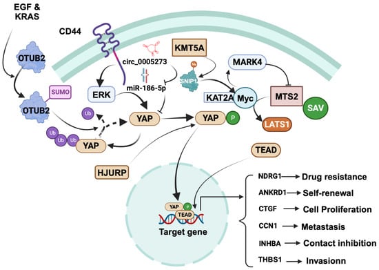
4.1. Tumor-Promoting Role of YAP in Breast Cancer

YAP plays a key role in focal adhesion, a key event of invasiveness and metastasis (Figure 2). Mechanistically, YAP promotes focal adhesion and invasion by inducing TEAD-dependent transcription of thrombospondin 1 (THBS1), which in turn activates FAK via phosphorylation at Tyr 397 [48]. TEAD is also essential for YAP-dependent oncogenic transformation as well as EMT [49], negatively regulated by the Hippo pathway [50].
Figure 2. Regulation of cancer-promoting role of YAP in breast cancer. YAP promotes various cellular processes, including proliferation, contact inhibition, invasion, self-renewal, metastasis, and drug resistance. Focusing on specific mechanisms, YAP induces focal adhesion and invasion by driving TEAD-dependent transcription of thrombospondin 1 (THBS1). YAP is positively regulated by CD44, OTUB2, KMT5A, and circRNA hsa_circ_0005273, while it is negatively regulated by SYNPO2 and RICHI1. CD44 activation leads to ERK-mediated YAP phosphorylation, translocating YAP to the nucleus to upregulate ankyrin repeat domain 1 (ANKRD1), connective tissue growth factor (CTGF), cysteine-rich angiogenic inducer 61 (Cyr61/CCN1), and inhibin βA (INHBA). Further regulatory events, such as the SUMOylation of OTUB2 by EGF and KRAS and the stabilization of YAP in the cytosol through deubiquitination, come into play. KMT5A regulates YAP nuclear translocation by catalyzing K301 methylation of SNIP1, releasing histone acetyltransferase KAT2A, and enhancing the c-MYC/KAT2A complex’s recruitment to c-MYC target promoters. This complex activates MARK4, promoting MST2, SAV, and LATS1 phosphorylation, thereby promoting YAP’s nuclear translocation. HJURP affects YAP1 stability and distribution and subsequently binds to the NDRG1 gene promoter, upregulating NDRG1 transcription. Notably, circRNA hsa_circ_0005273 upregulates YAP expression by counteracting miR-186-5p’s tumor-suppressive activity. The figure was created with BioRender.com.
In BC stem cells, an upstream mediator called CD44 is critical in regulating YAP expression. At a mechanistic level, the activation of YAP by CD44 involves the activation of ERK, which phosphorylates YAP. The phosphorylated YAP then moves from the cytosol to the nucleus, where it promotes the expression of genes that are tangled with contact inhibition of BC stem cells, including ankyrin repeat domain 1 (ANKRD1), connective tissue growth factor (CTGF), cysteine-rich angiogenic inducer 61 (Cyr61/CCN1), and inhibin βA (INHBA) [51]. Another factor, OTUB2, promotes stem cell formation and metastasis by stabilizing YAP in MDA-MB 231 TNBC cells via deubiquitination. This occurs through the binding of SUMOylated OTUB2 to YAP through its SUMO-interacting motif.

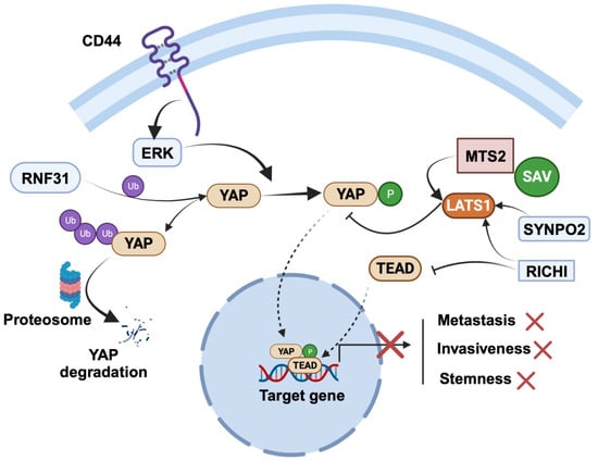
4.2. Tumor-Suppressor Role of YAP in Breast Cancer

YAP is negatively regulated by various mechanisms (Figure 3). YAP is negatively regulated by synaptopodin-2 (SYNPO2) through the stabilization of the LATS2 protein. This essentially inhibits stemness, invasiveness, and metastasis in BC cells via the LATS-mediated inhibition of YAP [52]. Similarly, RICHI1 negatively regulates YAP in BC cells [53]. Mechanistically, the RICHI1-dependent activation of Hippo kinases displaces Amot-p80 from Merlin phosphorylation at S518, resulting in the negative regulation of YAP/TAZ and promotion of transcription of CTGF as well as CYR61 through complexing with TEADs. Overexpression of RICH1 inhibits phosphorylation of MerlinS518 and promotes phosphorylation of LATS1T1079, YAPS127, and TAZS89 while significantly decreasing the total LATS1, YAP, and TAZ protein levels. ing their interplay may lead to targeted therapies.
Figure 3. Regulation of cancer-suppressing role of YAP in breast cancer. SYNPO2 and RICHI1 serve as negative regulators of YAP: SYNPO2 stabilizes LATS2, while RICHI1 activates Hippo kinases and inhibits TEAD complexation with YAP. RNF31 interacts with YAP, promoting polyubiquitination and subsequent degradation of YAP. The figure was created with BioRender.com.

5. Targeting YAP Signaling and Drug Resistance in BC

Over the past two decades, a multitude of potential natural compounds and synthetic derivatives have emerged as promising inhibitors of YAP (Table 1). These compounds demonstrate the ability to independently restrain growth and metastasis or sensitize TNBC cells to chemotherapeutic agents through either the inhibition of YAP signaling or the promotion of its degradation. Furthermore, select compounds exhibit the capacity to induce ferroptosis in TNBC cells by modulating YAP expression. 

Table 1. Natural compounds and their derivatives targeting YAP-dependent drug resistance and ferroptosis in TNBC.
Natural Compound Cellular Mechanism Molecular Mechanism Ref.
Apigenin
  • Reduces proliferation of TNBC cells.
  • Inhibits TNBC cell migration.
  • Blocks stemness of TNBC cells.
  • Reduces the activity of YAP/TAZ.
  • Reduces CTGF and CYR61 expression.
[54]
 
  • Sensitizes TNBC cells to TAZ silencing.
  • Disrupts protein–protein interaction of YAP/TAZ–TEAD.
[55]
Luteolin
  • Suppresses EMT in TNBC cells.
  • Reduces migration of TNBC cells.
  • Triggers the degradation of YAP/TAZ.
  • Diminishes vimentin and N-cadherin expression.
  • Enhances tE-cadherin and catenin expression.
[56]
Parthenolide
derivative
  • Triggers ferroptosis and apoptosis in TNBC cells.
  • Represses breast tumor growth.
  • Prolongs the lifespan of mice without noticeable toxicity.
  • Binds and ubiquitinates GPX4.
  • Controls GPX4-dependent EGR1 in TNBC cells.
[57]
Alantolactone
  • Inhibits TNBC growth.
  • Induces ROS in TNBC cells.
  • Sequesters YAP in cytosol by promoting phosphorylation.
  • Inhibits YAP1/TAZ activity.
  • Enhances LATS kinase activity.
  • Induces proteosomal degradation of YAP.
[58]
Curcumin
  • Demonstrates antitumorigenic effect on BC.
  • Induces ferroptosis in TNBC cells.
  • Modulates SLC1A5.
  • Increases ROS.
  • Enhances MDA and intracellular iron.
[59]
  • Promotes ferroptotic death.
  • Inhibits BC cell viability.
  • Increases intracellular Fe2+, ROS, lipid peroxides, and MDA.
  • Reduces GSH.
  • Upregulates HO-1.
[60]
Curcumin
derivative
  • Inhibits breast tumor growth.
  • Causes dysfunction of mitochondria.
  • Suppresses ROS generation.
  • Reduces YAP/JNK activation.
[61]
  • Shows potential antitumor activity against TNBC.
  • Induces apoptosis.
  • Triggers ROS generation.
  • Induces mitochondrial dysfunction.
  • Reduces phosphorylation of YAP.
  • Increases phosphorylation of JNK.
  • Promotes phosphorylation of 14-3-3 and facilitates the liberation of BAX and FOXO.
  • Reduces N-cadherin, MMP-2, and MMP-9 expression by activating FOXO.
[61]
Resveratrol
  • Attenuates the invasion of TNBC cells.
  • Reduce EGF-induced YAP-dependent expression of AREG, CTGF, and CYR61.
  • Activates LATS1 and promotes phosphorylation-dependent inactivation of YAP.
[62]
Caudatin
  • Reduce the growth of BC cells.
  • Suppresses the formation of mammospheres.
  • Reduces the proportion of CD44+/CD24− as well as ALDH+ BC cells.
  • Inhibits self-renewal of BCSCs.
  • Decreases CD44, Oct4, Sox2, and c-Myc expression.
  • Promotes the degradation of GR through ubiquitin-dependent pathways.
  • Inhibits the accumulation of YAP in the nucleus.
  • Blocks the transcription of YAP target genes.
[63]
Rosmarinic acid
  • Induces cytotoxicity.
  • Reduces the viability of TNBC cells.
  • Induced cell apoptosis.
  • Inhibit the Hippo-YAP/TAZ signaling pathway in MDA-MB 231 cells.
  • Induces LATS1/2-driven phosphorylation of YAP.
  • Promotes YAP retention in cytosol and its subsequent degradation.
  • Causes dissociation of YAP/TAZ–TEAD complex.
  • Reduces YAP–TEAD-mediated transcriptional activity.
[64]
Hydnocarpin
  • Inhibits TNBC cell proliferation, colony formation, invasion, and the EMT.
  • Downregulates CTGF and Cyr61 at both the protein and mRNA levels.
  • Reduces total YAP protein levels without affecting YAP mRNA.
  • Enhances YAP degradation via the proteasome pathway and increases YAP ubiquitination.
[65]

6. Conclusions

YAP protein plays a pivotal role in mediating the oncogenic effects of the Hippo signaling pathway and contributing to the development of drug-resistance mechanisms in different BC subtypes. In BC, the localization and activity of YAP are controlled by distinct mechanisms. YAP mediates drug resistance in the more challenging BC subtype TNBC by inducing self-renewal and differentiation mechanisms as well as activating multidrug-resistance-associated proteins. YAP also mediates resistance to ferroptosis, a key regulatory cell-death mechanism in TNBC, by modulating critical mediators of ferroptosis.
The potential therapeutic implications of targeting the Hippo-YAP pathway in drug resistance mechanisms are significant. Targeting these pathways could improve the efficacy of current treatments and provide new avenues for developing novel therapeutics. The targeting of the YAP pathway could induce ferroptosis in chemotherapy and prevent the development of drug resistance in BC. Similarly, activation of ferroptosis could induce cancer cell death and enhance the effectiveness of chemotherapy. Also, targeting CSCs could also enhance the effectiveness of chemotherapy by sensitization. These approaches, either alone or in combination with natural compounds that target YAP, could improve the outcomes of breast cancer treatment and provide new opportunities for the development of more effective therapies.

References

  1. Prat, A.; Pineda, E.; Adamo, B.; Galván, P.; Fernández, A.; Gaba, L.; Díez, M.; Viladot, M.; Arance, A.; Muñoz, M. Clinical implications of the intrinsic molecular subtypes of breast cancer. Breast 2015, 24 (Suppl. S2), S26–S35.
  2. Kobayashi, S. Basal-like subtype of breast cancer: A review of its unique characteristics and their clinical significance. Breast Cancer 2008, 15, 153–158.
  3. Li, X.; Yang, J.; Peng, L.; Sahin, A.A.; Huo, L.; Ward, K.C.; O’Regan, R.; Torres, M.A.; Meisel, J.L. Triple-negative breast cancer has worse overall survival and cause-specific survival than non-triple-negative breast cancer. Breast Cancer Res. Treat. 2017, 161, 279–287.
  4. Bai, X.; Ni, J.; Beretov, J.; Graham, P.; Li, Y. Triple-negative breast cancer therapeutic resistance: Where is the Achilles’ heel? Cancer Lett. 2021, 497, 100–111.
  5. Nedeljković, M.; Damjanović, A. Mechanisms of Chemotherapy Resistance in Triple-Negative Breast Cancer-How We Can Rise to the Challenge. Cells 2019, 8, 957.
  6. Yang, D.; Zhang, N.; Li, M.; Hong, T.; Meng, W.; Ouyang, T. The Hippo Signaling Pathway: The Trader of Tumor Microenvironment. Front. Oncol. 2021, 11, 772134.
  7. Wang, Z.; Wang, F.; Ding, X.Y.; Li, T.E.; Wang, H.Y.; Gao, Y.H.; Wang, W.J.; Liu, Y.F.; Chen, X.S.; Shen, K.W. Hippo/YAP signaling choreographs the tumor immune microenvironment to promote triple negative breast cancer progression via TAZ/IL-34 axis. Cancer Lett. 2022, 527, 174–190.
  8. Xu, N.; Li, B.; Liu, Y.; Yang, C.; Tang, S.; Cho, W.C.; Huang, Z. Ferroptosis and triple-negative breast cancer: Potential therapeutic targets. Front. Oncol. 2022, 12, 1017041.
  9. Ma, S.; Meng, Z.; Chen, R.; Guan, K.L. The Hippo Pathway: Biology and Pathophysiology. Annu. Rev. Biochem. 2019, 88, 577–604.
  10. Calses, P.C.; Crawford, J.J.; Lill, J.R.; Dey, A. Hippo Pathway in Cancer: Aberrant Regulation and Therapeutic Opportunities. Trends Cancer 2019, 5, 297–307.
  11. Luo, J.; Yu, F.X. GPCR-Hippo Signaling in Cancer. Cells 2019, 8, 426.
  12. Samji, P.; Rajendran, M.K.; Warrier, V.P.; Ganesh, A.; Devarajan, K. Regulation of Hippo signaling pathway in cancer: A MicroRNA perspective. Cell Signal. 2021, 78, 109858.
  13. Ouyang, T.; Meng, W.; Li, M.; Hong, T.; Zhang, N. Recent Advances of the Hippo/YAP Signaling Pathway in Brain Development and Glioma. Cell Mol. Neurobiol. 2020, 40, 495–510.
  14. Cao, L.; Sun, P.L.; Yao, M.; Jia, M.; Gao, H. Expression of YES-associated protein (YAP) and its clinical significance in breast cancer tissues. Hum. Pathol. 2017, 68, 166–174.
  15. Kim, S.K.; Jung, W.H.; Koo, J.S. Yes-associated protein (YAP) is differentially expressed in tumor and stroma according to the molecular subtype of breast cancer. Int. J. Clin. Exp. Pathol. 2014, 7, 3224–3234.
  16. Abylkassov, R.; Xie, Y. Role of Yes-associated protein in cancer: An update (Review). Oncol. Lett. 2016, 12, 2277–2282.
  17. Yu, Y.; Su, X.; Qin, Q.; Hou, Y.; Zhang, X.; Zhang, H.; Jia, M.; Chen, Y. Yes-associated protein and transcriptional coactivator with PDZ-binding motif as new targets in cardiovascular diseases. Pharmacol. Res. 2020, 159, 105009.
  18. Zhao, W.; Wang, M.; Cai, M.; Zhang, C.; Qiu, Y.; Wang, X.; Zhang, T.; Zhou, H.; Wang, J.; Zhao, W. Transcriptional co-activators YAP/TAZ: Potential therapeutic targets for metastatic breast cancer. Biomed. Pharmacother. 2021, 133, 110956.
  19. Morciano, G.; Vezzani, B.; Missiroli, S.; Boncompagni, C.; Pinton, P.; Giorgi, C. An Updated Understanding of the Role of YAP in Driving Oncogenic Responses. Cancers 2021, 13, 3100.
  20. Yang, C.E.; Lee, W.Y.; Cheng, H.W.; Chung, C.H.; Mi, F.L.; Lin, C.W. The antipsychotic chlorpromazine suppresses YAP signaling, stemness properties, and drug resistance in breast cancer cells. Chem. Biol. Interact. 2019, 302, 28–35.
  21. Maugeri-Saccà, M.; De Maria, R. Hippo pathway and breast cancer stem cells. Crit. Rev. Oncol. Hematol. 2016, 99, 115–122.
  22. Rong, X.; Liang, Y.; Han, Q.; Zhao, Y.; Jiang, G.; Zhang, X.; Lin, X.; Liu, Y.; Zhang, Y.; Han, X.; et al. Molecular Mechanisms of Tyrosine Kinase Inhibitor Resistance Induced by Membranous/Cytoplasmic/Nuclear Translocation of Epidermal Growth Factor Receptor. J. Thorac. Oncol. 2019, 14, 1766–1783.
  23. Sadri, F.; Hosseini, S.F.; Rezaei, Z.; Fereidouni, M. Hippo-YAP/TAZ signaling in breast cancer: Reciprocal regulation of microRNAs and implications in precision medicine. Genes Dis. 2024, 11, 760–771.
  24. Quinn, H.M.; Vogel, R.; Popp, O.; Mertins, P.; Lan, L.; Messerschmidt, C.; Landshammer, A.; Lisek, K.; Château-Joubert, S.; Marangoni, E.; et al. YAP and β-Catenin Cooperate to Drive Oncogenesis in Basal Breast Cancer. Cancer Res. 2021, 81, 2116–2127.
  25. Sun, H.L.; Men, J.R.; Liu, H.Y.; Liu, M.Y.; Zhang, H.S. FOXM1 facilitates breast cancer cell stemness and migration in YAP1-dependent manner. Arch. Biochem. Biophys. 2020, 685, 108349.
  26. Guo, Z.; Guo, A.; Zhou, C. Breast Cancer Stem Cell-Derived ANXA6-Containing Exosomes Sustain Paclitaxel Resistance and Cancer Aggressiveness in Breast Cancer. Front. Cell Dev. Biol. 2021, 9, 718721.
  27. Khanal, P.; Yeung, B.; Zhao, Y.; Yang, X. Identification of Prolyl isomerase Pin1 as a novel positive regulator of YAP/TAZ in breast cancer cells. Sci. Rep. 2019, 9, 6394.
  28. Kim, J.; Jang, G.; Sim, S.H.; Park, I.H.; Kim, K.; Park, C. SMARCA4 Depletion Induces Cisplatin Resistance by Activating YAP1-Mediated Epithelial-to-Mesenchymal Transition in Triple-Negative Breast Cancer. Cancers 2021, 13, 5474.
  29. Jiang, Y.; Ji, F.; Liu, Y.; He, M.; Zhang, Z.; Yang, J.; Wang, N.; Zhong, C.; Jin, Q.; Ye, X.; et al. Cisplatin-induced autophagy protects breast cancer cells from apoptosis by regulating yes-associated protein. Oncol. Rep. 2017, 38, 3668–3676.
  30. Elaimy, A.L.; Amante, J.J.; Zhu, L.J.; Wang, M.; Walmsley, C.S.; FitzGerald, T.J.; Goel, H.L.; Mercurio, A.M. The VEGF receptor neuropilin 2 promotes homologous recombination by stimulating YAP/TAZ-mediated Rad51 expression. Proc. Natl. Acad. Sci. USA 2019, 116, 14174–14180.
  31. Koh, S.B.; Ross, K.; Isakoff, S.J.; Melkonjan, N.; He, L.; Matissek, K.J.; Schultz, A.; Mayer, E.L.; Traina, T.A.; Carey, L.A.; et al. RASAL2 Confers Collateral MEK/EGFR Dependency in Chemoresistant Triple-Negative Breast Cancer. Clin. Cancer Res. 2021, 27, 4883–4897.
  32. Qin, X.; Lv, X.; Li, P.; Yang, R.; Xia, Q.; Chen, Y.; Peng, Y.; Li, L.; Li, S.; Li, T.; et al. Matrix stiffness modulates ILK-mediated YAP activation to control the drug resistance of breast cancer cells. Biochim. Biophys. Acta Mol. Basis Dis. 2020, 1866, 165625.
  33. Friedmann Angeli, J.P.; Krysko, D.V.; Conrad, M. Ferroptosis at the crossroads of cancer-acquired drug resistance and immune evasion. Nat. Rev. Cancer 2019, 19, 405–414.
  34. Yun, T.; Liu, Z.; Wang, J.; Wang, R.; Zhu, L.; Zhu, Z.; Wang, X. Microenvironment immune response induced by tumor ferroptosis-the application of nanomedicine. Front. Oncol. 2022, 12, 1019654.
  35. Jiang, L.; Gao, X.M.; Cao, J. The Achilles heel of TNBCs: Ferroptosis heterogeneity. Cell Metab. 2023, 35, 1–2.
  36. Yang, F.; Xiao, Y.; Ding, J.H.; Jin, X.; Ma, D.; Li, D.Q.; Shi, J.X.; Huang, W.; Wang, Y.P.; Jiang, Y.Z.; et al. Ferroptosis heterogeneity in triple-negative breast cancer reveals an innovative immunotherapy combination strategy. Cell Metab. 2023, 35, 84–100.e8.
  37. Brown, C.W.; Amante, J.J.; Chhoy, P.; Elaimy, A.L.; Liu, H.; Zhu, L.J.; Baer, C.E.; Dixon, S.J.; Mercurio, A.M. Prominin2 Drives Ferroptosis Resistance by Stimulating Iron Export. Dev. Cell 2019, 51, 575–586.e4.
  38. Wu, J.; Minikes, A.M.; Gao, M.; Bian, H.; Li, Y.; Stockwell, B.R.; Chen, Z.N.; Jiang, X. Intercellular interaction dictates cancer cell ferroptosis via NF2-YAP signalling. Nature 2019, 572, 402–406.
  39. Morrow, K.A.; Das, S.; Metge, B.J.; Ye, K.; Mulekar, M.S.; Tucker, J.A.; Samant, R.S.; Shevde, L.A. Loss of tumor suppressor Merlin in advanced breast cancer is due to post-translational regulation. J. Biol. Chem. 2011, 286, 40376–40385.
  40. White, S.M.; Avantaggiati, M.L.; Nemazanyy, I.; Di Poto, C.; Yang, Y.; Pende, M.; Gibney, G.T.; Ressom, H.W.; Field, J.; Atkins, M.B.; et al. YAP/TAZ Inhibition Induces Metabolic and Signaling Rewiring Resulting in Targetable Vulnerabilities in NF2-Deficient Tumor Cells. Dev. Cell 2019, 49, 425–443.e9.
  41. Petrilli, A.M.; Fernández-Valle, C. Role of Merlin/NF2 inactivation in tumor biology. Oncogene 2016, 35, 537–548.
  42. Mota, M.; Metge, B.J.; Hinshaw, D.C.; Alsheikh, H.A.; Chen, D.; Samant, R.S.; Shevde, L.A. Merlin deficiency alters the redox management program in breast cancer. Mol. Oncol. 2021, 15, 942–956.
  43. Wu, M.; Zhang, X.; Zhang, W.; Chiou, Y.S.; Qian, W.; Liu, X.; Zhang, M.; Yan, H.; Li, S.; Li, T.; et al. Cancer stem cell regulated phenotypic plasticity protects metastasized cancer cells from ferroptosis. Nat. Commun. 2022, 13, 1371.
  44. Koo, J.H.; Guan, K.-L. Interplay between YAP/TAZ and Metabolism. Cell Metab. 2018, 28, 196–206.
  45. Dai, J.Z.; Wang, Y.J.; Chen, C.H.; Tsai, I.L.; Chao, Y.C.; Lin, C.W. YAP Dictates Mitochondrial Redox Homeostasis to Facilitate Obesity-Associated Breast Cancer Progression. Adv. Sci. 2022, 9, e2103687.
  46. Sorrentino, G.; Ruggeri, N.; Zannini, A.; Ingallina, E.; Bertolio, R.; Marotta, C.; Neri, C.; Cappuzzello, E.; Forcato, M.; Rosato, A.; et al. Glucocorticoid receptor signalling activates YAP in breast cancer. Nat. Commun. 2017, 8, 14073.
  47. You, M.; Xie, Z.; Zhang, N.; Zhang, Y.; Xiao, D.; Liu, S.; Zhuang, W.; Li, L.; Tao, Y. Signaling pathways in cancer metabolism: Mechanisms and therapeutic targets. Signal Transduct. Target. Ther. 2023, 8, 196.
  48. Shen, J.; Cao, B.; Wang, Y.; Ma, C.; Zeng, Z.; Liu, L.; Li, X.; Tao, D.; Gong, J.; Xie, D. Hippo component YAP promotes focal adhesion and tumour aggressiveness via transcriptionally activating THBS1/FAK signalling in breast cancer. J. Exp. Clin. Cancer Res. 2018, 37, 175.
  49. Zhao, B.; Ye, X.; Yu, J.; Li, L.; Li, W.; Li, S.; Yu, J.; Lin, J.D.; Wang, C.Y.; Chinnaiyan, A.M.; et al. TEAD mediates YAP-dependent gene induction and growth control. Genes Dev. 2008, 22, 1962–1971.
  50. Lei, Q.Y.; Zhang, H.; Zhao, B.; Zha, Z.Y.; Bai, F.; Pei, X.H.; Zhao, S.; Xiong, Y.; Guan, K.L. TAZ promotes cell proliferation and epithelial-mesenchymal transition and is inhibited by the hippo pathway. Mol. Cell Biol. 2008, 28, 2426–2436.
  51. Yu, S.; Cai, X.; Wu, C.; Wu, L.; Wang, Y.; Liu, Y.; Yu, Z.; Qin, S.; Ma, F.; Thiery, J.P.; et al. Adhesion glycoprotein CD44 functions as an upstream regulator of a network connecting ERK, AKT and Hippo-YAP pathways in cancer progression. Oncotarget 2015, 6, 2951–2965.
  52. Liu, J.; Ye, L.; Li, Q.; Wu, X.; Wang, B.; Ouyang, Y.; Yuan, Z.; Li, J.; Lin, C. Synaptopodin-2 suppresses metastasis of triple-negative breast cancer via inhibition of YAP/TAZ activity. J. Pathol. 2018, 244, 71–83.
  53. Tian, Q.; Gao, H.; Zhou, Y.; Zhu, L.; Yang, J.; Wang, B.; Liu, P.; Yang, J. RICH1 inhibits breast cancer stem cell traits through activating kinases cascade of Hippo signaling by competing with Merlin for binding to Amot-p80. Cell Death Dis. 2022, 13, 71.
  54. Malla, R.R.; Deepak, K.G.K.; Merchant, N.; Dasari, V.R. Breast Tumor Microenvironment: Emerging target of therapeutic phytochemicals. Phytomedicine 2020, 70, 153227.
  55. Li, Y.W.; Xu, J.; Zhu, G.Y.; Huang, Z.J.; Lu, Y.; Li, X.Q.; Wang, N.; Zhang, F.X. Apigenin suppresses the stem cell-like properties of triple-negative breast cancer cells by inhibiting YAP/TAZ activity. Cell Death Discov. 2018, 4, 105.
  56. Cao, D.; Zhu, G.-Y.; Lu, Y.; Yang, A.; Chen, D.; Huang, H.-J.; Peng, S.-X.; Chen, L.-W.; Li, Y.-W. Luteolin suppresses epithelial-mesenchymal transition and migration of triple-negative breast cancer cells by inhibiting YAP/TAZ activity. Biomed. Pharmacother. 2020, 129, 110462.
  57. Ding, Y.; Chen, X.; Liu, C.; Ge, W.; Wang, Q.; Hao, X.; Wang, M.; Chen, Y.; Zhang, Q. Identification of a small molecule as inducer of ferroptosis and apoptosis through ubiquitination of GPX4 in triple negative breast cancer cells. J. Hematol. Oncol. 2021, 14, 19.
  58. Nakatani, K.; Maehama, T.; Nishio, M.; Otani, J.; Yamaguchi, K.; Fukumoto, M.; Hikasa, H.; Hagiwara, S.; Nishina, H.; Mak, T.W.; et al. Alantolactone is a natural product that potently inhibits YAP1/TAZ through promotion of reactive oxygen species accumulation. Cancer Sci. 2021, 112, 4303–4316.
  59. Cao, X.; Li, Y.; Wang, Y.; Yu, T.; Zhu, C.; Zhang, X.; Guan, J. Curcumin suppresses tumorigenesis by ferroptosis in breast cancer. PLoS ONE 2022, 17, e0261370.
  60. Li, R.; Zhang, J.; Zhou, Y.; Gao, Q.; Wang, R.; Fu, Y.; Zheng, L.; Yu, H. Transcriptome Investigation and In Vitro Verification of Curcumin-Induced HO-1 as a Feature of Ferroptosis in Breast Cancer Cells. Oxid. Med. Cell Longev. 2020, 2020, 3469840.
  61. Wang, L.; Wang, C.; Tao, Z.; Zhao, L.; Zhu, Z.; Wu, W.; He, Y.; Chen, H.; Zheng, B.; Huang, X.; et al. Curcumin derivative WZ35 inhibits tumor cell growth via ROS-YAP-JNK signaling pathway in breast cancer. J. Exp. Clin. Cancer Res. 2019, 38, 460.
  62. Kim, Y.N.; Choe, S.R.; Cho, K.H.; Cho, D.Y.; Kang, J.; Park, C.G.; Lee, H.Y. Resveratrol suppresses breast cancer cell invasion by inactivating a RhoA/YAP signaling axis. Exp. Mol. Med. 2017, 49, e296.
  63. Zhen, X.; Choi, H.S.; Kim, J.H.; Kim, S.L.; Liu, R.; Ko, Y.C.; Yun, B.S.; Lee, D.S. Caudatin Isolated from Cynanchum auriculatum Inhibits Breast Cancer Stem Cell Formation via a GR/YAP Signaling. Biomolecules 2020, 10, 925.
  64. Kim, C.-L.; Shin, Y.-S.; Choi, S.-H.; Oh, S.; Kim, K.; Jeong, H.-S.; Mo, J.-S. Extracts of Perilla frutescens var. Acuta (Odash.) Kudo Leaves Have Antitumor Effects on Breast Cancer Cells by Suppressing YAP Activity. Evid.-Based Complement. Alternat. Med. 2021, 2021, 5619761.
  65. Ou, H.L.; Wu, H.; Ren, Y.L.; Si, Y.; Duan, Z.Q.; Liu, X.W. Hydnocarpin inhibits malignant progression of triple negative breast cancer via CNOT4-mediated ubiquitination and degradation of YAP. Zhongguo Zhong Yao Za Zhi 2023, 48, 4483–4492.
More
Information
Contributors MDPI registered users' name will be linked to their SciProfiles pages. To register with us, please refer to https://encyclopedia.pub/register : , , , ,
View Times: 305
Revisions: 2 times (View History)
Update Date: 17 Jan 2024
1000/1000
Video Production Service