Your browser does not fully support modern features. Please upgrade for a smoother experience.
Submitted Successfully!
 +1 credit
Thank you for your contribution! You can also upload a video entry or images related to this topic.
For video creation, please contact our Academic Video Service.
| Version | Summary | Created by | Modification | Content Size | Created at | Operation |
|---|---|---|---|---|---|---|
| 1 | Francesca Cinelli | -- | 2007 | 2024-01-16 08:56:54 | | | |
| 2 | Fanny Huang | Meta information modification | 2007 | 2024-01-18 07:29:40 | | |
Video Upload Options
We provide professional Academic Video Service to translate complex research into visually appealing presentations. Would you like to try it?
Cite
If you have any further questions, please contact Encyclopedia Editorial Office.
Cinelli, F.; Piva, F.; Bertini, F.; Russo, D.S.; Giachetti, L. Maxillary Anterior Teeth Dimensions and Relative Width Proportions. Encyclopedia. Available online: https://encyclopedia.pub/entry/53866 (accessed on 11 June 2026).
Cinelli F, Piva F, Bertini F, Russo DS, Giachetti L. Maxillary Anterior Teeth Dimensions and Relative Width Proportions. Encyclopedia. Available at: https://encyclopedia.pub/entry/53866. Accessed June 11, 2026.
Cinelli, Francesca, Francesco Piva, Fabio Bertini, Daniele Scaminaci Russo, Luca Giachetti. "Maxillary Anterior Teeth Dimensions and Relative Width Proportions" Encyclopedia, https://encyclopedia.pub/entry/53866 (accessed June 11, 2026).
Cinelli, F., Piva, F., Bertini, F., Russo, D.S., & Giachetti, L. (2024, January 16). Maxillary Anterior Teeth Dimensions and Relative Width Proportions. In Encyclopedia. https://encyclopedia.pub/entry/53866
Cinelli, Francesca, et al. "Maxillary Anterior Teeth Dimensions and Relative Width Proportions." Encyclopedia. Web. 16 January, 2024.
Copy Citation
Predictable results in the aesthetic treatment of anterior teeth can be obtained by resorting to the concept of dental aesthetics and, in particular, defining the ideal tooth dimensions and proportions to obtain a harmonious smile.
teeth dimensions
golden proportions
golden percentage
1. Introduction
Which are the harmonious tooth dimensions? Which are the teeth proportions to produce a pleasant smile? What are the means available to plan a cosmetic treatment? Clinicians have long sought to answer these questions to obtain predictable results during the treatment process and to limit the “subjectivity” in achieving aesthetic goals. To summarize, update, and integrate current knowledge, literature was reviewed to find evidence regarding teeth dimensions and anterior teeth proportions theories. Aesthetic facial parameters were also considered, such as interpupil and inter-canine distance, nasal inter-alar width, and mesiodistal distance of the maxillary anterior teeth. Gender and ethnicity were also taken into account.
Among the most famous proportions theories, the “Golden Proportions theory” involves ancient Greek art and architectural mathematical relationships. Since the pre-Christian era, it has been established that the golden proportions, which are based on the ratio between the diagonal and the side of a square, represent absolute perfection. It is universally recognized and defined with the ratio 1.618:1. Richard Lombardi proposed the application of the “Golden Proportions theory” in dentistry [1] (Figure 1a). In particular, the mesiodistal width of the central and lateral incisors can be repeated in constant proportion [1]. Since then, numerous other theories on dental proportions have been proposed. In 1978, Levin [2] reviewed the concept: The width of the central incisor (1.618) is in “golden proportion” with the width of the lateral (1), which is in “golden proportion” with the canine (0.618) (Figure 1b). He stated that there is no relationship between the real measured widths of the incisors; hence, he proposed the golden proportion based on the apparent size, from a frontal point of view. Levin [2] also proposed the use of a segmented grid, based on the Golden Proportion, which would help to visualize dental proportions. Preston [3] proposed his own theory: The width of the maxillary lateral incisor should be 66% of the width of the central one, and the canines should be 84% of the lateral ones (or 55% of the central ones) [3] (Figure 1c). A few years later, Snow proposed the “Golden Percentage” or “Golden Mean” [4]: Within the inter-canine distance, each tooth corresponds to a percentage of space occupied. The percentages are the following (canine to canine): 10%, 15%, 25%, 25%, 15%, and 10% [4] (Figure 1d). The author [4] declared this method more accurate for determining symmetry, dominance, and proportion for esthetically pleasing smiles, but also that those percentages should be validated by further studies. In 2000, RED (Recurring Esthetic Dental) proportions were introduced by Ward [5], which are based on the constant reduction in the width of the next tooth as it progresses distally, in frontal view. The range of suggested RED proportions is between 62% and 80% [5] (Figure 1e). The Golden Proportion lead to a narrow lateral incisor and to a reduction in the display of the canine. So, he suggested those constant proportions moving distally. More recently, Marquardt proposed the concept of a Golden Rectangle in which the height of the central incisor is in golden proportion with the combined width of the maxillary central incisors (1:1.618) [6] (Figure 1f). He focused only on the central incisors.
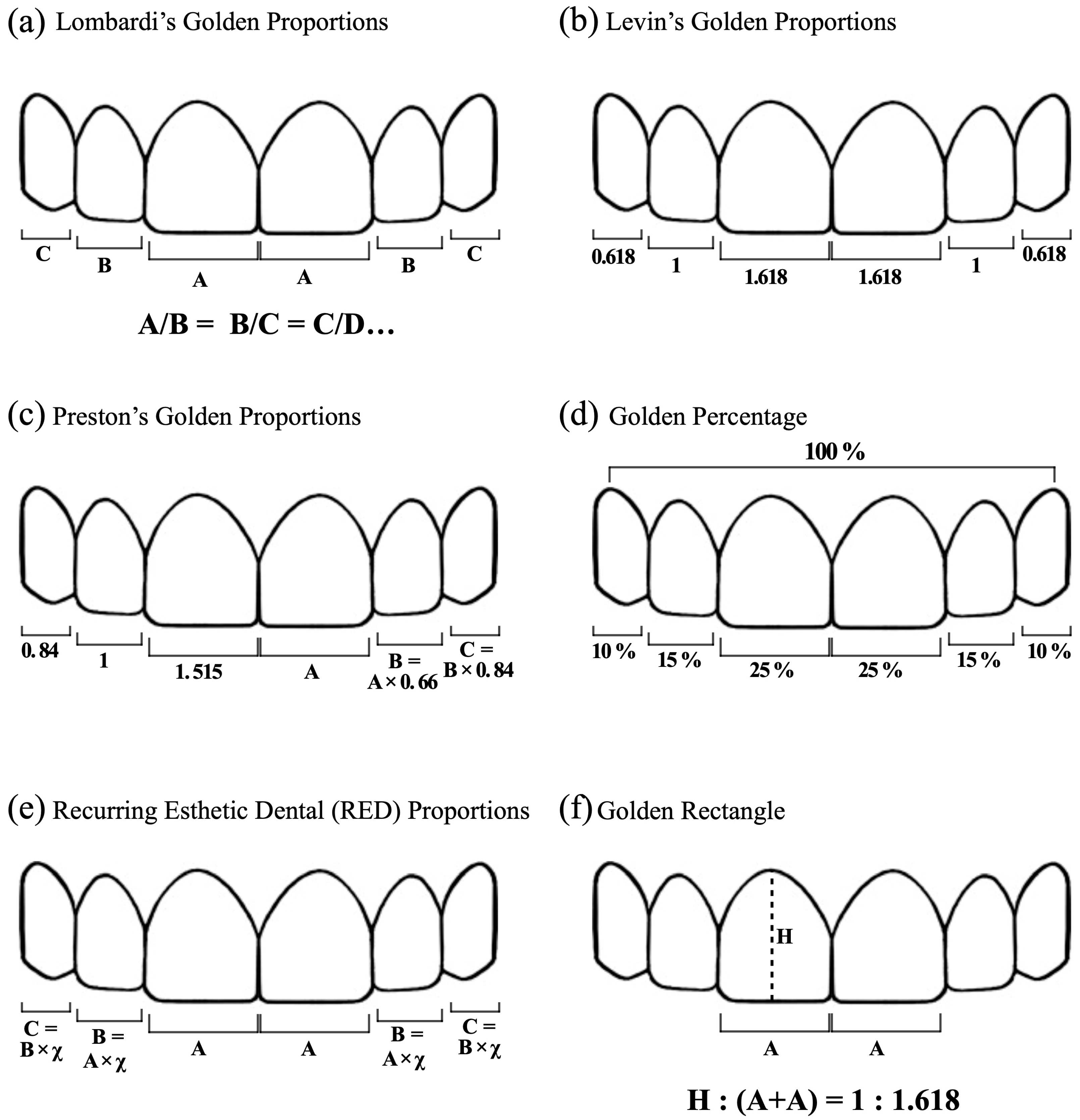
Figure 1. Various theories developed over the years regarding dental proportions.
Various theories have been proposed, but individual differences make it difficult to find a universal rule, exact “magic numbers”. However, interest in the aesthetics of the smile has not waned, meaning that there is an increasing need for treatment to achieve aesthetic standards. Today, the clinician’s concern is not to find and apply a universal rule to all patients but to find harmony in the individual smile. Therefore, considering the importance of references in the esthetic treatment of anterior teeth and the gaps that exist to achieve the best results, this narrative review aims to analyze the following main theme: the size of the anterior teeth and the relationships that bind them. In particular, size, symmetry, and proportion between central incisors and all the anterior teeth were first considered, and then these data were related to gender, ethnicity, and facial parameters.
2. Anterior Teeth Dimensions
Several studies can be found in the literature regarding the relative dental dimensions of the anterior teeth. In particular, the width, the length, and the ratio between them (W/L ratio) are measured to identify the ideal dimensions. This is in relation to some factors, such as extraoral aesthetic parameters, gender, and race. The results obtained can be useful mainly as guidelines in planning aesthetic treatment. Two main comparative studies [7][8] report the average values of length, width, and W/L ratio within the analyzed samples. The first study [7] takes measurements on photographs of extracted teeth, and the second one [8] on models. In both cases, the greater mesiodistal distances for the width and the greater apico-coronal distances for the length are measured. The sample is composed of European-origin adults. Magne et al. [7] do not include female sex in the analysis. However, it also reports the dimensions of worn teeth that logically have a width as the predominant dimension, so their W/L ratio is higher.
Some studies point out that there are different results depending on the sample populations: In a Pakistani population [9], smaller measurements are reported. The only similarity is the 78% mean W/L ratio for the central incisor. In Chinese populations, instead, the W/L ratio seems to be bigger [10].
Regarding the symmetry between the central incisors, the literature shows that perfect coincident dimensions are rare [8][11][12][13][14]. According to these studies, central incisors are identical in 10–13% of cases, similar in 27–29% of subjects (with a difference of a maximum of 0.2 mm), while the rest (60–61%) are different (with a difference of more than 0.2 mm). However, the literature does not agree on this topic: More recently, Wang’s systematic review with meta-analysis [15] reported that within the 23 studies analyzed, there were no differences in the size of right and left incisors. Width and length also appear greater in men than in women [15][16]. Ethnicity is an influencing factor: The Caucasian population shows larger W, L, and W/L ratio than the Asian population, but there is also a great variability within the same populations [15].
The correlation with facial parameters is not always present and constant, and it strongly depends on the sample’s ethnicity [15][17]. Correlation seems to be low, but significant for the inter-canthal distance, with the sum of the central incisors mesiodistal diameters or with the entire anterior group [18][19][20][21]. Regarding the inter-pupillary distance, there is no correlation between the dental dimensions in males and females [22], but in women, the inter-canine distance coincides with the inter-alar distance [22]. However, other articles [17][23] found a correlation between inter-pupillary distance, inter-commissural distance, and the sum of the mesiodistal diameters of the anterior teeth and also a correlation between inter-commissural distance and inter-canine distance. According to these authors, the correlations found can be used as a reference for anterior teeth rehabilitations. These conclusions disagree with other authors who claim, on the basis of the measurements performed, that the use of facial parameters is inaccurate in determining dental dimensions [24].
Regarding the height and the width of the face, the literature does not agree here either. In 2005, Hasanreisoglu [22] stated that there is no correlation between bizygomatic distance and dental dimensions in males, while in women, there is a ratio of 1:16 with central incisors width [22]. However, in a more recent study [14], it was found to be a ratio of 1:16 between the width of the central incisor and the bizygomatic distance. The same study found a ratio of 1:18 with the total facial height and 1:12 with the lower facial height [14]. It also shows how gender influences the correlations: The measurements in men are greater, but the ratios are similar in the two genders. A previous study [25][26], instead, investigated the existence of the 1:16 ratio (Trubyte Tooth Indicator) between the length of the central incisor and the face to produce artificial teeth: It was found that 14.5% of the participants exhibited it, while 14.3% of them have a shorter face and 71.9% a longer one. The 1:16 ratio between central incisor and face width appears in 23% of the population; 53% has a narrower face, and 23% has a larger one. The study also shows sex differences for each group (correct ratio, smaller ratio, and bigger ratio). It concludes that the 1:16 ratio is not precise: Artificial teeth produced with this ratio are generally narrower and longer. The choice of the dimensions of the artificial teeth depends on many factors: the dimension of the maxillary arch, the relationship between the mandible and the maxilla, the profile of the residual ridges, the vertical dimension, the dimensions of the lips at rest and when smiling, the face shape and contour, age, gender, and personality. The findings of the study can be used only as initial guidelines.
Regarding the ethnic differences, the articles comparing the dental dimensions of Asian and European subjects [27] seem to show that Caucasians have a greater width, and therefore also a greater W/L ratio, of the central incisors than Asians. The length, instead, is similar. Laterals and canines do not differ in width, but the length is greater in Asians and the W/L ratio is greater in White subjects. In the same study, a comparison is also made for worn teeth: The central incisor width and W/L ratio are greater in Caucasians. The length of the central incisors is greater in Asians, but the difference is not significant. A comparison of facial parameters and dental measurements in three ethnicities: Asian, African American, and European [28], shows that the bizygomatic width and inter-canthal distance are more constant in women and that the widest teeth are the central incisors of African American men and women. Consequently, the inter-canine gap in African American individuals is also greater than in other ethnic groups. The relationship between the width of the central incisor and the bi-zygomatic distance varies between African Americans and Asians but is similar in Asians and Caucasians of the same sex. Finally, in Asian women, there is a correlation between commissural distance and width of a single central incisor, two central incisors, four incisors, and the anterior group. A weak correlation between central incisor width and bizygomatic width exists in the Saudi population [29]. The Arab population, according to the article by Alqahtani, has similarities only with the Turkish population due to the similar cultural background and differs significantly from the other populations examined [13]. The same study also points out the differences between their populations (European, Chinese, Turkish, and White) and the ones in other studies [7][8][22][30][31].
3. Golden Proportions, Golden Percentage, RED Proportion, and Golden Rectangle
According to the literature, ideal dental proportions are either partially found or not found at all in natural teeth. The Golden Proportions, according to various articles [32][33], are not fully present in the analyzed samples from different populations. The same is true for Preston’s Golden Proportions [34][35][36][37]. A percentage of 62% can be found between central–lateral and between lateral–canine, but only in a very low percentage of the samples. Similarly, the Golden Percentage proposed by Snow (25–15–10% from centra lincisor to canine) is almost never found [32][36][37][38][39][40][41][42][43]. In particular, central incisors are wider and canines are narrower. However, a more recent study on an English population [35] proposes modified Golden Percentages: in particular, 22.5–15–12.5 percentages are indicated for central–lateral–canine incisor. The percentage for the central and lateral are found in about 71% of cases, while for the canine in 61% of them. Equally encouraging percentages emerge in the Spanish population [36]. Regarding the RED Proportions, their existence is limited to a very low percentage of subjects [32][35][36][37][38][40][41][44]. However, predicting central incisor width with 70% RED proportions and inter-alar distance is an accurate method to evaluate the width of maxillary anterior teeth [45] using specific formulas. A recent study [46] also identified formulas to determine central incisor width by modifying inner-canthal distance according to Golden Percentage, and interpupillary distance according to Golden Proportions. The Golden Rectangle theory, as suggested by a few articles found in the literature, seems to be applicable to the Indian populations investigated in the studies, both in men and women [47][48][49].
References
- Lombardi, R.E. The principles of visual perception and their clinical application to denture esthetics. J. Prosthet. Dent. 1973, 29, 358–382.
- Levin, E.I. Dental esthetics and the golden proportion. J. Prosthet. Dent. 1978, 40, 244–252.
- Preston, J.D. The Golden Proportion Revisited. J. Esthet. Restor. Dent. 1993, 5, 247–251.
- Snow, S.R. Esthetic smile analysis of maxillary anterior tooth width: The golden percentage. J. Esthet. Dent. 1999, 11, 177–184.
- Ward, D.H. Proportional smile design using the recurring esthetic dental (red) proportion. Dent. Clin. N. Am. 2001, 45, 143–154.
- Marquardt, S.R. Marquardt on the Golden Decagon and human facial beauty. Interview by Dr. Gottlieb. J. Clin. Orthod. 2002, 36, 339–347.
- Magne, P.; Gallucci, G.O.; Belser, U.C. Anatomic crown width/length ratios of unworn and worn maxillary teeth in white subjects. J. Prosthet. Dent. 2003, 89, 453–461.
- Orozco-Varo, A.; Arroyo-Cruz, G.; Martínez-de-Fuentes, R.; Jiménez-Castellanos, E. Biometric analysis of the clinical crown and the width/length ratio in the maxillary anterior region. J. Prosthet. Dent. 2015, 113, 565–570.
- Saleem, B.; Mahmood, A.; Butt, A.M.; Najmi, N.; Aslam, S.; Shah, S.M.; Shams, S. Analysis of width, height and width/height ratio of crowns of maxillary anterior teeth. Pak. J. Med. Health Sci. 2022, 32, 46–49.
- Zhao, Q.; Li, N.; Cao, J. Morphological features of maxillary anterior teeth in a sample of Chinese population. Homo 2015, 66, 448–454.
- Mavroskoufis, F.; Ritchie, G.M. Variation in size and form between left and right maxillary central incisor teeth. J. Prosthet. Dent. 1980, 43, 254–257.
- Vadavadagi, S.V.; Hombesh, M.N.; Choudhury, G.K.; Deshpande, S.; Anusha, C.V.; Murthy, D.K. Variation in Size and Form between Left and Right Maxillary Central Incisor Teeth. J. Int. Oral. Health 2015, 7, 33–36.
- Wang, Y.; Song, Y.; Zhong, Q.; Xu, C. Evaluation of influence factors on the width, length, and width to length ratio of the maxillary central incisor: A systematic review and meta-analysis. J. Esthet. Restor. Dent. 2021, 33, 351–363.
- Sterrett, J.D.; Oliver, T.; Robinson, F.; Fortson, W.; Knaak, B.; Russell, C.M. Width/length ratios of normal clinical crowns of the maxillary anterior dentition in man. J. Clin. Periodontol. 1999, 26, 153–157.
- Al Wazzan, K.A. The relationship between intercanthal dimension and the widths of maxillary anterior teeth. J. Prosthet. Dent. 2001, 86, 608–612.
- Attokaran, G.; Shenoy, K. Correlation between Innercanthal Distance and Mesiodistal Width of Maxillary Anterior Teeth in a Thrissur, Kerala, India, Population. J. Contemp. Dent. Pract. 2016, 17, 382–387.
- Arun Kumar, K.V.; Gupta, S.H.; Sandhu, H.S. Determination of mesiodistal width of maxillary anterior teeth using inner canthal distance. Med. J. Armed. Forces India 2015, 71, S376–S381.
- Ahmed, N.; Abbasi, M.; Khan, D.; Khalid, S.; Jawed, W.; Mahmood, M. Determination of the combined width of maxillary anterior teeth using innercanthal distance with respect to age gender and ethinicity. PAFMJ 2021, 71, S164–S169.
- Hasanreisoglu, U.; Berksun, S.; Aras, K.; Arslan, I. An analysis of maxillary anterior teeth: Facial and dental proportions. J. Prosthet. Dent. 2005, 94, 530–538.
- Barman, J.; Serin, S. Comparison of interpupillary distance and combined mesiodistal width of maxillary central incisor teeth in two ethnic groups of Northeast India: An in vivo study. Indian J. Dent. Res. 2018, 29, 155–160.
- Kini, A.Y.; Angadi, G.S. Biometric ratio in estimating widths of maxillary anterior teeth derived after correlating anthropometric measurements with dental measurements. Gerodontology 2013, 30, 105–111.
- Zlatarić, D.K.; Kristek, E.; Celebić, A. Analysis of width/length ratios of normal clinical crowns of the maxillary anterior dentition: Correlation between dental proportions and facial measurements. Int. J. Prosthodont. 2007, 20, 313–315.
- Radia, S.; Sherriff, M.; McDonald, F.; Naini, F.B. Relationship between maxillary central incisor proportions and facial proportions. J. Prosthet. Dent. 2016, 115, 741–748.
- LaVere, A.M.; Marcroft, K.R.; Smith, R.C.; Sarka, R.J. Denture tooth selection: An analysis of the natural maxillary central incisor compared to the length and width of the face. Part I. J. Prosthet. Dent. 1992, 67, 661–663.
- LaVere, A.M.; Marcroft, K.R.; Smith, R.C.; Sarka, R.J. Denture tooth selection: An analysis of the natural maxillary central incisor compared to the length and width of the face: Part II. J. Prosthet. Dent. 1992, 67, 810–812.
- Isa, Z.M.; Tawfiq, O.F.; Noor, N.M.; Shamsudheen, M.I.; Rijal, O.M. Regression methods to investigate the relationship between facial measurements and widths of the maxillary anterior teeth. J. Prosthet. Dent. 2010, 103, 182–188.
- Parciak, E.C.; Dahiya, A.T.; AlRumaih, H.S.; Kattadiyil, M.T.; Baba, N.Z.; Goodacre, C.J. Comparison of maxillary anterior tooth width and facial dimensions of 3 ethnicities. J. Prosthet. Dent. 2017, 118, 504–510.
- Mohammed, S.; Bakhsh, L. Evaluation of Relation between Bizygomatic Width and Mesiodistal Dimension of Maxillary Central Incisor in Saudi Population: An In-vivo Study. Study J. Clin. Diagn. Res. 2020, 14, ZC32–ZC36.
- Alqahtani, A.S.; Habib, S.R.; Ali, M.; Alshahrani, A.S.; Alotaibi, N.M.; Alahaidib, F.A. Maxillary anterior teeth dimension and relative width proportion in a Saudi subpopulation. J. Taibah. Univ. Med. Sci. 2021, 16, 209–216.
- Marcuschamer, E.; Tsukiyama, T.; Griffin, T.J.; Arguello, E.; Magne, P.; Gallucci, G.O. Anatomical crown width/length ratios of worn and unworn maxillary teeth in Asian subjects. Int. J Periodontics Restor. Dent. 2011, 31, 495–503.
- Sah, S.K.; Zhang, H.D.; Chang, T.; Dhungana, M.; Acharya, L.; Chen, L.L.; Ding, Y.M. Maxillary Anterior Teeth Dimensions and Proportions in a Central Mainland Chinese Population. Chin. J. Dent. Res. 2014, 17, 117–124.
- Akl, M.A.; Mansour, D.E.; Mays, K.; Wee, A.G. Mathematical Tooth Proportions: A Systematic Review. J. Prosthodont. 2022, 31, 289–298.
- Forster, A.; Velez, R.; Antal, M.; Nagy, K. Width ratios in the anterior maxillary region in a Hungarian population: Addition to the golden proportion debate. J. Prosthet. Dent. 2013, 110, 211–215.
- Mahshid, M.; Khoshvaghti, A.; Varshosaz, M.; Vallaei, N. Evaluation of “Golden Proportion” in Individuals with an Esthetic Smile. J. Esthet. Restor. Dent. 2004, 16, 185–192.
- Kalia, R. An analysis of the aesthetic proportions of anterior maxillary teeth in a UK population. Br. Dent. J. 2020, 228, 449–455.
- Rodríguez-López, S.; Martínez, M.F.E.; Velasco, J.P.; Junquera, L.; García-Pola, M. Analysis of dental esthetic proportions in a Spanish population sample. J. Oral. Sci. 2021, 63, 257–262.
- Melo, M.; Ata-Ali, F.; Huertas, J.; Cobo, T.; Shibli, J.A.; Galindo-Moreno, P.; Ata-Ali, J. Revisiting the Maxillary Teeth in 384 Subjects Reveals A Deviation from the Classical Aesthetic Dimensions. Sci. Rep. 2019, 9, 730.
- Calçada, D.; Correia, A.; Araújo, F. Anthropometric analysis of anterior maxillary teeth with digital photography—A study in a Portuguese sample. Int. J. Esthet. Dent. 2014, 9, 370–380.
- Maharjan, A.; Joshi, S. Clinical Evaluation of Maxillary Anterior Teeth in Relation to Golden Proportion, Red Proportion and Golden Percentage. J. Nepal. Health Res. Counc. 2018, 16, 11–15.
- Agrawal, V.S.; Kapoor, S.; Bhesania, D.; Shah, C. Comparative photographic evaluation of various geometric and mathematical proportions of maxillary anterior teeth: A clinical study. Indian J. Dent. Res. 2016, 27, 32–36.
- Fayyad, M.; Jamani, K.; Agrabawi, J. Geometric and Mathematical Proportions and their Relations to Maxillary Anterior Teeth. Contemp. Dent. Pract. 2006, 7, 62–70.
- Ahmed, N.; Halim, M.S.; Khalid, S.; Ghani, Z.A.; Jamayet, N.B. Evaluation of golden percentage in natural maxillary anterior teeth width: A systematic review. J. Prosthet. Dent. 2021, 127, e1–e845.
- Ahmed, N.; Halim, M.S.B.; Ghani, Z.A.; Khan, Z.A.; Abbasi, M.S.; Jamayet, N.B.; Alam, M.K. A 2D Photographic and 3D Digital Dental Model Analysis of Golden Percentage in Maxillary Anterior Teeth. Biomed. Res. Int. 2021, 2021, 6674400.
- Shetty, S.; Pitti, V.; Satish Babu, C.; Surendra Kumar, G.; Jnanadev, K. To evaluate the validity of Recurring Esthetic Dental proportion in natural dentition. J. Conserv. Dent. 2011, 14, 314–317.
- Liao, P.; Fan, Y.; Nathanson, D. Evaluation of maxillary anterior teeth width: A systematic review. J. Prosthet. Dent. 2019, 122, 275–281.e7.
- Ahmed, N.; Halim, M.S.; Ab-Ghani, Z.; Abbasi, M.S.; Aslam, A.; Safdar, J.; Das, G.; Ahmed, A.R.; Jamayet, N.B. The Analysis of Facio-Dental Proportions to Determine the Width of Maxillary Anterior Teeth: A Clinical Study. J. Clin. Med. 2022, 11, 7340.
- Chaudhari, D.; Dange, D.; Khalikar, S. Golden Rectangle Ratio—How Precious Is It?: A Clinical Study. IOSR-JDMS 2014, 13, 1–6.
- Singh, R.; Tripathi, A.; Singh, S.; Bhatnagar, A. A study on the practical applicability of the rule of golden rectangle in dental aesthetics. Eur. J. Prosthodont. Restor. Dent. 2011, 19, 85–89.
- Varghese, P.; Cherian, B.; Sukumaran, B.; Anu, S.; Jacob, B.M.; Raja, V.V. Analysis of Geometric Proportions on Maxillary Anterior Teeth for Esthetic Smile Design: An In vivo Study. J. Pharm. Bioallied. Sci. 2021, 13, s778–s782.
More
Information
Subjects:
Dentistry, Oral Surgery & Medicine
Contributors
MDPI registered users' name will be linked to their SciProfiles pages. To register with us, please refer to https://encyclopedia.pub/register
:
View Times:
2.3K
Revisions:
2 times
(View History)
Update Date:
18 Jan 2024
Table of Contents


Notice
You are not a member of the advisory board for this topic. If you want to update advisory board member profile, please contact office@encyclopedia.pub.
OK
Confirm
Only members of the Encyclopedia advisory board for this topic are allowed to note entries. Would you like to become an advisory board member of the Encyclopedia?
Yes
No
${ textCharacter }/${ maxCharacter }
Submit
Cancel
Back
Comments
${ item }
|
${ item.createdUser.fullName }
${ item.createdAt }
${ item.vote }
${ item.reply }
Delete
${ reply.createdUser.fullName }
${ reply.createdAt }
${ reply.vote }
Delete
There is no reply to this comment~
${ item.replyTextCharacter }/${ item.replyMaxCharacter }
Submit
Cancel
More
No more~
There is no comment~
${ textCharacter }/${ maxCharacter }
Submit
Cancel
${ selectedItem.replyTextCharacter }/${ selectedItem.replyMaxCharacter }
Submit
Cancel
Confirm
Are you sure to Delete?
Yes
No