Your browser does not fully support modern features. Please upgrade for a smoother experience.
Submitted Successfully!
 +1 credit
Thank you for your contribution! You can also upload a video entry or images related to this topic.
For video creation, please contact our Academic Video Service.
| Version | Summary | Created by | Modification | Content Size | Created at | Operation |
|---|---|---|---|---|---|---|
| 1 | Alexander Borbély | -- | 1650 | 2024-01-08 19:06:23 | | | |
| 2 | Mona Zou | Meta information modification | 1650 | 2024-01-09 09:19:35 | | |
Video Upload Options
We provide professional Academic Video Service to translate complex research into visually appealing presentations. Would you like to try it?
Cite
If you have any further questions, please contact Encyclopedia Editorial Office.
Borbély, A.; Tobler, I. Concepts in the Two-Process Model of Sleep Regulation. Encyclopedia. Available online: https://encyclopedia.pub/entry/53569 (accessed on 23 July 2026).
Borbély A, Tobler I. Concepts in the Two-Process Model of Sleep Regulation. Encyclopedia. Available at: https://encyclopedia.pub/entry/53569. Accessed July 23, 2026.
Borbély, Alexander, Irene Tobler. "Concepts in the Two-Process Model of Sleep Regulation" Encyclopedia, https://encyclopedia.pub/entry/53569 (accessed July 23, 2026).
Borbély, A., & Tobler, I. (2024, January 08). Concepts in the Two-Process Model of Sleep Regulation. In Encyclopedia. https://encyclopedia.pub/entry/53569
Borbély, Alexander and Irene Tobler. "Concepts in the Two-Process Model of Sleep Regulation." Encyclopedia. Web. 08 January, 2024.
Copy Citation
The two-process model of sleep regulation has served as a conceptual framework in the last four decades for understanding sleep physiology. In the 1970s, long-term recordings of sleep in rats were obtained thanks to EEG telemetry. NonREM sleep and REM sleep were found to differ in their time course and response to light-dark protocols. There were indications for their coupling to the circadian system, in particular the light-dark and the dark-light transitions. With the advent of quantitative EEG analysis, slow-wave activity in nonREM sleep was recognized as a sleep-wake-dependent variable. The term “sleep homeostasis” was coined to specify the regulated balance between sleep and waking. The regulatory homeostatic process was designated as “Process S”. In the two-process model, its interaction with the circadian pacemaker “Process C” can account for sleep duration under various experimental protocols. Local, use-dependent slow-wave activity changes were demonstrated in both humans and rats by the selective, unilateral activation of a cortical region prior to sleep. Finding that rest in invertebrates has sleep-like regulatory properties opened a new realm of animal studies. Comparative sleep studies in a broad variety of animal species confirmed the validity of the basic concepts of the two-process model.
two-process model
EEG analysis
sleep homeostasis
circadian rhythm
local sleep
comparative sleep studies
1. Recovery Process
Berger and Oswald [1] were the first to report an increase of slow-wave sleep after sleep deprivation in human subjects. Feinberg’s group [2], using period-amplitude analysis of the EEG, showed that during baseline sleep, delta activity shifted towards lower frequencies and declined in amplitude. They speculated that the changes may reflect the kinetics of metabolic processes underlying sleep and may be related to the “goodness of sleep”. By applying period-amplitude analysis of the EEG in rats, the researchers showed that the low-frequency fraction of nonREM sleep (SWS) declined over the course of a 12 h light period [3]. An additional important observation was that SWS was enhanced during recovery sleep after sleep deprivation and that this effect depended on the duration of prior wakefulness. This led to the proposition that SWS represents an intensity parameter of nonREM sleep and that its decline during sleep reflects a recovery process. In a preliminary version of the two-process model, the recovery process and the circadian system were considered to be two separate facets of sleep regulation [4].
2. Sleep Homeostasis
The term “sleep homeostasis” was first proposed in a book chapter on functional aspects of sleep [4]. It can be defined as a regulated balance between sleep and waking [5]. A subsequent article proposed that sleep homeostasis denotes the regulation of the sleep process relative to an internal reference level [6]. Slow waves were considered an indicator of sleep intensity that could reflect a restitutional function of sleep.
The term sleep homeostasis has gained popularity and is widely used. For example, recent reviews refer to the rising homeostatic sleep pressure during waking leading to a compensatory sleep rebound or recovery sleep, strengthening the idea that it reflects some important restorative process [7][8].
3. Process S
In the two-process model, Process S is designated as a regulatory process that is determined by preceding sleep and wakefulness [9]. Its time course was derived from EEG slow-wave activity, an electrophysiological variable that declines during the daily sleep period in humans [10] and rodents [3]. The time course of its rise during the daily waking period was derived from slow-wave activity during multiple naps in humans [11] and from interspersed sleep episodes in rats [3].
The term “process” in Process S is meant to indicate a regulatory homeostatic principle that can be operationalized by an EEG correlate. It has given rise to several hypotheses concerning the underlying neural mechanisms. One of the best known propositions is the synaptic homeostasis hypothesis [12][13]. The authors posit that neuronal circuits increase in synaptic strength during waking and weaken during sleep. Some have used the term “homeostat”, implying that it could be specified and localized. However, it is likely that all parts of the sleep circuitry in the brain can implement homeostasis [7] and even that factors from skeletal muscle may be involved [14]. Therefore, in view of our ignorance of the nature of sleep homeostasis, the general term “process” is still appropriate.
4. Interaction of S and C
Usually, the two processes S and C act in synergy and reinforce each other. By scheduling the recovery from sleep deprivation to the beginning of an animal’s circadian activity period, a conflict is created. Its consequences were apparent in a 24 h sleep deprivation schedule that ended at the beginning of the animal’s activity period [3]. A two-stage rebound of slow-wave sleep occurred, the second stage being delayed by 12 h from the light period. In the guinea pig, an animal with a weak circadian rest-activity rhythm [15], the sleep rebound was equal in the light and dark periods [16]. Forced desynchrony between the sleep-wake cycle and the circadian rhythm was applied to investigate the interaction between S and C in humans [17]. Sleep continuity was impaired when the enforced sleep period coincided with the circadian wake period.
In the two-process model, the duration of sleep is determined by the interaction of Process S with a threshold modulated by Process C [9]. In the extended version of the model, an upper threshold was added to define the duration of waking under free-running conditions [18]. The question of the type of continuous interaction of the two processes (linear or nonlinear) arose in conjunction with the forced desynchrony protocol [19]. While there are arguments for both types of interaction, the possibility of a nonlinearity in neurobehavioral metrics such as alertness and cognitive performance must also be considered.
5. NonREM-REM Sleep Cycle
The homeostatic part of the model was elaborated to include the ultradian nonREM-REM sleep cycle [20][21]. The initial buildup of slow-wave activity within a nonREM sleep episode was represented by the combined action of an exponentially increasing process and a saturation process. Based on a large set of empirical data, the model parameters were estimated and the performance tested on data from independent protocols [22]. A close fit was obtained between the empirical and simulated data.
6. Behavioral Homeostasis
Initially, EEG slow-wave activity served as the marker of sleep homeostasis. This led to the observation that its time course during sleep was comparable in humans and in rats. However, this approach was restricted to those animals in which the EEG could be recorded. In invertebrates, the usual electrographic criteria are absent. The question arose of whether immobility periods have sleep-like properties. In other words, does rest deprivation induce a compensatory increase in resting behavior? To examine this possibility, cockroaches, Leucophaea maderae, were prevented from resting by manual rotation and slight shaking of their dish for 3 h [23]. In comparison to the control condition, immobility was enhanced in the first hour following rest deprivation. Additionally, in another cockroach strain, a 6 h rest deprivation gave rise to a compensatory response [24]. A similar effect was obtained in scorpions, another arthropod, following a 12 h rest deprivation by mechanical stimulation [25]. These were the first studies showing that rest in invertebrates shows a similar homeostatic regulation as sleep in homeotherms. The correspondence was confirmed in rats, where the time course of sleep continuity, indexed by a behavioral variable, the number of brief awakenings, and of EEG slow-wave activity was comparable for baseline conditions and sleep deprivation [26].
These early studies opened the sleep field to the realm of invertebrates and in particular to Drosophila as the prime organism for the genetic dissection of sleep [27][28]. Since then, the field has witnessed an explosive growth, and the number of citations of “sleep and Drosophila” has skyrocketed. Recent studies showed a sleep-like state that may be homeostatically regulated, even in animals without a centralized nervous system, such as Cassiopea [29] and Hydra vulgaris [30].
7. Comparative Sleep Studies
The comparison of sleep among different species provides information about its variation in nature and allows insights into general regulatory principles. Due to her background in zoology, Irene Tobler was particularly interested in this approach (Figure 1).
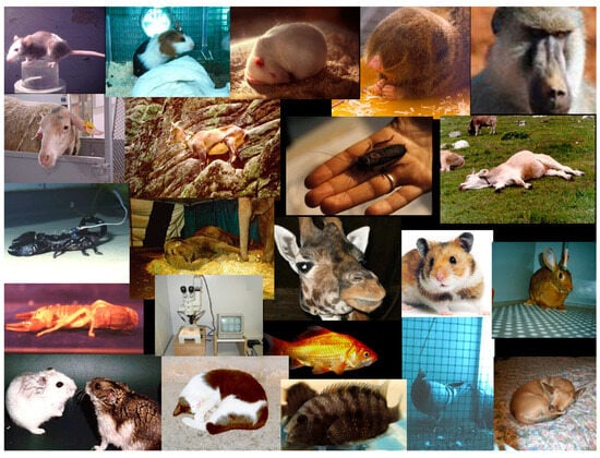
Figure 1. Composite picture of the animal species in which rest activity or sleep was analyzed by Irene Tobler.
Homeostasis of slow-wave activity that had been originally reported for rats was shown to also be present in diverse other rodent species: the Syrian hamster [31], the guinea pig [16], the Djungarian hamster [32], several mouse strains [33][34][35], and the blind mole rat, a fossorial animal [36]. In these studies, Tom Deboer and Vlad Vyazovskiy played a major role. Blind mole rats whose motor activity was recorded for several months showed a large intra- and inter-individual variability in their 24 h pattern [37]. In most animals, a free-running circadian rhythm was seen under continuous darkness. Despite the large ecological differences, their polysomnographic features were similar to those of other rodents.
Guinea pigs conformed to the predictions of the two-process model, but the low amplitude of the circadian rhythm caused equal effects of sleep deprivation in both the light and dark periods [15][16].
Cats studied in a light-dim cycle exhibited a low-amplitude circadian rhythm and responded with a rise in slow-wave activity to 14 h sleep deprivation [38]. Rabbits kept under light-dim conditions slept more in the light period, where they showed a declining trend of slow-wave activity in nonREM sleep [39]. Twenty-four hour sleep deprivation beginning at light onset caused an increase of slow-wave activity during recovery sleep.
In dogs, a behavioral index was used to study sleep homeostasis. Motor activity was measured continuously with an actometer worn on the collar [40]. Twenty-four hour sleep deprivation was followed by reduced motor activity and an increase in the number of low-activity episodes.
In perch and goldfish, light-induced activation was used to prevent the animals from having resting behavior for 6 or 12 h [41]. The interventions were followed by a prolongation of rest periods and an increase of low-activity states, evidence of homeostatic regulation.
In animals in which homeostatic challenges were not possible, observational studies focused on the daily time course of rest activity, sleep, its behavioral characteristics, and seasonal changes. Studies were carried out on ibex in the wild [42], elephants in captivity [43] and giraffes kept in a zoological garden [44].
References
- Berger, R.J.; Oswald, I. Effects of sleep deprivation on behaviour, subsequent sleep and dreaming. J. Ment. Sci. 1962, 108, 457–465.
- Church, M.W.; March, J.D.; Hibi, S.; Benson, K.; Cavness, C.; Feinberg, I. Changes in frequency and amplitude of delta activity during sleep. Electroencephalogr. Clin. Neurophysiol. 1975, 39, 1–7.
- Borbely, A.A.; Neuhaus, H.U. Sleep-deprivation—Effects on sleep and EEG in the rat. J. Comp. Physiol. 1979, 133, 71–87.
- Borbely, A.A. Sleep: Circadian rhythm versus recovery process. In Functional States of the Brain: Their Determinants; Koukkou, M., Lehmann, D., Angst, J., Eds.; Elsevier: Amsterdam, The Netherlands, 1980; pp. 151–161.
- Tobler, I.; Achermann, P. Sleep homeostasis. Scholarpedia 2007, 2, 2432.
- Borbély, A.A. The sleep process: Circadian and homeostatic aspects. In Advances in Physiological Science; Pergamon Press: London, UK, 1981; Volume 18, pp. 85–91.
- Franks, N.P.; Wisden, W. The inescapable drive to sleep: Overlapping mechanisms of sleep and sedation. Science 2021, 374, 556–559.
- Nollet, M.; Franks, N.P.; Wisden, W. Understanding sleep regulation in normal and pathological conditions, and why it matters. J. Huntingt. Dis. 2023, 12, 105–119.
- Borbely, A.A. A two process model of sleep regulation. Hum. Neurobiol. 1982, 1, 195–204.
- Borbely, A.A.; Baumann, F.; Brandeis, D.; Strauch, I.; Lehmann, D. Sleep-deprivation—Effect on sleep stages and EEG power-density in man. Electroencephalogr. Clin. Neurophysiol. 1981, 51, 483–493.
- Dijk, D.J.; Beersma, D.G.M.; Daan, S. EEG power density during nap sleep: Reflection of an hourglass measuring the duration of prior wakefulness. J. Biol. Rhythm. 1987, 2, 207–220.
- Tononi, G.; Cirelli, C. Sleep and synaptic homeostasis: A hypothesis. Brain Res. Bull. 2003, 62, 143–150.
- Tononi, G.; Cirelli, C. Steep function and synaptic homeostasis. Sleep Med. Rev. 2006, 10, 49–62.
- Ehlen, J.C.; Brager, A.J.; Baggs, J.; Pinckney, L.; Gray, C.L.; DeBruyne, J.P.; Esser, K.A.; Takahashi, J.S.; Paul, K.N. Bmal1 function in skeletal muscle regulates sleep. Elife 2017, 6, e26557.
- Tobler, I.; Franken, P. Sleep homeostasis in the guinea-pig—Similar response to sleep-deprivation in the light and dark period. Neurosci. Lett. 1993, 164, 105–108.
- Tobler, I.; Franken, P.; Jaggi, K. Vigilance states, EEG spectra, and cortical temperature in the guinea pig. Am. J. Physiol. 1993, 264, R1125–R1132.
- Dijk, D.J.; Czeisler, C.A. Contribution of the circadian pacemaker and the sleep homeostat to sleep propensity, sleep structure, electroencephalographic slow waves, and sleep spindle activity in humans. J. Neurosci. 1995, 15, 3526–3538.
- Daan, S.; Beersma, D.G.M.; Borbely, A.A. Timing of human sleep—Recovery process gated by a circadian pacemaker. Am. J. Physiol. 1984, 246, R161–R178.
- Achermann, P. Technical note: A problem with identifying nonlinear interactions of circadian and homeostatic processes. J. Biol. Rhythm. 1999, 14, 602–603.
- Achermann, P.; Borbely, A.A. Simulation of human sleep—Ultradian dynamics of electroencephalographic slow-wave activity. J. Biol. Rhythm. 1990, 5, 141–157.
- Achermann, P.; Beersma, D.G.M.; Borbély, A.A. The two-process model: Ultradian dynamics of sleep. In Sleep ’90; Horne, J.A., Ed.; Pontenagel Press: Bochum, Germany, 1990; pp. 296–300.
- Achermann, P.; Dijk, D.J.; Brunner, D.P.; Borbely, A.A. A model of human sleep homeostasis based on EEG slow-wave activity—Quantitative comparison of data and simulations. Brain Res. Bull. 1993, 31, 97–113.
- Tobler, I. Effect of forced locomotion on the rest activity cycle of the cockroach. Behav. Brain Res. 1983, 8, 351–360.
- Tobler, I.; Neunerjehle, M. 24-h variation of vigilance in the cockroach Blaberus-giganteus. J. Sleep Res. 1992, 1, 231–239.
- Tobler, I.; Stalder, J. Rest in the scorpion—A sleep-like state. J. Comp. Physiol. A Sens. Neural Behav. Physiol. 1988, 163, 227–235.
- Franken, P.; Dijk, D.J.; Tobler, I.; Borbely, A.A. Sleep-deprivation in rats—Effects on EEG power spectra, vigilance states, and cortical temperature. Am. J. Physiol. 1991, 261, R198–R208.
- Hendricks, J.C.; Finn, S.M.; Panckeri, K.A.; Chavkin, J.; Williams, J.A.; Sehgal, A.; Pack, A.I. Rest in Drosophila is a sleep-like state. Neuron 2000, 25, 129–138.
- Shaw, P.J.; Cirelli, C.; Greenspan, R.J.; Tononi, G. Correlates of sleep and waking in Drosophila melanogaster. Science 2000, 287, 1834–1837.
- Nath, R.D.; Bedbrook, C.N.; Abrams, M.J.; Basinger, T.; Bois, J.S.; Prober, D.A.; Sternberg, P.W.; Gradinaru, V.; Goentoro, L. The jellyfish Cassiopeia exhibits a sleep-like state. Curr. Biol. 2017, 27, 2984–2990.e3.
- Kanaya, H.J.; Park, S.; Kim, J.; Kusumi, J.; Krenenou, S.; Sawatari, E.; Sato, A.; Lee, J.; Bang, H.; Kobayakawa, Y.; et al. A sleep-like state in the Hydra unravels conserved sleep mechanisms during the evolutionary development of the central nervous system. Sci. Adv. 2020, 6, eabb9415.
- Tobler, I.; Jaggi, K. Sleep and EEG spectra in the Syrian-hamster (Mesocricetus-auratus) under baseline conditions and following sleep deprivation. J. Comp. Physiol. A Sens. Neural Behav. Physiol. 1987, 161, 449–459.
- Deboer, T.; Franken, P.; Tobler, I. Sleep and cortical temperature in the Djungarian hamster under baseline conditions and after sleep deprivation. J. Comp. Physiol. A Sens. Neural Behav. Physiol. 1994, 174, 145–155.
- Huber, R.; Deboer, T.; Tobler, I. Effects of sleep deprivation on sleep and sleep EEG in three mouse strains: Empirical data and simulations. Brain Res. 2000, 857, 8–19.
- Tobler, I.; Gaus, S.E.; Deboer, T.; Achermann, P.; Fischer, M.; Rulicke, T.; Moser, M.; Oesch, B.; McBride, P.A.; Manson, J.C. Altered circadian activity rhythms and sleep in mice devoid of prion protein. Nature 1996, 380, 639–642.
- Tobler, I.; Deboer, T.; Fischer, M. Sleep and sleep regulation in normal and prion protein-deficient mice. J. Neurosci. 1997, 17, 1869–1879.
- Tobler, I.; Deboer, T. Sleep in the blind mole rat Spalax Ehrenbergi. Sleep 2001, 24, 147–154.
- Tobler, I.; Herrmann, M.; Cooper, H.M.; Negroni, J.; Nevo, E.; Achermann, P. Rest-activity rhythm of the blind mole rat Spalax ehrenbergi under different lighting conditions. Behav. Brain Res. 1998, 96, 173–183.
- Tobler, I.; Scherschlicht, R. Sleep and EEG slow-wave activity in the domestic cat—Effect of sleep deprivation. Behav. Brain Res. 1990, 37, 109–118.
- Tobler, I.; Franken, P.; Scherschlicht, R. Sleep and EEG spectra in the rabbit under baseline conditions and following sleep deprivation. Physiol. Behav. 1990, 48, 121–129.
- Tobler, I.; Sigg, H. Long-term motor-activity recording of dogs and the effect of sleep deprivation. Experientia 1986, 42, 987–991.
- Tobler, I.; Borbely, A.A. Effect of rest deprivation on motor-activity of fish. J. Comp. Physiol. A Sens. Neural Behav. Physiol. 1985, 157, 817–822.
- Tobler, I.; Ruhlé, C.; Hindenlang, K. Long-term rest-activity recording in two female Ibex (Capra Ibex) in the wild. J. Sleep Res. 1994, 3, 255.
- Tobler, I. Behavioral sleep in the asian elephant incaptivity. Sleep 1992, 15, 1–12.
- Tobler, I.; Schwierin, B. Behavioural sleep in the giraffe (Giraffa camelopardalis) in a zoological garden. J. Sleep Res. 1996, 5, 21–32.
More
Information
Subjects:
Physiology
Contributors
MDPI registered users' name will be linked to their SciProfiles pages. To register with us, please refer to https://encyclopedia.pub/register
:
View Times:
1.6K
Revisions:
2 times
(View History)
Update Date:
17 Jan 2024
Table of Contents


Notice
You are not a member of the advisory board for this topic. If you want to update advisory board member profile, please contact office@encyclopedia.pub.
OK
Confirm
Only members of the Encyclopedia advisory board for this topic are allowed to note entries. Would you like to become an advisory board member of the Encyclopedia?
Yes
No
${ textCharacter }/${ maxCharacter }
Submit
Cancel
Back
Comments
${ item }
|
${ item.createdUser.fullName }
${ item.createdAt }
${ item.vote }
${ item.reply }
Delete
${ reply.createdUser.fullName }
${ reply.createdAt }
${ reply.vote }
Delete
There is no reply to this comment~
${ item.replyTextCharacter }/${ item.replyMaxCharacter }
Submit
Cancel
More
No more~
There is no comment~
${ textCharacter }/${ maxCharacter }
Submit
Cancel
${ selectedItem.replyTextCharacter }/${ selectedItem.replyMaxCharacter }
Submit
Cancel
Confirm
Are you sure to Delete?
Yes
No