Submitted Successfully!
To reward your contribution, here is a gift for you: A free trial for our video production service.
Thank you for your contribution! You can also upload a video entry or images related to this topic.
Version Summary Created by Modification Content Size Created at Operation
1 -- 5037 2024-01-04 11:25:23 |
2 format change + 1 word(s) 5038 2024-01-05 01:51:23 |

Video Upload Options

Do you have a full video?

Confirm

Are you sure to Delete?
Cite
If you have any further questions, please contact Encyclopedia Editorial Office.
Roy, B.; Amemasor, E.; Hussain, S.; Castro, K. Potential Biomarkers Related to UBE3A. Encyclopedia. Available online: https://encyclopedia.pub/entry/53421 (accessed on 07 July 2024).
Roy B, Amemasor E, Hussain S, Castro K. Potential Biomarkers Related to UBE3A. Encyclopedia. Available at: https://encyclopedia.pub/entry/53421. Accessed July 07, 2024.
Roy, Bidisha, Enyonam Amemasor, Suhail Hussain, Kimberly Castro. "Potential Biomarkers Related to UBE3A" Encyclopedia, https://encyclopedia.pub/entry/53421 (accessed July 07, 2024).
Roy, B., Amemasor, E., Hussain, S., & Castro, K. (2024, January 04). Potential Biomarkers Related to UBE3A. In Encyclopedia. https://encyclopedia.pub/entry/53421
Roy, Bidisha, et al. "Potential Biomarkers Related to UBE3A." Encyclopedia. Web. 04 January, 2024.
Potential Biomarkers Related to UBE3A
Edit

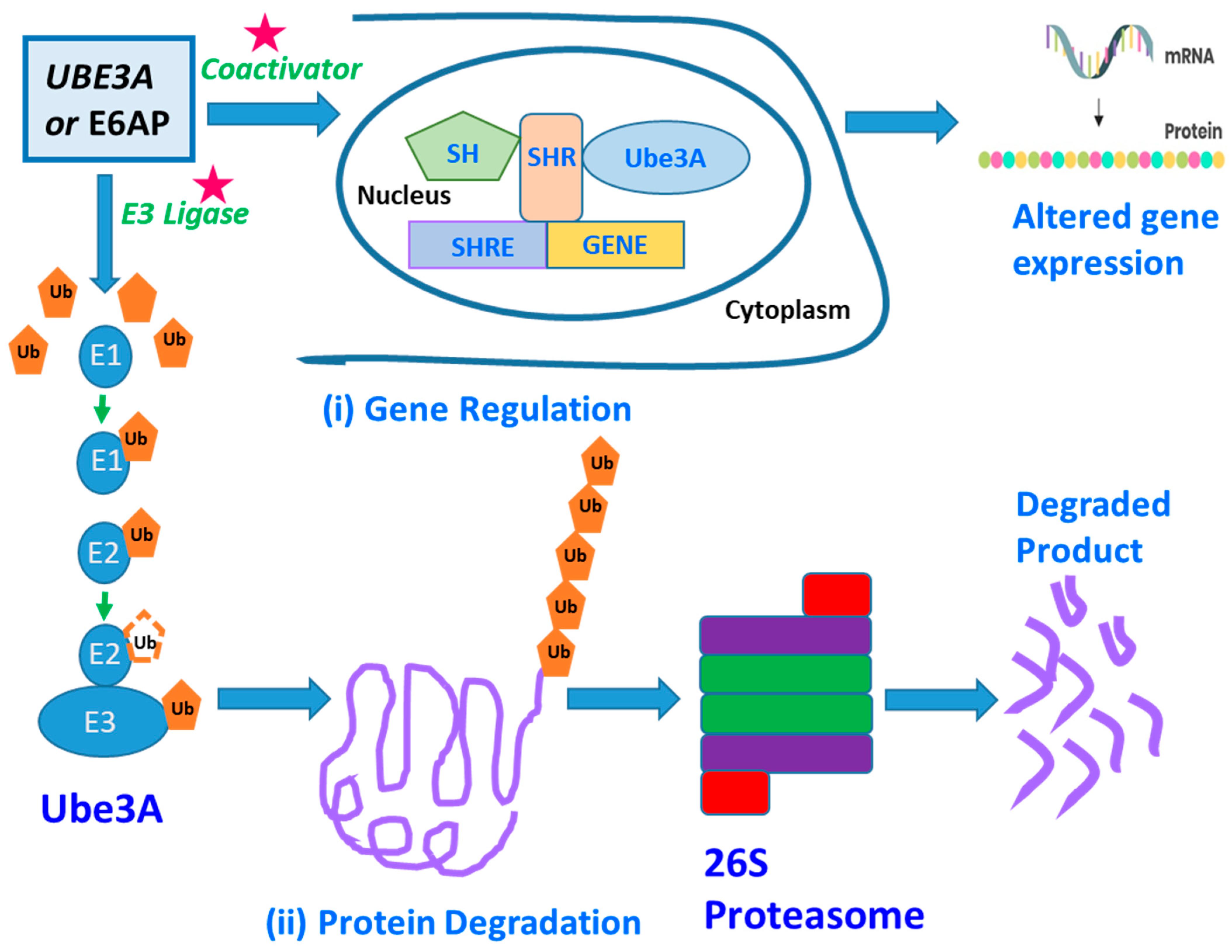
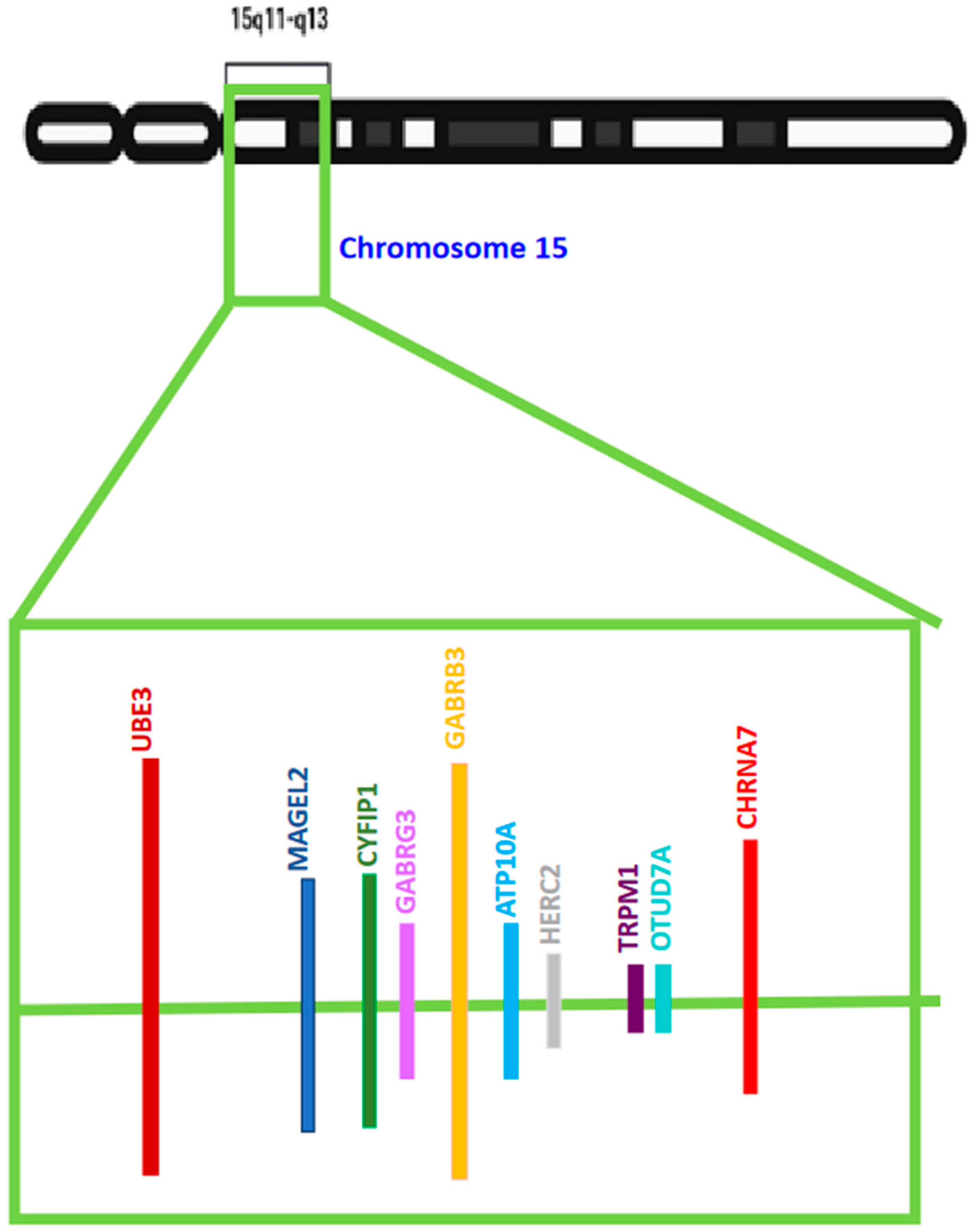
Many of the ASDs exhibiting varying degrees of autism-like phenotypes have chromosomal anomalies in the Chr15q11–q13 region. Numerous potential candidate genes linked with ASD reside in this chromosomal segment. However, several clinical, in vivo, and in vitro studies selected one gene more frequently than others randomly and unbiasedly. This gene codes for UBE3A or Ubiquitin protein ligase E3A [also known as E6AP ubiquitin-protein ligase (E6AP)], an enzyme involved in the cellular degradation of proteins. This gene has been listed as one of the several genes with a high potential of causing ASD in the Autism Database. The gain of function mutations, triplication, or duplication in the UBE3A gene is also associated with ASDs like Angelman Syndrome (AS) and Dup15q Syndrome. The genetic imprinting of UBE3A in the brain and a preference for neuronal maternal-specific expression are the key features of various ASDs. Since the UBE3A gene is involved in two main important diseases associated with autism-like symptoms, there has been widespread research going on in understanding the link between this gene and autism. 

UBE3A mechanisms biomarker

1. Introduction

Published reports from the CDC’s Autism and Development Disabilities Monitoring (ADDM) Networks predicted an average of 1 in every 44 (2.3%) 8-year-old children to have an ASD in 2018. However, reports from the World Health Organization and worldwide estimates suggested 1 out of 100 children suffered from autism in the year 2020, and its occurrence shows a bias towards males, as the median male-to-female ratio was found to be 4.2 [1]. The ADDM Network data suggests that the prevalence of autism per 1000 children rose from 11.3 to 27.6 between 2000–2020 (https://www.cdc.gov/ncbddd/autism/data.html, 22 May 2023).
Many of the ASDs with reported autism-like phenotypes of varying degrees have been attributed to the involvement of chromosomal anomalies in the chromosome 15q11–q13 segment. Several putative candidate genes associated with ASD lie within this chromosomal segment. This suggests that epigenetic modifications in the chromosome 15q11–q13 region might be a contributing factor in the pathophysiology of ASD. However, several scientific analyses and research studies using clinical, in vivo, and in vitro samples and varied techniques have shown that the UBE3A gene was selected in an unbiased and random manner more frequently than other genes.
UBE3A or Ubiquitin protein ligase E3A is an enzyme involved in targeted protein degradation and maintains protein homeostasis within the cell. This gene has been listed as one of the numerous genes with a high potential of causing ASD in the Autism Database, [http://autism.mindspec.org/RescueModel/UBE3A/F_UBE3A_29_KO_HT_G-UBE3A, 22 May 2023; [2][3][4]]. Various mutations including the gain of function mutations, triplication, or duplication in the UBE3A gene are also associated with various ASDs like Angelman Syndrome (AS) and Duplication 15q Syndrome (Dup15Q Syndrome). These syndromes are identified by clinical symptoms like abnormalities in social interactions, intellectual disability, and various behavioral defects, like AS and Dup15q Syndrome [5][6][7][8][9][10][11][12]. The genetic imprinting of UBE3A in the brain and a preference for neuronal maternal-specific expression are the key features of various ASDs including AS and Dup15q Syndrome [13][14][15].
The Dup15q syndrome is a sporadic congenital disease affecting 1 in 30,000 to 1 in 60,000 children globally [16]. AS is also another uncommon neurodevelopmental disorder with an estimated prevalence of 1 in 12,000 to 1 in 20,000 people in the United States (National Organization for Rare Disorders, 2018, retrieved from https://rarediseases.org/rare-diseases/angelman-syndrome, 22 May 2023. Both of these diseases with autistic-like clinical manifestations have been extensively investigated and several genes have been linked and associated with them. However, a few genes like UBE3A and HERC2 were modulated in both these diseases at the transcriptional level [17].
UBE3A or Ubiquitin protein ligase E3A [also known as E6AP ubiquitin-protein ligase (E6AP)] is an enzyme involved in the cellular degradation of proteins. It is transcribed and translated from the UBE3A gene in humans. The degradation of proteins is a cell biological process and involves the removal of dysfunctional, worn out, abnormal, and unnecessary proteins and helps maintain cellular protein homeostasis and normal function. UBE3A labels all damaged or dysfunctional proteins with a small tag or marker protein called ubiquitin. These ubiquitin-tagged proteins are destined for a fate of degradation. Protein complexes such as proteasomes degrade dysfunctional proteins tagged with ubiquitin by proteolytic cleavage of peptide bonds [18][19][20].
Usually, the UBE3A gene actively functions in a large proportion of somatic cells in two copies. However, in neurons, only the copy inherited from the mother [referred to as the maternal copy] is functionally active. This phenomenon is also known as paternal imprinting [14][21][22]. On the other hand, some neurons and glial cells may exhibit a bi-allelic expression of UBE3A [23][24][25]. The silencing of the UBE3A gene on the paternal allele is thought to occur via a Large Non-Coding Antisense Transcript, known as the UBE3A-ATS part of a lincRNA called “LNCAT” [26].
The location of the UBE3A gene, on the long arm [also referred to as the q arm] of chromosome 15 between positions 11 and 13, is an important locus and has been widely studied for deciphering the link between the gene’s chromosomal location and the severity of ASDs [15]. Some neurodevelopmental disorders arise from genetic rearrangements (like duplications and deletions) occurring at the 15q11–q13 locus. These include various forms of ASDs like Prader–Willi syndrome (PWS), AS, and Dup15q Syndrome. The chromosome’s 15q11–q13 region contains several genes which are controlled by genomic imprinting, a genetic event in which genes are specifically expressed from one parental allele. Therefore, the genes that are subjected to modulation by genomic imprinting are functionally haploid and possess only a single functional copy of the respective gene [27]. The three previously mentioned neurodevelopmental disorders arise primarily from deletions or duplications that occur at the 15q11–q13 locus. PWS, AS, and Dup15q Syndrome are disorders that arise from the loss of function or over-expression of at least one imprinted gene. They each occur with a frequency of approximately 1/15,000 to 1/30,000 live births globally [27][28].
PWS is caused by the loss of the paternally inherited chromosome 15q11.2–q13. The loss of expression from this chromosomal region generally occurs by one of the following three mechanisms: (1) 70% of individuals with PWS have a deletion of the entire genetically imprinted 15q11–q13 region, (2) 25% of individuals with PWS have maternal uniparental disomy (matUPD), in which both copies of chromosome 15 have been inherited from the mother, and (3) around 5% of individuals with PWS have an imprinting defect that causes paternal 15q11–q13 to function as though it were inherited from the mother [29]. PWS is characterized by hypotonia, thriving failure, feeding difficulties, hypogonadism, obesity, and hyperphagia [27]. There is no example of a point mutation in any gene causing PWS, suggesting that PWS is a contiguous gene syndrome, caused due to the loss of more than one gene.

2. UBE3A—Structure and Function

An individual’s brain contains billions of neurons connected via trillions of synapses. This requires proper connection and development of the neurons for one’s brain to function normally. The disruption of these developmental processes leads to the development of neuron abnormalities in neurodevelopmental disorders like ASDs [30]. The genetic basis of ASDs is highly diverse because many heterogeneous genes are involved in causing the disease. However, many of these genes express themselves at the initial developmental stage of the disorder. A UBE3/E6-associated protein (E6AP) is one of the many genes with a predisposition to cause ASDs. The UBE3A, Ubiquitin Protein Ligase E3A, is one of the major genes implicated in autism spectrum disorder (ASD) [31] and encodes the E6AP protein, a protein that manifests itself in the brain in an imprinted manner. UBE3A is a gene, and E6AP is a UBE3A-protein product (Figure 1). This research focuses on UBE3A as a potential biomarker by examining UBE3A’s neurobiological functions as a ligase of the Ubiquitin–Proteosome pathway. Several of E6AP’s target proteins with known functions are discussed, including SK2, p53, and Ephexin5 [32][33][34]. The UBE3A gene’s protein product is the E3 ligase or the E6-associated protein (E6AP), and genetic imprinting regulates its specific expression in the brain. Copy number variations (CNVs) leading to E6AP’s overexpression are strongly linked with ASD’s development, followed by clinical manifestations like reduced social communication, diminished social attentiveness, and increased monotonous behavior. Conversely, the loss of or reduction in E6AP’s expression leads to Angelman syndrome (AS), clinically described by speech anomalies, delayed motor development and function, and the occurrence of frequent, repetitive epileptic seizures [32][35][36][37].
Figure 1. This figure represents the functional role of UBE3A or E6AP: UBE3A selectively targets various Ub-tagged proteins in the cytoplasm through the Ubiquitin–proteasome pathway and this degradation pathway regulates various basic neuronal functions (ii). Alternatively, UBE3A could modulate various neuronal genes’ expressions in the nucleus, owing to its ability to function as a co-activator in the mRNA transcription pathway. Altered mRNA levels in the cytoplasm affect protein levels (i). Over-activation or loss of UBE3A’s activity could alter the function and plasticity of the synapse. Altered synaptic function and plasticity could potentially serve as one of the fundamental causes of many behavioral defects and clinical manifestations observed in autism and AS. Abbreviations: SH stands for steroid hormone; SHR stands for steroid hormone receptor; SHRE stands for steroid hormone receptor element; Ub stands for Ubiquitin; E1 stands for Ubiquitin-activating enzyme; E2 stands for Ubiquitin-conjugating enzyme; and E3 for Ubiquitin ligase.
UBE3A is a HECT domain E3 ubiquitin ligase responsible for protein ubiquitination, targeting them for proteomic degradation (Figure 1), [38]. It is generally associated with a neurodevelopmental disorder. E3 ligase, or E6-associated protein (E6AP), is the protein product of UBE3A, whose brain specificity is regulated through genomic imprinting as previously discussed. Differential splicing generates three potential E6AP proteins encoded by the UBE3A gene. The E6AP-coding region is 2.7 Kb long and contains ten exons or coding regions that encode 865 amino acids [39]. Neuronal activities change E6AP’s expression, particularly in cultured neurons, which is enhanced by either glutamate receptor activation or membrane depolarization. Various stimuli which are known to induce synaptic developmental processes in an experience-dependent manner also trigger E6AP’s expression.
The UBE3A gene instructs the synthesis of a protein known as ubiquitin-protein ligase E3A. These are enzymes that catalyze the degradation of other proteins within cells. Ubiquitin-protein ligases attach ubiquitin to proteins that require degradation. The ubiquitin-tagged proteins are then recognized and digested by cellular structures known as proteasomes (Figure 1). Proteolysis or protein degradation involves the removal of unnecessary and damaged proteins. This cellular process aids in maintaining protein homeostasis within cells [40][41]. Scientific research has shown that UBE3A plays an essential role in maintaining the normal function and development of the nervous system. Several studies suggest that UBE3A controls the balance of protein degradation and synthesis at the synapses [31]. Proteostasis regulation is crucial for synapses to adapt and change in response to experience and environment, a trait known as synaptic plasticity. Studies have found that synaptic plasticity is essential for memory and learning. Individuals inherit both copies of the UBE3A gene, with each parent contributing one copy each. These copies are activated in most of the body tissues. However, only the maternal copies are active in the spinal cord and the brain, with paternal copies lying in an active mode. This phenomenon is known as parent-specific gene activation and is also termed genomic imprinting. Normal brain development and functioning depend on proper UBE3A gene dosage, as shown by neurodevelopmental issues associated with mutation, deletion, and UBE3A copy number variations [5][6][42][43][44][45][46].
UBE3A gene dosage plays a role in ASD and AS [39]. The leading cause of Angelman Syndrome (AS) is mutations and deletions in UBE3A.
UBE3A’s functional loss in the brain leads to the development of Angelman Syndrome (AS), a sophisticated neuronal disease primarily affecting the neuronal system. UBE3A loses its function due to gene mutation or chromosomal changes that impact the maternal copy. Various genetic mechanisms can delete or turn off the UBE3A gene. In most cases involving Angelman syndrome a section of maternal chromosome 15 that contains the UBE3A gene is deleted. Other studies have also found Angelman Syndrome to result from UBE3A gene mutation [31]. UBE3A mutation results in the production of various versions of non-functional UBE3A. The copies inherited from the father are often inactive in some parts of the brain; therefore, when the maternal copy is deleted, it prevents the production of an enzyme that leads to the disease. The UBE3A gene’s gain-of-function mutation, triplication, and duplication are associated with autism, a condition clinically described by seizures, speech anomalies, and intellectual disability. Studies have found that UBE3A’s activity and expression should be controlled during the formation and growth of an individual’s brain. The reason behind this can be attributed to the pivotal role this gene plays in regulating synaptic plasticity and functions via proteasome-mediated degradation and targeted protein transcriptional regulation [39]. A genome sequencing study by Iossifov and colleagues [47] found that autism can be associated with a missense mutation [T485A] in the UBE3A gene. The T485A missense mutation affects the phosphorylation site for protein kinase A, known to impede UBE3A activities towards its substrates and itself. According to the International Classification of Disease, autism is a developmental disorder affecting the neuronal network with three important clinical features: repetitive, stereotyped behavior, impaired social skills, and interpersonal communication defects [39]. Most autism cases result from deleting maternally acquired copies of the 15q11–q13 chromosomal region. The 15q11–q13 chromosomal region consists of numerous genes, including the UBE3A gene (Figure 2). The ubiquitin–proteasome pathway, which is also known as the UPP, selectively regulates the levels and quality of different proteins by determining the quality and quantity of these proteins [48] and selectively removing damaged or unnecessary proteins to maintain normal cellular function. This pathway is important for the many functions of cells and the nervous system. During UPP processing and development, the protein substrate is identified by the bonding of a protein ubiquitin via the proteasome which breaks down the molecules of proteins. After initiating the transfer of the E2 enzymes, the E3 ligases which are 100 kDa proteins determine specific aspects of the ubiquitination. One of the major divisions of E3 ligases, HECT, plays an essential role in the regulation of sufficient amounts of UBE3A in brain processes. UBE3A is a gene that is also classified as a ubiquitin ligase [18]. Without proper levels of UBE3A by the HECT domain, an individual is likely to develop serious neurological disorders. Low and high levels of UBE3A or a lack of UBE3A function are linked to disorders such as Angelman syndrome (AS). Angelman syndrome is usually identified by intellectual disability, speech defects, and social and psychological deficits and can often present itself in hyperactive and abnormal patterns [49]. The UBE3A gene is located on the 15th chromosome of the human body [50]. The deletion of this chromosome often results in mental disabilities and developmental delays. An example of a disorder that is believed to be caused by an increased expression of UBE3A is ASD. UBE3A has also been linked to the control of Golgi acidification [51], levels of Cbln1 [52], and function of the mitochondria [53][54][55], and although the function and precision of UBE3A have been studied, the understanding of this brain function is still minimal. UBE3A is not only located on the 15th chromosome but it is specifically found on the 15q11–q13 part of the chromosome. The development of Angelman syndrome is not solely due to the mutation or deletion of UBE3A as many other genes are present spanning this chromosomal region. It was also identified in past studies that the mutation of the 15q11–q13 segment of the chromosome also affects the levels of autistic phenotypes that are displayed. For example, individuals who have an inverted duplicated 15q11–q13 segment usually show mild to moderate forms of autism whereas individuals with three copies of this part of the chromosome display more severe forms of autism spectrum disorder [5][6][42]. The initial finding regarding UBE3A was that this molecule was discovered to degrade the tumor suppressor P53 protein by interacting with the viral E6 oncoprotein in cells invaded by the human papillomavirus [38]. In addition to its ligase activity, UBE3A is known to act as a transcriptional co-activator [56][57][58], a cell cycle regulator [59][60][61], synaptic function, plasticity regulator [32][33][34][62][63][64][65], and cellular protein quality controller [66][67][68].
Figure 2. This figure displays the schematic showing some of the important genes for ASD situated in the Chr15q11–q13 segment. Even though the position and the size of genes have not been appropriately scaled, the researchers have represented several genes that span the 15q11–q13 chromosome segment. The gene of interest, UBE3A, is one of the candidates with a genetic location between 25,333,728 and 25,346,031.
Alternative splicing of the UBE3A gene generates three E6AP protein isoforms. The coding region of the UBE3A gene is 2.7 Kb long, consisting of 10 exons and coding for a protein product comprising 865 amino acids. The second and the third isoforms have an extra 20 and 23 amino acids, respectively, at their amino terminuses. Isoforms 2/3 are similar in their E3 ligase catalytic site and activity. However, the correlation between the variable amino terminus and differential ubiquitination substrate specificity has not been elucidated and still needs further research. Recently, it was reported that UBE3A isoform 1 RNA is coded by a shortened sequence of the gene and lacks the sequence of the E3 catalytic domain. This UBE3A isoform 1 RNA is not translated into a protein within the detectable range. However, this RNA of UBE3A isoform 1 is an important regulator of spine maturation and complex dendrite formation. It was speculated that small non-coding RNAs, like micro-RNA 134 [69], might be a regulator of the UBE3A1 RNA, providing a unique mechanism for regulating the levels of UBE3A in the cells. Optimum levels of UBE3A are important for healthy brain development, as observed in neurodevelopmental disorders linked with various genetic aberrations like the deletions, mutations, and copy number variations (CNVs) of UBE3A.
A fly was also studied to understand UBE3A’s role in the glial function. Glia are known to act as accessory and supporting cells to the neurons in the nervous system [70]. The UBE3A gene in flies is known as the DUBE3A gene, and after the manipulation of the gene, the results observed in the fly are like that found in the animal model monitored. An overexpression of UBE3A (DUBE3A) also produces epileptic seizures, which is consistent with the overexpression or deletion of this gene in humans. In a study that mirrored the same method of increasing the expression of UBE3A in an animal, excessive amounts of UBE3A were found in the nucleus of mice, which affected the glutamatergic transmission negatively and resulted in an impairment in social skills, memory deficits, and other symptoms of Angelman syndrome [71][72]. These findings provide possible explanations of how an overabundance of UBE3A can be linked to autism spectrum disorder. However, it is important to acknowledge that although many of these findings in animal research are consistent with human responses to the manipulation of UBE3A, there are some differences in the expression of UBE3A between animals and humans. The functions of UBE3A extend beyond its link to autism spectrum disorder. For example, it has been observed that UBE3A also functions as a method of regulating synaptic function and synaptic plasticity, which encourages an expansion in memory and learning. UBE3A plays a role in these functions by interacting with the actions of the potassium channel and Arc protein by regulating its transcription process. The link between UBE3A and AS and other ASDs triggered a strong interest amongst researchers to understand the regulation of the synaptic function and plasticity of UBE3A. Numerous protein targets like cytoskeleton-associated protein (Arc), a Rho guanine nucleotide exchange factor (Ephexin5), and a small conductance calcium-activated potassium channel (SK2) [32][33][34] came to be noticed during this research investigation. It was found that UBE3A’s expression is enhanced during experience-dependent neuronal activity. The increased level of UBE3A thereby upregulates the formation of excitatory synapses by regulating the level of Arc, a synaptic protein that induces the internalization of AMPA types of glutamate receptor [32]. However, it was later found that Arc failed to act as a substrate of UBE3A; but, on the other hand, it was transcriptionally regulated by UBE3A [73]. UBE3A is known to tag SK2 with Ubiquitin and stimulate its endocytosis, resulting in increased NMDA receptor activation, thus modulating synaptic plasticity [34]. In the maternal UBE3A-deficient AS-modelled mice, many characteristic features of AS including cognitive and motor dysfunction, seizures, anxiety, aberrant circadian rhythm, and irregular sleep-wake cycles were exhibited [74][75][76][77]. Additionally, impairments in calcium/calmodulin-dependent protein kinase-II activity, long-term potentiation, experience-dependent synaptic plasticity, and imbalance of excitatory/inhibitory circuitry in the brain hippocampal regions [78][79][80][81] were also observed. This suggested that these abnormalities in AS mice could be due to aberrant levels of SK2, Ephexin5, Arc, or some other novel substrate of UBE3A.

3. Potential Biomarkers Related to UBE3A

Duplications and triplications of 15q11.2–q13, are causal factors of autism spectrum disorder (ASD), intellectual disability (ID), delays in developmental processes, and epileptic seizures observed in Dup15q syndrome. Designing targeted therapies for varied forms of ASD and Dup15q syndrome requires biomarkers with the potential to aid in evaluating treatment response at the clinical and bio-molecular levels. Biomarkers can be used as potential stimulators for clinical trials and are computable measurement parameters of drug target efficacy, which, in turn, can influence assessments regarding the continuation of these clinical trials. Several biomarkers associated with UBE3A-mediated ASDs are listed in Table 1, and this table lists the methodology associated with biomarker detection in various research findings.
Table 1. Biomarkers associated with UBE3A-mediated ASDs: Table listing the biomarkers and the methodology associated with the biomarker detection in various research findings.
Electroencephalogram (EEG) forms one of the important characteristic quantifiable features of Dup15q syndrome that possibly reflects molecular pathological characteristics [31][84][85]. Frohlick and coworkers [31] quantified the EEG phenotype as spontaneous beta band (12–30 Hz) oscillations in children with Dup15q syndrome. These children were not taking benzodiazepines or other pharmaceutical drugs known to induce beta activity. The encouraging result of this study shows that the EEG phenotype in patients with Dup15q is a potential optimistic biomarker that might help in quantifying disease pathophysiology and drug–target efficacy, and shows optimistic potential for Dup15q syndrome therapies.
The effective application of the Dup15q syndrome biomarker requires an understanding of the underlying genes, their function, and Dup15q syndrome pathophysiology. There are several 15q genes associated with the disease progression, including UBE3A and a cluster of gamma-aminobutyric acid type-A (GABAA) receptors β3, α5, and γ3 subunit genes. To understand the role of UBE3A or the GABRB3/GABRA5/GABRG3 gene group’s role in mediating the beta EEG phenotype in Dup15q syndrome, Frohlick and coworkers [31] performed some studies. The prior characterization of the beta EEG phenotype showed strong, robust, and reproducible phenotypes in a larger Dup15q syndrome sample. In this study, the healthy and developing children cohort’s beta power was compared with the Dup15q syndrome children cohort with both interstitial and iso-dicentric duplications. The hypothesis is that the dysfunction of the GABAergic system is necessary to produce the beta EEG phenotype. To execute this, the GABAergic dysfunction in patients with Dup15q syndrome was compared to beta oscillations induced by midazolam [pharmacological GABAA modulator] in healthy adults. Additionally, they also evaluated whether UBE3A dysregulation was necessary for the beta EEG phenotype. For this, they compared two cases of paternal Dup15q syndrome to the reference cohort of children with Dup15q syndrome previously mentioned. Their studies were a step forward in efforts toward improving clinical trials in Dup15q syndrome since knowledge regarding the mechanistic underpinnings of the EEG biomarker paves the way for its application in pharmacological trials. These methodologies could therefore serve as an effective and potent quantitative methods for treatment response or drug-target engagement.
Studies have shown that abnormal sleep cycles occur in 40–80% of ASDs [86][87][88][89]. Poor sleep has been associated with enhanced autism severity, cognitive anomalies, and behavioral impairment [90][91]. Sleep physiology has its quantifiable structural attributes like spindles and slow wave sleep (SWS). Abnormal spindle number and morphology are associated with epilepsy, as well as with neurodevelopmental and neuropsychiatric disorders [92][93][94][95][96][97]. Abnormalities in SWS have been observed in neurodevelopmental disorders such as Rett syndrome ([98][99], in both genetic forms of ASD and non-syndromic forms of ASD [100]). One common methodology to quantify sleep physiology is an awake EEG.
Saravanapandian and coworkers [101] have used awake electroencephalography (EEG), as a methodology for screening for patients with Dup15q syndrome. The increased frequency of beta band waves (12–30 Hz) is an electrographic biomarker generated in this methodology which helps in distinguishing the patients with Dup15q from healthy developing and non-syndromic ASD children. The EEG signature matched with the pattern observed in patients placed on a benzodiazepine or allosteric modulators of GABAA receptor therapies. This validates that the EEG biomarker mimics abnormal GABAergic neuronal transmission. Additionally, in this research, the authors investigated the role of sleep physiology in Dup15q syndromic patients. Several metrics [beta band oscillations, spindle density, and percentage of SWS (slow wave sleeps)] were quantified from clinical EEG and polysomnogram (PSG) recordings of patients with Dup15q syndrome and age-matched controls. The results exhibited that Dup15q syndromic children showed aberrant sleep physiology with an elevated beta power, reduced spindle density, and reduced or absent SWS compared to age-matched controls. All these findings pointed towards the strong association between sleep EEG and cognition, identification, and the validation of potential pharmacological targets to improve sleep cycle defects and neurodevelopmental clinical manifestations in this syndrome. This study aided in establishing that abnormal sleep physiology might impede the healthy development of cognitive abilities. Additionally, this study also established that sleep EEG might serve as a robust computable target for behavioral and pharmacological interventions.
Godler and coworkers [83] evaluated differential alterations in the mRNA levels of UBE3A and SNORD116 located within the 15q11–q13 region between Chromosome 15-imprinting disorders (Prader–Willi (PWS), Angelman (AS) and chromosome 15 duplication (Dup15q) syndromes) and their subtypes in correlation with clinical phenotypes. In this study, 58 participants were affected with either PWS, AS, or Dup15q, and 20 healthy controls were included. Utilizing the advanced and sophisticated reverse transcription droplet digital polymerase chain reaction (PCR) technique, a semi-quantitative analysis of UBE3A and SNORD116 mRNA from peripheral blood mononuclear cells (PBMCs) was performed. The normalization of UBE3A and SNORD116 gene levels was performed concerning a panel of internal control genes (housekeeping constitutively active genes) using the geNorm approach. As a step forward, in this study, a functional evaluation of the intellectual development of the experimental subjects was also performed. The parameters set for this evaluation were set according to the standard and reliable guidelines laid by the Autism Diagnostic Observation Schedule-2nd Edition. The researchers found that the Dup15q group showed significantly more elevated levels of UBE3A mRNA than its healthy control counterparts (p < 0.001). The AS and Dup15q groups also had significantly elevated SNORD116 mRNA levels compared to controls (AS: p < 0.0001; Dup15q: p = 0.002). The mRNA levels of UBE3A and SNORD116 also exhibited a positive correlation with the functional intellectual development scores in the deletion AS subjects (p < 0.001) and the clinical autistic manifestations (p < 0.001) in the non-deletion PWS patients. These experimental results hinted towards the existence of novel molecular mechanisms underlying UBE3A and SNORD116 expression in PBMCs and brain-related processes associated with motor and language impairments and autism-like features in these disorders. Additionally, they hypothesized that distinct gene expression profiles in the PBMCs possibly will identify novel immune system-related biological processes in the brain. It is known that the specific expression of genes in the brain microglial cells modulates neuronal processes associated with the intellectual development of an individual. Any form of abnormal alteration in the expression levels of these genes in microglial cells leads to autism features in these disorders. Since direct gene expression profiling from brain neurons or glia is technically more cumbersome and challenging and can only be implemented in post mortem tissues, this study optimistically also facilitates the use of UBE3A and SNORD116 mRNA from PBMCs as potential molecular biomarkers for the clinical diagnosis of these neurodevelopmental disorders and readouts for testing the ameliorating effects of potent pharmacological drugs and gene therapies.
The neurophysiological underpinnings of UBE3A-mediated neurodevelopmental disorders are an unexplored arena and very little information is available for review. Nash and coworkers [39] generated a full-length UBE3A deletion in an AS rat model, with the aim and objective to elucidate novel mechanisms and therapeutic targets. This report demonstrated a novel finding that the UBE3A protein is detectable in an active form in the cerebrospinal fluid (CSF) of wild-type rats but is absent from the CSF of the AS rat. Within the rat’s hippocampal region, micro-dialysis was performed. It was found that the UBE3A protein was concentrated in the interstitial fluid of wild-type rat brains but was significantly absent in AS animals. These findings established that the UBE3A protein maintains its activity and is modulated in a way that strongly relies on its catalytic activity. Their findings proved that CSF can also act as a potential biomarker for UBE3A-mediated ASDs.

References

  1. Zeidan, J.; Fombonne, E.; Scorah, J.; Ibrahim, A.; Durkin, M.S.; Saxena, S.; Yusuf, A.; Shih, A.; Elsabbagh, M. Global prevalence of autism: A systematic review update. Autism Res. 2022, 15, 778–790.
  2. Masini, E.; Loi, E.; Vega-Benedetti, A.F.; Carta, M.; Doneddu, G.; Fadda, R.; Zavattari, P. An Overview of the Main Genetic, Epigenetic and Environmental Factors Involved in Autism Spectrum Disorder Focusing on Synaptic Activity. Int. J. Mol. Sci. 2020, 21, 8290.
  3. Satterstrom, F.K.; Kosmicki, J.A.; Wang, J.; Breen, M.S.; De Rubeis, S.; An, J.Y.; Peng, M.; Collins, R.; Grove, J.; Klei, L.; et al. Large-Scale Exome Sequencing Study Implicates Both Developmental and Functional Changes in the Neurobiology of Autism. Cell 2020, 180, 568–584.e523.
  4. Herrero, M.J.; Velmeshev, D.; Hernandez-Pineda, D.; Sethi, S.; Sorrells, S.; Banerjee, P.; Sullivan, C.; Gupta, A.R.; Kriegstein, A.R.; Corbin, J.G. Identification of amygdala-expressed genes associated with autism spectrum disorder. Mol. Autism 2020, 11, 39.
  5. Malzac, P.; Webber, H.; Moncla, A.; Graham, J.M.; Kukolich, M.; Williams, C.; Pagon, R.A.; Ramsdell, L.A.; Kishino, T.; Wagstaff, J. Mutation analysis of UBE3A in Angelman syndrome patients. Am. J. Hum. Genet. 1998, 62, 1353–1360.
  6. Lossie, A.C.; Whitney, M.M.; Amidon, D.; Dong, H.J.; Chen, P.; Theriaque, D.; Hutson, A.; Nicholls, R.D.; Zori, R.T.; Williams, C.A.; et al. Distinct phenotypes distinguish the molecular classes of Angelman syndrome. J. Med. Genet. 2001, 38, 834–845.
  7. Cook, E.H., Jr.; Lindgren, V.; Leventhal, B.L.; Courchesne, R.; Lincoln, A.; Shulman, C.; Lord, C.; Courchesne, E. Autism or atypical autism in maternally but not paternally derived proximal 15q duplication. Am. J. Hum. Genet. 1997, 60, 928–934.
  8. Schroer, R.J.; Phelan, M.C.; Michaelis, R.C.; Crawford, E.C.; Skinner, S.A.; Cuccaro, M.; Simensen, R.J.; Bishop, J.; Skinner, C.; Fender, D.; et al. Autism and maternally derived aberrations of chromosome 15q. Am. J. Med. Genet. 1998, 76, 327–336.
  9. Thomas, J.A.; Johnson, J.; Peterson Kraai, T.L.; Wilson, R.; Tartaglia, N.; LeRoux, J.; Beischel, L.; McGavran, L.; Hagerman, R.J. Genetic and clinical characterization of patients with an interstitial duplication 15q11–q13, emphasizing behavioral phenotype and response to treatment. Am. J. Med. Genet. A 2003, 119, 111–120.
  10. Hogart, A.; Wu, D.; LaSalle, J.M.; Schanen, N.C. The comorbidity of autism with the genomic disorders of chromosome 15q11.2–q13. Neurobiol. Dis. 2010, 38, 181–191.
  11. Borgatti, R.; Piccinelli, P.; Passoni, D.; Dalpra, L.; Miozzo, M.; Micheli, R.; Gagliardi, C.; Balottin, U. Relationship between clinical and genetic features in “inverted duplicated chromosome 15” patients. Pediatr. Neurol. 2001, 24, 111–116.
  12. Khatri, N.; Man, H.Y. The Autism and Angelman Syndrome Protein Ube3A/E6AP: The Gene, E3 Ligase Ubiquitination Targets and Neurobiological Functions. Front. Mol. Neurosci. 2019, 12, 109.
  13. Dindot, S.V.; Antalffy, B.A.; Bhattacharjee, M.B.; Beaudet, A.L. The Angelman syndrome ubiquitin ligase localizes to the synapse and nucleus, and maternal deficiency results in abnormal dendritic spine morphology. Hum. Mol. Genet. 2008, 17, 111–118.
  14. Rougeulle, C.; Glatt, H.; Lalande, M. The Angelman syndrome candidate gene, UBE3A/E6-AP, is imprinted in brain. Nat. Genet. 1997, 17, 14–15.
  15. Yamasaki, K.; Joh, K.; Ohta, T.; Masuzaki, H.; Ishimaru, T.; Mukai, T.; Niikawa, N.; Ogawa, M.; Wagstaff, J.; Kishino, T. Neurons but not glial cells show reciprocal imprinting of sense and antisense transcripts of Ube3a. Hum. Mol. Genet. 2003, 12, 837–847.
  16. Ortiz-Prado, E.; Iturralde, A.L.; Simbana-Rivera, K.; Gomez-Barreno, L.; Hidalgo, I.; Rubio-Neira, M.; Espinosa, N.; Izquierdo-Condoy, J.; Arteaga-Espinosa, M.E.; Lister, A.; et al. 15q Duplication Syndrome: Report on the First Patient from Ecuador with an Unusual Clinical Presentation. Case Rep. Med. 2021, 2021, 6662054.
  17. Urraca, N.; Hope, K.; Victor, A.K.; Belgard, T.G.; Memon, R.; Goorha, S.; Valdez, C.; Tran, Q.T.; Sanchez, S.; Ramirez, J.; et al. Significant transcriptional changes in 15q duplication but not Angelman syndrome deletion stem cell-derived neurons. Mol. Autism 2018, 9, 6.
  18. Hershko, A.; Ciechanover, A. The ubiquitin system for protein degradation. Annu. Rev. Biochem. 1992, 61, 761–807.
  19. Pelzer, C.; Kassner, I.; Matentzoglu, K.; Singh, R.K.; Wollscheid, H.P.; Scheffner, M.; Schmidtke, G.; Groettrup, M. UBE1L2, a novel E1 enzyme specific for ubiquitin. J. Biol. Chem. 2007, 282, 23010–23014.
  20. Shang, F.; Deng, G.; Obin, M.; Wu, C.C.; Gong, X.; Smith, D.; Laursen, R.A.; Andley, U.P.; Reddan, J.R.; Taylor, A. Ubiquitin-activating enzyme (E1) isoforms in lens epithelial cells: Origin of translation, E2 specificity and cellular localization determined with novel site-specific antibodies. Exp. Eye Res. 2001, 73, 827–836.
  21. LaSalle, J.M.; Reiter, L.T.; Chamberlain, S.J. Epigenetic regulation of UBE3A and roles in human neurodevelopmental disorders. Epigenomics 2015, 7, 1213–1228.
  22. Vu, T.H.; Hoffman, A.R. Imprinting of the Angelman syndrome gene, UBE3A, is restricted to brain. Nat. Genet. 1997, 17, 12–13.
  23. Jones, K.A.; Han, J.E.; DeBruyne, J.P.; Philpot, B.D. Persistent neuronal Ube3a expression in the suprachiasmatic nucleus of Angelman syndrome model mice. Sci. Rep. 2016, 6, 28238.
  24. Grier, M.D.; Carson, R.P.; Lagrange, A.H. Toward a Broader View of Ube3a in a Mouse Model of Angelman Syndrome: Expression in Brain, Spinal Cord, Sciatic Nerve and Glial Cells. PLoS ONE 2015, 10, e0124649.
  25. Judson, M.C.; Sosa-Pagan, J.O.; Del Cid, W.A.; Han, J.E.; Philpot, B.D. Allelic specificity of Ube3a expression in the mouse brain during postnatal development. J. Comp. Neurol. 2014, 522, 1874–1896.
  26. Runte, M.; Huttenhofer, A.; Gross, S.; Kiefmann, M.; Horsthemke, B.; Buiting, K. The IC-SNURF-SNRPN transcript serves as a host for multiple small nucleolar RNA species and as an antisense RNA for UBE3A. Hum. Mol. Genet. 2001, 10, 2687–2700.
  27. Kalsner, L.; Chamberlain, S.J. Prader-Willi, Angelman, and 15q11–q13 Duplication Syndromes. Pediatr. Clin. N. Am. 2015, 62, 587–606.
  28. Whittington, J.E.; Holland, A.J.; Webb, T.; Butler, J.; Clarke, D.; Boer, H. Population prevalence and estimated birth incidence and mortality rate for people with Prader-Willi syndrome in one UK Health Region. J. Med. Genet. 2001, 38, 792–798.
  29. Cassidy, S.B.; Schwartz, S.; Miller, J.L.; Driscoll, D.J. Prader-Willi syndrome. Genet. Med. 2012, 14, 10–26.
  30. Dierssen, M.; Ramakers, G.J. Dendritic pathology in mental retardation: From molecular genetics to neurobiology. Genes Brain Behav. 2006, 5 (Suppl. S2), 48–60.
  31. Frohlich, J.; Reiter, L.T.; Saravanapandian, V.; DiStefano, C.; Huberty, S.; Hyde, C.; Chamberlain, S.; Bearden, C.E.; Golshani, P.; Irimia, A.; et al. Mechanisms underlying the EEG biomarker in Dup15q syndrome. Mol. Autism 2019, 10, 29.
  32. Greer, P.L.; Hanayama, R.; Bloodgood, B.L.; Mardinly, A.R.; Lipton, D.M.; Flavell, S.W.; Kim, T.K.; Griffith, E.C.; Waldon, Z.; Maehr, R.; et al. The Angelman Syndrome protein Ube3A regulates synapse development by ubiquitinating arc. Cell 2010, 140, 704–716.
  33. Margolis, S.S.; Salogiannis, J.; Lipton, D.M.; Mandel-Brehm, C.; Wills, Z.P.; Mardinly, A.R.; Hu, L.; Greer, P.L.; Bikoff, J.B.; Ho, H.Y.; et al. EphB-mediated degradation of the RhoA GEF Ephexin5 relieves a developmental brake on excitatory synapse formation. Cell 2010, 143, 442–455.
  34. Sun, J.; Zhu, G.; Liu, Y.; Standley, S.; Ji, A.; Tunuguntla, R.; Wang, Y.; Claus, C.; Luo, Y.; Baudry, M.; et al. UBE3A Regulates Synaptic Plasticity and Learning and Memory by Controlling SK2 Channel Endocytosis. Cell Rep. 2015, 12, 449–461.
  35. Sadikovic, B.; Fernandes, P.; Zhang, V.W.; Ward, P.A.; Miloslavskaya, I.; Rhead, W.; Rosenbaum, R.; Gin, R.; Roa, B.; Fang, P. Mutation Update for UBE3A variants in Angelman syndrome. Hum. Mutat. 2014, 35, 1407–1417.
  36. Mishra, A.; Prabha, P.K.; Singla, R.; Kaur, G.; Sharma, A.R.; Joshi, R.; Suroy, B.; Medhi, B. Epigenetic Interface of Autism Spectrum Disorders (ASDs): Implications of Chromosome 15q11–q13 Segment. ACS Chem. Neurosci. 2022, 13, 1684–1696.
  37. Burette, A.C.; Judson, M.C.; Li, A.N.; Chang, E.F.; Seeley, W.W.; Philpot, B.D.; Weinberg, R.J. Subcellular organization of UBE3A in human cerebral cortex. Mol. Autism 2018, 9, 54.
  38. Beer-Romero, P.; Glass, S.; Rolfe, M. Antisense targeting of E6AP elevates p53 in HPV-infected cells but not in normal cells. Oncogene 1997, 14, 595–602.
  39. Dodge, A.; Willman, J.; Willman, M.; Nenninger, A.W.; Morrill, N.K.; Lamens, K.; Greene, H.; Weeber, E.J.; Nash, K.R. Identification of UBE3A Protein in CSF and Extracellular Space of the Hippocampus Suggest a Potential Novel Function in Synaptic Plasticity. Autism Res. 2021, 14, 645–655.
  40. Hegde, A.N.; Haynes, K.A.; Bach, S.V.; Beckelman, B.C. Local ubiquitin-proteasome-mediated proteolysis and long-term synaptic plasticity. Front. Mol. Neurosci. 2014, 7, 96.
  41. Hegde, A.N. The ubiquitin-proteasome pathway and synaptic plasticity. Learn Mem. 2010, 17, 314–327.
  42. Saitoh, S.; Buiting, K.; Cassidy, S.B.; Conroy, J.M.; Driscoll, D.J.; Gabriel, J.M.; Gillessen-Kaesbach, G.; Glenn, C.C.; Greenswag, L.R.; Horsthemke, B.; et al. Clinical spectrum and molecular diagnosis of Angelman and Prader-Willi syndrome patients with an imprinting mutation. Am. J. Med. Genet. 1997, 68, 195–206.
  43. Sebat, J.; Lakshmi, B.; Malhotra, D.; Troge, J.; Lese-Martin, C.; Walsh, T.; Yamrom, B.; Yoon, S.; Krasnitz, A.; Kendall, J.; et al. Strong association of de novo copy number mutations with autism. Science 2007, 316, 445–449.
  44. Morrow, E.M.; Yoo, S.Y.; Flavell, S.W.; Kim, T.K.; Lin, Y.; Hill, R.S.; Mukaddes, N.M.; Balkhy, S.; Gascon, G.; Hashmi, A.; et al. Identifying autism loci and genes by tracing recent shared ancestry. Science 2008, 321, 218–223.
  45. Glessner, J.T.; Wang, K.; Cai, G.; Korvatska, O.; Kim, C.E.; Wood, S.; Zhang, H.; Estes, A.; Brune, C.W.; Bradfield, J.P.; et al. Autism genome-wide copy number variation reveals ubiquitin and neuronal genes. Nature 2009, 459, 569–573.
  46. Weiss, L.A.; Shen, Y.; Korn, J.M.; Arking, D.E.; Miller, D.T.; Fossdal, R.; Saemundsen, E.; Stefansson, H.; Ferreira, M.A.; Green, T.; et al. Association between microdeletion and microduplication at 16p11.2 and autism. N. Engl. J. Med. 2008, 358, 667–675.
  47. Iossifov, I.; O’Roak, B.J.; Sanders, S.J.; Ronemus, M.; Krumm, N.; Levy, D.; Stessman, H.A.; Witherspoon, K.T.; Vives, L.; Patterson, K.E.; et al. The contribution of de novo coding mutations to autism spectrum disorder. Nature 2014, 515, 216–221.
  48. Cheon, S.; Dean, M.; Chahrour, M. The ubiquitin proteasome pathway in neuropsychiatric disorders. Neurobiol. Learn. Mem. 2019, 165, 106791.
  49. Buiting, K.; Williams, C.; Horsthemke, B. Angelman syndrome—insights into a rare neurogenetic disorder. Nat. Rev. Neurol. 2016, 12, 584–593.
  50. Kishino, T.; Wagstaff, J. Genomic organization of the UBE3A/E6-AP gene and related pseudogenes. Genomics 1998, 47, 101–107.
  51. Condon, K.H.; Ho, J.; Robinson, C.G.; Hanus, C.; Ehlers, M.D. The Angelman syndrome protein Ube3a/E6AP is required for Golgi acidification and surface protein sialylation. J. Neurosci. 2013, 33, 3799–3814.
  52. Krishnan, V.; Stoppel, D.C.; Nong, Y.; Johnson, M.A.; Nadler, M.J.; Ozkaynak, E.; Teng, B.L.; Nagakura, I.; Mohammad, F.; Silva, M.A.; et al. Autism gene Ube3a and seizures impair sociability by repressing VTA Cbln1. Nature 2017, 543, 507–512.
  53. Su, H.; Fan, W.; Coskun, P.E.; Vesa, J.; Gold, J.A.; Jiang, Y.H.; Potluri, P.; Procaccio, V.; Acab, A.; Weiss, J.H.; et al. Mitochondrial dysfunction in CA1 hippocampal neurons of the UBE3A deficient mouse model for Angelman syndrome. Neurosci. Lett. 2011, 487, 129–133.
  54. Llewellyn, K.J.; Nalbandian, A.; Gomez, A.; Wei, D.; Walker, N.; Kimonis, V.E. Administration of CoQ10 analogue ameliorates dysfunction of the mitochondrial respiratory chain in a mouse model of Angelman syndrome. Neurobiol. Dis. 2015, 76, 77–86.
  55. Santini, E.; Turner, K.L.; Ramaraj, A.B.; Murphy, M.P.; Klann, E.; Kaphzan, H. Mitochondrial Superoxide Contributes to Hippocampal Synaptic Dysfunction and Memory Deficits in Angelman Syndrome Model Mice. J. Neurosci. 2015, 35, 16213–16220.
  56. Nawaz, Z.; Lonard, D.M.; Smith, C.L.; Lev-Lehman, E.; Tsai, S.Y.; Tsai, M.J.; O’Malley, B.W. The Angelman syndrome-associated protein, E6-AP, is a coactivator for the nuclear hormone receptor superfamily. Mol. Cell. Biol. 1999, 19, 1182–1189.
  57. Smith, C.L.; DeVera, D.G.; Lamb, D.J.; Nawaz, Z.; Jiang, Y.H.; Beaudet, A.L.; O’Malley, B.W. Genetic ablation of the steroid receptor coactivator-ubiquitin ligase, E6-AP, results in tissue-selective steroid hormone resistance and defects in reproduction. Mol. Cell. Biol. 2002, 22, 525–535.
  58. Khan, O.Y.; Fu, G.; Ismail, A.; Srinivasan, S.; Cao, X.; Tu, Y.; Lu, S.; Nawaz, Z. Multifunction steroid receptor coactivator, E6-associated protein, is involved in development of the prostate gland. Mol. Endocrinol. 2006, 20, 544–559.
  59. Oda, H.; Kumar, S.; Howley, P.M. Regulation of the Src family tyrosine kinase Blk through E6AP-mediated ubiquitination. Proc. Natl. Acad. Sci. USA 1999, 96, 9557–9562.
  60. Mishra, A.; Jana, N.R. Regulation of turnover of tumor suppressor p53 and cell growth by E6-AP, a ubiquitin protein ligase mutated in Angelman mental retardation syndrome. Cell. Mol. Life Sci. 2008, 65, 656–666.
  61. Mishra, A.; Godavarthi, S.K.; Jana, N.R. UBE3A/E6-AP regulates cell proliferation by promoting proteasomal degradation of p27. Neurobiol. Dis. 2009, 36, 26–34.
  62. Reiter, L.T.; Seagroves, T.N.; Bowers, M.; Bier, E. Expression of the Rho-GEF Pbl/ECT2 is regulated by the UBE3A E3 ubiquitin ligase. Hum. Mol. Genet. 2006, 15, 2825–2835.
  63. Kaphzan, H.; Buffington, S.A.; Jung, J.I.; Rasband, M.N.; Klann, E. Alterations in intrinsic membrane properties and the axon initial segment in a mouse model of Angelman syndrome. J. Neurosci. 2011, 31, 17637–17648.
  64. Kaphzan, H.; Hernandez, P.; Jung, J.I.; Cowansage, K.K.; Deinhardt, K.; Chao, M.V.; Abel, T.; Klann, E. Reversal of impaired hippocampal long-term potentiation and contextual fear memory deficits in Angelman syndrome model mice by ErbB inhibitors. Biol. Psychiatry 2012, 72, 182–190.
  65. Mabb, A.M.; Judson, M.C.; Zylka, M.J.; Philpot, B.D. Angelman syndrome: Insights into genomic imprinting and neurodevelopmental phenotypes. Trends Neurosci. 2011, 34, 293–303.
  66. Mishra, A.; Dikshit, P.; Purkayastha, S.; Sharma, J.; Nukina, N.; Jana, N.R. E6-AP promotes misfolded polyglutamine proteins for proteasomal degradation and suppresses polyglutamine protein aggregation and toxicity. J. Biol. Chem. 2008, 283, 7648–7656.
  67. Mishra, A.; Godavarthi, S.K.; Maheshwari, M.; Goswami, A.; Jana, N.R. The ubiquitin ligase E6-AP is induced and recruited to aggresomes in response to proteasome inhibition and may be involved in the ubiquitination of Hsp70-bound misfolded proteins. J. Biol. Chem. 2009, 284, 10537–10545.
  68. Mulherkar, S.A.; Sharma, J.; Jana, N.R. The ubiquitin ligase E6-AP promotes degradation of alpha-synuclein. J. Neurochem. 2009, 110, 1955–1964.
  69. Valluy, J.; Bicker, S.; Aksoy-Aksel, A.; Lackinger, M.; Sumer, S.; Fiore, R.; Wust, T.; Seffer, D.; Metge, F.; Dieterich, C.; et al. A coding-independent function of an alternative Ube3a transcript during neuronal development. Nat. Neurosci. 2015, 18, 666–673.
  70. Jessen, K.R. Glial cells. Int. J. Biochem. Cell Biol. 2004, 36, 1861–1867.
  71. Nakatani, J.; Tamada, K.; Hatanaka, F.; Ise, S.; Ohta, H.; Inoue, K.; Tomonaga, S.; Watanabe, Y.; Chung, Y.J.; Banerjee, R.; et al. Abnormal behavior in a chromosome-engineered mouse model for human 15q11–q13 duplication seen in autism. Cell 2009, 137, 1235–1246.
  72. Smith, S.E.; Zhou, Y.D.; Zhang, G.; Jin, Z.; Stoppel, D.C.; Anderson, M.P. Increased gene dosage of Ube3a results in autism traits and decreased glutamate synaptic transmission in mice. Sci. Transl. Med. 2011, 3, 103ra197.
  73. Kuhnle, S.; Mothes, B.; Matentzoglu, K.; Scheffner, M. Role of the ubiquitin ligase E6AP/UBE3A in controlling levels of the synaptic protein Arc. Proc. Natl. Acad. Sci. USA 2013, 110, 8888–8893.
  74. Jiang, Y.H.; Armstrong, D.; Albrecht, U.; Atkins, C.M.; Noebels, J.L.; Eichele, G.; Sweatt, J.D.; Beaudet, A.L. Mutation of the Angelman ubiquitin ligase in mice causes increased cytoplasmic p53 and deficits of contextual learning and long-term potentiation. Neuron 1998, 21, 799–811.
  75. Heck, D.H.; Zhao, Y.; Roy, S.; LeDoux, M.S.; Reiter, L.T. Analysis of cerebellar function in Ube3a-deficient mice reveals novel genotype-specific behaviors. Hum. Mol. Genet. 2008, 17, 2181–2189.
  76. Godavarthi, S.K.; Dey, P.; Maheshwari, M.; Jana, N.R. Defective glucocorticoid hormone receptor signaling leads to increased stress and anxiety in a mouse model of Angelman syndrome. Hum. Mol. Genet. 2012, 21, 1824–1834.
  77. Shi, S.Q.; Bichell, T.J.; Ihrie, R.A.; Johnson, C.H. Ube3a imprinting impairs circadian robustness in Angelman syndrome models. Curr. Biol. 2015, 25, 537–545.
  78. Weeber, E.J.; Jiang, Y.H.; Elgersma, Y.; Varga, A.W.; Carrasquillo, Y.; Brown, S.E.; Christian, J.M.; Mirnikjoo, B.; Silva, A.; Beaudet, A.L.; et al. Derangements of hippocampal calcium/calmodulin-dependent protein kinase II in a mouse model for Angelman mental retardation syndrome. J. Neurosci. 2003, 23, 2634–2644.
  79. Yashiro, K.; Riday, T.T.; Condon, K.H.; Roberts, A.C.; Bernardo, D.R.; Prakash, R.; Weinberg, R.J.; Ehlers, M.D.; Philpot, B.D. Ube3a is required for experience-dependent maturation of the neocortex. Nat. Neurosci. 2009, 12, 777–783.
  80. Sato, M.; Stryker, M.P. Genomic imprinting of experience-dependent cortical plasticity by the ubiquitin ligase gene Ube3a. Proc. Natl. Acad. Sci. USA 2010, 107, 5611–5616.
  81. Wallace, M.L.; Burette, A.C.; Weinberg, R.J.; Philpot, B.D. Maternal loss of Ube3a produces an excitatory/inhibitory imbalance through neuron type-specific synaptic defects. Neuron 2012, 74, 793–800.
  82. Gregory, S.G.; Connelly, J.J.; Towers, A.J.; Johnson, J.; Biscocho, D.; Markunas, C.A.; Lintas, C.; Abramson, R.K.; Wright, H.H.; Ellis, P.; et al. Genomic and epigenetic evidence for oxytocin receptor deficiency in autism. BMC Med. 2009, 7, 62.
  83. Baker, E.K.; Butler, M.G.; Hartin, S.N.; Ling, L.; Bui, M.; Francis, D.; Rogers, C.; Field, M.J.; Slee, J.; Gamage, D.; et al. Relationships between UBE3A and SNORD116 expression and features of autism in chromosome 15 imprinting disorders. Transl. Psychiatry 2020, 10, 362.
  84. Urraca, N.; Cleary, J.; Brewer, V.; Pivnick, E.K.; McVicar, K.; Thibert, R.L.; Schanen, N.C.; Esmer, C.; Lamport, D.; Reiter, L.T. The interstitial duplication 15q11.2–q13 syndrome includes autism, mild facial anomalies and a characteristic EEG signature. Autism Res. 2013, 6, 268–279.
  85. Al Ageeli, E.; Drunat, S.; Delanoe, C.; Perrin, L.; Baumann, C.; Capri, Y.; Fabre-Teste, J.; Aboura, A.; Dupont, C.; Auvin, S.; et al. Duplication of the 15q11–q13 region: Clinical and genetic study of 30 new cases. Eur. J. Med. Genet. 2014, 57, 5–14.
  86. Patzold, L.M.; Richdale, A.L.; Tonge, B.J. An investigation into sleep characteristics of children with autism and Asperger’s Disorder. J. Paediatr. Child Health 1998, 34, 528–533.
  87. Wiggs, L.; Stores, G. Sleep patterns and sleep disorders in children with autistic spectrum disorders: Insights using parent report and actigraphy. Dev. Med. Child Neurol. 2004, 46, 372–380.
  88. Liu, X.; Hubbard, J.A.; Fabes, R.A.; Adam, J.B. Sleep disturbances and correlates of children with autism spectrum disorders. Child Psychiatry Hum. Dev. 2006, 37, 179–191.
  89. Souders, M.C.; Mason, T.B.; Valladares, O.; Bucan, M.; Levy, S.E.; Mandell, D.S.; Weaver, T.E.; Pinto-Martin, J. Sleep behaviors and sleep quality in children with autism spectrum disorders. Sleep 2009, 32, 1566–1578.
  90. Devnani, P.A.; Hegde, A.U. Autism and sleep disorders. J. Pediatr. Neurosci. 2015, 10, 304–307.
  91. Cohen, S.; Conduit, R.; Lockley, S.W.; Rajaratnam, S.M.; Cornish, K.M. The relationship between sleep and behavior in autism spectrum disorder (ASD): A review. J. Neurodev. Disord. 2014, 6, 44.
  92. Ferrarelli, F.; Tononi, G. Reduced sleep spindle activity point to a TRN-MD thalamus-PFC circuit dysfunction in schizophrenia. Schizophr. Res. 2017, 180, 36–43.
  93. Wamsley, E.J.; Tucker, M.A.; Shinn, A.K.; Ono, K.E.; McKinley, S.K.; Ely, A.V.; Goff, D.C.; Stickgold, R.; Manoach, D.S. Reduced sleep spindles and spindle coherence in schizophrenia: Mechanisms of impaired memory consolidation? Biol. Psychiatry 2012, 71, 154–161.
  94. Limoges, E.; Mottron, L.; Bolduc, C.; Berthiaume, C.; Godbout, R. Atypical sleep architecture and the autism phenotype. Brain 2005, 128 Pt 5, 1049–1061.
  95. Tessier, S.; Lambert, A.; Chicoine, M.; Scherzer, P.; Soulieres, I.; Godbout, R. Intelligence measures and stage 2 sleep in typically-developing and autistic children. Int. J. Psychophysiol. 2015, 97, 58–65.
  96. Christensen, J.A.; Kempfner, J.; Zoetmulder, M.; Leonthin, H.L.; Arvastson, L.; Christensen, S.R.; Sorensen, H.B.; Jennum, P. Decreased sleep spindle density in patients with idiopathic REM sleep behavior disorder and patients with Parkinson’s disease. Clin. Neurophysiol. 2014, 125, 512–519.
  97. Fernandez, L.M.J.; Luthi, A. Sleep Spindles: Mechanisms and Functions. Physiol. Rev. 2020, 100, 805–868.
  98. Johnston, M.V.; Ammanuel, S.; O’Driscoll, C.; Wozniak, A.; Naidu, S.; Kadam, S.D. Twenty-four hour quantitative-EEG and in-vivo glutamate biosensor detects activity and circadian rhythm dependent biomarkers of pathogenesis in Mecp2 null mice. Front. Syst. Neurosci. 2014, 8, 118.
  99. Ammanuel, S.; Chan, W.C.; Adler, D.A.; Lakshamanan, B.M.; Gupta, S.S.; Ewen, J.B.; Johnston, M.V.; Marcus, C.L.; Naidu, S.; Kadam, S.D. Heightened Delta Power during Slow-Wave-Sleep in Patients with Rett Syndrome Associated with Poor Sleep Efficiency. PLoS ONE 2015, 10, e0138113.
  100. Arazi, A.; Meiri, G.; Danan, D.; Michaelovski, A.; Flusser, H.; Menashe, I.; Tarasiuk, A.; Dinstein, I. Reduced sleep pressure in young children with autism. Sleep 2020, 43, zsz309.
  101. Saravanapandian, V.; Nadkarni, D.; Hsu, S.H.; Hussain, S.A.; Maski, K.; Golshani, P.; Colwell, C.S.; Balasubramanian, S.; Dixon, A.; Geschwind, D.H.; et al. Abnormal sleep physiology in children with 15q11.2–q13.1 duplication (Dup15q) syndrome. Mol. Autism 2021, 12, 54.
More
Information
Subjects: Biology
Contributors MDPI registered users' name will be linked to their SciProfiles pages. To register with us, please refer to https://encyclopedia.pub/register : , , ,
View Times: 249
Revisions: 2 times (View History)
Update Date: 05 Jan 2024
1000/1000
Video Production Service