Submitted Successfully!
To reward your contribution, here is a gift for you: A free trial for our video production service.
Thank you for your contribution! You can also upload a video entry or images related to this topic.
Version Summary Created by Modification Content Size Created at Operation
1 -- 3501 2024-01-02 13:02:24 |
2 Reference format revised. + 2 word(s) 3503 2024-01-03 02:12:26 |

Video Upload Options

Do you have a full video?

Confirm

Are you sure to Delete?
Cite
If you have any further questions, please contact Encyclopedia Editorial Office.
Martins, M.; Vieira, J.; Pereira-Leite, C.; Saraiva, N.; Fernandes, A.S. The Golgi Apparatus as an Anticancer Therapeutic Target. Encyclopedia. Available online: https://encyclopedia.pub/entry/53321 (accessed on 02 May 2024).
Martins M, Vieira J, Pereira-Leite C, Saraiva N, Fernandes AS. The Golgi Apparatus as an Anticancer Therapeutic Target. Encyclopedia. Available at: https://encyclopedia.pub/entry/53321. Accessed May 02, 2024.
Martins, Marta, João Vieira, Catarina Pereira-Leite, Nuno Saraiva, Ana Sofia Fernandes. "The Golgi Apparatus as an Anticancer Therapeutic Target" Encyclopedia, https://encyclopedia.pub/entry/53321 (accessed May 02, 2024).
Martins, M., Vieira, J., Pereira-Leite, C., Saraiva, N., & Fernandes, A.S. (2024, January 02). The Golgi Apparatus as an Anticancer Therapeutic Target. In Encyclopedia. https://encyclopedia.pub/entry/53321
Martins, Marta, et al. "The Golgi Apparatus as an Anticancer Therapeutic Target." Encyclopedia. Web. 02 January, 2024.
The Golgi Apparatus as an Anticancer Therapeutic Target
Edit

The Golgi apparatus (GA) is a central hub in our cells, helping modify and move proteins and lipids. When the GA does not work correctly, it can affect cell processes linked to cancer. This dysregulation can impact how proteins are changed, where they go in and outside the cell, how cells use energy, or even the structure of the extracellular matrix and the environment. That is why targeting the GA could be an appealing approach to treat cancer. Surprisingly, there are no anticancer drugs approved that specifically target the GA.

cancer Golgi apparatus therapy nanotechnology subcellular targeting

1. Introduction

The Golgi apparatus (GA) is an ancient and fundamental eukaryotic organelle found in almost all eukaryotic organisms and cell types [1]. Situated at the vital intersection of the endocytic and exocytic pathways, the GA assumes a pivotal role as a crucial transport center for eukaryotic cells. This organelle serves as a dynamic site for protein and lipid modification and acts as the key orchestrator for lipid and protein sorting and as a store for several molecules and ions [2]. Given its central involvement in membrane and protein trafficking and secretion, and in the storage capacity of second messengers (such as Ca2+), it comes as no surprise that dysregulation of GA function is often associated with human pathologies, such as Batten’s disease [3] or more prevalent conditions like Alzheimer’s disease [4] or cancer [5].

The GA involvement in various cancer hallmarks is of paramount importance, shaping the malignant phenotype of cancer and tumor stromal cells (reviewed in [6]). From aberrant glycosylation to enhanced secretion, the GA apparatus orchestrates a series of intricate molecular events that can contribute to shape cancer progression. It acts as a central hub for the modification and trafficking of cell surface and secreted proteins, allowing cancer cells to evade apoptosis and sustain proliferative signaling. Additionally, it can promote angiogenesis and shape the tumor microenvironment, namely by controlling extracellular matrix (ECM) composition and stromal cell behavior. By regulating the processing and maturation of critical signaling molecules, the GA can have a profound impact on the activation of key oncogenic pathways, thus driving the uncontrolled growth of cancer cells. Moreover, the GA plays a crucial role in the acquisition of invasive and metastatic capabilities by cancer cells. It modulates the synthesis and trafficking of most ECM components and of ECM-modifying enzymes such as matrix metalloproteinases (MMPs), which are crucial enzymes involved in ECM remodeling [7]. By facilitating the secretion and activation of this type of protein, the GA enables cancer cells to breach the physical barriers of the surrounding tissues [5] and initiate the metastatic cascade. Furthermore, the GA is involved in the dysregulated post-translational protein modifications, such as the glycosylation patterns, observed in cancer cells [8]. Aberrant glycosylation of proteins and lipids, catalyzed by GA-resident enzymes, leads to the generation of unique carbohydrate structures on the cell surface. These altered glycan structures play a significant role in cancer cell adhesion, migration, and immune evasion, further fueling tumor progression and metastasis [8]. The GA acts as a key regulator of these glycosylation processes, thus having a profound influence on the functional properties of cancer cells. Some less explored facets of this enigmatic organelle also have the potential to play a significant role in influencing tumor cells by impacting signal transduction, redox regulation, and ion homeostasis.

2. Pharmacological Approaches

2.1. Golgi Apparatus Trafficking Disrupting Agents

The GA is a pivotal element in the secretory pathway where it participates in protein and lipid synthesis, quality control, processing, and transport [2]. The dynamic nature of this organelle implies that it is continuously built by fusion of vesicles and, at the same time, it is used to produce new membrane structures. Vesicles arriving from the ER and other cellular membrane components contribute to create the GA, while, at the same time, parts of this organelle are lost to vesicle trafficking to the plasma membrane through the secretory pathway, to the ER via the retrograde movement of GA-derived vesicles, and to other destinations. The GA receives proteins from the endoplasmic reticulum (ER) and modifies them through glycosylation, phosphorylation, and proteolytic cleavage. The GA also acts as a sorting center, packaging proteins into vesicles for transport to different cellular compartments, and ensures quality control by retaining misfolded proteins for refolding or degradation. The secretory pathway, involved in protein secretion and trafficking, plays a crucial role in cancer progression. The dysregulation of this pathway affects important aspects of cancer cells, including sustained proliferative signaling, the inhibition of cell death (including apoptosis and ferroptosis), the induction of angiogenesis, and the acquisition of invasive and metastatic capabilities, and it can also modulate the amplitude and type of immune response [6].
Understanding and targeting the dysregulated aspects of the secretory pathway in cancer cells may contribute to the development of effective anticancer therapies. In this context, ADP-ribosylation factor 1 (ARF1), a small GTPase protein, plays a key role in the ER-GA secretory pathway by regulating vesicle formation and cargo sorting. It controls the budding of transport vesicles from the GA, facilitating the transport of proteins and lipids to their respective destinations [9]. ARF1 can be activated by GEFs (Guanine Nucleotide Exchange Factors) by catalyzing the exchange of GDP for GTP, enabling ARF1 to carry out its functions in the secretory pathway [9]. Brefeldin A (BFA), a macrocyclic lactone isolated from fungi, is amongst the most studied GA-dispersing compounds. This compound hampers the interaction between members of the ARF1 guanosine triphosphatase (GTPase) family and their associated large GEFs, leading to the tubulation of early endosomes (EEs) and fusion of the GA with the ER [10] (Figure 1). BFA has shown antiproliferative and pro-apoptotic effects in different tumor cell lines [11][12]. It also decreased the invasiveness of bladder cancer cells, an effect that could be related to an increased synthesis of the glycosphingolipid GM3 [13]. BFA, and also monensin, impact the intracellular trafficking of proteins, contributing to anticancer effects. By blocking the trafficking of the multidrug resistance protein P-gp to the cell surface, this protein accumulates intracellularly. In HeLa cells, such a re-localization of P-gp increased the intracellular accumulation of daunorubicin, suggesting that this could constitute a strategy to overcome multidrug resistance [14]. Other combinatory strategies comprising BFA have been proposed. The silencing of ERGIC3 (ER-GA intermediate compartment 3, a transmembrane protein located in the ER and GA often over-expressed in cancer cells), in combination with BFA, additively inhibited lung cancer cell growth [15]. PTEN (phosphatase and tensin homolog on chromosome 10) knockout promotes GA extension and significantly sensitizes cancer cells to secretion inhibitors BFA and golgicide A (GCA) [16].
Figure 1. ARF1 GEF interaction inhibitors. ARFs can be activated by GEFs by allowing the exchange of GDP for GTP, enabling ARFs to control vesicular trafficking. Molecules like brefeldin A (BFA), AMF-26, and golgicide A (GCA) hamper the activation of ARFs and consequently affect vesicular trafficking and protein transport across the GA.
Although the interaction of ARF1 with ARF-GEFs is an attractive target for cancer treatment, BFA did not progress beyond the preclinical stage of drug development, due to its poor bioavailability [11]. Several other small-molecule effectors of endomembrane trafficking were previously identified through large-scale chemical genetic screens, including GCA and AMF-26 [10] (Figure 1). GCA specifically targets Golgi Brefeldin A-Resistant Guanine Nucleotide Exchange Factor 1 (GBF1), a cis-Golgi-residing ARF1-GEF [10][17]. AMF-26, an octahydronaphthalene derivative, is an exocytosis inhibitor that impairs the interaction between the ARF1 GTPase and its GEFs in mammals, disrupting the cis- and trans Golgi [18][19]. AMF-26 showed strong growth inhibition in several cell lines [11][20].
Several protein kinases play key roles in the secretory pathway at the trans-Golgi network and are generally hyperactivated in cancer cells, contributing to cancer progression [21]. Their inhibition has thus been studied as a GA trafficking disruption strategy with potential anticancer effects. For example, IN-9, an antagonist of the phosphatidylinositol (PI)-4-kinase IIIβ (PI4KIIIβ), decreased cell proliferation and migration and increased apoptosis in a model of chromosome 1q-amplified lung adenocarcinoma [22]. Protein kinase D (PKD) inhibitors, such as CID2011756 or CRT0066101, were shown to sensitize the drug-resistant cancer cells to platinum-based anticancer drugs [23] and to suppresses bladder cancer in vitro and in vivo [24]. Despite the importance of other kinases that are associated with the GA for the correct function of this organelle, several of these (namely, protein kinase A, protein kinase C, and Src) are also found at other subcellular localizations where they participate in other Golgi-unrelated pathways. The lack of a Golgi-restricted localization and activity in these cases means that an anticancer application that requires the specific modulation of the Golgi-localized subpopulations of any of the abovementioned kinases would be challenging.

2.2. Golgi-Milieu-Disrupting Agents

Monensin is a polyether monocarboxylic acid obtained from fermentation using Streptomyces cinnamonensin that has been extensively described as a GA-disrupting agent [25][26] (Figure 2). It binds to membranes and acts as an ionophore, inducing cation imbalance and, in turn, leading to a rapid swelling of the GA and a perturbation of intracellular vesicular trafficking [25][26]. In addition, monensin alters the GA pH, affecting post-translational GA-associated processes. These alterations may trigger a GA stress response leading to apoptosis. Monensin has a potent and selective cytotoxicity to cancer cell lines with EMT-like characteristics [26].
Figure 2. GA cation content modulators. The levels of various cations (namely, Ca2+ and H+) in the GA lumen are essential for its function. Molecules such as monensin andsSalinomycin act as cation ionophores, thus allowing the flow of cations across the GA membrane. The inhibition of H+ flow from the Golgi lumen by compounds such as bafilomycin A1 or concanamycin A leads to the alkalinization of this organelle. Changes in GA luminal cation concentrations affect various GA-associated processes like vesicular trafficking, glycosylation, and protein secretion.
Salinomycin is a monovalent cation ionophore similar to monensin, isolated from Streptomyces albus (Figure 2). This compound is highly selective towards cancer stem cells in many cancer types, including breast, ovarian, lung, prostate, and colorectal cancers [27][28]. Multiple mechanisms and molecular targets are described to support this activity, including mechanisms related to apoptosis, autophagy, and necrosis [27]. Among such mechanisms, salinomycin may disturb the GA, altering the expression of GA-related proteins, GA morphology, post-translational modifications of proteins, and secretion [28]
Prodigiosin is a bacterial secondary metabolite that also exerts anticancer effects in different in vitro models (e.g., breast, lung, and oropharyngeal cancers), as well as in a mouse model of lung carcinoma [29]. This compound acts as an H+/Cl symporter, leading to an alkalization of acidic organelles such as the GA. In parallel, as demonstrated in HeLa cells, prodigiosin interferes with Golgi reassembly-stacking protein of 55 kDa (GRASP55), a protein responsible for establishing the stacked structure of the Golgi. 
GA alkalinization may also be achieved by inhibition of vacuolar H+-ATPase (V-ATPase; Figure 2). V-ATPase is generally overexpressed in cancers and its inhibition limits the growth and metastatic properties of cancer cells [30]. V-ATPase inhibitors, such as bafilomycin A1 or concanamycin A, were shown to suppress cancer cell proliferation and invasion, as well as to produce synergistic effects when combined with chemotherapy [30][31].

2.3. Inhibitors of the Synthetic Glycosilation Machinery

The inhibition of glycosidases interferes with the biosynthesis of glycans on cell surface glycoproteins and has thus been explored as an anticancer strategy of great interest. Some examples of glycosylation inhibitors studied as anticancer agents are given below. Targeting the GA α-mannosidase (GM) prevents the formation of ß1,6-branched-complex-type N-glycans. However, all known potent GM inhibitors also interfere with lysosomal α-mannosidase (Lman). The lack of selectivity of the inhibitors developed so far has precluded their clinical use [32]. Swainsonine, mannostatin A, and 1,4-dideoxy-1,4-imino-D-mannitol (DIM) are naturally occurring inhibitors of α-mannosidase II that are effective in nanomolar concentrations (Figure 3). Swainsonine was developed in hydrochloride salt form, under the product code GD0039, which is orally available. This product showed significant in vitro and in vivo antitumor activities and beneficial immunomodulatory effects. However, in a clinical trial enrolling 17 patients with locally advanced or metastatic renal cell carcinoma, no antitumor activity was observed [32].
Figure 3. GA-glycan-processing modulators. Several steps of the glycosylation process occur in the GA. Modulation of this process can alter glycosylation patterns and consequently protein function. Several molecules have been initially identified as glycan-processing modulators (yellow box), and drugs used for other purposes were also reported to possess this type of activity (red box).
Eisenberg-Lerner et al. have studied the impact of inducing GA stress in multiple myeloma cells, which are characterized by a heavy load of glycoprotein production and secretion. Two pharmacological modulators were used: monensin, an ionophore that neutralizes luminal pH and blocks intra-GA trafficking described above; and lithocholylglycine (LCG), which inhibits 2–3 linkage sialyltransferase activity (Figure 3). 
Through a cell-based screening assay using a small-molecule compound library, Sorensen et al. identified two glycosylation inhibitors. The compounds NSC80997 and NSC255112 inhibited glycosylation steps at the GA, including those involved in the formation of O-glycans, N-glycans, and glycosaminoglycans (Figure 3).

2.4. Inhibitors of Peripheral Golgi-Associated Proteins

Many gastrointestinal stromal tumors (GISTs) are caused by gain-of-function mutations in the Kit receptor tyrosine kinase. Imatinib improves the prognosis of GIST patients, but often becomes ineffective with prolonged use because of secondary mutations in the Kit kinase domain. Mutant Kit accumulates predominantly on the GA, whereas normal Kit localizes to the plasma membrane [33][34]. Heat-shock protein 90 (HSP90) is required for the appropriate folding of the KIT protein. TAS-116, an orally active HSP90 inhibitor, decreased GA-localized KIT in both imatinib-naïve and imatinib-resistant GIST cells (Figure 4).
Figure 4. GA protein localization modulators. The precise location of proteins within the GA is essential for the correct function of this organelle. Some compounds can alter the location or change the concentration of specific GA proteins. Several molecules have been initially identified as capable of altering GA protein location (yellow box) while drugs used for other purposes also possess this action (red box).
Other anticancer agents acting on GA are the 2-(substituted phenyl)-benzimidazole (2-PB) compounds (Figure 4). These agents displace resident GA proteins from the juxtanuclear region, resulting in their degradation. 2-PB compounds have been shown to decrease proliferation in a variety of cell lines and suppress tumor growth in xenograft models of human melanoma, breast carcinoma, and renal carcinoma [35].

2.5. Drug Repurposing

Drug repurposing, i.e., the identification of clinically approved drugs with known safety and pharmacokinetic profiles for new indications, has emerged as a useful approach for the search for novel cancer treatments. Such a strategy has also been applied to GA-targeted drugs. For example, drug repositioning plays an important role in identifying potential inhibitors of GA mannosidases. A cell-based screen of a compound library identified three approved drugs, tamoxifen, raloxifene, and sulindac, as potential GA mannosidases inhibitors that induced accumulation of mannose-type N-glycan in HeLa cells [36] (Figure 3). In addition, the approved drug fluvastatin inhibits the mevalonate pathway and N-glycosylation at both the ER and GA (Figure 3). In a mouse model of post-surgical metastatic breast cancer, adjuvant treatment with fluvastatin decreased metastasis and improved overall survival [37].
Memantine is a non-competitive N-methyl-D-aspartate (NMDA) receptor antagonist used in Alzheimer’s disease, which has been suggested as a potential anticancer drug (Figure 4). While its tumor-suppressive effect has been attributed to the blockage of the NMDA-receptor, another mechanism was recently proposed involving the Golgi glycoprotein 1 (GLG1), an intracellular fibroblast growth factor receptor (FGFR). 
Using an image-based screening platform, Gendarme et al. identified 12 novel GA-fragmentation-inducing agents, essentially belonging to two classes of compounds: histone deacetylase inhibitors (HDACi) and DNA-damaging agents. Panobinostat, an HDACi used in the treatment of multiple myeloma, increases the protein trafficking rate without negatively affecting glycosylation or GA dynamics (Figure 4).

3. Nanotechnological Approaches

3.1. Passive Targeting

Passive targeting to tackle cancer cells is based on the intrinsic physicochemical properties of the nanocarriers (size and surface chemistry) and the specific features of the tumor microenvironment, including the enhanced permeability and retention effect. This phenomenon leads to the accumulation of nanocarriers within tumor tissues, thanks to the upregulated angiogenesis that results in an impaired vasculature [38]. Passive targeting strategies have been investigated for the delivery of a wide range of drugs, including those with a primary mechanism of action centered in the GA, such as BFA, monensin, and salinomycin and prodigiosin.
Polymeric nanocarriers have been explored to upgrade the delivery of BFA [39][40], not only because it displays poor bioavailability [11] but also to better target the tumor tissues.
The poor aqueous solubility of salinomycin and its remarkable potency to kill cancer stem-like cells (CSCs) triggered the development of various nanosystems to encapsulate it and to improve salinomycin biodistribution and bioavailability, namely, polymeric micelles, nanofibers and nanoparticles, pegylated liposomes, and nanoemulsions [41][42][43][44][45][46]. Passive-targeting polymeric micelles made of PEG-b-poly(ε-caprolactone) (PEG-b-PCL) to load salinomycin were developed and combined with octreotide-modified paclitaxel-loaded PEG-b-PCL micelles as an alternative approach to improve the treatment of breast cancer. This dual-acting strategy was effective considering both in vitro cytotoxicity studies using the MCF-7 cell line and in vivo studies using MCF-7 xenografts [45].

3.2. Active Targeting

Active targeting is based on ligand-conjugated nanosystems that interact with molecules expressed at the surface or specific organelles of tumor cells. Antibodies and aptamers are widely recognized as highly specific targeting moieties. Other active strategies often explored in the field of anticancer therapy comprise ligand–receptor interactions and peptides [38]. Various active-targeting approaches have been used to specifically deliver GA-acting drugs to cancer cells or to the specific organelle (Figure 5).
Figure 5. Active strategies for drug nanodelivery targeting cancer cells or GA. Antibodies, aptamers, and ligand–receptor interactions have been explored as cancer-cell-specific approaches, whereas polysaccharides (chondroitin sulfate), peptides, and membrane camouflage have been investigated as GA-specific approaches.

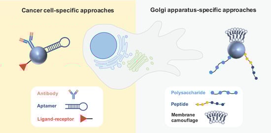
3.2.1. Cancer Cell-Specific Approaches

Active targeting using antibodies was considered for the delivery of monensin and salinomycin to tumor cells. Singh et al. [47] developed monensin-loaded liposomes functionalized with monoclonal antibodies (mAbs) targeting the murine anticarcinoembryonic antigen (CEA), which is well known to be overexpressed in cancer cells. In vitro experiments revealed that mAb-targeted monensin liposomes exhibited a potency 100 times greater than that of monensin liposomes, significantly enhancing the activity of ricin immunotoxins against diverse tumor cell lines. To specifically target human epidermal growth factor receptor 2 (HER2)-positive breast cancer cells, anti-HER2 nanoparticles were developed to deliver salinomycin.
Aptamers were also considered as an active targeting strategy for the nanodelivery of salinomycin to treat melanoma and osteosarcoma. The salinomycin therapeutic efficacy against CSCs was the rationale to develop lipid-polymer nanoparticles functionalized with anti-CD20 aptamers, as CD20-positive stem cells are key for the development and metastatic behavior of melanoma. The selectivity of these nanoparticles was demonstrated and they were more cytotoxic than the non-decorated ones. 

3.2.2. Golgi-Apparatus-Specific Approaches

The central involvement of the GA in orchestrating signaling pathways governing migration, invasion, and angiogenesis has captivated the interest of various research groups, leading to the development of GA-specific active targeting nanoparticles based on ligand–receptor interactions, peptides, and membrane camouflage. These nanosystems are intended to disrupt both the structural characteristics and functional aspects of the GA that underlie metastatic processes in cancer.
Chondroitin sulfate (CS)—a biocompatible, non-immunogenic polysaccharide—was described as an interesting antitumor vector due to its affinity for the surface receptor CD44, which mediates the uptake by tumor cells. Interestingly, this polysaccharide was also found to accumulate in the GA, leading to the design of nanocarriers to selectively target the GA for cancer treatment [48][49]. This strategy was employed to co-deliver a chemotherapeutic agent (doxorubicin or paclitaxel) along with retinoic acid (RA). This derivative of vitamin A was chosen because it was observed to induce changes in the morphology of the GA.

3.3. Physical Targeting

Physical targeting encompasses stimulus-responsive nanosystems that release the cargo upon the effect of an endogenous (pH, hydrogen peroxide concentration, redox state, and hypoxia) or exogenous (light, magnetic fields, heat, and ultrasonic radiation) stimulus [50].
An interesting strategy proposed is a smart nanosystem that takes advantage of the acidic microenvironment of the GA. Bovine serum albumin (BSA) nanoparticles containing a pH-responsive photothermal ablation agent (pH-PTT) based on cyanine dyes for photothermal therapy (PTT) were developed to ultimately kill cancer cells through hyperthermia upon incidence of light. These nanosystems preferentially accumulated in the GA of cancer cells compared to normal cells, and thus can be specifically activated by the acidic environment of the GA in cancer cells. This resulted in a higher photothermal toxicity towards cancer cells, as observed in vitro. In addition, a remarkable photothermal anticancer effect was observed in HePG-2-tumor-bearing mice [51].

4. Conclusions

Various nanotechnological approaches have been proposed for the delivery of GA-process-modulating drugs to cancer cells, making use of passive, active, and physical targeting strategies with successful outcomes in cellular and animal studies. In the last 4 years, elegant targeting approaches, making use of ligand–receptor interactions, transformable peptides, and membrane camouflage, have been suggested for the specific delivery of anticancer compounds to the GA. This may have only been the beginning of the field of organelle-specific targeting, which may be an asset to specifically modulate the GA functions with benefits in terms of anticancer efficacy, particularly related to the control of metastasis and chemoresistance. Thus, it is expected that some of these approaches may reach clinical trials in the near-future. Despite the results of the foreseen trials, questions remain regarding the potential translation of these nanoplatforms into clinical practice as these advanced delivery systems are associated with laborious and costly production methods. Nevertheless, the journey to develop a cost-effective, clinical-translatable, GA-targeting nanoplatform with remarkable anticancer efficacy and proper safety profiles is ongoing with promising initial breakthroughs.

References

  1. Klute, M.J.; Melancon, P.; Dacks, J.B. Evolution and Diversity of the Golgi. Cold Spring Harb. Perspect. Biol. 2011, 3, a007849.
  2. Ma, W.; Mayr, C. A Membraneless Organelle Associated with the Endoplasmic Reticulum Enables 3′UTR-Mediated Protein-Protein Interactions. Cell 2018, 175, 1492–1506.e19.
  3. Kremmidiotis, G. The Batten disease gene product (CLN3p) is a Golgi integral membrane protein. Hum. Mol. Genet. 1999, 8, 523–531.
  4. Crawford, K.; Leonenko, G.; Baker, E.; Grozeva, D.; Lan-Leung, B.; Holmans, P.; Williams, J.; O’Donovan, M.C.; Escott-Price, V.; Ivanov, D.K. Golgi apparatus, endoplasmic reticulum and mitochondrial function implicated in Alzheimer’s disease through polygenic risk and RNA sequencing. Mol. Psychiatry 2023, 28, 1327–1336.
  5. Almeida, N.; Carrara, G.; Palmeira, C.M.; Fernandes, A.S.; Parsons, M.; Smith, G.L.; Saraiva, N. Stimulation of cell invasion by the Golgi Ion Channel GAAP/TMBIM4 via an H2O2-Dependent Mechanism. Redox Biol. 2020, 28, 101361.
  6. Martins, M.; Fernandes, A.S.; Saraiva, N. GOLGI: Cancer cell fate control. Int. J. Biochem. Cell Biol. 2022, 145, 106174.
  7. Kajiho, H.; Kajiho, Y.; Frittoli, E.; Confalonieri, S.; Bertalot, G.; Viale, G.; Di Fiore, P.P.; Oldani, A.; Garre, M.; Beznoussenko, G.V.; et al. RAB2A controls MT1-MMP endocytic and E-cadherin polarized Golgi trafficking to promote invasive breast cancer programs. EMBO Rep. 2016, 17, 1061–1080.
  8. Magalhães, A.; Duarte, H.O.; Reis, C.A. Aberrant Glycosylation in Cancer: A Novel Molecular Mechanism Controlling Metastasis. Cancer Cell 2017, 31, 733–735.
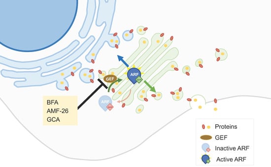
  9. Donaldson, J.G.; Jackson, C.L. ARF family G proteins and their regulators: Roles in membrane transport, development and disease. Nat. Rev. Mol. Cell Biol. 2011, 12, 362–375.
  10. Mishev, K.; Dejonghe, W.; Russinova, E. Small Molecules for Dissecting Endomembrane Trafficking: A Cross-Systems View. Chem. Biol. 2013, 20, 475–486.
  11. Ohashi, Y.; Iijima, H.; Yamaotsu, N.; Yamazaki, K.; Sato, S.; Okamura, M.; Sugimoto, K.; Dan, S.; Hirono, S.; Yamori, T. AMF-26, a Novel Inhibitor of the Golgi System, Targeting ADP-ribosylation Factor 1 (Arf1) with Potential for Cancer Therapy. J. Biol. Chem. 2012, 287, 3885–3897.
  12. Shao, R.-G.; Shimizu, T.; Pommier, Y. Brefeldin A Is a Potent Inducer of Apoptosis in Human Cancer Cells Independently of p53. Exp. Cell Res. 1996, 227, 190–196.
  13. Satoh, M.; Ito, A.; Nojiri, H.; Handa, K.; Numahata, K.; Ohyama, C.; Saito, S.; Hoshi, S.; Hakomori, S.-I. Enhanced GM3 expression, associated with decreased invasiveness, is induced by brefeldin A in bladder cancer cells. Int. J. Oncol. 2001, 19, 723–731.
  14. Fu, D.; Bebawy, M.; Kable, E.P.W.; Roufogalis, B.D. Dynamic and intracellular trafficking of P-glycoprotein-EGFP fusion protein: Implications in multidrug resistance in cancer. Int. J. Cancer 2004, 109, 174–181.
  15. Zhao, Q.; Wu, M.; Zheng, X.; Yang, L.; Zhang, Z.; Li, X.; Chen, J. ERGIC3 Silencing Additively Enhances the Growth Inhibition of BFA on Lung Adenocarcinoma Cells. Curr. Cancer Drug Targets 2020, 20, 67–75.
  16. Shen, S.-M.; Ji, Y.; Zhang, C.; Dong, S.-S.; Yang, S.; Xiong, Z.; Ge, M.-K.; Yu, Y.; Xia, L.; Guo, M.; et al. Nuclear PTEN safeguards pre-mRNA splicing to link Golgi apparatus for its tumor suppressive role. Nat. Commun. 2018, 9, 2392.
  17. Sáenz, J.B.; Sun, W.J.; Chang, J.W.; Li, J.; Bursulaya, B.; Gray, N.S.; Haslam, D.B. Golgicide A reveals essential roles for GBF1 in Golgi assembly and function. Nat. Chem. Biol. 2009, 5, 157–165.
  18. Yang, X.-Z.; Cui, S.-Z.; Zeng, L.-S.; Cheng, T.-T.; Li, X.-X.; Chi, J.; Wang, R.; Zheng, X.F.S.; Wang, H.-Y. Overexpression of Rab1B and MMP9 predicts poor survival and good response to chemotherapy in patients with colorectal cancer. Aging 2017, 9, 914–931.
  19. Yao, M.; Wang, L.; Leung, P.S.C.; Li, Y.; Liu, S.; Wang, L.; Guo, X.; Zhou, G.; Yan, Y.; Guan, G.; et al. The Clinical Significance of GP73 in Immunologically Mediated Chronic Liver Diseases: Experimental Data and Literature Review. Clin. Rev. Allergy Immunol. 2018, 54, 282–294.
  20. Shiina, I.; Umezaki, Y.; Ohashi, Y.; Yamazaki, Y.; Dan, S.; Yamori, T. Total Synthesis of AMF-26, an Antitumor Agent for Inhibition of the Golgi System, Targeting ADP-Ribosylation Factor 1. J. Med. Chem. 2013, 56, 150–159.
  21. Del Giudice, S.; De Luca, V.; Parizadeh, S.; Russo, D.; Luini, A.; Di Martino, R. Endogenous and Exogenous Regulatory Signaling in the Secretory Pathway: Role of Golgi Signaling Molecules in Cancer. Front. Cell Dev. Biol. 2022, 10, 611.
  22. Tan, X.; Banerjee, P.; Pham, E.A.; Rutaganira, F.U.N.; Basu, K.; Bota-Rabassedas, N.; Guo, H.-F.; Grzeskowiak, C.L.; Liu, X.; Yu, J.; et al. PI4KIIIβ is a therapeutic target in chromosome 1q–amplified lung adenocarcinoma. Sci. Transl. Med. 2020, 12, eaax3772.
  23. Janardhanan, P.; Somasundaran, A.K.; Balakrishnan, A.J.; Pilankatta, R. Sensitization of cancer cells towards Cisplatin and Carboplatin by protein kinase D inhibitors through modulation of ATP7A/B (copper transport ATPases). Cancer Treat. Res. Commun. 2022, 32, 100613.
  24. Li, Q.Q.; Hsu, I.; Sanford, T.; Railkar, R.; Balaji, N.; Sourbier, C.; Vocke, C.; Balaji, K.C.; Agarwal, P.K. Protein kinase D inhibitor CRT0066101 suppresses bladder cancer growth in vitro and xenografts via blockade of the cell cycle at G2/M. Cell. Mol. Life Sci. 2018, 75, 939–963.
  25. Zhang, M.; Xu, N.; Xu, W.; Ling, G.; Zhang, P. Potential therapies and diagnosis based on Golgi-targeted nano drug delivery systems. Pharmacol. Res. 2022, 175, 105861.
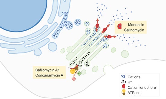
  26. Vanneste, M.; Huang, Q.; Li, M.; Moose, D.; Zhao, L.; Stamnes, M.A.; Schultz, M.; Wu, M.; Henry, M.D. High content screening identifies monensin as an EMT-selective cytotoxic compound. Sci. Rep. 2019, 9, 1200.
  27. Wang, H.; Zhang, H.; Zhu, Y.; Wu, Z.; Cui, C.; Cai, F. Anticancer Mechanisms of Salinomycin in Breast Cancer and Its Clinical Applications. Front. Oncol. 2021, 11, 654428.
  28. Marjanović, M.; Mikecin Dražić, A.-M.; Mioč, M.; Paradžik, M.; Kliček, F.; Novokmet, M.; Lauc, G.; Kralj, M. Salinomycin disturbs Golgi function and specifically affects cells in epithelial-to-mesenchymal transition. J. Cell Sci. 2023, 136, jcs260934.
  29. Nguyen, S.L.T.; Nguyen, T.C.; Do, T.T.; Vu, T.L.; Nguyen, T.T.; Do, T.T.; Nguyen, T.H.T.; Le, T.H.; Trinh, D.K.; Nguyen, T.A.T. Study on the Anticancer Activity of Prodigiosin from Variants of Serratia Marcescens QBN VTCC 910026. Biomed Res. Int. 2022, 2022, 4053074.
  30. Chen, F.; Kang, R.; Liu, J.; Tang, D. The V-ATPases in cancer and cell death. Cancer Gene Ther. 2022, 29, 1529–1541.
  31. Lu, X.; Chen, L.; Chen, Y.; Shao, Q.; Qin, W. Bafilomycin A1 inhibits the growth and metastatic potential of the BEL-7402 liver cancer and HO-8910 ovarian cancer cell lines and induces alterations in their microRNA expression. Exp. Ther. Med. 2015, 10, 1829–1834.
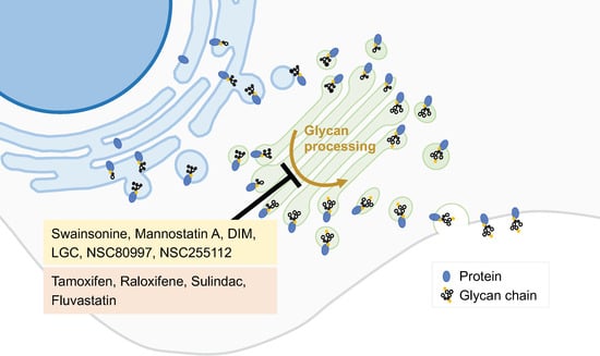
  32. Lee, Z.Y.; Loo, J.S.E.; Wibowo, A.; Mohammat, M.F.; Foo, J.B. Targeting cancer via Golgi α-mannosidase II inhibition: How far have we come in developing effective inhibitors? Carbohydr. Res. 2021, 508, 108395.
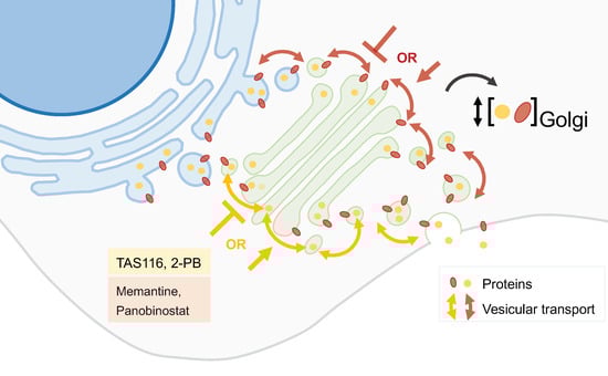
  33. Obata, Y.; Horikawa, K.; Takahashi, T.; Akieda, Y.; Tsujimoto, M.; Fletcher, J.A.; Esumi, H.; Nishida, T.; Abe, R. Oncogenic signaling by Kit tyrosine kinase occurs selectively on the Golgi apparatus in gastrointestinal stromal tumors. Oncogene 2017, 36, 3661–3672.
  34. Saito, Y.; Takahashi, T.; Obata, Y.; Nishida, T.; Ohkubo, S.; Nakagawa, F.; Serada, S.; Fujimoto, M.; Ohkawara, T.; Nishigaki, T.; et al. TAS-116 inhibits oncogenic KIT signalling on the Golgi in both imatinib-naïve and imatinib-resistant gastrointestinal stromal tumours. Br. J. Cancer 2020, 122, 658–667.
  35. Lio, S.C.; Johnson, J.; Chatterjee, A.; Ludwig, J.W.; Millis, D.; Banie, H.; Sircar, J.C.; Sinha, A.; Richards, M.L. Disruption of Golgi processing by 2-phenyl benzimidazole analogs blocks cell proliferation and slows tumor growth. Cancer Chemother. Pharmacol. 2008, 61, 1045–1058.
  36. Koyama, R.; Hakamata, W.; Hirano, T.; Nishio, T. Identification of Small-Molecule Inhibitors of Human Golgi Mannosidase via a Drug Repositioning Screen. Chem. Pharm. Bull. 2018, 66, 678–681.
  37. Yu, R.; Longo, J.; van Leeuwen, J.E.; Zhang, C.; Branchard, E.; Elbaz, M.; Cescon, D.W.; Drake, R.R.; Dennis, J.W.; Penn, L.Z. Mevalonate Pathway Inhibition Slows Breast Cancer Metastasis via Reduced N -glycosylation Abundance and Branching. Cancer Res. 2021, 81, 2625–2635.
  38. Tiwari, H.; Rai, N.; Singh, S.; Gupta, P.; Verma, A.; Singh, A.K.; Kajal; Salvi, P.; Singh, S.K.; Gautam, V. Recent Advances in Nanomaterials-Based Targeted Drug Delivery for Preclinical Cancer Diagnosis and Therapeutics. Bioengineering 2023, 10, 760.
  39. Yu, R.-Y.; Xing, L.; Cui, P.-F.; Qiao, J.-B.; He, Y.-J.; Chang, X.; Zhou, T.-J.; Jin, Q.-R.; Jiang, H.-L.; Xiao, Y. Regulating the Golgi apparatus by co-delivery of a COX-2 inhibitor and Brefeldin A for suppression of tumor metastasis. Biomater. Sci. 2018, 6, 2144–2155.
  40. Liu, W.; Wei, J.; Huo, P.; Lu, Y.; Chen, Y.; Wei, Y. Controlled release of brefeldin A from electrospun PEG–PLLA nanofibers and their in vitro antitumor activity against HepG2 cells. Mater. Sci. Eng. C 2013, 33, 2513–2518.
  41. Basu, S.M.; Yadava, S.K.; Singh, R.; Giri, J. Lipid nanocapsules co-encapsulating paclitaxel and salinomycin for eradicating breast cancer and cancer stem cells. Colloids Surfaces B Biointerfaces 2021, 204, 111775.
  42. Gao, J.; Liu, J.; Xie, F.; Lu, Y.; Yin, C.; Shen, X. Co-Delivery of Docetaxel and Salinomycin to Target Both Breast Cancer Cells and Stem Cells by PLGA/TPGS Nanoparticles. Int. J. Nanomed. 2019, 14, 9199–9216.
  43. Anees, M.; Mehrotra, N.; Tiwari, S.; Kumar, D.; Kharbanda, S.; Singh, H. Polylactic acid based biodegradable hybrid block copolymeric nanoparticle mediated co-delivery of salinomycin and doxorubicin for cancer therapy. Int. J. Pharm. 2023, 635, 122779.
  44. Norouzi, M.; Abdali, Z.; Liu, S.; Miller, D.W. Salinomycin-loaded Nanofibers for Glioblastoma Therapy. Sci. Rep. 2018, 8, 9377.
  45. Zhang, Y.; Zhang, H.; Wang, X.; Wang, J.; Zhang, X.; Zhang, Q. The eradication of breast cancer and cancer stem cells using octreotide modified paclitaxel active targeting micelles and salinomycin passive targeting micelles. Biomaterials 2012, 33, 679–691.
  46. Tefas, L.R.; Toma, I.; Sesarman, A.; Banciu, M.; Jurj, A.; Berindan-Neagoe, I.; Rus, L.; Stiufiuc, R.; Tomuta, I. Co-delivery of gemcitabine and salinomycin in PEGylated liposomes for enhanced anticancer efficacy against colorectal cancer. J. Liposome Res. 2023, 33, 234–250.
  47. Singh, M.; Griffin, T.; Salimi, A.; Micetich, R.G.; Atwal, H. Potentiation of ricin A immunotoxin by monoclonal antibody targeted monensin containing small unilamellar vesicles. Cancer Lett. 1994, 84, 15–21.
  48. Li, H.; Zhang, P.; Luo, J.; Hu, D.; Huang, Y.; Zhang, Z.-R.; Fu, Y.; Gong, T. Chondroitin Sulfate-Linked Prodrug Nanoparticles Target the Golgi Apparatus for Cancer Metastasis Treatment. ACS Nano 2019, 13, 9386–9396.
  49. Luo, J.; Gong, T.; Ma, L. Chondroitin-modified lipid nanoparticles target the Golgi to degrade extracellular matrix for liver cancer management. Carbohydr. Polym. 2020, 249, 116887.
  50. Zhang, C.; Yan, L.; Wang, X.; Zhu, S.; Chen, C.; Gu, Z.; Zhao, Y. Progress, challenges, and future of nanomedicine. Nano Today 2020, 35, 101008.
  51. Xue, F.; Wen, Y.; Wei, P.; Gao, Y.; Zhou, Z.; Xiao, S.; Yi, T. A smart drug: A pH-responsive photothermal ablation agent for Golgi apparatus activated cancer therapy. Chem. Commun. 2017, 53, 6424–6427.
More
Information
Subjects: Oncology
Contributors MDPI registered users' name will be linked to their SciProfiles pages. To register with us, please refer to https://encyclopedia.pub/register : , , , ,
View Times: 152
Revisions: 2 times (View History)
Update Date: 03 Jan 2024
1000/1000