Submitted Successfully!
To reward your contribution, here is a gift for you: A free trial for our video production service.
Thank you for your contribution! You can also upload a video entry or images related to this topic.
Version Summary Created by Modification Content Size Created at Operation
1 -- 3643 2023-12-22 21:29:57 |
2 Reference format revised. Meta information modification 3643 2023-12-25 02:17:10 |

Video Upload Options

Do you have a full video?

Confirm

Are you sure to Delete?
Cite
If you have any further questions, please contact Encyclopedia Editorial Office.
Bautista-Perez, S.M.; Silva-Islas, C.A.; Sandoval-Marquez, O.U.; Toledo-Toledo, J.; Bello-Martínez, J.M.; Barrera-Oviedo, D.; Maldonado, P.D. Antioxidant/Anti-Inflammatory Effects of Garlic in Ischemic Stroke. Encyclopedia. Available online: https://encyclopedia.pub/entry/53081 (accessed on 27 July 2024).
Bautista-Perez SM, Silva-Islas CA, Sandoval-Marquez OU, Toledo-Toledo J, Bello-Martínez JM, Barrera-Oviedo D, et al. Antioxidant/Anti-Inflammatory Effects of Garlic in Ischemic Stroke. Encyclopedia. Available at: https://encyclopedia.pub/entry/53081. Accessed July 27, 2024.
Bautista-Perez, Sandra Monserrat, Carlos Alfredo Silva-Islas, Oscar Uriel Sandoval-Marquez, Jesús Toledo-Toledo, José Manuel Bello-Martínez, Diana Barrera-Oviedo, Perla D. Maldonado. "Antioxidant/Anti-Inflammatory Effects of Garlic in Ischemic Stroke" Encyclopedia, https://encyclopedia.pub/entry/53081 (accessed July 27, 2024).
Bautista-Perez, S.M., Silva-Islas, C.A., Sandoval-Marquez, O.U., Toledo-Toledo, J., Bello-Martínez, J.M., Barrera-Oviedo, D., & Maldonado, P.D. (2023, December 22). Antioxidant/Anti-Inflammatory Effects of Garlic in Ischemic Stroke. In Encyclopedia. https://encyclopedia.pub/entry/53081
Bautista-Perez, Sandra Monserrat, et al. "Antioxidant/Anti-Inflammatory Effects of Garlic in Ischemic Stroke." Encyclopedia. Web. 22 December, 2023.
Antioxidant/Anti-Inflammatory Effects of Garlic in Ischemic Stroke
Edit

Stroke represents one of the main causes of death and disability in the world; despite this, pharmacological therapies against stroke remain insufficient. Ischemic stroke is the leading etiology of stroke. Different molecular mechanisms, such as excitotoxicity, oxidative stress, and inflammation, participate in cell death and tissue damage. At a preclinical level, different garlic compounds have been evaluated against these mechanisms. Additionally, there is evidence supporting the participation of garlic compounds in other mechanisms that contribute to brain tissue recovery, such as neuroplasticity. After ischemia, neuroplasticity is activated to recover cognitive and motor function. Some garlic-derived compounds and preparations have shown the ability to promote neuroplasticity under physiological conditions and, more importantly, in cerebral damage models. 

garlic S-allylcysteine cerebral ischemia neuroplasticity neurogenesis synaptogenesis neurotrophins antioxidant properties anti-inflammatory properties

1. Introduction

Stroke significantly impacts a large segment of the population and stands as one of the leading causes of death and disability. Currently, fibrinolytics and endovascular therapies that induce reperfusion are the only treatments available, yet they are often insufficient and can even result in further brain damage. Consequently, research is focused on identifying new therapeutic targets and protective molecules. Key mechanisms implicated in ischemic stroke-related injury include excitotoxicity, oxidative stress, and inflammation. Numerous molecules demonstrate potent antioxidant and anti-inflammatory properties; however, they frequently fail in clinical trials as effective stroke treatments. On the other hand, there are repair mechanisms such as neuroplasticity that are potential targets for ischemic stroke treatment. Neuroplasticity is a repair mechanism that comprises changes that generate new cells and synaptic connections. Thus, the discovery of novel mechanisms related to recovery in therapeutic stroke research is essential. Garlic and its preparations are a source of antioxidant, anti-inflammatory, and neurotrophic molecules.

2. Stroke

2.1. Stroke Epidemiology and Risk Factors

Amongst neurological diseases, stroke represents one of the leading causes of death and disability worldwide [1]. Furthermore, people affected by stroke require temporary or lifelong assistance, resulting in a huge burden at the human and economic cost levels [2][3].
Stroke is classified into ischemic and hemorrhagic, with a higher prevalence of the ischemic condition. Ischemic stroke occurs when the blood supply decreases under the tissue demand requirements for normal function, resulting in deficiencies in oxygen, glucose, and other molecules required for brain metabolism [4].
Despite the heterogeneity of this disease, some non-modifiable risk factors such as age and gender contribute importantly to the incidence of ischemic stroke. Aging is the strongest non-modifiable risk factor; three quarters of all strokes occur in persons aged >65 years, and the risk doubles every 10 years after the age of 55 [5][6][7]. Moreover, aged patients with stroke have higher mortality and morbidity rates and present poorer functional recovery than their young counterparts [5][6][7]. It is estimated that the increase in the size of the aged population represents an important factor that will contribute to the increase in ischemic stroke cases in the future [8].

2.2. Damage Mechanisms in Ischemic Stroke

Ischemic tissue damage is caused by a disruption in blood supply (ischemia) to the brain, whereas the restoration of blood flow (reperfusion) sometimes leads to an additional form of damage called reperfusion injury [9][10][11]. These two phases trigger a rapid loss of brain function and the development of an infarct region, caused by excitotoxicity, oxidative stress, inflammation, synaptic deficits, the disintegration of neural networks, cell death, and ultimately, failure of neurological functions [9][10][11].
Excitotoxicity is one of the first mechanisms activated after blood vessel occlusion [12][13][14]. This process is mediated by the excitatory neurotransmitter glutamate [12][13][14]. During ischemia, the decrease in ATP levels promotes neuron depolarization, causing a rapid and massive release of glutamate to the synaptic cleft. Additionally, its clearance, mediated by astrocytes, is compromised due to a decrease in cell energy. Glutamate accumulation induces the overactivation of the N-methyl-D-aspartate receptor (NMDAR), resulting in an increase in neuron cytoplasmic calcium levels. These (1) promote the activation of different enzymes such as endonucleases, lipases, and proteases; (2) increase reactive oxygen species (ROS) production; and (3) induce cell damage and death [12][13][14].
Oxidative stress is an imbalance between prooxidants and antioxidants due to an increase in oxidant agents, a decrease in antioxidant systems, or a combination of both conditions [15]. During ischemia, ROS production increases due to the activation of calcium-dependent enzymes such as xanthine oxidase. Additionally, during reperfusion, the increases in tissue oxygen level promote a second and major burst of ROS, generated mainly by mitochondria and NADPH oxidase [16][17][18]. The increase in ROS levels promotes their interaction with biomolecules, leading to the aberrant regulation, altering, or destroying of the function.

2.3. Neuroprotective Mechanisms in Ischemic Stroke: Neuroplasticity

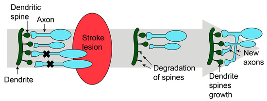
2.3.1. Synaptogenesis
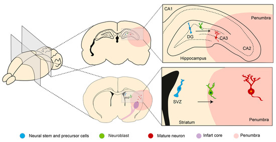
Synaptic plasticity appears during animal development and continues throughout life, but is decreased in aging [19]. Even during ischemic stroke, the formation of new synapses occurs in the damaged tissue (Figure 1) [20][21]. Two mechanisms have been described. First, dendritic spines undergo remodeling in the peri-infarct zone [20][21]. Within the first two weeks, there is an increase in the number and the turnover of dendritic spines [20]. Then, in the peri-infarct area, neurons develop branches and establish new connections [22]. The new synapses can be developed at the local level or reach longer distances, forming new circuits [22][23]. Synaptogenesis and axonal sprouting occur simultaneously [22][23].
Figure 1. Synaptogenesis after stroke. After injury, new dendritic spines grow and new axons are formed, resulting in new mature synapses. Figure was made in Illustrator 2022.

2.3.2. Neurogenesis

The other mechanism of repair is neurogenesis, leading to the generation of new functional neurons from the neural stem and precursor cells (NS/PS). Like synaptogenesis, neurogenesis occurs in mammals throughout life in restricted brain regions. It is activated after stroke and starts at the neurogenic niches where the NS/PC are located (Figure 2) [24].
Figure 2. Neurogenesis after stroke. Neural stem and precursor cells (NS/PC) reside in two neurogenic regions in the adult mammalian brain: the subventricular zone (SVZ) and subgranular zone (SGZ) of the dentate gyrus (DG) in the hippocampus. After stroke, neurogenesis could be activated, generating new mature neurons that migrate to CA3 or the stroke lesion. Arrows indicated the direction cell migration. Figure was made in Illustrator 2022.

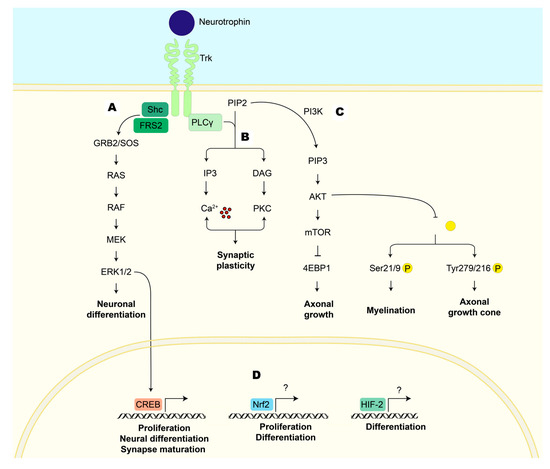
2.3.3. Neurotrophic Factors

Neurotrophic factors are a group of soluble polypeptides delivered by cells, with a wide range of functions in the nervous system, including neuronal survival and repair, synaptic plasticity, and the formation of long-lasting memories [25]. They are divided into different families according to their structure and function:
(1)
NTs promote neuronal survival, neuronal differentiation, axonal and dendritic growth, synaptic plasticity, and synaptogenesis [26]. Some examples are nerve growth factor (NGF), brain-derived neurotrophic factor (BDNF), and neurotrophin-3 (NT-3).
(2)
Members of the transforming growth factor family (TGF) stimulate astrocyte proliferation, migration, and transformation to the axon growth-supportive phenotype [27]. They stimulate neural cell proliferation and differentiation and the synthesis of NGF in astrocytes [27]. After stroke, they promote neurogenesis, angiogenesis, and provide oligodendrocyte protection [28], e.g., glia-derived neurotrophic factor (GDNF).
(3)
Neurokines, such as interleukin 6 (IL6), play critical roles in immunity, brain-regulating neurodevelopment, food intake, body temperature, learning, and memory [29].
(4)
Non-neuronal factor families have neurotrophic and angiogenic activity [30]. They act as neuroprotective signals against acute ischemic brain injury [30], e.g., insulin growth factor (IGF).
Other proteins called angioneurins act as neurotrophic factors and regulate angiogenesis. They act on neurons and vascular cells directly (promoting their proliferation and migration and altering the composition of the extracellular matrix to facilitate angiogenesis) or indirectly (recruiting pro-angiogenic cells like mesenchymal stem cells and promoting the release of angiogenic factors by neurons and astrocytes) [31].
Neurotrophic factors exert their biological activities through tyrosine kinase activity receptors. The tropomyosin-related kinase (Trk) receptor family is the main target of NTs (each NT has a preference for a specific Trk receptor). The binding NT/Trk receptor activates different pathways, including (A) mitogen-activated protein kinase/extracellular signal-regulated kinase (MEK/ERK), (B) phospholipase C gamma (PLCγ), and (C) PI3K/AKT. This activation induces transcription factors such as CREB that increase the expression of proteins involved in promoting neuronal survival, differentiation, cytoskeletal rearrangement, synapse formation, and synaptic plasticity (Figure 3) [32].
Figure 3. Cellular pathways activated by the union of neurotrophin to the tropomyosin-related kinase (Trk) receptor that induces neuroplasticity. (A) MEK/ERK: The adapter protein GRB2 binds to the phosphorylated Trk receptor. GRB2 is associated with the SOS protein, which promotes the activation of RAS, which initiates the kinase cascade that includes RAF, MEK, and ERK. ERK can get into the nucleus to activate transcription factors (CREB), promoting neuronal differentiation and synapse maturation. (B) PLCγ: The phosphorylated Trk receptor activates PLCγ, which hydrolyzes PIP2 into two secondary messengers: IP3 and DAG. The first binds to its receptor on the endoplasmic reticulum, causing the release of calcium into the cytoplasm, whereas DAG activates PKC. Calcium and PKC modulate ion channels, affecting membrane potential and excitability and modulating synaptic plasticity. (C) PI3K/AKT: The phosphorylated Trk receptor recruits PI3K, which, in turn, phosphorylates PIP2 to generate PIP3. PIP3 serves as a secondary messenger that recruits AKT. AKT activation leads to the activation of the mTOR pathway, which plays a role in protein synthesis and impacts axonal growth. AKT can phosphorylate GSK3β, inhibiting its kinase activity; this could favor the survival and differentiation of oligodendrocytes, which are critical for myelination, or affect neuronal structure, stimulating the axonal growth cone. (D) Other transcription factors that regulate neuroplasticity after stroke are Nrf2 and HIF-2. Both transcription factors are stabilized and translocated into the nucleus, where they induce the transcription of genes involved in proliferation (Nrf2) and differentiation (HIF-2). AKT: serine/threonine protein kinase; CREB: cyclic AMP response-element-binding protein; DAG: diacylglycerol; 4E-BP1: eukaryotic translation initiation factor 4E-binding protein 1; ERK1/2: extracellular signal-regulated kinase; FRS2: factor receptor substrate 2; GRB2: growth factor receptor-bound protein-2; GSK3 β: glycogen synthase kinase-3β; HIF-2: hypoxia-inducible factor 2; IP3: inositol 1,4,5-trisphosphate; MEK: mitogen-activated protein kinase kinase; mTOR: mechanistic target of rapamycin; Nrf2: nuclear factor erythroid 2-related factor 2; PI3K: phosphoinositide 3-kinase; PIP2: phosphatidylinositol 4,5-bisphosphate; PIP3: phosphatidylinositol 3,4,5-trisphosphate; PKC: protein kinase C; PLCγ: phospholipase C gamma; RAF: rapidly accelerated fibrosarcoma kinases; RAS: rat sarcoma virus proteins; Shc: Src homology and collagen; SOS: RAS activator son of sevenless; Trk: tropomyosin-related kinase. Figure was made in Illustrator 2022.
MEK/ERK pathway activation occurs after ligand–receptor dimerization, leading to the phosphorylation of tyrosine residues of the carboxyl terminal of the receptor, which acts as docking site for Shc (Src homology and collagen) and fibroblast growth factor receptor substrate 2 (FRS2) and forms a complex with growth factor receptor binding protein 2 (GRB2). This complex is constitutively associated with rat sarcoma virus proteins (RAS) and the activator son of sevenless (SOS), forming the GRB2/SOS complex [33]. The recruitment of this complex activates RAS and rapidly accelerates the fibrosarcoma kinase (RAF)/MEK/ERK cascade [34].

2.4. Treatments for Ischemic Stroke

The main objective of ischemic stroke treatment is to provide safe revascularization and, therefore, limit the neuronal damage. Additionally, the proper management of patients is mandatory and includes early hemodynamic stabilization and monitoring of possible complications. Revascularization of the affected brain area could be carried out by intravenous drug thrombolysis and endovascular thrombectomy under imaging guidance [35].

Intravenous Thrombolysis

The only drug approved by the United States Food and Drug Administration (FDA) for the treatment of acute ischemic stroke is alteplase, a recombinant tissue plasminogen activator (rtPA). rtPA is an enzyme that converts plasminogen to plasmin, dissolving the blood clot responsible for blood flow obstruction. However, its use is limited due to the exclusion criteria defined by each country [36][37]. Due to the differences in criteria and other cultural and economic factors, there are variations between countries and the percentage of patients who receive thrombolytic therapy.
Primary prevention includes strategies to prevent a first stroke or transient ischemic attack (TIA) in patients. There are modifiable risk factors and non-modifiable risk factors. Nevertheless, 90% of risk for stroke worldwide is attributable to modifiable risk factors. Hence, the management of these risk factors is the best strategy for preventing first-ever stroke [38].

3. Garlic

Garlic (Allium sativum L.) is a vegetable that has been used worldwide since ancient times in folk medicine and gastronomy in many cultures [39][40]. Garlic cloves are commonly used for the treatment of fungal and bacterial infectious diseases, and as a cardiovascular protective measure for the prevention of stroke. Garlic extracts have been used for blood sugar maintenance, to reduce serum cholesterol levels, and for the treatment of rheumatism, toothache, and earache [41].
Garlic cloves contain (1) 62–68% water; (2) 26–30% carbohydrates (it has a high content of fructans, such as fructose polymers); (3) 1.5–2.1% proteins; (4) 1–1.5% free amino acids (which is like its protein content); (5) 1.5% fibers; and (6) 1.1–3.5% organosulfur compounds (OSCs) [42][43].
The medicinal properties of garlic are mainly associated with its OSC, like allicin, diallyl sulfide (DAS), diallyl disulfide (DADS), diallyl trisulfide (DATS), and S-allylcysteine (SAC). The effects of these compounds have been evaluated in several preclinical models and some clinical trials in the treatment of different diseases [44][45].
In fresh garlic, the principal OSCs are S-allylcysteine sulfoxide (alliin, 6–14 mg/g fresh weight), γ-glutamyl-S-trans-1-propenylcysteine (3–9 mg/g fresh weight), γ-glutamyl-S-allylcysteine (2–6 mg/g fresh weight), methylcysteine sulfoxide (methiin, 0.5–2 mg/g fresh weight), cycloalliin (0.5–1.5 mg/g fresh weight), and trans-1-propenylcysteine sulfoxide (isoalliin, 0.1–1.2 mg/g fresh weight) (Figure 4A) [46][47][48][49]. These cysteine sulfoxides are odorless compounds; however, when garlic cloves are cut, crushed, or chewed, they are transformed to thiosulfinates [49][50]. The formation of these compounds occurs when cysteine sulfoxides, located in clove mesophyll storage cells, are metabolized by allinase or alliin lyase (10 mg/g fresh), an enzyme localized in the vacuoles of vascular bundle sheath cells. Due to the abundance of alliin in cloves, the main thiosulfinate formed is allicin (Figure 4B). Thiosulfinates are reactive and unstable compounds, and when they are processed in oils or by aging (commercial garlic products), other OSCs are produced [44][47].
Figure 4. Main organosulfur compounds (OSCs) in garlic cloves and garlic products. (A) In garlic cloves, the main OSCs are γ-glutamyl-S-cysteines (γ-glutamyl-S-allylcysteine and γ-glutamyl-S-t-1-propenylcysteine) and cysteine sulfoxides (alliin, methiin, and cycloallin). (B) When garlic cloves are cut, cooked, or crushed, new compounds are formed such as the thiosulfinates (allicin and allylmethanethiosulfinate) by the interaction between alliin and alliinase. (C) In commercial garlic products, the transformation of OSCs (γ-glutamyl-S-cysteines and thiosulfinates) depends on the enzymatic reactions and extraction conditions. Adapted from [48]. The image was made in Inkscape.

3.1. Garlic Preparations

In addition to garlic cloves, several commercial garlic products are consumed: (1) garlic powder (dried garlic), (2) aged garlic extract (AGE), (3) steam-distilled garlic oils, and (4) garlic oil macerate. The OSC content in each product is different, and its transformation depends on the enzymatic reactions and extraction conditions (Figure 4C) [48].
Garlic powder is the most identical product to garlic cloves since it dehydrated at low oven temperatures (50–60 °C) and pulverized. The amount of alliin will depend on the care used in slicing and handling the cloves [48].
AGEs are obtained from the prolonged (aging) extraction (18–24 months) of chopped garlic in 20% ethanol (12 mL/g) in a closed stainless-steel container at room temperature [48][51]. Under these conditions, the main changes are: (1) the complete loss of thiosulfinates after 3 months, converted to volatile allyl sulfides, and (2) the complete hydrolysis of γ-glutamyl-S-alkylcysteines to form SAC (7.2 mg/g dry extract) and S-1-propenylcysteine (4.4 mg/g dry extract), the main OSCs in AGE. SAC content remains constant after 3 months, but S-1-propenylcysteine decreases from 12 months. Additionally, the cysteine (1.2 mg/g dry extract) and S-allylmercaptocysteine (1.9 mg/g dry extract) content increases at 24 months [48]. In fresh garlic, the γ-glutamyl-S-allylcysteine (localized in vacuoles) is metabolized by γ-glutamyltranspeptidase (bound to cell membranes) to form SAC [44][52]. In fresh garlic cloves, the SAC levels are low (0.27–0.68 mg/g of dry weight) [49]; however, it is the main OSC in AGE [44].

3.2. Garlic Compounds as Treatment for Ischemic Stroke

Compounds derived from garlic are known to have antioxidant and anti-inflammatory properties. They can scavenge different ROS [53][54], and some (SAC, DATS, and DADS) show the ability to promote the activation of Nrf2 transcription factor, increasing endogenous antioxidant defense [55][56][57][58]. Also, SAC, DATS, and DAS inhibit the nuclear factor kappa-light-chain-enhancer of activated B cells (NF-κB) transcription factor, decreasing the expression of different proinflammatory cytokines, such as tumor necrosis factor alfa (TNFα), interleukin (IL)1β, IL6, monocyte chemoattractant protein-1 (MCP-1), and IL-12 [59][60]. Due to these properties, garlic-derived compounds have been evaluated in different ischemic stroke models, showing a neuroprotective effect against the damage induced by brain ischemia [53][60].
In vitro models, SAC shows protection against OGD/reoxygenation insult, increasing viability [61] and decreasing apoptosis [58] through the inhibition of the ERK [53], c-Jun N-terminal kinase (JNK), and 38-kDa mitogen-activated protein kinase (p38) pathways and the activation of the Nrf2 pathway [58]. Allicin and alliin prevent the decline of cellular viability induced by OGD/reoxygenation [61][62]
Furthermore, garlic OSCs also promote brain protection in focal brain ischemia models. SAC administered before ischemia decreases neurological deficit and infarct volume, preventing the activation of the ERK1/2 [53], JNK, and p38 pathways [58]. Additionally, it reduces oxidative stress, and increases glutathione (GSH) [63] and antioxidant defense levels (HO-1, glutamate-cysteine ligase catalytic subunit (GCLC), and glutamate-cysteine ligase regulatory subunit (GCLM)) through the Nrf2 pathway [58], as well as the activity of the antioxidant enzymes glutathione reductase (GR), glutathione peroxidase (GPx), SOD, and CAT [64]. Also, SAC reduces the increase in glial fribillary acidic protein (GFAP) and inducible nitric oxide synthase (iNOS) levels [64], resulting in the improvement of neurological deficits [58][63] and a reduction in infarct volume and brain edema [58][63][64][65].

3.3. Garlic Preparations as Treatment for Ischemic Stroke

Commercial garlic products, which contain a mixture of different OSCs, also show protection against global brain ischemia. Pretreatment with aqueous garlic extract reduces inflammation [66], whereas garlic oil decreases infarct volume and lipoperoxidation, and improves short-term memory and motor coordination [67]. In focal brain ischemia, AGE, aqueous garlic extract, and garlic clove and skin extracts (GCE and GSE) show brain tissue protection. AGE decreases neurological impairment, infarct area, and brain edema by reducing oxidative stress and inflammation [65][68][69] and increasing GLUT3 transporter [70]. Aqueous garlic extract improves neurobehavioral problems, diminishes cell death, and enhances antioxidant defense [71]. GCE and GSE reduce cell damage and increase mitochondrial membrane potential and ATP levels, which could be associated with its scavenging activity against superoxide anions, peroxynitrite, hydroxyl radicals, and peroxyl radicals [72].

3.4. Garlic Compounds and Neuroplasticity

The neurotrophic effects of SAC include an increase in axonal branching, neurite length, and the number of neurites in hippocampal neuron cultures. The changes in the morphology of neurons are related to better efficiency of the transmission and information processing ability of the neural network [73][74]. Also, after cell damage triggered by excitotoxic insult with quinolinic acid, SAC treatment increases the levels of the neurotrophin BDNF, antioxidant defenses (HO-1) through the Nrf2 pathway, and ERK1/2 phosphorylation levels [75].
SAC is the OSC that is most studied in vivo, and its trophic effects have been proven in different models. Treatment administered for 21 days to young healthy animals increases the number of positive cells to marker of proliferation Ki67 (Ki67) and the marker of neuroblast differentiation (doublecortin) in the SGZ of the dentate gyrus in the hippocampus. Furthermore, SAC increases serotonin 1 A receptor levels, and the activation of these receptors increases neurogenesis in the dentate gyrus [76]. Also, it improves memory in senescence-accelerated animals, or damage due to streptozotocin or lipopolysaccharide [74][77][78]. Senescence-accelerated mouse prone is a model for aging and age-related disorders that has a short lifespan and age-dependent pathologies like impairment in learning and memories. The improvement in memory in senescence-accelerated mouse prone treated with SAC was accompanied by the preservation of α-amino-3-hydroxy-5-methyl-4-isoxazolepropionic acid receptor (AMPAR), NMDAR, and phosphorylated α-calcium/calmodulin-dependent protein kinase II (CaMKII) in the hippocampus; these proteins are related to the maintenance of learning and memory functions [74].
The intraventricular streptozotocin administration model produces cognitive deficits and oxidative damage in the hippocampus. SAC prevents cognitive and neurobehavioral impairments, increases the antioxidant state (GSH, GPx and GR), and diminishes thiobarbituric acid-reactive substances (TBARS) and apoptotic parameters (DNA fragmentation, the expression of B cell lymphoma 2 (Bcl-2) and tumor protein p53 (p53)) [77]. Similarly, lipopolysaccharide administration induces learning and memory impairment and neuroinflammation. SAC improves memory, mitigates lipid peroxidation (malondyaldehyde) and augments SOD, GSH, and acetylcholinesterase activity. Furthermore, it downregulated hippocampal NF-κB, Toll like receptor 4 (TLR4), GFAP, IL-1β, and ionized calcium-binding adaptor molecule (Iba1) and upregulated Nrf2 [78].
The other OSCs that have shown an increase in memory performance after injury are the allicin and Z-ajone [79][80]. The effects of allicin have been mainly related to morphological modifications, increasing the density of the dendritic spine, and synaptophysin and glutamate receptor-1 levels, indicating the formation of new synapses [79]. As mentioned before, the formation of new synapses after stroke has been linked to functional and cognitive recovery. Z-ajone has inhibitory effects against memory impairment induced by scopolamine [80].
Essential oils from two Allium species administered for 21 days to healthy animals increase memory, cell proliferation, and neuroblast differentiation in the dentate gyrus by increasing BDNF and acetylcholinesterase levels [81]. Also, after chronic mild stress, treatment with garlic oil diminishes depressive-like behavior, increasing serotonin and dopamine levels through the activate BDNF/AKT/CREB pathway in the hippocampus [82]. Aqueous garlic extract decreased blood lead levels and increased the neuroblast number (doublecortin-positive cells) in the dentate gyrus of 21-day-old offspring rats [83]. In the case of memory deficits caused by diabetes, cognitive impairment was related to the alteration of the fluidity of the membranes, inhibiting Na+/K+ ATPase and Ca2+ATPase. In that work, ethanolic garlic extract augmented the activity of both ATPases and glutamine synthetase in animals with diabetes [84]. Glutamine synthetase is an enzyme that is important in controlling the intracellular concentration of glutamate. The accumulation of glutamate in the extracellular fluid decreases the levels of glutamine synthetase, which may lead to seizures [84].

4. Conclusions

Different garlic OSCs and preparations have been extensively utilized in preclinical studies for treating stroke. Their protective properties are principally attributed to their antioxidant and anti-inflammatory capacities assessed during short periods of ischemia and/or reperfusion. However, the mechanisms activated over longer periods, such as neuroplasticity, that are essential for effective patient recovery have not been studied. Despite this, both garlic compounds and preparations can stimulate neuroplasticity in healthy animals and models of neurological damage, suggesting that garlic compounds and preparations might stimulate neuroplasticity in ischemic stroke. Although this is a process that occurs after ischemic stroke, it requires an antioxidant and anti-inflammatory environment to ensure the survival of the new neurons and the proper functioning of connections between pre-existing neurons. 

References

  1. Wang, X.; Wang, Y.; Ding, Z.J.; Yue, B.; Zhang, P.Z.; Chen, X.D.; Chen, X.; Chen, J.; Chen, F.Q.; Chen, Y.; et al. The role of RIP3 mediated necroptosis in ouabain-induced spiral ganglion neurons injuries. Neurosci. Lett. 2014, 2014, 111–116.
  2. Avan, A.; Digaleh, H.; di Napoli, M.; Stranges, S.; Behrouz, R.; Shojaeianbabaei, G.; Amiri, A.; Tabrizi, R.; Mokhber, N.; Spence, J.D.; et al. Socioeconomic status and stroke incidence, prevalence, mortality, and worldwide burden: An ecological analysis from the Global Burden of Disease Study 2017. BMC Med. 2019, 17, 191.
  3. Godwin, K.M.; Wasserman, J.; Ostwald, S.K. Cost associated with stroke: Outpatient rehabilitative services and medication. Top. Stroke Rehabil. 2011, 18, 676–684.
  4. Cowled, P.; Fitridge, R. Pathophysiology of reperfusion injury. In Mechanisms of Vascular Disease: A Reference Book for Vascular Specialists, 1st ed.; Fitridge, R., Matthew, T., Eds.; University of Adelaide Press: Adelaide, Australia, 2011; pp. 331–350.
  5. Hou, Y.; Dan, X.; Babbar, M.; Wei, Y.; Hasselbalch, S.G.; Croteau, D.L.; Bohr, V.A. Ageing as a risk factor for neurodegenerative disease. Nat. Rev. Neurol. 2019, 15, 565–581.
  6. Roy-O’Reilly, M.A.; Ahnstedt, H.; Spychala, M.S.; Munshi, Y.; Aronowski, J.; Sansing, L.H.; McCullough, L.D. Aging exacerbates neutrophil pathogenicity in ischemic stroke. Aging 2020, 12, 436–461.
  7. Yousufuddin, M.; Young, N. Aging and ischemic stroke. Aging 2019, 11, 2542–2544.
  8. Beard, J.R.; Officer, A.; de Carvalho, I.A.; Sadana, R.; Pot, A.M.; Michel, J.P.; Lloyd-Sherlock, P.; Epping-Jordan, J.E.; Peeters, G.M.E.E.; Mahanani, W.R.; et al. The World report on ageing and health: A policy framework for healthy ageing. Lancet 2016, 387, 2145–2154.
  9. Eltzschig, H.K.; Eckle, T. Ischemia and reperfusion-from mechanism to translation. Nat. Med. 2011, 17, 1391–1401.
  10. Palop, J.J.; Chin, J.; Mucke, L. A network dysfunction perspective on neurodegenerative diseases. Nature 2006, 443, 768–773.
  11. Zhao, H.; Jaffer, T.; Eguchi, S.; Wang, Z.; Linkermann, A.; Ma, D. Role of necroptosis in the pathogenesis of solid organ injury. Cell Death Dis. 2015, 6, e1975.
  12. Chamorro, Á.; Dirnagl, U.; Urra, X.; Planas, A.M. Neuroprotection in acute stroke: Targeting excitotoxicity, oxidative and nitrosative stress, and inflammation. Lancet Neurol. 2016, 15, 869–881.
  13. Domercq, M.; Matute, C. Excitotoxicity therapy for stroke patients still alive. EBioMedicine 2019, 39, 3–4.
  14. Lai, T.W.; Zhang, S.; Wang, Y.T. Excitotoxicity and stroke: Identifying novel targets for neuroprotection. Prog. Neurobiol. 2014, 115, 157–188.
  15. Allen, C.L.; Bayraktutan, U. Oxidative stress and its role in the pathogenesis of ischaemic stroke. Int. J. Stroke 2009, 4, 461–470.
  16. Crack, P.J.; Taylor, J.M. Reactive oxygen species and the modulation of stroke. Free Radic. Biol. Med. 2005, 38, 1433–1444.
  17. Rodrigo, R.; Fernandez-Gajardo, R.; Gutierrez, R.; Matamala, J.; Carrasco, R.; Miranda-Merchak, A.; Feuerhake, W. Oxidative Stress and Pathophysiology of Ischemic Stroke: Novel Therapeutic Opportunities. CNS Neurol. Disord. Drug Targets 2013, 12, 698–714.
  18. Yang, J. The role of reactive oxygen species in angiogenesis and preventing tissue injury after brain ischemia. Microvasc. Res. 2019, 123, 62–67.
  19. Burke, S.; Barnes, C. Neural plasticity in the ageing brain. Nat. Rev. Neurosci. 2006, 7, 30–40.
  20. Brown, C.E.; Li, P.; Boyd, J.D.; Delaney, K.R.; Murphy, T.H. Extensive turnover of dendritic spines and vascular remodeling in cortical tissues recovering from stroke. J. Neurosci. 2007, 27, 4101–4109.
  21. Brown, C.E.; Wong, C.; Murphy, T.H. Rapid morphologic plasticity of peri-infarct dendritic spines after focal ischemic stroke. Stroke 2008, 39, 1286–1291.
  22. Cotman, C.W. Axon Sprouting and Reactive Synaptogenesis. In Basic Neurochemistry: Molecular, Cellular and Medical Aspects; Siegel, G.J., Agranoff, B.W., Albers, R.W., Eds.; Lippincott-Raven: Philadelphia, PA, USA, 1999. Available online: https://www.ncbi.nlm.nih.gov/books/NBK28183/ (accessed on 17 July 2023).
  23. Koh, S.H.; Park, H.H. Neurogenesis in Stroke Recovery. Transl. Stroke Res. 2017, 8, 3–13.
  24. Ming, G.-L.; Song, H. Adult neurogenesis in the mammalian brain: Significant answers and significant questions. Neuron 2012, 70, 687–702.
  25. Rocha, N.P.; Teixeira, A.L. Neurotrophic Factors in Aging. In Encyclopedia of Geropsychology; Pachana, N.A., Ed.; Springer: Singapore, 2017; pp. 1628–1638.
  26. Al-Qudah, M.A.; Al-Dwairi, A. Mechanisms and regulation of neurotrophin synthesis and secretion. Neurosciences 2016, 21, 306–313.
  27. White, R.E.; Rao, M.; Gensel, J.C.; Mctigue, D.M.; Kaspar, B.K.; Jakeman, L.B. Transforming Growth Factor α Transforms Astrocytes to a Growth-Supportive Phenotype after Spinal Cord Injury. J. Neurosci. 2011, 31, 15173–15187.
  28. Dai, X.; Chen, J.; Xu, F.; Zhao, J.; Cai, W.; Sun, Z.; Hitchens, T.K.; Foley, L.M.; Leak, R.K.; Chen, J.; et al. TGFα preserves oligodendrocyte lineage cells and improves white matter integrity after cerebral ischemia. J. Cereb. Blood Flow Metab. 2020, 40, 639–655.
  29. Sarver, D.C.; Lei, X.; Wong, G.W. FAM19A (TAFA): An Emerging Family of Neurokines with Diverse Functions in the Central and Peripheral Nervous System. ACS Chem. Neurosci. 2021, 12, 945–958.
  30. Lanfranconi, S.; Locatelli, F.; Corti, S.; Candelise, L.; Comi, G.P.; Baron, P.L.; Strazzer, S.; Bresolin, N.; Bersano, A. Growth factors in ischemic stroke. J. Cell. Mol. Med. 2011, 15, 1645–1687.
  31. Zacchigna, S.; Lambrechts, D.; Carmeliet, P. Neurovascular signalling defects in neurodegeneration. Nat. Rev. Neurosci. 2008, 9, 169–181.
  32. Pramanik, S.; Yanuar, I.; Sulistio, A.; Heese, K. Neurotrophin Signaling and Stem Cells-Implications for Neurodegenerative Diseases and Stem Cell Therapy. Mol. Neurobiol. 2017, 54, 7401–7459.
  33. Minichiello, L. Long-term potentiation Synaptic plasticity TrkB signalling pathways in LTP and learning. Nat. Rev. Neurosci. 2009, 10, 850–860.
  34. Ehrlich, D.E.; Josselyn, S.A. Plasticity-related genes in brain development and amygdala-dependent learning. Genes Brain Behav. 2016, 15, 125–143.
  35. Prabhakaran, S.; Ruff, I.; Bernstein, R.S. Acute Stroke Intervention: A Systematic Review. JAMA 2015, 313, 1451–1462.
  36. Craig, L.E.; Middleton, S.; Hamilton, H.; Cudlip, F.; Swatzell, V.; Alexandrov, A.V.; Lightbody, E.; Watkins, D.C.; Philip, S.; Cadilhac, D.A.; et al. Does the Addition of Non-Approved Inclusion and Exclusion Criteria for rtPA Impact Treatment Rates? Findings in Australia, the UK, and the USA. Interv. Neurol. 2020, 8, 1–12.
  37. Demaerschalk, B.M.; Kleindorfer, D.O.; Adeoye, O.M.; Demchuk, A.M.; Fugate, J.E.; Grotta, J.C.; Khalessi, A.A.; Levy, E.I.; Palesch, Y.Y.; Prabhakaran, S.; et al. Scientific Rationale for the Inclusion and Exclusion Criteria for Intravenous Alteplase in Acute Ischemic Stroke A Statement for Healthcare Professionals from the American Heart Association/American Stroke Association. Stroke 2016, 47, 581–641.
  38. Caprio, F.Z.; Sorond, F.A. Cerebrovascular Disease Primary and Secondary Stroke Prevention. Med. Clin. North Am. 2019, 103, 295–308.
  39. Petrovska, B.; Cekovska, S. Extracts from the history and medical properties of garlic. Pharmacogn. Rev. 2010, 4, 106–110.
  40. Rivlin, R.S. Historical perspective on the use of garlic. J Nutr. 2001, 131, 951–954.
  41. Ekşi, G.; Mine, A.; Özkan, G.; Koyuncu, M. Garlic and onions: An eastern tale. J. Ethnopharmacol. 2020, 253, 112675.
  42. Shang, A.; Cao, S.Y.; Xu, X.Y.; Gan, R.Y.; Tang, G.Y.; Corke, H.; Mavumengwana, V.; Li, H.B. Bioactive Compounds and Biological Functions of Garlic (Allium sativum L.). Foods 2019, 8, 246.
  43. Omar, S.H.; Al-Wabel, N.A. Organosulfur compounds and possible mechanism of garlic in cancer. Saudi Pharm. J. 2010, 18, 51–58.
  44. Amagase, H. Clarifying the Real Bioactive Constituents of Garlic. J. Nutr. 2006, 136, 716–725.
  45. Ribeiro, M.; Alvarenga, L.; Cardozo, L.F.M.F.; Chermut, T.R.; Sequeira, J.; de Souza Gouveia Moreira, L.; Teixeira KT, R.; Shiels, P.G.; Stenvinkel, P.; Mafra, D. From the distinctive smell to therapeutic effects: Garlic for cardiovascular, hepatic, gut, diabetes and chronic kidney disease. Clin. Nutr. 2021, 40, 4807–4819.
  46. Abe, K.; Hori, Y.; Myoda, Y. Volatile compounds of fresh and processed garlic. Exp. Ther. Med. 2020, 19, 1585–1593.
  47. Aviello, G.; Abenavoli, L.; Borrelli, F.; Capasso, R.; Izzo, A.A.; Lembo, F.; Romano, B.; Capasso, F. Garlic: Empiricism or science? Nat. Prod. Commun. 2009, 4, 1785–1796.
  48. Lawson, L.D. Garlic: A Review of Its Medicinal Effects and Indicated Active Compounds. Avicenna J. Phytomed. 2014, 4, 1–14.
  49. Yoo, M.; Lee, S.; Kim, S.; Hwang, J.B.; Choe, J.; Shin, D. Composition of organosulfur compounds from cool- and warm-type garlic (Allium sativum L.) in Korea. Food Sci. Biotechnol. 2014, 23, 337–344.
  50. Yamaguchi, Y.; Kumagai, H. Characteristics, biosynthesis, decomposition, metabolism and functions of the garlic odour precursor, S-allyl-L-cysteine sulfoxide. Exp. Ther. Med. 2020, 19, 1528–1535.
  51. Borek, C. Antioxidant Health Effects of Aged Garlic Extract. J. Nutr. 2001, 131, 1010–1015.
  52. Xu, X.; Miao, Y.; Chen, J.Y.; Zhang, Q.; Wang, J. Effective production of S-allyl-L-cysteine through a homogeneous reaction with activated endogenous γ-glutamyltranspeptidase in garlic (Allium Sativum). J. Food Sci. Technol. 2015, 52, 1724–1729.
  53. Kim, J.M.; Lee, J.C.; Chang, N.; Chun, H.S.; Kim, W.K. S-Allyl-l-cysteine attenuates cerebral ischemic injury by scavenging peroxynitrite and inhibiting the activity of extracellular signal-regulated kinase. Free Radic. Res. 2006, 40, 827–835.
  54. Maldonado, P.D.; Alvarez-Idaboy, J.R.; Aguilar-González, A.; Lira-Rocha, A.; Jung-Cook, H.; Medina-Campos, O.N.; Pedraza-Chaverrí, J.; Galano, A. Role of allyl group in the hydroxyl and peroxyl radical scavenging activity of S-allylcysteine. J. Phys. Chem. B 2011, 115, 13408–13417.
  55. Kärkkäinen, V.; Pomeshchik, Y.; Savchenko, E.; Dhungana, H.; Kurronen, A.; Lehtonen, S.; Naumenko, N.; Tavi, P.; Levonen, A.L.; Yamamoto, M.; et al. Nrf2 Regulates Neurogenesis and Protects Neural Progenitor Cells Against Aβ Toxicity. Stem Cells 2014, 32, 1904–1916.
  56. Chen, C.; Pung, D.; Leong, V.; Hebbar, V.; Shen, G.; Nair, S.; Li, W.; Tony Kong, A.N. Induction of detoxifying enzymes by garlic organosulfur compounds through transcription factor Nrf2: Effect of chemical structure and stress signals. Free Radic. Biol. Med. 2004, 37, 1578–1590.
  57. Fisher, C.D.; Augustine, L.M.; Maher, J.M.; Nelson, D.M.; Slitt, A.L.; Klaassen, C.D.; Lehman-McKeeman, L.D.; Cherrington, N.J. Induction of Drug-Metabolizing Enzymes by Garlic and Allyl Sulfide Compounds via Activation of Constitutive Androstane Receptor and Nuclear Factor E2-Related Factor 2. Drug Metab. Dispos. 2007, 35, 995–1000.
  58. Shi, H.; Jing, X.; Wei, X.; Perez, R.G.; Ren, M.; Zhang, X.; Lou, H. S-allyl cysteine activates the Nrf2-dependent antioxidant response and protects neurons against ischemic injury in vitro and in vivo. J. Neurochem. 2015, 133, 298–308.
  59. Arreola, R.; Quintero-Fabián, S.; Lopez-Roa, R.L.; Flores-Gutierrez, E.O.; Reyes-Grajeda, J.P.; Carrera-Quintanar, L.; Ortuno-Sahagun, D. Immunomodulation and Anti-Inflammatory Effects of Garlic Compounds. J. Immunol. Res. 2015, 2015, 401630.
  60. Moutia, M.; Habti, N.; Badou, A. In Vitro and In Vivo Immunomodulator Activities of Allium sativum L. Evid. Based Complement. Alternat. Med. 2018, 2018, 4984659.
  61. Kim, J.M.; Hyun, J.C.; Kim, W.K.; Chang, N.; Hyang, S.C. Structure−Activity Relationship of Neuroprotective and Reactive Oxygen Species Scavenging Activities for Allium Organosulfur Compounds. J. Agric. Food Chem. 2006, 54, 6547–6553.
  62. Lin, J.J.; Chang, T.; Cai, W.K.; Zhang, Z.; Yang, Y.X.; Sun, C.; Li, X.Y.; Li, W.X. Post-injury administration of allicin attenuates ischemic brain injury through sphingosine kinase 2: In vivo and in vitro studies. Neurochem. Int. 2015, 89, 92–100.
  63. Atif, F.; Yousuf, S.; Agrawal, S.K. S-Allyl L-cysteine diminishes cerebral ischemia-induced mitochondrial dysfunctions in hippocampus. Brain Res. 2009, 1265, 128–137.
  64. Ashafaq, M.; Khan, M.M.; Shadab Raza, S.; Ahmad, A.; Khuwaja, G.; Javed, H.; Khan, A.; Islam, F.; Siddiqui, M.S.; Safhi, M.M.; et al. S-allyl cysteine mitigates oxidative damage and improves neurologic deficit in a rat model of focal cerebral ischemia. Nutr. Res. 2012, 32, 133–143.
  65. Numagami, Y.; Sato, S.; Ohnishi, S.T. Attenuation of rat ischemic brain damage by aged garlic extracts: A possible protecting mechanism as antioxidants. Neurochem. Int. 1996, 29, 135–143.
  66. Batirel, H.F.; Aktan, S.; Aykut, C.; Yeǧen, B.C.; Coşkun, T. The effect of aqueous garlic extract on the levels of arachidonic acid metabolites (leukotriene C4 and prostaglandin E2) in rat forebrain after ischemia-reperfusion injury. Prostaglandins Leukot. Essent. Fat. Acids 1996, 54, 289–291.
  67. Gupta, R.; Singh, M.; Sharma, A. Neuroprotective effect of antioxidants on ischaemia and reperfusion-induced cerebral injury. Pharmacol. Res. 2003, 48, 209–215.
  68. Aguilera, P.; Chánez-Cárdenas, M.E.; Ortiz-Plata, A.; León-Aparicio, D.; Barrera, D.; Espinoza-Rojo, M.; Villeda-Hernández, J.; Sánchez-García, A.; Maldonado, P.D. Aged garlic extract delays the appearance of infarct area in a cerebral ischemia model, an effect likely conditioned by the cellular antioxidant systems. Phytomedicine 2010, 17, 241–247.
  69. Colín-González, A.L.; Ortiz-Plata, A.; Villeda-Hernández, J.; Barrera, D.; Molina-Jijón, E.; Pedraza-Chaverrí, J.; Maldonado, P.D. Aged Garlic Extract Attenuates Cerebral Damage and Cyclooxygenase-2 Induction after Ischemia and Reperfusion in Rats. Plant Foods Hum. Nut. 2011, 66, 348–354.
  70. Gomez, C.D.; Aguilera, P.; Ortiz-Plata, A.; López, F.N.; Chánez-Cárdenas, M.E.; Flores-Alfaro, E.; Ruiz-Tachiquín, M.E.; Espinoza-Rojo, M. Aged garlic extract and S-allylcysteine increase the GLUT3 and GCLC expression levels in cerebral ischemia. Adv. Clin. Exp. Med. 2019, 28, 1609–1614.
  71. Saleem, S.; Ahmad, M.; Ahmad, A.S.; Yousuf, S.; Ansari, M.A.; Khan, M.B.; Ishrat, T.; Islam, F. Behavioral and Histologic Neuroprotection of Aqueous Garlic Extract After Reversible Focal Cerebral Ischemia. J. Med. Food 2007, 9, 537–544.
  72. Cervantes, M.I.; De Oca Balderas, P.M.; de Jesús Gutiérrez-Baños, J.; Orozco-Ibarra, M.; Fernández-Rojas, B.; Medina-Campos, O.M.; Espinoza-Rojo, M.; Ruiz-Tachiquín, M.; Ortiz-Plata, A.; Salazar, M.I.; et al. Comparison of antioxidant activity of hydroethanolic fresh and aged garlic extracts and their effects on cerebral ischemia. Food Chem. 2013, 140, 343–352.
  73. Moriguchi, T.; Matsuura, H.; Kodera, Y.; Itakura, Y.; Katsuki, H.; Saito, H.; Nishiyama, N. Neurotrophic activity of organosulfur compounds having a thioallyl group on cultured rat hippocampal neurons. Neurochem. Res. 1997, 22, 1449–1452.
  74. Hashimoto, M.; Nakai, T.; Masutani, T.; Unno, K.; Akao, Y. Improvement of Learning and Memory in Senescence-Accelerated Mice by S-Allylcysteine in Mature Garlic Extract. Nutrients 2020, 12, 1834.
  75. Reyes-Soto, C.Y.; Rangel-López, E.; Galván-Arzate, S.; Colín-González, A.L.; Silva-Palacios, A.; Zazueta, C.; Pedraza-Chaverri, J.; Ramírez, J.; Chavarria, A.; Túnez, I.; et al. S-Allylcysteine Protects Against Excitotoxic Damage in Rat Cortical Slices Via Reduction of Oxidative Damage, Activation of Nrf2/ARE Binding, and BDNF Preservation. Neurotox. Res. 2020, 38, 929–940.
  76. Nam, S.M.; Yoo, Y.; Kim, W.; Yoo, M.; Kim, D.W.; Won, M.H.; Hwang, I.K.; Yoon, Y.S. Effects of S-Allyl-L-Cysteine on Cell Proliferation and Neuroblast Differentiation in the Mouse Dentate Gyrus. J. Vet. Med. Sci. 2011, 73, 1071–1075.
  77. Javed, H.; Khan, M.M.; Khan, A.; Vaibhav, K.; Ahmad, A.; Khuwaja, G.; Ahmed, M.E.; Raza, S.S.; Ashafaq, M.; Tabassum, R.; et al. S-allyl cysteine attenuates oxidative stress associated cognitive impairment and neurodegeneration in mouse model of streptozotocin-induced experimental dementia of Alzheimer’s type. Brain Res. 2011, 1389, 133–142.
  78. Zarezadeh, M.; Baluchnejadmojarad, T.; Kiasalari, Z.; Afshin-Majd, S.; Roghani, M. Garlic active constituent s-allyl cysteine protects against lipopolysaccharide-induced cognitive deficits in the rat: Possible involved mechanisms. Eur. J. Pharmacol. 2017, 795, 13–21.
  79. Xiang, Q.; Li, X.; Yang, B.; Fang, X.; Jia, J.; Ren, J.; Dong, Y.; Ou-Yang, C.; Wang, G. Allicin attenuates tunicamycin-induced cognitive deficits in rats via its synaptic plasticity regulatory activity. Iran. J. Basic Med. Sci. 2017, 20, 676–682.
  80. Yamada, N.; Hattori, A.; Hayashi, T.; Nishikawa, T.; Fukuda, H.; Fujino, T. Improvement of scopolamine-induced memory impairment by Z-ajoene in the water maze in mice. Pharmacol. Biochem. Behav. 2004, 78, 787–791.
  81. Jung, H.Y.; Lee, K.Y.; Yoo, D.Y.; Kim, J.W.; Yoo, M.; Lee, S.; Yoo, K.Y.; Yoon, Y.S.; Hoon Choi, J.; Hwang, I.K. Essential oils from two Allium species exert effects on cell proliferation and neuroblast differentiation in the mouse dentate gyrus by modulating brain-derived neurotrophic factor and acetylcholinesterase. BMC Complement. Med. Ther. 2016, 16, 431.
  82. Huang, Y.J.; Lu, K.H.; Lin, Y.E.; Panyod, S.; Wu, H.Y.; Chang, W.T.; Sheen, Y. Garlic essential oil mediates acute and chronic mild stress-induced depression in rats via modulation of monoaminergic neurotransmission and brain-derived neurotrophic factor levels. Food Funct. 2019, 10, 8094–8105.
  83. Alipour, F.; Bideskan, A.E.; Fazel, A.; Sadeghi, A.; Hami, J.; Kheradmand, H.; Haghir, H. Protective effects of ascorbic acid and garlic extract against neurogenesis inhibition caused by developmental lead exposure in the dentate gyrus of rat. Comp. Clin. Pathol. 2014, 23, 1681–1687.
  84. Semuyaba, I.; Alao Safiriyu, A.; Ayikobua Tiyo, E.; Figueredo Niurka, R. Memory Improvement Effect of Ethanol Garlic (A. sativum) Extract in Streptozotocin-Nicotinamide Induced Diabetic Wistar Rats Is Mediated through Increasing of Hippocampal Sodium-Potassium ATPase, Glutamine Synthetase, and Calcium ATPase Activities. Evid. Based Complement. Alternat. Med. 2017, 2017, 3720380.
More
Information
Subjects: Neurosciences
Contributors MDPI registered users' name will be linked to their SciProfiles pages. To register with us, please refer to https://encyclopedia.pub/register : , , , , , ,
View Times: 230
Revisions: 2 times (View History)
Update Date: 25 Dec 2023
1000/1000
Video Production Service