Submitted Successfully!
To reward your contribution, here is a gift for you: A free trial for our video production service.
Thank you for your contribution! You can also upload a video entry or images related to this topic.
Version Summary Created by Modification Content Size Created at Operation
1 -- 2152 2023-12-20 10:57:47 |
2 Reference format revised. + 1 word(s) 2153 2023-12-22 02:37:49 |

Video Upload Options

Do you have a full video?

Confirm

Are you sure to Delete?
Cite
If you have any further questions, please contact Encyclopedia Editorial Office.
Stecco, A.; Bonaldi, L.; Fontanella, C.G.; Stecco, C.; Pirri, C. Mechanical Stress on Hyaluronan Fragments’ Inflammatory Cascade. Encyclopedia. Available online: https://encyclopedia.pub/entry/52972 (accessed on 28 April 2024).
Stecco A, Bonaldi L, Fontanella CG, Stecco C, Pirri C. Mechanical Stress on Hyaluronan Fragments’ Inflammatory Cascade. Encyclopedia. Available at: https://encyclopedia.pub/entry/52972. Accessed April 28, 2024.
Stecco, Antonio, Lorenza Bonaldi, Chiara Giulia Fontanella, Carla Stecco, Carmelo Pirri. "Mechanical Stress on Hyaluronan Fragments’ Inflammatory Cascade" Encyclopedia, https://encyclopedia.pub/entry/52972 (accessed April 28, 2024).
Stecco, A., Bonaldi, L., Fontanella, C.G., Stecco, C., & Pirri, C. (2023, December 20). Mechanical Stress on Hyaluronan Fragments’ Inflammatory Cascade. In Encyclopedia. https://encyclopedia.pub/entry/52972
Stecco, Antonio, et al. "Mechanical Stress on Hyaluronan Fragments’ Inflammatory Cascade." Encyclopedia. Web. 20 December, 2023.
Mechanical Stress on Hyaluronan Fragments’ Inflammatory Cascade
Edit

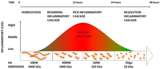
The mechanical stress can depolymerize into small pieces at low molecular weight and have a high inflammatory capacity. Many of these pieces are then further degraded into small oligosaccharides. Recently, it has been demonstrated that oligosaccharides are able to stop this inflammatory process. These data support that deep friction could metabolize self-aggregated hyaluronan (HA) chains responsible for increasing loose connective tissue viscosity, catalyzing a local HA fragment cascade that will generate soreness but, at the same time, facilitate the reconstitution of the physiological loose connective tissue properties. This information can help to explain the meaning of the inflammatory process as well as the requirement for it for the long-lasting resolution of these alterations.

mechanical stress hyaluronan hyaluronan fragment inflammatory reaction soreness

1. Introduction

It is a common experience, reported by patients who have undergone manual therapy, to perceive soreness in treatment areas that lasts for around 24 h, with a numeric rating scale of < 3/10 [1]. However, this reaction cannot merely be considered as a side effect of treatment since it is supposed to be the expression of a fundamental inflammatory phenomenon that permits physiological tissue restoration [2]. One hypothesis is that the mechanical stresses of manual therapy, through deep friction, catalyze an inflammatory reaction that is key to restoring the proper viscosity of loose connective tissues with benefits in the physiology and functionality of the areas previously densified. In fact, from established nomenclature, the term densification defines an area perceived as rigid, rough, not sliding properly, and incompressible due to a viscosity alteration in the loose connective tissue typically localized between interfaces such as muscle and deep fascia or fascial system layers [3]. One method that supports the reorganization of the extracellular matrix and that has been demonstrated to be effective for densification treatment, thanks to deep friction manipulation, is the fascial manipulation (FM) method.
Whilst the treatment modality of the FM method can be compared to other techniques, the reasoning process for the choice of point to be treated presents major differences. The points are selected after a specific assessment process involving clinical history taking, clinical examination of specific movements, and, not least, palpatory verifications [4][5]. During the examination of clinical history, segments with dysfunctions are identified with an emphasis on chronology in order to generate a treatment hypothesis based on the current symptomatology of the patient and previous musculoskeletal events, which may be causing compensations. The selection of points to treat is guided by a specific assessment chart (FM chart) [5]. The choice of points of where to apply the deep friction is based on the information collected through the FM chart, movement, and, overall, palpatory verifications to define the presence of “densification”. These consecutive steps should limit the overall clinician’s subjectivity in the decision process [6]. The treatment must be performed over specific areas, called the center of coordination (CC) and the center of fusion (CF) which are anatomically safe and do not overlap major superficial nerves and veins. Additional guidance for point selection includes avoiding the patient’s excessively painful areas where inflammation, lesions, or even fractures could be present. Absolute contraindications are thrombosis, phlebitis, skin lesions, and fever [3]. The manipulation of the CC and CF has the aim of restoring the gliding of the underlying tissue layers [7].
HA, historically regarded as a mere “space-filler” within the extracellular matrix (ECM), has undergone a remarkable transformation in our understanding of its significance. Emerging research has unveiled its profound structural and signaling roles [8]. This seemingly unassuming molecule is universally distributed among vertebrates and plays a pivotal role, especially within the ECM of soft connective tissues [9]. HA, the simplest glycosaminoglycan (GAG), is composed of a non-sulfated linear polymer, consisting of as many as 20,000 repeats of its disaccharide unit, which combines D-glucuronic acid and N-acetyl-D-glucosamine [10]. Thanks to its carboxyl groups, HA carries a negative charge and exhibits remarkable hydrophilicity, enabling it to retain water molecules at an astonishing 1000-fold of its own molecular weight [10]. This newfound understanding opens doors to exciting avenues of research and applications in various fields. 
At high molecular weights (HMWs), HA forms a substantial, viscous network. When it interacts with various proteoglycans, such as aggrecan, it leads to the creation of molecular composites that occupy significant volumes within the ECM. These complexes contribute to the gel-like state of the matrix. Additionally, these extensive HA–HA binding proteoglycan complexes also crosslink with other matrix proteins, including collagen. This crosslinking results in the formation of supermolecular structures that significantly enhance tissue stiffness [11]. The intricate interplay between HA, proteoglycans, and collagen within the ECM has profound implications for tissue structure and function. HA’s HMW and its ability to form these complex supermolecular structures make it a critical regulator of tissue rigidity. Moreover, the dynamic turnover of HA in different tissues underscores its versatility and adaptability, allowing it to fulfill specific roles tailored to the needs of each tissue type [11]
The ECM undergoes significant alterations in its physical properties, particularly concerning the presence of free water and the entangled HA chains. This leads to a substantial increase in viscosity within the ECM, which, in turn, has profound implications for the behavior of loose connective tissue and the mechanisms governing interactions between adjacent tissue interfaces [12]. It is crucial to recognize that the viscosity of HA is inherently temperature dependent. As the temperature surpasses the threshold of 40 °C, the three-dimensional superstructure of HA chains progressively disintegrates [12]. This disintegration results in a subsequent reduction in viscosity. 
Understanding the temperature-induced alterations in HA viscosity provides valuable insights into the physiological responses of connective tissues and the dynamic nature of the ECM. These insights are particularly pertinent in clinical contexts, where interventions and treatments are designed to mitigate the adverse effects of temperature-induced changes [13]. By delving into the intricacies of HA behavior in response to temperature fluctuations, researchers and medical professionals can devise targeted strategies to address continuously involved connective tissue, pain management, and mobility impairment more effectively. 
For instance, Menon et al. (2019) demonstrated the direct correlation between water-bound HA and range of motion in spastic patients [14]. These authors injected human recombinant hyaluronidase, which has the capacity to fragmentize the long chains of HA, to drain and metabolize the exceeded amount of self-aggregate HA with poor hydrophilic capacity. As a result, a more homogenous fluid was formed, stimulating the local cells to produce new HA with regular water-bound capacity and allowing proper sliding between interfaces [14].

2. Mechanical Stress on Hyaluronan Fragments’ Inflammatory Cascade

2.1. The Role of HA Weight: A Decremental Cascade during Inflammation

HA, in normal constitution, provides viscoelasticity and lubrication of liquid connective tissues [15]. Because of these properties, HA is able to lubricate and space-fill tissues [16] with a fundamental role for the constitutional ECM organization [17] present within endomysium, perimysium, epimysium interfaces, and deep fascia layers. The variability of HA concentration in the human body can range from less than 40 ng/mL in blood serum [18] to about 2–3 mg/mL in the knee synovial joint [19]. HA synthesis is driven by different enzymes [20]; one of them is hyaluronan synthase 2 which has been reported to be able to generate HA as large as 6000 kDa, the typical average size for newly synthesized HA in healthy tissues [21]. Indeed, the HA between 1800–3000 kDa, also known as high-molecular-weight hyaluronan (HMW HA), is responsible for tissue hydration due to its ability to bind high amounts of water. Nonetheless, HA can retain not only water up to almost 1000 times its weight but also self-aggregate and bind many proteins [22][23][24]. When ECM homeostasis is altered, endogenous HMW HA is disrupted, unbalancing the equilibrium toward a higher concentration of medium-molecular-weight HA (MMW HA, 250–1000 kDa) to low-molecular-weight HA (LMW HA, ≤250 kDa). Then, LMW HA can be further fragmented into shorter oligomers (o-HA, <10 kDa) [25]. A variety of different authors have demonstrated how mechanical stresses can be the root cause of HA depolymerization from high to low molecular weight [26].

2.2. The Inflammation Cascade: Influencing Factors

HA can also undergo depolymerization via non-specific mechanisms. Inflammatory processes can lead to the generation of free radicals within tissues undergoing widespread inflammation [27]. When the natural antioxidant defenses prove insufficient to counter the substantial influx of ROS, these radicals directly interact with native HA, resulting in significant production of HA fragments [28].
In a proposed catabolic pathway [29], it is suggested that the high-molecular-mass HA polymer undergoes stepwise cleavage by a series of enzymes, with the product of one reaction becoming the substrate for the subsequent one. These successive enzymatic events result in the generation of increasingly smaller HA fragments. Small HA components are able to exacerbate the inflammatory response by inducing the release of various detrimental mediators such as ROS, cytokines, chemokines, and destructive enzymes (i.e., hyaluronidase) and by facilitating the recruitment of leukocytes [30]. HA fragments, with a molecular size ≤ 500 kDa, have been shown to exhibit several proinflammatory effects. They can stimulate the expression of proinflammatory genes including TNFa, IL-1β, IL-1, and MMPs [31]. LMW HA can further maintain and strengthen the inflammatory response [30].
Yamasaki et al. [32] also showed that during sterile inflammatory processes, HA is able to activate the interleukin 1β (IL-1β) pathway and the cryopyrin mechanism. Previous reports have shown that HA fragments can stimulate an inflammatory response through their interaction with the TLR-4 and CD44 receptors [30]. The same authors explain how HA fragments, produced from native HA degradation, mediate a response made by IL-1β that produces an inflammatory response through the CD44 receptor [30]. The activation of these receptors mediated the activation of the nuclear factor kB (NF-kB) which in turn activates the release of several proinflammatory cytokines. HA fragments are also generated by hyal2, which is present on the cell membrane together with CD44. [33]. Hyal2 translocation is required for the degradation of extracellular hyaluronan. 

2.3. HA Polymer Fragments: Diverse Biological Activities

While commonly categorized as proinflammatory, it is more appropriate to consider HA fragments as pro-defensive entities in specific environments [9]. HA polymer fragments exhibit diverse biological activities depending on their size and are integral to numerous essential processes [29]. Studies have shown that short oligosaccharides often play a role in the body’s alarm system [34]. Moreover, some of the smaller HA oligosaccharides appear to alleviate the effects of these stress signals [29]. For instance, six-unit oligosaccharides derived from HA exhibit the capacity to stimulate fibroblast motility and expedite wound closure [35]. In contrast, HMW HA (1500 kDa) inhibits platelet adhesion and the activation of endothelial cell layers. HA oligomers also possess the ability to Impede the proliferation and migration of vascular smooth muscle cells in response to platelet-derived growth factor, as elucidated by Tavianatou et al. [36].
All of these studies help to explain how the inflammatory cascade, when catalyzed by manual therapy or free radicals, is then able to self-resolve (Figure 1). To finalize the process, HA pieces will then either be further depolymerized locally or drained from the tissue via the lymphatic system [30]. Most of the HA fragments leave the tissue with the lymph and are cleared in the lymph nodes. All that remains, after passage through the nodes, is degraded by the liver [37].
Figure 1. Schematization of hyaluronan fragments and corresponding inflammation.

2.4. Summary

Manual therapies and physical therapies are the most common therapeutic options for non-specific musculoskeletal pain even if a great variety of treatment options are available. Furthermore, the duration of the results is often questionable due to relative short-term effects [38]. While McDevitt et al. [38] in their recent review proved the efficacy of physical therapies within 6 weeks, only a few therapies have demonstrated long-term effects [6][39].
Matteini et al. [40] showed how HA aggregation breaks down progressively when the temperature increases over ~40 °C. These values are compatible with weak non-covalent interactions like those characteristics of van der Waals and hydrophobic forces localized between oligo HA fragments. This information can easily explain how low energy therapies (exothermic or light manual therapies) can decrease HA aggregation so the entire viscosity of the extracellular matrix as a consequence improves the range of motion and decreases the irritation of the free nerve ending [40]. However, the long chains of HA cannot be washed out without being fragmented first. This could explain the recurrence of symptoms due to consequent re-aggregation of the HA that will still be retained in the compartment due to the lack of mechanical forces that will stimulate the drainage and consequently metabolization of the fragments.
On the other hand, high-intensity therapy such as focus shock wave and deep friction manipulation can not only warm up the area, separating the single HA chains and giving an instantaneous result, but also fragmentize the HA. The latter catalyzes an inflammatory process that metabolizes entangled HA chains that are more susceptible to absorbing physical forces due to their overstructured conformation.
In a nutshell, in the course of inflammation, HA undergoes depolymerization, breaking down into smaller fragments with LMW. A significant portion of these fragments subsequently undergo further degradation, resulting in the formation of small oligosaccharides. These oligosaccharides possess the capacity to exacerbate the inflammatory process by stimulating the generation of various inflammation mediators and agents, including ROS, cytokines, chemokines, and destructive enzymes (such as hyaluronidase). These enzymes serve to initiate and intensify the inflammatory response [41].

References

  1. Paanalahti, K.; Holm, L.W.; Nordin, M.; Asker, M.; Lyander, J.; Skillgate, E. Adverse events after manual therapy among patients seeking care for neck and/or back pain: A randomized controlled trial. BMC Musculoskelet. Disord. 2014, 15, 77.
  2. Fidut-Wrońska, J.; Chołuj, K.; Chmiel, J.; Pikto-Pitkiewicz, K.; Majcher, P. Observation using thermography of post-operative reaction after fascial manipulation®. Ann. Agric. Environ. Med. 2019, 26, 468–471.
  3. Stecco, C.; Day, J.A. The fascial manipulation technique and its biomechanical model: A guide to the human fascial system. Int. J. Ther. Massage Bodyw. 2010, 3, 38–40.
  4. Day, J.A.; Copetti, L.; Rucli, G. From clinical experience to a model for the human fascial system. J. Bodyw. Mov. Ther. 2012, 16, 372–380.
  5. Pintucci, M.; Simis, M.; Imamura, M.; Pratelli, E.; Stecco, A.; Ozcakar, L.; Battistella, L.R. Successful treatment of rotator cuff tear using Fascial Manipulation® in a stroke patient. J. Bodyw. Mov. Ther. 2017, 21, 653–657.
  6. Branchini, M.; Lopopolo, F.; Andreoli, E.; Loreti, I.; Marchand, A.M.; Stecco, A. Fascial Manipulation® for chronic aspecific low back pain: A single blinded randomized controlled trial. F1000Res 2015, 4, 1208.
  7. Cowman, M.K.; Schmidt, T.A.; Raghavan, P.; Stecco, A. Viscoelastic Properties of Hyaluronan in Physiological Conditions. F1000Res 2015, 4, 622.
  8. Lee, J.Y.; Spicer, A.P. Hyaluronan: A multifunctional, megaDalton, stealth molecule. Curr. Opin. Cell Biol. 2000, 12, 581–586.
  9. Abatangelo, G.; Vindigni, V.; Avruscio, G.; Pandis, L.; Brun, P. Hyaluronic Acid: Redefining Its Role. Cells 2020, 9, 1743.
  10. Anderegg, U.; Simon, J.C.; Averbeck, M. More than just a filler—The role of hyaluronan for skin homeostasis. Exp. Dermatol. 2014, 23, 295–303.
  11. Lee, D.H.; Oh, J.H.; Chung, J.H. Glycosaminoglycan and proteoglycan in skin aging. J. Dermatol. Sci. 2016, 83, 174–181.
  12. Tømmeraas, K.; Melander, C. Kinetics of hyaluronan hydrolysis in acidic solution at various pH values. Biomacromolecules 2008, 9, 1535–1540.
  13. Menon, R.G.; Oswald, S.F.; Raghavan, P.; Regatte, R.R.; Stecco, A. T1ρ-Mapping for Musculoskeletal Pain Diagnosis: Case Series of Variation of Water Bound Glycosaminoglycans Quantification before and after Fascial Manipulation® in Subjects with Elbow Pain. Int. J. Environ. Res. Public Health 2020, 17, 708.
  14. Menon, R.G.; Raghavan, P.; Regatte, R.R. Quantifying muscle glycosaminoglycan levels in patients with post-stroke muscle stiffness using T1ρ MRI. Sci. Rep. 2019, 9, 14513.
  15. Han, W.; Lv, Y.; Sun, Y.; Wang, Y.; Zhao, Z.; Shi, C.; Chen, X.; Wang, L.; Zhang, M.; Wei, B.; et al. The anti-inflammatory activity of specific-sized hyaluronic acid oligosaccharides. Carbohydr. Polym. 2022, 276, 118699, Erratum in Carbohydr. Polym. 2022, 282, 119101.
  16. Bohaumilitzky, L.; Huber, A.K.; Stork, E.M.; Wengert, S.; Woelfl, F.; Boehm, H. A Trickster in Disguise: Hyaluronan’s Ambivalent Roles in the Matrix. Front. Oncol. 2017, 7, 242.
  17. Laurent, T.C.; Fraser, J.R. Hyaluronan. FASEB J. 1992, 6, 2397–2404.
  18. Cowman, M.K. Hyaluronan and Hyaluronan Fragments. Adv. Carbohydr. Chem. Biochem. 2017, 74, 1–59.
  19. Balazs, E.A. Viscoelastic properties of hyaluronic acid and biological lubrication. Univ. Mich. Med. Cent. J. 1968, 1, 255–259.
  20. Anderegg, U.; Halfter, N.; Schnabelrauch, M.; Hintze, V. Collagen/glycosaminoglycan-based matrices for controlling skin cell responses. Biol. Chem. 2021, 402, 1325–1335.
  21. Spicer, A.P.; Tien, J.Y. Hyaluronan and morphogenesis. Birth Defects Res. C Embryo Today 2004, 72, 89–108.
  22. Garg, H.; Hales, C. Chemistry and Biology of Hyaluronan, 1st ed.; Elsevier: Amsterdam, The Netherlands, 2004.
  23. Yu, L.; Quinn, D.A.; Garg, H.G.; Hales, C.A. Cyclin-dependent kinase inhibitor p27Kip1, but not p21WAF1/Cip1, is required for inhibition of hypoxia-induced pulmonary hypertension and remodeling by heparin in mice. Circ. Res. 2005, 97, 937–945.
  24. Heldin, P. Chemistry and Biology of Hyaluronan. Edited by Hari G. Garg and Charles A. Hales. ChemBioChem 2005, 6, 1288–1289.
  25. Tian, X.; Azpurua, J.; Hine, C.; Vaidya, A.; Myakishev-Rempel, M.; Ablaeva, J.; Mao, Z.; Nevo, E.; Gorbunova, V.; Seluanov, A. High-molecular-mass hyaluronan mediates the cancer resistance of the naked mole rat. Nature 2013, 499, 346–349.
  26. Itano, N.; Sawai, T.; Yoshida, M.; Lenas, P.; Yamada, Y.; Imagawa, M.; Shinomura, T.; Hamaguchi, M.; Yoshida, Y.; Ohnuki, Y.; et al. Three isoforms of mammalian hyaluronan synthases have distinct enzymatic properties. J. Biol. Chem. 1999, 274, 25085–25092.
  27. Parsons, B.J.; Spickett, C.M. Special issue on “Analytical methods for the detection of oxidized biomolecules and antioxidants”. Free Radic. Res. 2015, 49, 473–476.
  28. Kasai, S.; Furuichi, Y.; Ando, N.; Kagami, K.; Abe, M.; Nakane, T.; Goi, K.; Inukai, T.; Saitoh, S.; Ohno, S.; et al. Inflammatory mediator ultra-low-molecular-weight hyaluronan triggers necrosis of B-precursor leukemia cells with high surface CD44 expression. Cell Death Dis. 2017, 8, e2857.
  29. Stern, R. Hyaluronan catabolism: A new metabolic pathway. Eur. J. Cell Biol. 2004, 83, 317–325.
  30. Avenoso, A.; Bruschetta, G.; D’Ascola, A.; Scuruchi, M.; Mandraffino, G.; Saitta, A.; Campo, S.; Campo, G.M. Hyaluronan Fragmentation During Inflammatory Pathologies: A Signal that Empowers Tissue Damage. Mini Rev. Med. Chem. 2020, 20, 54–65.
  31. Horton, M.R.; Shapiro, S.; Bao, C.; Lowenstein, C.J.; Noble, P.W. Induction and regulation of macrophage metalloelastase by hyaluronan fragments in mouse macrophages. J. Immunol. 1999, 162, 4171–4176.
  32. Yamasaki, K.; Muto, J.; Taylor, K.R.; Cogen, A.L.; Audish, D.; Bertin, J.; Grant, E.P.; Coyle, A.J.; Misaghi, A.; Hoffman, H.M.; et al. NLRP3/cryopyrin is necessary for interleukin-1beta (IL-1beta) release in response to hyaluronan, an endogenous trigger of inflammation in response to injury. J. Biol. Chem. 2009, 284, 12762–12771.
  33. Harada, H.; Takahashi, M. CD44-dependent intracellular and extracellular catabolism of hyaluronic acid by hyaluronidase-1 and -2. J. Biol. Chem. 2007, 282, 5597–5607.
  34. Powell, J.D.; Horton, M.R. Threat matrix: Low-molecular-weight hyaluronan (HA) as a danger signal. Immunol. Res. 2005, 31, 207–218.
  35. Tolg, C.; McCarthy, J.B.; Yazdani, A.; Turley, E.A. Hyaluronan and RHAMM in wound repair and the “cancerization” of stromal tissues. Biomed. Res. Int. 2014, 2014, 103923.
  36. Tavianatou, A.G.; Caon, I.; Franchi, M.; Piperigkou, Z.; Galesso, D.; Karamanos, N.K. Hyaluronan: Molecular size-dependent signaling and biological functions in inflammation and cancer. FEBS J. 2019, 286, 2883–2908.
  37. Tammi, M.I.; Oikari, S.; Pasonen-Seppänen, S.; Rilla, K.; Auvinen, P.; Tammi, R.H. Activated hyaluronan metabolism in the tumor matrix—Causes and consequences. Matrix Biol. 2019, 78–79, 147–164.
  38. McDevitt, A.W.; Cooper, C.G.; Friedrich, J.M.; Anderson, D.J.M.; Arnold, E.A.; Clewley, D.J. Effect of Physical Therapy Timing on Patient Reported Outcomes for Individuals with Acute Low Back Pain: A Systematic Review with Meta Analysis of Randomized Controlled Trials. PM&R 2023, 15, 1466–1477.
  39. Daecke, W.; Kusnierczak, D.; Loew, M. Long-term effects of extracorporeal shockwave therapy in chronic calcific tendinitis of the shoulder. J. Shoulder Elbow Surg. 2002, 11, 476–480.
  40. Matteini, P.; Dei, L.; Carretti, E.; Volpi, N.; Goti, A.; Pini, R. Structural behavior of highly concentrated hyaluronan. Biomacromolecules 2009, 10, 1516–1522.
  41. Nagy, N.; Kuipers, H.F.; Marshall, P.L.; Wang, E.; Kaber, G.; Bollyky, P.L. Hyaluronan in immune dysregulation and autoimmune diseases. Matrix Biol. 2019, 78–79, 292–313.
More
Information
Contributors MDPI registered users' name will be linked to their SciProfiles pages. To register with us, please refer to https://encyclopedia.pub/register : , , , ,
View Times: 79
Revisions: 2 times (View History)
Update Date: 22 Dec 2023
1000/1000