Submitted Successfully!
To reward your contribution, here is a gift for you: A free trial for our video production service.
Thank you for your contribution! You can also upload a video entry or images related to this topic.
Version Summary Created by Modification Content Size Created at Operation
1 -- 3991 2023-11-24 09:00:40 |
2 Reference format revised. + 1 word(s) 3992 2023-11-27 06:19:59 |

Video Upload Options

Do you have a full video?

Confirm

Are you sure to Delete?
Cite
If you have any further questions, please contact Encyclopedia Editorial Office.
Sciaccotta, R.; Murdaca, G.; Caserta, S.; Rizzo, V.; Gangemi, S.; Allegra, A. Circular RNAs and Multiple Sclerosis. Encyclopedia. Available online: https://encyclopedia.pub/entry/52019 (accessed on 27 July 2024).
Sciaccotta R, Murdaca G, Caserta S, Rizzo V, Gangemi S, Allegra A. Circular RNAs and Multiple Sclerosis. Encyclopedia. Available at: https://encyclopedia.pub/entry/52019. Accessed July 27, 2024.
Sciaccotta, Raffaele, Giuseppe Murdaca, Santino Caserta, Vincenzo Rizzo, Sebastiano Gangemi, Alessandro Allegra. "Circular RNAs and Multiple Sclerosis" Encyclopedia, https://encyclopedia.pub/entry/52019 (accessed July 27, 2024).
Sciaccotta, R., Murdaca, G., Caserta, S., Rizzo, V., Gangemi, S., & Allegra, A. (2023, November 24). Circular RNAs and Multiple Sclerosis. In Encyclopedia. https://encyclopedia.pub/entry/52019
Sciaccotta, Raffaele, et al. "Circular RNAs and Multiple Sclerosis." Encyclopedia. Web. 24 November, 2023.
Circular RNAs and Multiple Sclerosis
Edit

Multiple sclerosis (MS), a condition characterised by demyelination and axonal damage in the central nervous system, is due to autoreactive immune cells that recognise myelin antigens. Alteration of the immune balance can promote the onset of immune deficiencies, loss of immunosurveillance, and/or development of autoimmune disorders such as MS. Numerous enzymes, transcription factors, signal transducers, and membrane proteins contribute to the control of immune system activity. The “transcriptional machine” of eukaryotic cells is a complex system composed not only of mRNA but also of non-coding elements grouped together in the set of non-coding RNAs. ncRNAs play a crucial role in numerous cellular functions, gene expression, and the pathogenesis of many immune disorders.

circRNA multiple sclerosis non-coding RNA

1. Multiple Sclerosis

The most common autoimmune condition that results in non-traumatic neurological impairment in young people is multiple sclerosis (MS) [1]. MS has the highest prevalence in North America, Western Europe, and Australasia (>100 cases per 100,000 people) and lowest in countries close to the equator (30 cases per 100,000 people) [2]. Females are three times as likely as males to develop the condition, and although MS can manifest at any age, the average occurrence is between twenty and forty years old, with about ten percent of the cases starting under the age of eighteen [3]. Cognitive dysfunction, bladder and bowel incontinence, tingling, and numbness, as well as vision impairment, are some of the several neurological symptoms that might occur in MS, and according to their severity and evolution, the disease can take a variety of clinical courses. Relapsing–remitting (RR) MS, which affects 80% of patients, is characterised by attacks accompanied by full or partial remissions [4]. The term “progressive MS” refers to a subtle decline in neurological function accompanied by new symptoms and signs that have lasted at least a year. The main cause is neurodegeneration, which is manifested by extensive neuroaxonal degeneration in the white and grey matter [5]. A combination of clinical and radiographic (magnetic resonance) characteristics are used to diagnose MS; in detail, the guidelines for the recognition of all forms of MS are the McDonald criteria from 2017 [6]. The interaction of genetic human leukocyte antigen (HLA) DRB1, vitamin D deficiency, Epstein Barr Virus (EBV) infection, and epigenetic variables is a key component of the pathogenesis of MS [7][8][9][10]. The primary proposed aetiology of MS is an autoimmune attack on the central nervous system (CNS); in fact, studies have suggested that autoreactive T cells, mainly T helper (Th)-1 CD4+ T cells and Th17 cells, play a key role in the pathogenesis, especially since they secrete cytokines and trigger the inflammatory cascade [11]. Microglia activation and prolonged oxidative stress caused by the inflammatory demyelination process contribute to neurodegeneration and subsequently axonal and neuronal destruction [12]. Both inflammation and demyelination, as well as astroglial growth (gliosis) and neurodegeneration, define MS. Scientists identified metabolites differently active in MS during the four seasons, establishing the role of the citrate cycle, sphingolipid metabolism, and pyruvate metabolism. The fall and spring seasons were shown to have the most metabolites influenced by these changes, while summer had the least amount of metabolite changes. Histidine and its metabolite, methyl histidine, serum level seem to be lower in MS than in control: histidine is a precursor to histamine, a potent neurotransmitter and immune-modulator; the inverse relationship between the blood level of this amino acid and IL-6, a cytokine that promotes inflammation, may help to explain the immune-modulating role of histidine in the pathophysiology of MS. Similar to this, reduced levels of histidine were linked to elevated levels of oxidative stress indicators and serum C-reactive proteins. According to the scientific literature, low levels of histidine during the spring and fall may diminish the anti-inflammatory effects of this amino acid and its metabolites, which may lead to MS flare-ups.

2. Non-Coding RNAs

Although most of the human genome is actively involved in genetic transcription processes, eukaryotic cells are quantitatively poor in coding RNA (less than 1.4%), resulting in a large part of human DNA being transcribed into ncRNAs [13]. This concept partially overturns the biological dogma proposed by Francis Crick, according to which proteins represent the main and final product of genetic information [14]. On the other hand, ncRNAs, although not involved in ribosomal proteosynthesis, play a key role in many physiological and pathological cellular processes [15]. The optimization and innovation of gene profiling techniques have made it possible to isolate and sequence an ever-increasing number of ncRNA “species”. Gene sequencing methods based on next-generation sequencing (NGS), such as RNA-Seq or small RNA-sequencing (smRNA-Seq), ideal for identifying small RNA species using reverse transcription and PCR amplification, have implemented our knowledge about RNA, allowing the drafting of dedicated genetic databases [16]. The consultation of these “genetic libraries”, such as GENCODE, the RefSeq project, NONCODE, HUGO, LNCipedia, and the ENCODE project, represents an obligatory step to define the taxonomy of ncRNAs [13][17][18][19][20][21]. One of the parameters used to classify ncRNAs concerns the length and structure of the non-coding segment [22][23]. It can, therefore, distinguish the “small-ncRNAs”, composed of a few nucleotides, and the “long-ncRNAs” (lncRNA), whose length exceeds 200 nucleotides [24]. Small-ncRNAs include micro-RNAs (miRNAs, 22 nucleotides) and PIWI-interacting RNAs (piRNAs, 24–30 nucleotides). Circular-RNAs (circ-RNAs) are also grouped within the class of lncRNAs, although these have a circular structure unlike the other linear lncRNAs [24]. The ncRNAs can also be classified according to the function they cover within the eukaryotic cell, differentiating them into “Housekeeping ncRNAs” and “Regulatory ncRNAs” [25][26]. Housekeeping ncRNAs are generally small in size and perform basic cellular functions such as protein synthesis (tRNAs,rRNAs), RNA splicing (snRNAs), and post-transcriptional modification of rRNA (sno-RNA) [27][28]. Furthermore, among the Housekeeping ncRNAs, scholars include the tRNA-derived fragments, cleavage products of tRNAs, involved in the regulation of gene expression and apoptosis [29]. Regulatory ncRNAs perform functions of control and modulation of gene expression, both at the epigenetic and transcriptional levels [30][31]. The length of regulatory ncRNAs varies from a few bases (<200), as in miRNAs, siRNAs, and piRNAs, up to larger nucleotide sequences (>200), as in lncRNAs and circRNAs [32]. miRNAs are small portions of non-coding RNA, highly expressed within eukaryotic cells, which, through the formation of a biological aggregate called the “RNA-induced silencing complex (RISC)”, modulate the post-transcriptional processes of the mRNA, silencing specific target sequences [33][34]. Similarly to miRNAs, siRNAs also perform an RNA interference function through the activation of the RISC; PiRNAs, short non-coding elements composed of single-stranded RNA nucleotide sequences, form cytoplasmic complexes with PIWI proteins and migrating into the cell nucleus, actively modulate gene transcription [35].
Circular RNAs (circRNAs) are nuclear biomolecules composed of ribonucleotide sequences with a circular structure and no polyadenylated tails [36]. They are classified among the Housekeeping ncRNAs, although some forms possess the ability to code for specific proteins. A total of 1976 small portions of non-coding material with a circular structure, considered splicing errors, were isolated from viroids infecting plants and, subsequently, identified in the hepatotropic virus δ and eukaryotic cells, as in the mitochondrial RNAs of some yeasts [37][38][39][40]. In the 1990s, the casual discovery of circular and non-polyadenylated RNAs derived from non-canonical splicing of the human EST-1 gene and the isolation of the same molecules in the cytoplasm of mouse testicular cells aroused the interest of the scientific community towards this biotype of RNAs [41][42][43].

3. Multiple Sclerosis and circRNAs

3.1. Epigenetic Mechanisms

DNA methylation is a potential regulatory mechanism that might modulate the production of circRNA. Recent studies in genetics and epigenetics have indicated that a significant proportion of potential causative variations for autoimmune disorders are located in non-coding regions of the genome [44]. These variants are believed to have a regulatory function in the expression of genes associated with these illnesses. Epigenetic processes have the ability to govern the expression of genes through the alteration of DNA, a process that may be inherited across successive cell divisions [45]. The epigenetic mechanism that has been extensively researched is the covalent attachment of a methyl group to cytosines within the CpG dinucleotide context. This particular process has garnered significant attention because of the well-established and steady process by which mCpG is propagated across DNA, facilitated by DNA (Cytosine-5)-Methyltransferase 1 [46]. DNA methylation changes have been seen in blood, CD4+, and CD8+ T lymphocytes, as well as in unaffected brain areas of individuals with MS [47]. Numerous research has been conducted to elucidate the role of epigenetic processes in the modulation of circRNA production, particularly within the domain of cancer. Evidence indicates that hypermethylation of cancer-specific promoter CpG islands is linked to reduced production of both circRNAs and their corresponding host gene counterparts [48]; additionally, it is well-established that intragenic methylation plays a role in the regulation of AS [49]. Furthermore, another work demonstrated that there were differential expressions of circRNAs with distinct methylated sites in tumour samples compared to adjacent normal samples; interestingly, this differential expression was not observed in the parental genes of these circRNAs: this finding suggests the possibility that certain abnormal DNA methylation events may specifically impact the processing of pre-mRNA to produce circRNAs, while not affecting the generation of linear RNAs [50].

3.2. circRNAs Biomarkers in MS

The exceptional stability of circRNAs, attributed to their resistance to exonucleases responsible for degrading linear transcripts, makes these non-coding molecules ideal as potential disease biomarkers. Additionally, circRNAs have expression patterns that are specific to different tissues and developmental stages, which may be a good way to address the lack of specificity seen in a number of existing biomarkers [51]. It is important to note that circRNAs exhibit stronger interspecies conservation, which makes it easier to transfer biomarkers from animal models to people [52], and this fact suggests that GSDMD circRNA may have the ability to serve as an important biomarker for MS [53]. In rare instances, the existence of a pathological disease may be indicated by a reduced or entirely absent marker. In the scientific literature, an example of a particularly significant dysregulated circular RNA, hsa_circ_0007990, is presented to its particular relevance due to the association with the PGAP3 host gene, which encodes a protein known to play a significant role in regulating autoimmune reactions; it is responsible for the modification of fatty acids in glycosylphosphatidylinositol (GPI)-anchored proteins [52]. Notably, when PGAP3 is absent in mice, there is an increased T cell response and a worsened experimental immune-mediated encephalomyelitis phenotype, along with various symptoms resembling autoimmune disorders [53]. A recent study confirmed the finding of PGAP3 circRNA downregulation in MS patients, representing not only a possible disease biomarker but also being considered, in the future, as a “disease progression biomarker” [54]. Another noteworthy example involves the annexin A2 gene (ANXA2) [55]. Annexin A2 (ANXA2), a member of the annexin family, is a calcium-dependent phospholipid-binding protein with a molecular weight of 36 kilodaltons, mostly found on the surface of the majority of eukaryotic cells. It plays a significant role in several biological processes, such as exerting anti-inflammatory effects, facilitating Ca2+-dependent exocytosis, regulating immunological responses, and controlling phospholipase A2 activity [56][57].

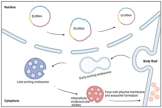
3.3. Extracellular Vesicles and circRNAs

Extracellular vesicles (EVs) refer to entities that are covered with a membrane and originate from either endosomes or the plasma membrane, then released into the extracellular environment [58]. The EVs were initially discovered four decades ago when they were observed in the form of reticulocytes; subsequently, it has been demonstrated that EVs may be found in many bodily fluids [59]. Typically, researchers have identified three distinct categories of extracellular vesicles (EVs): exosomes, which have a diameter ranging from 40 to 100 nm, microvesicles, and apoptotic bodies, with a diameter ranging from 50 to 2000 nm [60]. EVs are of paramount importance in facilitating intercellular relationships through the transfer of lipids, proteins, and genetic material between cells; in fact, their involvement in extracellular vesicle (EV)-mediated communication has been demonstrated in the modulation of multiple biological processes (Figure 1), such as the immune system’s response [61][62]. The RNA payload within EVs exhibits a great degree of complexity, with just a few research having comprehensively characterised the whole EV transcriptome in the context of MS [63][64]. In recent times, it has been demonstrated that circRNAs exhibit a high concentration within EVs and that exosomal circRNAs possess the ability to act as miRNA sponges, suppress protein activity, regulate splicing and transcription processes, and interact with RBPs [65][66]. These molecular functions have been found to have a role in the development and progression of neurodegenerative illnesses, although to a certain extent [67]. Regarding multiple sclerosis, Iparraguirre and colleagues revealed that circRNAs constitute the second most prevalent transcript in EV samples obtained from both MS patients and healthy individuals, showing disparities in the RNA biotypes present in leukocytes and EVs [68]. Interestingly, circRNAs have a higher representation in EVs, accounting for 4.2% of the total reads and 58.4% of the reads associated with ncRNAs, in contrast to leukocytes, where they constitute just 0.2% of the total reads and 0.9% of the ncRNA reads. Various parameters have been documented to influence the incorporation of RNA into EVs. These factors include cell abundance, particular sequence motifs, secondary structure, length, differential affinity for membrane lipids, and connection with RBPs [69]. In addition to the inherent characteristics of circRNAs that may facilitate or impede their inclusion inside EVs, the cellular state also influences the profiles of EV RNA. Consequently, a specific RNA molecule may be selectively encapsulated or excluded from EVs based on its physiologic or pathological consequences [68]. It is noteworthy to mention circNEIL3, one of the circRNAs identified as being differentially expressed in EVs of patients with RR-MS compared to EVs of healthy controls, and for this reason, has been proposed as a potential biomarker in leukocytes of RR-MS patients [70].
Figure 1. Extracellular vesicles and circRNAs: Exosomes are extracellular vesicles covered with a membrane and released into the extracellular environment. They modulate multiple biological processes, such as the immune system’s response, and contain a high concentration of circRNAs capable of acting as miRNA sponges, suppress protein activity, regulate splicing and transcription processes, and interact with RNA-binding proteins. Created with BioRender.com (accessed on 19 October 2023).

3.4. circRNAs Genetic Variation in MS

Paraboschi and his group conducted a bioinformatic investigation to examine the enrichment of circRNAs originating from non-coding regions in the genome related to MS and proposed that these circRNAs may have a role in the susceptibility to the illness. The authors of the study have shown a significant increase in non-coding elements, particularly circRNAs, that are located inside the 73 Linkage Disequilibrium (LD) blocks containing single-nucleotide polymorphisms (SNPs) linked with MS: in detail, a collective count of 482 circRNAs was identified in publically accessible databases and then compared to an average of 194.65 circRNAs found in randomly selected sets of LD blocks, using 1000 iterations. Through the assessment of RNA sequencing data obtained from two distinct cell lines, namely SH-SY5Y and Jurkat cells, a total of 18 circRNAs were discovered: among them, two were found to be unique and originated from genes related to MS. Furthermore, Paraboschi conducted an investigation to validate the levels of expression of a circRNA originating from a genomic region linked with MS, namely hsa_circ_0043813 originated from the STAT3 (Signal Transducer and Activator of Transcription 3) gene [67]: the activation of STAT3 is mediated by a diverse range of cytokines, which in turn, elicits a multitude of crucial biological activities and it has recently shown to be involved in the control of Th17 cell development, which is known to be a critical factor in the pathogenesis of MS [68]. Scientists examined the relationship between STAT3 hsa_circ_0043813 and the expression levels of certain circRNAs, which may be altered by disease-associated SNPs [71][72]. This finding aligns with the previously reported evidence on circANRIL, which stands as the sole instance of a circRNA where a connection between disease-associated SNPs and circRNA formation has been established [73]. In conclusion, researchers found unique evidence indicating that the top hits from genome-wide association studies (GWAS) in MS are located inside LD blocks that are enriched in circRNAs, suggesting that they may potentially play a role in the pathogenesis of MS, representing a new avenue for further investigation [72].

3.5. circRNAs and B-Cell Function

B-cell function plays a crucial role in the autoimmune aberrations observed in RRMS. In recent years, there has been substantial evidence supporting the significant involvement of B lymphocytes in several processes inside the CNS. These processes include antigen presentation, the release of cytokines that induce inflammation, and the generation of antimyelin antibodies [74][75]. There is also a proposition suggesting that ectopic lymphoid cell aggregation found in the leptomeninges consists of B lymphocytes and could have a role in determining the chronic nature of MS illness disease [76]. Recent studies have provided confirmation that the reduction of B-cells is an effective treatment strategy for treating RRMS and primary progressive multiple sclerosis (PPMS) and specific circRNA particles (hsa_circRNA_101348, hsa_circRNA_102611, and hsa_circRNA_104361) that are overproduced in PBMCs of patients with RRMS have been identified [77][78]. Importantly, the authors of this study have discovered two transcripts, AK2 and IKZF3 (Aiolos), which are associated with B-cell function and are regulated by these circRNA molecules. The results were validated by the direct demonstration of an increase in AK2 and IKZF3 expression in the peripheral blood mononuclear cells (PBMCs) of individuals experiencing a relapse of MS. The AK2 gene is recognised for its role in encoding phosphotransferase adenylate kinase 2, which fulfils the unique cellular needs related to mitochondrial processes, notably in the context of B-cell activation and antibody generation [79]. A special emphasis on the involvement of AK2 has been observed in B cells that are infected with the Epstein–Barr virus (EBV). This association has been extensively explored in the context of MS as a viral infection that is linked to MS [80]. The IKZF3 gene encodes the proteins Aiolos and Ikaros, which serve as crucial transcription elements belonging to the Ikaros family of zinc-finger proteins. These proteins play a pivotal role in regulating the development of lymphoid and myeloid cells, as well as maintaining immunological homeostasis [81]. It is noteworthy that a mutation of the IKZF3 gene has been identified as one of the risk alleles associated with MS [82]. The involvement of Aiolos in the functioning of mature B cells has been demonstrated, highlighting its crucial importance. Moreover, Aiolos is essential for the development of high-affinity antibody-secreting plasma cells [83].

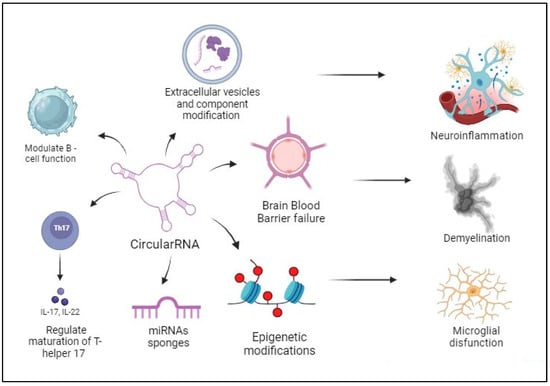
3.6. Roles of circRNAs in MS

3.6.1. hsa_circ_0106803 of GSDMB Gene

The available evidence suggests that circRNAs play important roles in controlling how the immune system and central nervous system (CNS) function. Only a few research have looked at this issue; thus, the precise roles played by these RNAs in the onset of MS are yet unknown. In contrast to PBMCs from healthy people, patients with RRMS showed differential expression of approximately 400 circRNAs in their peripheral blood mononuclear cells (PBMCs), according to the research of Cardamone and colleagues about “alternative splicing” (AS) and, specifically, the detection and description of GSDMB, a 17q12-locus alternatively spliced gene that has been frequently linked to autoimmune disorders and asthma susceptibility and that encodes Gasdermin B, a member of the family of proteins that contain gasdermin domains [84][85][86]. Alternative splicing (AS) has become more and more significant in the pathogenesis of autoimmune diseases in recent years since numerous immune system processes, including T-cell activation and migration, cytokine response, and T-cell stability and apoptosis, are impacted by it [87]. These processes are all necessary to prevent the suppression of self-tolerance and the emergence of autoimmune conditions.

3.6.2. circ_HECW2 and the Dysfunction of the Blood–Brain Barrier

Additionally, it was discovered that circular RNA HECW2 (circ_HECW2) contributed to the pathogenesis of multiple sclerosis. In both in vitro and in vivo MS experimental models, increased expression of circ_HECW2 results in EndoMT, which is crucial for BBB failure and contributes to BBB leakage. In order to boost the expression of ATG5 (autophagy-related 5), activate the NOTCH pathway, and ultimately favorably regulate LPS-induced EndoMT, Yang and colleagues demonstrated that circ_HECW2 served as a miR-30D sponge.

3.6.3. hsa_circ_0106803 Modulates the Expression of ASIC1a mRNA

MS patients had been discovered to have an increase of the lncRNA MALAT1: changes in circRNA back-splicing and aberrant splicing of MS-related genes, including IL7R and SP140, have been linked to changes in MALAT1 expression [54]. MiR-1275 and miR-149, two miRNAs that were among those predicted by hsa_circ_0106803 to have multiple target sites, exhibit differential expression in blood from MS patients; moreover, ASIC1a is said to bind to miR-149, which lowers its levels. The acid-sensing ion channel subunit ASIC1a is overexpressed in acute MS lesions and may play a role in the neuronal pathophysiology of the illness: by controlling the expression of ASIC1a mRNA through miR-149, hsa_circ_0106803 has been suggested to affect the progression of MS in light of this data [88].

3.6.4. circINPP4B Regulates Th17 Cell Differentiation

CD4+ T helper (Th) cells, particularly Th17 cells that secrete interleukin-17 (IL-17), play a significant role in the initiation and progression of both MS and experimental autoimmune encephalomyelitis (EAE), which is a mouse model used to study MS [89][90]. To support this concept, it was observed that there was a notable rise in the proportion of Th17 cells in the cerebrospinal fluid (CSF) of individuals diagnosed with MS. Furthermore, it was shown that the proportion of Th17 cells in MS patients during relapse was larger compared to the ratio observed during the remission phase [68]. The induction of EAE may be achieved by the infusion of activated CD4+ Th17 cells that are responsive to myelin. Additionally, the administration of IL-17 exacerbates the progression of EAE, whereas animals lacking IL-17 demonstrate resistance to this illness [91]. A recent investigation has demonstrated that the inhibition of Th17 differentiation in immune-deficient Rag1−/− mice who were administered CD4+ naïve T cells (CD4+ Tn) led to the reduction of the signs of EAE. In contrast, the promotion of Th17 differentiation worsened the EAE condition [89].

3.6.5. hsa_circRNA_101145 and hsa_circRNA_001896 in Patients with Relapse-Remitting MS

A complex analysis of the RNA of the peripheral blood mononuclear cells from patients with multiple sclerosis demonstrated several hundred circRNAs differentially expressed respect healthy controls (HCs). Mycko and colleagues evaluated 102 Caucasian participants, comprising 37 HCs and 65 patients with multiple sclerosis. All patients had relapsing-remitting multiple sclerosis, fulfilled the McDonald criteria, had not received disease-modifying drugs from any less than 6 months, and never had anti-CD20, cladribine, and anti-CD52 treatment. In order to distinguish RRMS patients in remission from HCs, this study identified two circRNAs and a collection of 10 miRNAs that may be targeted by circRNAs. All 10 of these miRNAs would be more active if circRNA were to express itself less. Numerous miRNAs have been connected to autoimmune mechanisms causing demyelination in MS. MiRNA has been linked by researchers to the growth and upkeep of populations of T helper cells that are encephalitogenic. MiRNAs are indeed variably expressed in PBMCs, whole blood, T cells, and B cells in MS patients, according to reports on the subject. The greater concentration of miR-181c in the cerebral fluid has been associated with a higher risk for the development of confirmed MS from the group of miRNAs discovered in this investigation. Almost all of the other miRNAs have been discovered to be involved in processes like autophagy, apoptosis, neural stem cell proliferation, and neurodegeneration, which may function in autoimmunity, brain recovery, and MS-related processes even if they have not been explicitly linked to the disease. The results gathered here provide a solid MS-related backdrop for the actions of the two highlighted circRNAs.

4. Conclusions

The structural attributes of circRNAs, their distribution across the CNS, and their involvement in regulating the immune response render them promising candidates as “non-invasive” biomarkers for disease detection in individuals with MS, as well as prospective targets for novel treatment strategies [66]. Multiple studies have provided evidence of the widespread presence and differential expression of these molecules in individuals with MS in comparison to individuals without the disease. CircRNAs play a role in regulating the expression of cytokines, growth factors, and enzymes involved in the maintenance of neuroinflammation in patients with MS [92][93]. They can directly or indirectly influence gene expression, for example, by acting as miRNA sponges and regulating epigenetic mechanisms [94][95]. Additionally, circRNAs can modulate the activity of immune cells that are involved in the pathogenic mechanisms of MS (Figure 2).
Figure 2. CircRNAs and pathogenetic mechanisms in MS: CircRNAs exert their effects through many pathogenic processes that promote the occurrence of characteristic alterations seen in MS, including neuroinflammation, demyelination, and microglial dysfunction.

References

  1. Browne, P.; Chandraratna, D.; Angood, C.; Tremlett, H.; Baker, C.; Taylor, B.V.; Thompson, A.J. Atlas of Multiple Sclerosis 2013: A growing global problem with widespread inequity. Neurology 2014, 83, 1022–1024.
  2. GBD 2016 Neurology Collaborators. Global, regional, and national burden of neurological disorders, 1990–2016: A systematic analysis for the Global Burden of Disease Study 2016. Lancet Neurol. 2019, 18, 459–480.
  3. Goldenberg, M.M. Multiple sclerosis review. Pharm. Ther. 2012, 37, 175–184.
  4. Lublin, F.D.; Reingold, S.C.; Cohen, J.A.; Cutter, G.R.; Sørensen, P.S.; Thompson, A.J.; Wolinsky, J.S.; Balcer, L.J.; Banwell, B.; Barkhof, F.; et al. Defining the clinical course of multiple sclerosis: The 2013 revisions. Neurology 2014, 83, 278–286.
  5. Cree, B.A.C.; Arnold, D.L.; Chataway, J.; Chitnis, T.; Fox, R.J.; Pozo Ramajo, A.; Murphy, N.; Lassmann, H. Secondary Progressive Multiple Sclerosis: New Insights. Neurology 2021, 97, 378–388.
  6. Thompson, A.J.; Banwell, B.L.; Barkhof, F.; Carroll, W.M.; Coetzee, T.; Comi, G.; Correale, J.; Fazekas, F.; Filippi, M.; Freedman, M.S.; et al. Diagnosis of multiple sclerosis: 2017 revisions of the McDonald criteria. Lancet Neurol. 2018, 17, 162–173.
  7. Alcina, A.; Abad-Grau Mdel, M.; Fedetz, M.; Izquierdo, G.; Lucas, M.; Fernández, O.; Ndagire, D.; Catalá-Rabasa, A.; Ruiz, A.; Gayán, J.; et al. Multiple sclerosis risk variant HLA-DRB1*1501 associates with high expression of DRB1 gene in different human populations. PLoS ONE 2012, 7, e29819.
  8. Sintzel, M.B.; Rametta, M.; Reder, A.T. Vitamin D and Multiple Sclerosis: A Comprehensive Review. Neurol. Ther. 2018, 7, 59–85.
  9. Guan, Y.; Jakimovski, D.; Ramanathan, M.; Weinstock-Guttman, B.; Zivadinov, R. The role of Epstein-Barr virus in multiple sclerosis: From molecular pathophysiology to in vivo imaging. Neural Regen. Res. 2019, 14, 373–386.
  10. Olsson, T.; Barcellos, L.F.; Alfredsson, L. Interactions between genetic, lifestyle and environmental risk factors for multiple sclerosis. Nat. Rev. Neurol. 2017, 13, 25–36.
  11. Liu, R.; Du, S.; Zhao, L.; Jain, S.; Sahay, K.; Rizvanov, A.; Lezhnyova, V.; Khaibullin, T.; Martynova, E.; Khaiboullina, S.; et al. Autoreactive lymphocytes in multiple sclerosis: Pathogenesis and treatment target. Front. Immunol. 2022, 13, 996469.
  12. Mahad, D.H.; Trapp, B.D.; Lassmann, H. Pathological mechanisms in progressive multiple sclerosis. Lancet Neurol. 2015, 14, 183–193.
  13. Djebali, S.; Davis, C.A.; Merkel, A.; Dobin, A.; Lassmann, T.; Mortazavi, A.; Tanzer, A.; Lagarde, J.; Lin, W.; Schlesinger, F.; et al. Landscape of transcription in human cells. Nature 2012, 489, 101–108.
  14. Crick, F. Central dogma of molecular biology. Nature 1970, 227, 561–563.
  15. Rodriguez, P.D.; Paculova, H.; Kogut, S.; Heath, J.; Schjerven, H.; Frietze, S. Non-Coding RNA Signatures of B-Cell Acute Lymphoblastic Leukemia. Int. J. Mol. Sci. 2021, 22, 2683.
  16. Romano, G.; Veneziano, D.; Acunzo, M.; Croce, C.M. Small non-coding RNA and cancer. Carcinogenesis 2017, 38, 485–491.
  17. Frankish, A.; Diekhans, M.; Ferreira, A.M.; Johnson, R.; Jungreis, I.; Loveland, J.; Mudge, J.M.; Sisu, C.; Wright, J.; Armstrong, J.; et al. GENCODE reference annotation for the human and mouse genomes. Nucleic Acids Res. 2019, 47, D766–D773.
  18. O’Leary, N.A.; Wright, M.W.; Brister, J.R.; Ciufo, S.; Haddad, D.; McVeigh, R.; Rajput, B.; Robbertse, B.; Smith-White, B.; Ako-Adjei, D.; et al. Reference sequence (RefSeq) database at NCBI: Current status, taxonomic expansion, and functional annotation. Nucleic Acids Res. 2016, 44, D733–D745.
  19. Fang, S.; Zhang, L.; Guo, J.; Niu, Y.; Wu, Y.; Li, H.; Zhao, L.; Li, X.; Teng, X.; Sun, X.; et al. NONCODEV5: A comprehensive annotation database for long non-coding RNAs. Nucleic Acids Res. 2018, 46, D308–D314.
  20. Seal, R.L.; Chen, L.L.; Griffiths-Jones, S.; Lowe, T.M.; Mathews, M.B.; O’Reilly, D.; Pierce, A.J.; Stadler, P.F.; Ulitsky, I.; Wolin, S.L.; et al. A guide to naming human non-coding RNA genes. EMBO J. 2020, 39, e103777.
  21. Volders, P.J.; Anckaert, J.; Verheggen, K.; Nuytens, J.; Martens, L.; Mestdagh, P.; Vandesompele, J. LNCipedia 5: Towards a reference set of human long non-coding RNAs. Nucleic Acids Res. 2019, 47, D135–D139.
  22. Dozmorov, M.G.; Giles, C.B.; Koelsch, K.A.; Wren, J.D. Systematic classification of non-coding RNAs by epigenomic similarity. BMC Bioinform. 2013, 14 (Suppl. S14), S2.
  23. Avenoso, A.; Campo, S.; Scuruchi, M.; Mania, M.; Innao, V.; D’Ascola, A.; Mandraffino, G.; Allegra, A.G.; Musolino, C.; Allegra, A. Quantitative polymerase Chain reaction profiling of microRNAs in peripheral lymph-monocytes from MGUS subjects. Pathol. Res. Pract. 2021, 218, 153317.
  24. Yan, H.; Bu, P. Non-coding RNA in cancer. Essays Biochem. 2021, 65, 625–639.
  25. Chen, Q.; Meng, X.; Liao, Q.; Chen, M. Versatile interactions and bioinformatics analysis of noncoding RNAs. Brief. Bioinform. 2019, 20, 1781–1794.
  26. Zhang, P.; Wu, W.; Chen, Q.; Chen, M. Non-Coding RNAs and their Integrated Networks. J. Integr. Bioinform. 2019, 16, 20190027.
  27. Adachi, H.; Yu, Y.T. Insight into the mechanisms and functions of spliceosomal snRNA pseudouridylation. World J. Biol. Chem. 2014, 5, 398–408.
  28. Scott, M.S.; Ono, M. From snoRNA to miRNA: Dual function regulatory non-coding RNAs. Biochimie 2011, 93, 1987–1992.
  29. Kumar, P.; Kuscu, C.; Dutta, A. Biogenesis and Function of Transfer RNA-Related Fragments (tRFs). Trends Biochem. Sci. 2016, 41, 679–689.
  30. Peschansky, V.J.; Wahlestedt, C. Non-coding RNAs as direct and indirect modulators of epigenetic regulation. Epigenetics 2014, 9, 3–12.
  31. Ponjavic, J.; Ponting, C.P.; Lunter, G. Functionality or transcriptional noise? Evidence for selection within long noncoding RNAs. Genome Res. 2007, 17, 556–565.
  32. Heffler, E.; Allegra, A.; Pioggia, G.; Picardi, G.; Musolino, C.; Gangemi, S. MicroRNA Profiling in Asthma: Potential Biomarkers and Therapeutic Targets. Am. J. Respir. Cell Mol. Biol. 2017, 57, 642–650.
  33. Caserta, S.; Gangemi, S.; Murdaca, G.; Allegra, A. Gender Differences and miRNAs Expression in Cancer: Implications on Prognosis and Susceptibility. Int. J. Mol. Sci. 2023, 24, 11544.
  34. Han, B.W.; Zamore, P.D. piRNAs. Curr. Biol. 2014, 24, R730–R733.
  35. Musolino, C.; Oteri, G.; Allegra, A.; Mania, M.; D’Ascola, A.; Avenoso, A.; Innao, V.; Allegra, A.G.; Campo, S. Altered microRNA expression profile in the peripheral lymphoid compartment of multiple myeloma patients with bisphosphonate-induced osteonecrosis of the jaw. Ann. Hematol. 2018, 97, 1259–1269.
  36. Allegra, A.; Cicero, N.; Tonacci, A.; Musolino, C.; Gangemi, S. Circular RNA as a Novel Biomarker for Diagnosis and Prognosis and Potential Therapeutic Targets in Multiple Myeloma. Cancers 2022, 14, 1700.
  37. Chen, C.Y.; Sarnow, P. Initiation of protein synthesis by the eukaryotic translational apparatus on circular RNAs. Science 1995, 268, 415–417.
  38. Sanger, H.L.; Klotz, G.; Riesner, D.; Gross, H.J.; Kleinschmidt, A.K. Viroids are single-stranded covalently closed circular RNA molecules existing as highly base-paired rod-like structures. Proc. Natl. Acad. Sci. USA 1976, 73, 3852–3856.
  39. Kos, A.; Dijkema, R.; Arnberg, A.C.; van der Meide, P.H.; Schellekens, H. The hepatitis delta (delta) virus possesses a circular RNA. Nature 1986, 323, 558–560.
  40. Arnberg, A.C.; Van Ommen, G.J.; Grivell, L.A.; Van Bruggen, E.F.; Borst, P. Some yeast mitochondrial RNAs are circular. Cell 1980, 19, 313–319.
  41. Cocquerelle, C.; Daubersies, P.; Majérus, M.A.; Kerckaert, J.P.; Bailleul, B. Splicing with inverted order of exons occurs proximal to large introns. EMBO J. 1992, 11, 1095–1098.
  42. Cocquerelle, C.; Mascrez, B.; Hétuin, D.; Bailleul, B. Mis-splicing yields circular RNA molecules. FASEB J. 1993, 7, 155–160.
  43. Capel, B.; Swain, A.; Nicolis, S.; Hacker, A.; Walter, M.; Koopman, P.; Goodfellow, P.; Lovell-Badge, R. Circular transcripts of the testis-determining gene Sry in adult mouse testis. Cell 1993, 73, 1019–1030.
  44. Bos, S.D.; Page, C.M.; Andreassen, B.K.; Elboudwarej, E.; Gustavsen, M.W.; Briggs, F.; Quach, H.; Leikfoss, I.S.; Bjølgerud, A.; Berge, T.; et al. Genome-wide DNA methylation profiles indicate CD8+ T cell hypermethylation in multiple sclerosis. PLoS ONE 2015, 10, e0117403.
  45. Ferreira, H.J.; Davalos, V.; de Moura, M.C.; Soler, M.; Perez-Salvia, M.; Bueno-Costa, A.; Setien, F.; Moran, S.; Villanueva, A.; Esteller, M. Circular RNA CpG island hypermethylation-associated silencing in human cancer. Oncotarget 2018, 9, 29208–29219.
  46. Shayevitch, R.; Askayo, D.; Keydar, I.; Ast, G. The importance of DNA methylation of exons on alternative splicing. RNA 2018, 24, 1351–1362.
  47. Xu, T.; Wang, L.; Jia, P.; Song, X.; Zhao, Z. An Integrative Transcriptomic and Methylation Approach for Identifying Differentially Expressed Circular RNAs Associated with DNA Methylation Change. Biomedicines 2021, 9, 657.
  48. Cardamone, G.; Paraboschi, E.M.; Soldà, G.; Liberatore, G.; Rimoldi, V.; Cibella, J.; Airi, F.; Tisato, V.; Cantoni, C.; Gallia, F.; et al. The circular RNA landscape in multiple sclerosis: Disease-specific associated variants and exon methylation shape circular RNA expression profile. Mult. Scler. Relat. Disord. 2023, 69, 104426.
  49. Kular, L.; Liu, Y.; Ruhrmann, S.; Zheleznyakova, G.; Marabita, F.; Gomez-Cabrero, D.; James, T.; Ewing, E.; Lindén, M.; Górnikiewicz, B.; et al. DNA methylation as a mediator of HLA-DRB1*15:01 and a protective variant in multiple sclerosis. Nat. Commun. 2018, 9, 2397.
  50. Wang, Y.; Murakami, Y.; Yasui, T.; Wakana, S.; Kikutani, H.; Kinoshita, T.; Maeda, Y. Significance of glycosylphosphatidylinositol-anchored protein enrichment in lipid rafts for the control of autoimmunity. J. Biol. Chem. 2013, 288, 25490–25499.
  51. Gerke, V.; Moss, S.E. Annexins: From structure to function. Physiol. Rev. 2002, 82, 331–371.
  52. El-Abd, N.; Fawzy, A.; Elbaz, T.; Hamdy, S. Evaluation of annexin A2 and as potential biomarkers for hepatocellular carcinoma. Tumour Biol. 2016, 37, 211–216.
  53. Cañas, F.; Simonin, L.; Couturaud, F.; Renaudineau, Y. Annexin A2 autoantibodies in thrombosis and autoimmune diseases. Thromb. Res. 2015, 135, 226–230.
  54. Murakami, H.; Wang, Y.; Hasuwa, H.; Maeda, Y.; Kinoshita, T.; Murakami, Y. Enhanced response of T lymphocytes from Pgap3 knockout mouse: Insight into roles of fatty acid remodeling of GPI anchored proteins. Biochem. Biophys. Res. Commun. 2012, 417, 1235–1241.
  55. Iparraguirre, L.; Muñoz-Culla, M.; Prada-Luengo, I.; Castillo-Triviño, T.; Olascoaga, J.; Otaegui, D. Circular RNA profiling reveals that circular RNAs from ANXA2 can be used as new biomarkers for multiple sclerosis. Hum. Mol. Genet. 2017, 26, 3564–3572.
  56. Fang, W.; Fa, Z.Z.; Xie, Q.; Wang, G.Z.; Yi, J.; Zhang, C.; Meng, G.X.; Gu, J.L.; Liao, W.Q. Complex Roles of Annexin A2 in Host Blood-Brain Barrier Invasion by Cryptococcus neoformans. CNS Neurosci. Ther. 2017, 23, 291–300.
  57. Lopez-Ramirez, M.A.; Wu, D.; Pryce, G.; Simpson, J.E.; Reijerkerk, A.; King-Robson, J.; Kay, O.; de Vries, H.E.; Hirst, M.C.; Sharrack, B.; et al. MicroRNA-155 negatively affects blood-brain barrier function during neuroinflammation. FASEB J. 2014, 28, 2551–2565.
  58. Yáñez-Mó, M.; Siljander, P.R.; Andreu, Z.; Zavec, A.B.; Borràs, F.E.; Buzas, E.I.; Buzas, K.; Casal, E.; Cappello, F.; Carvalho, J.; et al. Biological properties of extracellular vesicles and their physiological functions. J. Extracell. Vesicles 2015, 4, 27066.
  59. Selmaj, I.; Cichalewska, M.; Namiecinska, M.; Galazka, G.; Horzelski, W.; Selmaj, K.W.; Mycko, M.P. Global exosome transcriptome profiling reveals biomarkers for multiple sclerosis. Ann. Neurol. 2017, 81, 703–717.
  60. Fanale, D.; Taverna, S.; Russo, A.; Bazan, V. Circular RNA in Exosomes. Adv. Exp. Med. Biol. 2018, 1087, 109–117.
  61. Lukiw, W.J. Circular RNA (circRNA) in Alzheimer’s disease (AD). Front. Genet. 2013, 4, 307.
  62. Iparraguirre, L.; Alberro, A.; Hansen, T.B.; Castillo-Triviño, T.; Muñoz-Culla, M.; Otaegui, D. Profiling of Plasma Extracellular Vesicle Transcriptome Reveals That circRNAs Are Prevalent and Differ between Multiple Sclerosis Patients and Healthy Controls. Biomedicines 2021, 9, 1850.
  63. O’Brien, K.; Breyne, K.; Ughetto, S.; Laurent, L.C.; Breakefield, X.O. RNA delivery by extracellular vesicles in mammalian cells and its applications. Nat. Rev. Mol. Cell Biol. 2020, 21, 585–606.
  64. Iranifar, E.; Seresht, B.M.; Momeni, F.; Fadaei, E.; Mehr, M.H.; Ebrahimi, Z.; Rahmati, M.; Kharazinejad, E.; Mirzaei, H. Exosomes and microRNAs: New potential therapeutic candidates in Alzheimer disease therapy. J. Cell Physiol. 2019, 234, 2296–2305.
  65. Li, Y.; Zheng, Q.; Bao, C.; Li, S.; Guo, W.; Zhao, J.; Chen, D.; Gu, J.; He, X.; Huang, S. Circular RNA is enriched and stable in exosomes: A promising biomarker for cancer diagnosis. Cell Res. 2015, 25, 981–984.
  66. Mattingly, J.; Li, Y.; Bihl, J.C.; Wang, J. The promise of exosome applications in treating central nervous system diseases. CNS Neurosci. Ther. 2021, 27, 1437–1445.
  67. Paraboschi, E.M.; Cardamone, G.; Soldà, G.; Duga, S.; Asselta, R. Interpreting Non-coding Genetic Variation in Multiple Sclerosis Genome-Wide Associated Regions. Front. Genet. 2018, 9, 647.
  68. Brucklacher-Waldert, V.; Stuerner, K.; Kolster, M.; Wolthausen, J.; Tolosa, E. Phenotypical and functional characterization of T helper 17 cells in multiple sclerosis. Brain 2009, 132 Pt 12, 3329–3341.
  69. Abels, E.R.; Breakefield, X.O. Introduction to Extracellular Vesicles: Biogenesis, RNA Cargo Selection, Content, Release, and Uptake. Cell. Mol. Neurobiol. 2016, 36, 301–312.
  70. Raposo, G.; Stoorvogel, W. Extracellular vesicles: Exosomes, microvesicles, and friends. J. Cell Biol. 2013, 200, 373–383.
  71. Krishna Swaroop, A.; Krishnan Namboori, P.K.; Esakkimuthukumar, M.; Praveen, T.K.; Nagarjuna, P.; Patnaik, S.K.; Selvaraj, J. Leveraging decagonal in-silico strategies for uncovering IL-6 inhibitors with precision. Comput. Biol. Med. 2023, 163, 107231.
  72. Najafi, S.; Aghaei Zarch, S.M.; Majidpoor, J.; Pordel, S.; Aghamiri, S.; Fatih Rasul, M.; Asemani, Y.; Vakili, O.; Mohammadi, V.; Movahedpour, A.; et al. Recent insights into the roles of circular RNAs in human brain development and neurologic diseases. Int. J. Biol. Macromol. 2023, 225, 1038–1048.
  73. Holdt, L.M.; Stahringer, A.; Sass, K.; Pichler, G.; Kulak, N.A.; Wilfert, W.; Kohlmaier, A.; Herbst, A.; Northoff, B.H.; Nicolaou, A.; et al. Circular non-coding RNA ANRIL modulates ribosomal RNA maturation and atherosclerosis in humans. Nat. Commun. 2016, 7, 12429.
  74. Berger, T.; Rubner, P.; Schautzer, F.; Egg, R.; Ulmer, H.; Mayringer, I.; Dilitz, E.; Deisenhammer, F.; Reindl, M. Antimyelin antibodies as a predictor of clinically definite multiple sclerosis after a first demyelinating event. N. Engl. J. Med. 2003, 349, 139–145.
  75. Allegra, A.; Caserta, S.; Genovese, S.; Pioggia, G.; Gangemi, S. Gender Differences in Oxidative Stress in Relation to Cancer Susceptibility and Survival. Antioxidants 2023, 12, 1255.
  76. Magliozzi, R.; Howell, O.; Vora, A.; Serafini, B.; Nicholas, R.; Puopolo, M.; Reynolds, R.; Aloisi, F. Meningeal B-cell follicles in secondary progressive multiple sclerosis associate with early onset of disease and severe cortical pathology. Brain 2007, 130 Pt 4, 1089–1104.
  77. Hauser, S.L.; Bar-Or, A.; Cohen, J.A.; Comi, G.; Correale, J.; Coyle, P.K.; Cross, A.H.; de Seze, J.; Leppert, D.; Montalban, X.; et al. Ofatumumab versus Teriflunomide in Multiple Sclerosis. N. Engl. J. Med. 2020, 383, 546–557.
  78. Montalban, X.; Hauser, S.L.; Kappos, L.; Arnold, D.L.; Bar-Or, A.; Comi, G.; de Seze, J.; Giovannoni, G.; Hartung, H.P.; Hemmer, B.; et al. Ocrelizumab versus Placebo in Primary Progressive Multiple Sclerosis. N. Engl. J. Med. 2017, 376, 209–220.
  79. Zurawska, A.E.; Mycko, M.P.; Selmaj, I.; Raine, C.S.; Selmaj, K.W. Multiple Sclerosis: circRNA Profile Defined Reveals Links to B-Cell Function. Neurol. Neuroimmunol. Neuroinflamm. 2021, 8, e1041.
  80. Chou, J.; Alazami, A.M.; Jaber, F.; Hoyos-Bachiloglu, R.; Jones, J.; Weeks, S.; Alosaimi, M.F.; Bainter, W.; Cangemi, B.; Badran, Y.R.; et al. Hypomorphic variants in AK2 reveal the contribution of mitochondrial function to B-cell activation. J. Allergy Clin. Immunol. 2020, 146, 192–202.
  81. Heizmann, B.; Kastner, P.; Chan, S. The Ikaros family in lymphocyte development. Curr. Opin. Immunol. 2018, 51, 14–23.
  82. Keshari, P.K.; Harbo, H.F.; Myhr, K.M.; Aarseth, J.H.; Bos, S.D.; Berge, T. Allelic imbalance of multiple sclerosis susceptibility genes IKZF3 and IQGAP1 in human peripheral blood. BMC Genet. 2016, 17, 59.
  83. Cortés, M.; Georgopoulos, K. Aiolos is required for the generation of high affinity bone marrow plasma cells responsible for long-term immunity. J. Exp. Med. 2004, 199, 209–219.
  84. Marinho, S.; Custovic, A.; Marsden, P.; Smith, J.A.; Simpson, A. 17q12-21 variants are associated with asthma and interact with active smoking in an adult population from the United Kingdom. Ann. Allergy Asthma. Immunol. 2012, 108, 402–411.e9.
  85. Barrett, J.C.; Clayton, D.G.; Concannon, P.; Akolkar, B.; Cooper, J.D.; Erlich, H.A.; Julier, C.; Morahan, G.; Nerup, J.; Nierras, C.; et al. Genome-wide association study and meta-analysis find that over 40 loci affect risk of type 1 diabetes. Nat. Genet. 2009, 41, 703–707.
  86. Tamura, M.; Tanaka, S.; Fujii, T.; Aoki, A.; Komiyama, H.; Ezawa, K.; Sumiyama, K.; Sagai, T.; Shiroishi, T. Members of a novel gene family, Gsdm, are expressed exclusively in the epithelium of the skin and gastrointestinal tract in a highly tissue-specific manner. Genomics 2007, 89, 618–629.
  87. Evsyukova, I.; Somarelli, J.A.; Gregory, S.G.; Garcia-Blanco, M.A. Alternative splicing in multiple sclerosis and other autoimmune diseases. RNA Biol. 2010, 7, 462–473.
  88. Cardamone, G.; Paraboschi, E.M.; Rimoldi, V.; Duga, S.; Soldà, G.; Asselta, R. The Characterization of GSDMB Splicing and Backsplicing Profiles Identifies Novel Isoforms and a Circular RNA That Are Dysregulated in Multiple Sclerosis. Int. J. Mol. Sci. 2017, 18, 576.
  89. Qu, X.; Han, J.; Zhang, Y.; Wang, X.; Fan, H.; Hua, F.; Yao, R. TLR4-RelA-miR-30a signal pathway regulates Th17 differentiation during experimental autoimmune encephalomyelitis development. J. Neuroinflamm. 2019, 16, 183.
  90. Quinn, J.L.; Kumar, G.; Agasing, A.; Ko, R.M.; Axtell, R.C. Role of TFH Cells in Promoting T Helper 17-Induced Neuroinflammation. Front. Immunol. 2018, 9, 382.
  91. Qu, X.; Zhou, J.; Wang, T.; Han, J.; Ma, L.; Yu, H.; Geng, D.; Fan, H.; Zhang, Q.; Hua, F.; et al. MiR-30a inhibits Th17 differentiation and demyelination of EAE mice by targeting the IL-21R. Brain Behav. Immun. 2016, 57, 193–199.
  92. Baecher-Allan, C.; Kaskow, B.J.; Weiner, H.L. Multiple Sclerosis: Mechanisms and Immunotherapy. Neuron 2018, 97, 742–768.
  93. Hemmer, B.; Kerschensteiner, M.; Korn, T. Role of the innate and adaptive immune responses in the course of multiple sclerosis. Lancet Neurol. 2015, 14, 406–419.
  94. Du, W.W.; Fang, L.; Yang, W.; Wu, N.; Awan, F.M.; Yang, Z.; Yang, B.B. Induction of tumor apoptosis through a circular RNA enhancing Foxo3 activity. Cell Death Differ. 2017, 24, 357–370.
  95. Cencioni, M.T.; Mattoscio, M.; Magliozzi, R.; Bar-Or, A.; Muraro, P.A. B cells in multiple sclerosis—From targeted depletion to immune reconstitution therapies. Nat. Rev. Neurol. 2021, 17, 399–414.
More
Information
Subjects: Neurosciences
Contributors MDPI registered users' name will be linked to their SciProfiles pages. To register with us, please refer to https://encyclopedia.pub/register : , , , , ,
View Times: 189
Revisions: 2 times (View History)
Update Date: 27 Nov 2023
1000/1000
Video Production Service