Submitted Successfully!
To reward your contribution, here is a gift for you: A free trial for our video production service.
Thank you for your contribution! You can also upload a video entry or images related to this topic.
Version Summary Created by Modification Content Size Created at Operation
1 -- 1614 2023-11-22 08:54:41 |
2 layout -1 word(s) 1613 2023-11-22 09:19:10 | |
3 layout + 4 word(s) 1617 2023-11-22 09:28:10 |

Video Upload Options

Do you have a full video?

Confirm

Are you sure to Delete?
Cite
If you have any further questions, please contact Encyclopedia Editorial Office.
Kiriyama, Y.; Nochi, H. LCA, DCA, and Their Derivatives from Gut Microbiota. Encyclopedia. Available online: https://encyclopedia.pub/entry/51895 (accessed on 27 July 2024).
Kiriyama Y, Nochi H. LCA, DCA, and Their Derivatives from Gut Microbiota. Encyclopedia. Available at: https://encyclopedia.pub/entry/51895. Accessed July 27, 2024.
Kiriyama, Yoshimitsu, Hiromi Nochi. "LCA, DCA, and Their Derivatives from Gut Microbiota" Encyclopedia, https://encyclopedia.pub/entry/51895 (accessed July 27, 2024).
Kiriyama, Y., & Nochi, H. (2023, November 22). LCA, DCA, and Their Derivatives from Gut Microbiota. In Encyclopedia. https://encyclopedia.pub/entry/51895
Kiriyama, Yoshimitsu and Hiromi Nochi. "LCA, DCA, and Their Derivatives from Gut Microbiota." Encyclopedia. Web. 22 November, 2023.
LCA, DCA, and Their Derivatives from Gut Microbiota
Edit

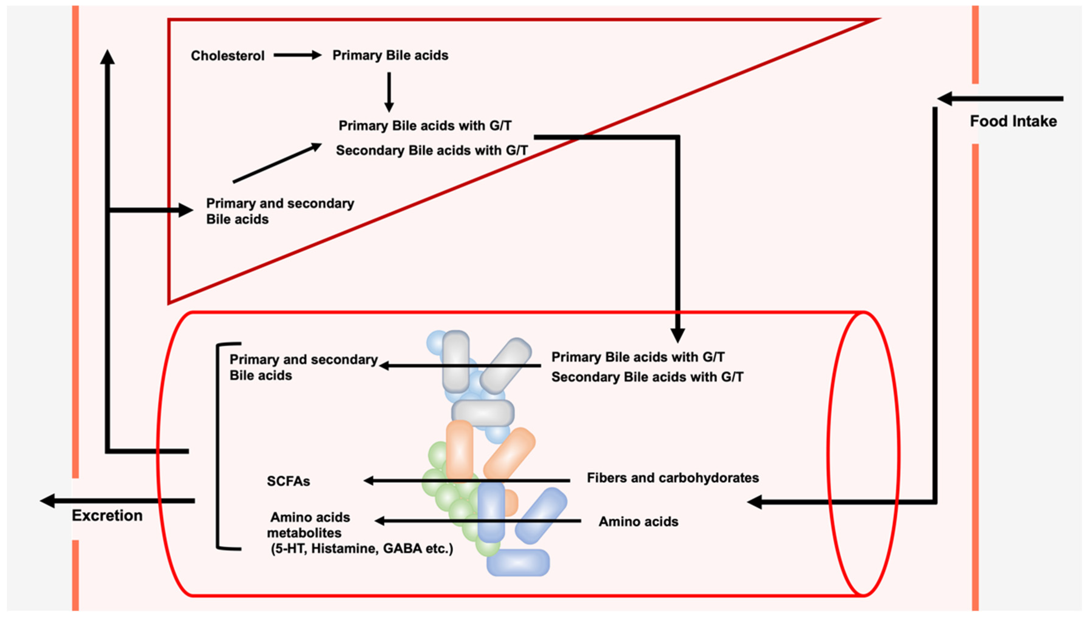
A wide variety and large number of bacterial species live in the gut, forming the gut microbiota. Gut microbiota not only coexist harmoniously with their hosts, but they also induce significant effects on each other. The composition of the gut microbiota can be changed due to environmental factors such as diet and antibiotic intake. In contrast, alterations in the composition of the gut microbiota have been reported in a variety of diseases, including intestinal, allergic, and autoimmune diseases and cancer. The gut microbiota metabolize exogenous dietary components ingested from outside the body to produce short-chain fatty acids (SCFAs) and amino acid metabolites. Unlike SCFAs and amino acid metabolites, the source of bile acids (BAs) produced by the gut microbiota is endogenous BAs from the liver. The gut microbiota metabolize BAs to generate secondary bile acids, such as lithocholic acid (LCA), deoxycholic acid (DCA), and their derivatives, which have recently been shown to play important roles in immune cells. 

bile acids DCA LCA isoalloLCA isoDCA FXR RORγt NR4A1 FOXP3 TGR5

1. Introduction

The mucous membranes of the oral cavity, stomach, gut, and skin form the boundary between the inside and outside of the human body. These mucosal and skin surfaces are inhabited by numerous bacteria [1][2]. In particular, a wide variety and large number of bacterial species live in the gut and form the gut microbiota. The estimated number of bacterial cells in the human body is approximately 3.8 × 10¹³ [1]. Furthermore, the estimated number of human cells is approximately 3 × 10¹³. Thus, the number of bacteria in the human body is equivalent to the number of human cells. In addition, the total mass of bacteria in the human body is estimated to be approximately 0.2 kg [1]. Although a wide variety of bacteria are present in the human gut microbiota, the predominant bacteria are Bacteroidetes, Firmicutes, Actinomycetes, Proteobacteria, and Velcomicrobia at the phylum level. Moreover, the environments of the small and large intestines are significantly different, with varying types of intestinal bacteria inhabiting each part. More specifically, the predominant bacterial families in the small intestine are Lactobacillaceae and Enterobacteriaceae, while those in the colon are Bacteroidaceae, Prevotellaceae, Rikenellaceae, Lachnospiraceae, and Ruminococcaceae [3]. Human gut microbiota and humans not only coexist, but they also influence each other. The composition of the gut microbiota in humans can be affected by environmental factors such as diet and antibiotic intake. The development of metagenomic analysis of the gut microbiota has allowed researchers to perform comparative analyses of the gut microbiota between healthy and diseased individuals, who have in turn identified a correlation between various diseases and changes in the gut microbiota composition. For instance, alterations in the composition of the gut microbiota have been reported in a variety of diseases, including intestinal diseases (e.g., inflammatory bowel disease and colorectal cancer), liver diseases (e.g., nonalcoholic fatty liver disease and liver cancer), systemic metabolic diseases (e.g., diabetes and atherosclerosis), immune-related diseases (e.g., allergic and autoimmune diseases), and neuropsychiatric diseases (e.g., Alzheimer’s and depression) [4][5][6]. In addition, changes in the composition of gut microbiota have been associated with the persistence of symptoms in many patients with post-coronavirus disease 2019 (COVID-19) symptoms, also known as acute post-COVID-19 syndrome (PACS) or long-term COVID [7][8][9].
However, various metabolites produced by the activity of the gut microbiota have also been found to affect their respective hosts. The gut microbiota metabolize exogenous dietary components ingested from outside the body to produce short-chain fatty acids (SCFAs), such as propionic acid, butyric acid, and acetic acid, and amino acid metabolites, such as serotonin (5-HT), dopamine, histamine, and gamma-amino butyric acid (GABA). These SCFAs have a variety of important physiological and pathological roles [10][11]. In addition, 5-HT, dopamine, histamine, and GABA, which are involved in neurotransmission, are produced by the gut microbiota as amino acid metabolites [12][13].
Unlike SCFAs and amino acid metabolites stemming from diets, bile acids (BAs) are synthesized from cholesterol in the liver (Figure 1). The newly produced BAs in the liver are called primary BAs, and chenodeoxycholic acid (CDCA) and cholic acid (CA) are the major primary BAs in humans [14][15]. In addition to these two BAs, previous research has shown that muricholic acids and hyocholic acids are produced in mice [15][16] and pigs [17], respectively. The gut microbiota metabolize these primary BAs to produce secondary bile acids, such as lithocholic acid (LCA), deoxycholic acid (DCA), and ursodeoxycholic acid (UDCA) [18][19][20], which play important roles in mitochondria and autophagy at the cellular level [21][22]. Furthermore, they are physiologically and pathologically important to the host as well as to the composition of the gut microbiota [14][23]. In particular, among secondary BAs, LCA, DCA, and their derivatives have recently been shown to have a significant effect on immune cells [24][25][26][27][28][29][30]. This research focuses on current knowledge of the role of LCA, DCA, and their derivatives on immune cells.
Figure 1. Modification of exogenous dietary components and endogenous bile acids by the gut microbiota. The gut microbiota metabolize exogenous dietary components ingested from outside the body to produce short-chain fatty acids (SCFAs), such as propionic acid, butyric acid, and acetic acid, and amino acid metabolites, such as serotonin (5-HT), dopamine, histamine, and gamma-amino butyric acid (GABA). The gut microbiota also metabolize endogenous bile acids (BAs). Cholesterol is converted to cholic acid (CA) or chenodeoxycholic acid (CDCA) in the liver. These newly produced BAs in the liver are called primary BAs, which are then conjugated with glycine or taurine (G/T). Conjugated primary BAs are secreted into the intestine upon food intake, and they are subsequently deconjugated and then converted to secondary BAs. Approximately 95% of BA in the intestine is reabsorbed and transported to the liver, while the remaining BA is excreted in the feces. A portion of BA reabsorbed from the intestine is transferred to the systemic circulation.

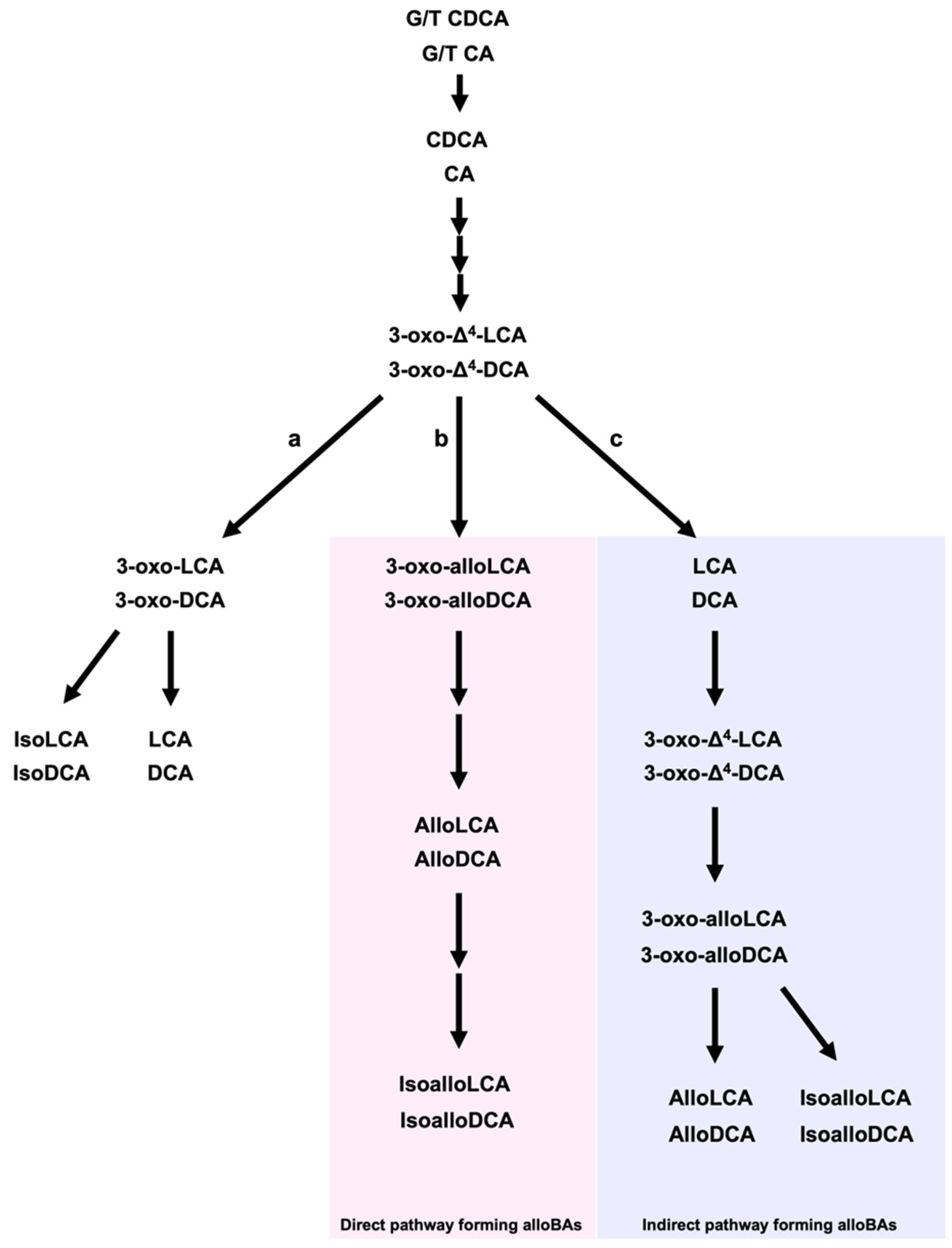
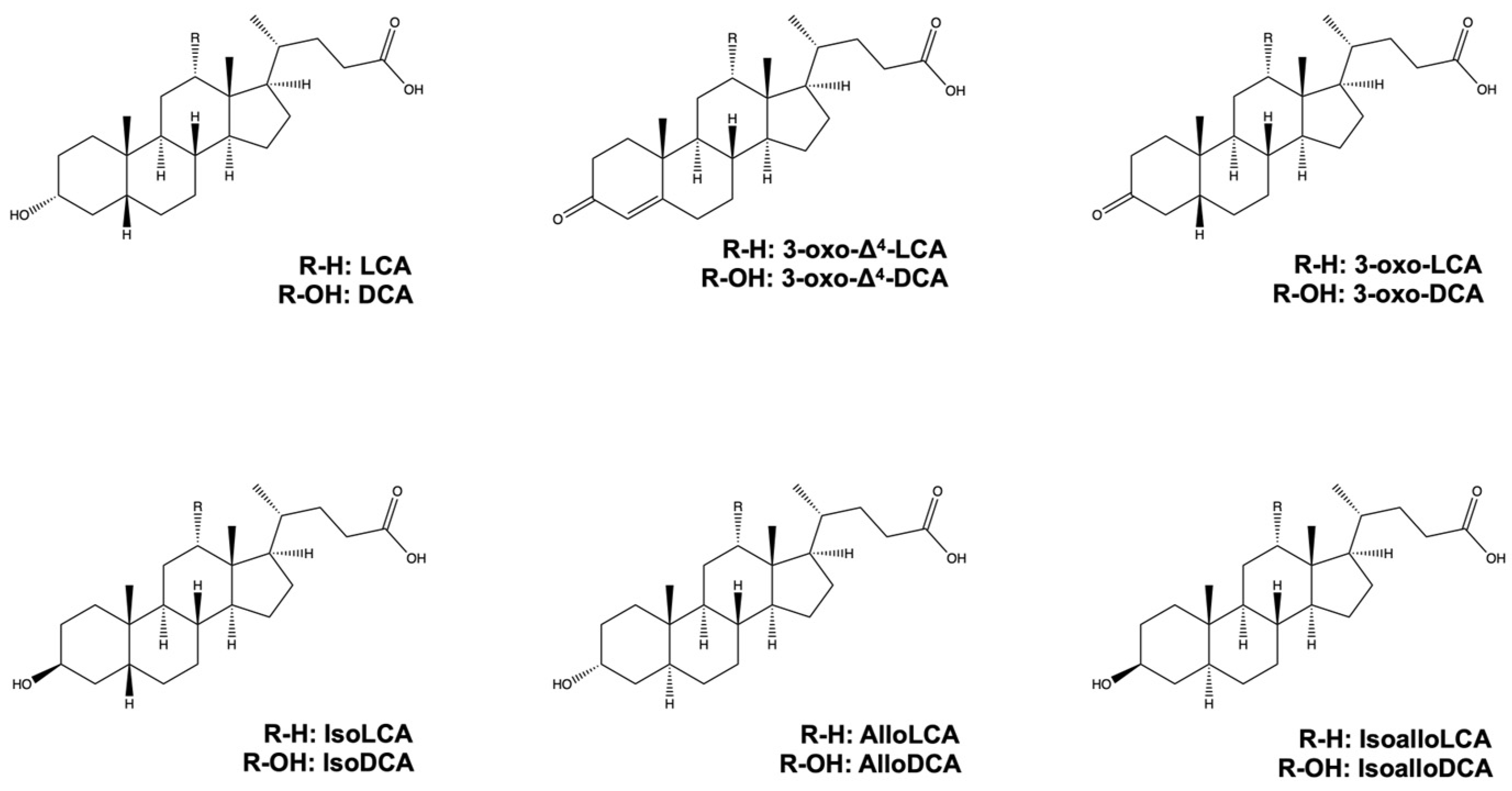
2. Lithocholic Acid, Deoxycholic Acid, and Their Derivatives from Gut Microbiota

Primary BAs, such as CDCA and CA, are primarily synthesized in the liver from cholesterol via two synthetic pathways, namely the classical (or neutral) pathway and the alternative (or acidic) pathway [14][31]. More than 16 enzymes, including cytochrome P450 7A1 (CYP7A1), CYP8B1, and CYP27A1, are involved in primary BA synthesis in the liver [32][33]. Primary BAs are then conjugated with glycine or taurine by bile acid-CoA amino acid N-acyltransferase in the liver [34], and they are transferred to hepatic bile canaliculi via the bile salt export pump or multidrug resistance-associated protein 2. Consequently, BAs in the gallbladder are secreted into the small intestine in response to food intake [35][36], whereas glycine or taurine-conjugated BAs are deconjugated by bile salt hydrolases (BSHs) in gut bacteria (Figure 2) [37]. Various gut bacteria in the ileum and colon, including Lactobacillus, Bifidobacterium, Enterococcus, Clostridium, and Bacteroides spp., have been shown to possess BSH enzyme activity [38][39][40][41][42][43]. Lachnoclostridium scindens (formerly Clostridium scindens), Peptacetobacter hiranonis (formerly Clostridium hiranonis), and Lachnoclostridum hylemonae (formerly Clostridium hylemonae) can 7α-dehydroxylation of BAs. 7α-dehydroxylation of the CDCA and CA is carried away by enzymes encoded in the bile acid-inducible (bai) operon to produce 3-oxo-Δ4-LCA and 3-oxo-Δ4-DCA, respectively [19][20][44]. Both 3-xo-Δ4-LCA and 3-oxo-Δ4-DCA are intracellular intermediates that are not released in appreciable amounts into the environment.
Figure 2. The metabolism of bile acids by gut microbiota to produce LCA, DCA, and their derivatives. In the intestine, glycine or taurine-conjugated primary BAs (CDCA and CA) are deconjugated by gut bacteria. CDCA and CA are then converted to 3-oxo-Δ4-LCA and 3-oxo-Δ4-DCA, respectively. (a) 3-oxo-Δ4-LCA and 3-oxo-Δ4-DCA are converted to 3-oxoLCA and 3-oxoDCA, respectively, which are then converted to LCA and DCA, respectively. There are direct (b) and indirect (c) pathways to produce alloBAs. The direct pathway is conducted by a single bacterial strain, while the indirect pathway is carried out by multiple bacterial strains. In the direct pathway (b), 3-oxo-Δ4-LCA and 3-oxo-Δ4-DCA are converted to alloLCA and alloDCA, respectively, which are in turn converted to isoalloLCA and isoalloDCA, respectively. In the indirect pathway (c), 3-oxo-Δ4-LCA and 3-oxo-Δ4-DCA are converted to LCA and DCA, and then to 3-oxo-Δ4-LCA and 3-oxo-Δ4-DCA, respectively. 3-oxo-Δ4-LCA and 3-oxo-Δ4-DCA are then converted to alloBAs.
There are both direct and indirect pathways to produce alloBAs, which are essentially BAs with a flat shape due to the trans-orientation of the A and B rings of BAs [45]. On the one hand, the direct pathway is conducted by a single bacterial strain, while on the other hand, the indirect pathway is carried out by multiple bacterial strains [19][45]. In the direct pathway, 5ɑ-reductase and BaiA1 (3ɑ-hydroxy bile acid-CoA-ester 3-dehydrogenase 1) convert 3-oxo-Δ4-LCA and 3-oxo-Δ4-DCA to alloLCA and alloDCA, respectively. AlloDCA is subsequently converted to isoalloDCA by 3α-HSDH and 3β-HSDH in Lachnoclostridium scindens. In addition, isoalloLCA can be indirectly produced from 3-oxo-LCA by eleven bacterial genera, namely Bacillus, Bacteroides, Bifidobacterium, Catenibacterium, Collinsella, Eggerthella, Lachnospira, Lactobacillus, Parabacteroides, Peptoniphilus, and Mediterraneibacter [29]. Moreover, a mixture of Lachnoclostridium scindens, which can produce LCA from CDCA [46], Eggerthella lenta, which can produce 3-oxo-LCA from LCA [47], and Parabacteroides merdae, which can produce isoalloLCA from 3-oxo-LCA [29] can synthesize isoalloLCA from LCA. In addition, 5β-reductase, 5α-reductase, and 3β-HSDH enzymes can synthesize isoalloLCA from 3-oxo-LCA, while 3-oxo-Δ4-LCA can be produced from 3-oxo-LCA by 5β-reductase. 3-oxo-alloLCA is then produced from 3-oxo-Δ4-LCA by 5α-reductase. IsoalloLCA is produced from 3-oxo-alloLCA by 3β-HSDH [29][48]. 5α-reductase converts 3-oxo-Δ4-DCA to 3-oxo-DCA and 5β-reductase converts 3-oxo-Δ4-DCA to 3-oxo-alloDCA. 3α-HSDH converts 3-oxo-DCA and 3-oxo-alloDCA to deoxycholic acid (DCA) and alloDCA, respectively [19][45]. IsoDCA and isoLCA are generated from DCA and LCA by 3β-HSDH, respectively [20]. The chemical structures of LCA, DCA, and their derivatives are shown in Figure 3.
Figure 3. Chemical structures of LCA, DCA, and their derivatives.
Approximately 95% of BA in the intestine is reabsorbed and transported to the liver, and the remainder is excreted in the feces. LCA can be sulfonated and converted to LCA 3-sulfate (LCA-3-S) by sulfotransferase in the liver [49][50].

References

  1. Sender, R.; Fuchs, S.; Milo, R. Revised Estimates for the Number of Human and Bacteria Cells in the Body. PLoS Biol. 2016, 14, e1002533.
  2. Dekaboruah, E.; Suryavanshi, M.V.; Chettri, D.; Verma, A.K. Human microbiome: An academic update on human body site specific surveillance and its possible role. Arch. Microbiol. 2020, 202, 2147–2167.
  3. Donaldson, G.P.; Lee, S.M.; Mazmanian, S.K. Gut biogeography of the bacterial microbiota. Nat. Rev. Microbiol. 2016, 14, 20–32.
  4. Mishra, K.; Bukavina, L.; Ghannoum, M. Symbiosis and Dysbiosis of the Human Mycobiome. Front. Microbiol. 2021, 12, 636131.
  5. Hou, K.; Wu, Z.X.; Chen, X.Y.; Wang, J.Q.; Zhang, D.; Xiao, C.; Zhu, D.; Koya, J.B.; Wei, L.; Li, J.; et al. Microbiota in health and diseases. Signal Transduct. Target Ther. 2022, 7, 135.
  6. Zhao, M.; Chu, J.; Feng, S.; Guo, C.; Xue, B.; He, K.; Li, L. Immunological mechanisms of inflammatory diseases caused by gut microbiota dysbiosis: A review. Biomed. Pharmacother. 2023, 164, 114985.
  7. Liu, Q.; Su, Q.; Zhang, F.; Tun, H.M.; Mak, J.W.Y.; Lui, G.C.; Ng, S.S.S.; Ching, J.Y.L.; Li, A.; Lu, W.; et al. Multi-kingdom gut microbiota analyses define COVID-19 severity and post-acute COVID-19 syndrome. Nat. Commun. 2022, 13, 6806.
  8. Zhang, F.; Lau, R.I.; Liu, Q.; Su, Q.; Chan, F.K.L.; Ng, S.C. Gut microbiota in COVID-19: Key microbial changes, potential mechanisms and clinical applications. Nat. Rev. Gastroenterol. Hepatol. 2023, 20, 323–337.
  9. Wang, B.; Zhang, L.; Wang, Y.; Dai, T.; Qin, Z.; Zhou, F.; Zhang, L. Alterations in microbiota of patients with COVID-19: Potential mechanisms and therapeutic interventions. Signal Transduct. Target Ther. 2022, 7, 143.
  10. Van der Hee, B.; Wells, J.M. Microbial Regulation of Host Physiology by Short-chain Fatty Acids. Trends Microbiol. 2021, 29, 700–712.
  11. Portincasa, P.; Bonfrate, L.; Vacca, M.; De Angelis, M.; Farella, I.; Lanza, E.; Khalil, M.; Wang, D.Q.; Sperandio, M.; Di Ciaula, A. Gut Microbiota and Short Chain Fatty Acids: Implications in Glucose Homeostasis. Int. J. Mol. Sci. 2022, 23, 1105.
  12. Jameson, K.G.; Olson, C.A.; Kazmi, S.A.; Hsiao, E.Y. Toward Understanding Microbiome-Neuronal Signaling. Mol. Cell 2020, 78, 577–583.
  13. Sun, P.; Su, L.; Zhu, H.; Li, X.; Guo, Y.; Du, X.; Zhang, L.; Qin, C. Gut Microbiota Regulation and Their Implication in the Development of Neurodegenerative Disease. Microorganisms 2021, 9, 2281.
  14. Kiriyama, Y.; Nochi, H. The Biosynthesis, Signaling, and Neurological Functions of Bile Acids. Biomolecules 2019, 9, 232.
  15. Li, J.; Dawson, P.A. Animal models to study bile acid metabolism. Biochim. Biophys. Acta Mol. Basis Dis. 2019, 1865, 895–911.
  16. Straniero, S.; Laskar, A.; Savva, C.; Hardfeldt, J.; Angelin, B.; Rudling, M. Of mice and men: Murine bile acids explain species differences in the regulation of bile acid and cholesterol metabolism. J. Lipid Res. 2020, 61, 480–491.
  17. Sun, J.; Li, M.; Zhou, H.; Chong, J.; Zhang, J.; Yu, B.; Chen, D.; Ge, L. Importance of gut microbiota for bile acid composition and concentration in pigs. Front. Anim. Sci. 2022, 3, 951840.
  18. Kiriyama, Y.; Nochi, H. Physiological Role of Bile Acids Modified by the Gut Microbiome. Microorganisms 2021, 10, 68.
  19. Lee, J.W.; Cowley, E.S.; Wolf, P.G.; Doden, H.L.; Murai, T.; Caicedo, K.Y.O.; Ly, L.K.; Sun, F.; Takei, H.; Nittono, H.; et al. Formation of secondary allo-bile acids by novel enzymes from gut Firmicutes. Gut. Microbes 2022, 14, 2132903.
  20. Doden, H.L.; Ridlon, J.M. Microbial Hydroxysteroid Dehydrogenases: From Alpha to Omega. Microorganisms 2021, 9, 469.
  21. Kiriyama, Y.; Nochi, H. Role of Microbiota-Modified Bile Acids in the Regulation of Intracellular Organelles and Neurodegenerative Diseases. Genes 2023, 14, 825.
  22. Che, Y.; Xu, W.; Ding, C.; He, T.; Xu, X.; Shuai, Y.; Huang, H.; Wu, J.; Wang, Y.; Wang, C.; et al. Bile acids target mitofusin 2 to differentially regulate innate immunity in physiological versus cholestatic conditions. Cell Rep. 2023, 42, 112011.
  23. Jia, W.; Xie, G.; Jia, W. Bile acid-microbiota crosstalk in gastrointestinal inflammation and carcinogenesis. Nat. Rev. Gastroenterol. Hepatol. 2018, 15, 111–128.
  24. Campbell, C.; McKenney, P.T.; Konstantinovsky, D.; Isaeva, O.I.; Schizas, M.; Verter, J.; Mai, C.; Jin, W.B.; Guo, C.J.; Violante, S.; et al. Bacterial metabolism of bile acids promotes generation of peripheral regulatory T cells. Nature 2020, 581, 475–479.
  25. Guo, C.; Xie, S.; Chi, Z.; Zhang, J.; Liu, Y.; Zhang, L.; Zheng, M.; Zhang, X.; Xia, D.; Ke, Y.; et al. Bile Acids Control Inflammation and Metabolic Disorder through Inhibition of NLRP3 Inflammasome. Immunity 2016, 45, 802–816.
  26. Hang, S.; Paik, D.; Yao, L.; Kim, E.; Trinath, J.; Lu, J.; Ha, S.; Nelson, B.N.; Kelly, S.P.; Wu, L.; et al. Bile acid metabolites control T(H)17 and T(reg) cell differentiation. Nature 2019, 576, 143–148.
  27. Hu, J.; Wang, C.; Huang, X.; Yi, S.; Pan, S.; Zhang, Y.; Yuan, G.; Cao, Q.; Ye, X.; Li, H. Gut microbiota-mediated secondary bile acids regulate dendritic cells to attenuate autoimmune uveitis through TGR5 signaling. Cell Rep. 2021, 36, 109726.
  28. Hu, J.; Zhang, Y.; Yi, S.; Wang, C.; Huang, X.; Pan, S.; Yang, J.; Yuan, G.; Tan, S.; Li, H. Lithocholic acid inhibits dendritic cell activation by reducing intracellular glutathione via TGR5 signaling. Int. J. Biol. Sci. 2022, 18, 4545–4559.
  29. Li, W.; Hang, S.; Fang, Y.; Bae, S.; Zhang, Y.; Zhang, M.; Wang, G.; McCurry, M.D.; Bae, M.; Paik, D.; et al. A bacterial bile acid metabolite modulates T(reg) activity through the nuclear hormone receptor NR4A1. Cell Host Microbe 2021, 29, 1366–1377.e9.
  30. Roggeri, A.; Schepers, M.; Tiane, A.; Rombaut, B.; van Veggel, L.; Hellings, N.; Prickaerts, J.; Pittaluga, A.; Vanmierlo, T. Sphingosine-1-Phosphate Receptor Modulators and Oligodendroglial Cells: Beyond Immunomodulation. Int. J. Mol. Sci. 2020, 21, 7537.
  31. Pandak, W.M.; Kakiyama, G. The acidic pathway of bile acid synthesis: Not just an alternative pathway. Liver Res. 2019, 3, 88–98.
  32. Russell, D.W. Fifty years of advances in bile acid synthesis and metabolism. J. Lipid Res. 2009, 50, S120–S125.
  33. Chiang, J.Y.L.; Ferrell, J.M. Up to date on cholesterol 7 alpha-hydroxylase (CYP7A1) in bile acid synthesis. Liver Res. 2020, 4, 47–63.
  34. Chiang, J.Y.L.; Ferrell, J.M. Bile Acids as Metabolic Regulators and Nutrient Sensors. Annu. Rev. Nutr. 2019, 39, 175–200.
  35. Xue, R.; Su, L.; Lai, S.; Wang, Y.; Zhao, D.; Fan, J.; Chen, W.; Hylemon, P.B.; Zhou, H. Bile Acid Receptors and the Gut-Liver Axis in Nonalcoholic Fatty Liver Disease. Cells 2021, 10, 2806.
  36. Kenna, J.G.; Taskar, K.S.; Battista, C.; Bourdet, D.L.; Brouwer, K.L.R.; Brouwer, K.R.; Dai, D.; Funk, C.; Hafey, M.J.; Lai, Y.; et al. Can Bile Salt Export Pump Inhibition Testing in Drug Discovery and Development Reduce Liver Injury Risk? An International Transporter Consortium Perspective. Clin. Pharmacol. Ther. 2018, 104, 916–932.
  37. Daly, J.W.; Keely, S.J.; Gahan, C.G.M. Functional and Phylogenetic Diversity of BSH and PVA Enzymes. Microorganisms 2021, 9, 732.
  38. Jones, B.V.; Begley, M.; Hill, C.; Gahan, C.G.; Marchesi, J.R. Functional and comparative metagenomic analysis of bile salt hydrolase activity in the human gut microbiome. Proc. Natl. Acad. Sci. USA 2008, 105, 13580–13585.
  39. O’Flaherty, S.; Briner Crawley, A.; Theriot, C.M.; Barrangou, R. The Lactobacillus Bile Salt Hydrolase Repertoire Reveals Niche-Specific Adaptation. mSphere 2018, 3, 10–1128.
  40. Clarke, G.; Sandhu, K.V.; Griffin, B.T.; Dinan, T.G.; Cryan, J.F.; Hyland, N.P. Gut Reactions: Breaking Down Xenobiotic-Microbiome Interactions. Pharmacol. Rev. 2019, 71, 198–224.
  41. Riviere, A.; Selak, M.; Lantin, D.; Leroy, F.; De Vuyst, L. Bifidobacteria and Butyrate-Producing Colon Bacteria: Importance and Strategies for Their Stimulation in the Human Gut. Front. Microbiol. 2016, 7, 979.
  42. Guo, X.; Okpara, E.S.; Hu, W.; Yan, C.; Wang, Y.; Liang, Q.; Chiang, J.Y.L.; Han, S. Interactive Relationships between Intestinal Flora and Bile Acids. Int. J. Mol. Sci. 2022, 23, 8343.
  43. Begley, M.; Hill, C.; Gahan, C.G. Bile salt hydrolase activity in probiotics. Appl. Environ. Microbiol. 2006, 72, 1729–1738.
  44. Ridlon, J.M.; Harris, S.C.; Bhowmik, S.; Kang, D.J.; Hylemon, P.B. Consequences of bile salt biotransformations by intestinal bacteria. Gut. Microbes 2016, 7, 22–39.
  45. Ridlon, J.M.; Daniel, S.L.; Gaskins, H.R. The Hylemon-Bjorkhem pathway of bile acid 7-dehydroxylation: History, biochemistry, and microbiology. J. Lipid Res. 2023, 64, 100392.
  46. Ridlon, J.M.; Kang, D.J.; Hylemon, P.B. Bile salt biotransformations by human intestinal bacteria. J. Lipid Res. 2006, 47, 241–259.
  47. Paik, D.; Yao, L.; Zhang, Y.; Bae, S.; D’Agostino, G.D.; Zhang, M.; Kim, E.; Franzosa, E.A.; Avila-Pacheco, J.; Bisanz, J.E.; et al. Human gut bacteria produce Tau(Eta)17-modulating bile acid metabolites. Nature 2022, 603, 907–912.
  48. Sato, Y.; Atarashi, K.; Plichta, D.R.; Arai, Y.; Sasajima, S.; Kearney, S.M.; Suda, W.; Takeshita, K.; Sasaki, T.; Okamoto, S.; et al. Novel bile acid biosynthetic pathways are enriched in the microbiome of centenarians. Nature 2021, 599, 458–464.
  49. Camilleri, M. Bile acid detergency: Permeability, inflammation, and effects of sulfation. Am. J. Physiol. Gastrointest. Liver Physiol. 2022, 322, G480–G488.
  50. Sheng, W.; Ji, G.; Zhang, L. The Effect of Lithocholic Acid on the Gut-Liver Axis. Front. Pharmacol. 2022, 13, 910493.
More
Information
Contributors MDPI registered users' name will be linked to their SciProfiles pages. To register with us, please refer to https://encyclopedia.pub/register : ,
View Times: 260
Revisions: 3 times (View History)
Update Date: 22 Nov 2023
1000/1000
Video Production Service