Submitted Successfully!
To reward your contribution, here is a gift for you: A free trial for our video production service.
Thank you for your contribution! You can also upload a video entry or images related to this topic.
Version Summary Created by Modification Content Size Created at Operation
1 -- 4674 2023-11-16 10:26:59 |
2 format Meta information modification 4674 2023-11-17 03:36:25 |

Video Upload Options

Do you have a full video?

Confirm

Are you sure to Delete?
Cite
If you have any further questions, please contact Encyclopedia Editorial Office.
Sultana, S.; Viet, T.D.; Amin, T.; Kazi, E.; Micolucci, L.; Mollah, A.K.M.M.; Akhtar, M.M.; Islam, M.S. Inflammasomes in Inflammatory Diseases. Encyclopedia. Available online: https://encyclopedia.pub/entry/51662 (accessed on 03 September 2024).
Sultana S, Viet TD, Amin T, Kazi E, Micolucci L, Mollah AKMM, et al. Inflammasomes in Inflammatory Diseases. Encyclopedia. Available at: https://encyclopedia.pub/entry/51662. Accessed September 03, 2024.
Sultana, Sharmim, Thanh Doan Viet, Tasmiha Amin, Esha Kazi, Luigina Micolucci, Abul Kalam Mohammad Moniruzzaman Mollah, Most Mauluda Akhtar, Md Soriful Islam. "Inflammasomes in Inflammatory Diseases" Encyclopedia, https://encyclopedia.pub/entry/51662 (accessed September 03, 2024).
Sultana, S., Viet, T.D., Amin, T., Kazi, E., Micolucci, L., Mollah, A.K.M.M., Akhtar, M.M., & Islam, M.S. (2023, November 16). Inflammasomes in Inflammatory Diseases. In Encyclopedia. https://encyclopedia.pub/entry/51662
Sultana, Sharmim, et al. "Inflammasomes in Inflammatory Diseases." Encyclopedia. Web. 16 November, 2023.
Inflammasomes in Inflammatory Diseases
Edit

Inflammasomes, a group of multiprotein complexes, are essential in regulating inflammation and immune responses. Several inflammasomes, including nucleotide-binding domain leucine-rich repeat-containing protein 1 (NLRP1), NLRP3, NLRP6, NLRP7, NLRP12, interferon-inducible protein 16 (IFI16), NOD-like receptor family CARD domain-containing protein 4 (NLRC4), absent in melanoma 2 (AIM2), and pyrin, have been studied in various inflammatory diseases. Activating inflammasomes leads to the processing and production of proinflammatory cytokines, such as interleukin (IL)-1β and IL-18. The NLRP3 inflammasome is the most extensively studied and well characterized. Consequently, targeting inflammasomes (particularly NLRP3) with several compounds, including small molecule inhibitors and natural compounds, has been studied as a potential therapeutic strategy. 

inflammasome inflammatory disease NLRP3 multiple sclerosis Alzheimer’s disease Parkinson’s disease

1. Introduction

Inflammasomes play a significant role in various inflammatory diseases [1] (Figure 1). The involvement of inflammasomes in a wide range of inflammatory diseases has been reported [1][2][3]. Here discuss the role of inflammasome complexes in various inflammatory diseases, including multiple sclerosis (MS), experimental autoimmune encephalomyelitis, Alzheimer’s disease (AD), Parkinson’s disease (PD), atherosclerosis, type 2 diabetes (T2D), and obesity (Figure 1). Limited but emerging data suggest that inflammasomes may play a role in the development and progression of other inflammatory conditions, including rheumatoid arthritis (RA), inflammatory bowel disease (IBD), gout, psoriasis, and systemic lupus erythematosus (SLE) [4].
Figure 1. Inflammasomes in major inflammatory diseases. The figure presents an overview of the major inflammasomes activated in the indicated diseases and an overview of the major products of inflammasome activity.

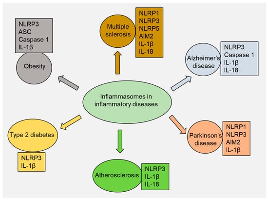
2. Multiple Sclerosis

MS is a chronic inflammatory disease that influences the central nervous system (CNS) and is characterized by neurodegenerative symptoms and disability, for example, weakness, tiredness, incontinence, and paralysis. The immune system damages the myelin sheath that covers nerve fibers, leading to demyelination, inflammation, and axonal damage. Genetic and environmental factors are believed to be involved in the development of MS, although the exact cause of the disease remains unknown. There is no cure for MS, and the central focus of existing treatments revolves around using immunomodulatory medications to lessen the severity and frequency of relapses [5].
The role of inflammasomes in the pathogenesis of MS has been emphasized in recent years, with particular attention paid to the NLRP3 inflammasome. In MS, the NLRP3 inflammasome is activated in various cell types, including microglia, astrocytes, and CD4+ T cells [6]. Activation of the NLRP3 inflammasome leads to the release of IL-1β and IL-18, which promote inflammation and contributes to myelin destruction [7]. The NLRP3 inflammasome also regulates the blood–brain barrier (BBB), which is disrupted in MS, allowing immune cells to enter the CNS [8]. The role of NLRP3 in MS was supported by the observation that numerous small-molecule NLRP3 inhibitors can reduce disease severity. For example, MCC950 is a small-molecule NLRP3 inhibitor that binds to the NLRP3 NACDHT domain and blocks NLRP3 conformational changes and oligomerization [9]. Ketotifen, an antihistamine, has been shown to inhibit NLRP3 inflammasome action and reduce oxidative stress and the infiltration of T cells in the CNS [10]. IC100, a humanized antibody against the ASC component of inflammasomes, has also been developed to block NLRP3 [11]. These findings suggest that targeting the NLRP3 inflammasome is a promising approach for MS treatment.
Another inflammasome linked to MS is the AIM2 inflammasome. AIM2 is activated by cytosolic double-stranded DNA and leads to the maturation and release of IL-1β and IL-18, as well as caspase-1, which promotes the proteolytic cleavage of the cytokines, as mentioned earlier [12]. AIM2 is linked to the development of experimental autoimmune encephalomyelitis, an animal model of MS [13]. AIM2 inflammasome plays a role in the demyelination process in MS as it induces the expression of matrix metalloproteinases (MMPs) in microglia, a key contributor to the breakdown of the BBB [14].
Many inflammasomes linked to MS remain understudied. This list includes inflammasomes such as NLRP1, NLRC5, and others. NLRP1 is known to play a role in the activation of caspase-1 and the production of IL-1β and IL-18 in response to bacterial toxins, and multiple studies have also identified its involvement in numerous sclerosis cases [15][16]. Similarly, NLRC5 deficiency reduces the severity of experimental autoimmune encephalomyelitis, with lower inflammation and demyelination in the CNS [17]. As a result of these limitations, more research is needed to fully elucidate the role of inflammasomes in the pathogenesis of MS.
Experimental autoimmune encephalomyelitis (EAE) is a group of disorders characterized by axonal damage and demyelination of the CNS and has been used as a model for MS [17]. The EAE model leads to neurological symptoms, with motor deficits (from weakness and spasticity to complete paralysis) being the most prevalent. Additionally, sensory disturbances such as numbness, tingling, pain, and autonomic dysfunction affecting blood pressure, heart rate, and bladder and bowel function are commonly observed [18]. In EAE, the immune system is activated against self-antigens within the CNS, leading to the recruitment and activation of immune cells such as T cells and macrophages [19][20]. In addition to T cells and macrophages, B cells and natural killer (NK) cells have also been found to be implicated in the pathogenesis of EAE [21][22].
In EAE, inflammasomes are thought to be activated by DAMPs released by damaged or dying CNS cells. These DAMPs include extracellular ATP, uric acid crystals, and the high mobility group box (HMGB1) protein [23]. The existing literature shows that inflammasomes contribute to the pathogenesis of EAE primarily by promoting the activation of microglia [24]. Microglia, the resident immune cells of the CNS, play a vital role in maintaining tissue homeostasis. However, they can become activated in response to injury or inflammation. Activated microglia generate proinflammatory cytokines such as IL-1β and IL-18, which further activate T cells and recruit other immune cells to sites of inflammation [25]. In addition to promoting microglial activation, inflammasomes can directly contribute to the production of proinflammatory cytokines. For example, the NLRP3 inflammasome promotes the activation of the NF-κB pathway, which leads to the production of proinflammatory cytokines, such as IL-1β and TNF-α, by microglia in EAE. These cytokines further activate T cells and promote the recruitment of other immune cells to sites of inflammation in the CNS, exacerbating tissue damage and disease progression [26]. Inflammasomes also contribute to the pathogenesis of EAE by activating other cell types in the CNS, such as astrocytes and oligodendrocytes [27][28]. AIM2 inflammasome activates astrocytes in EAE by producingIL-1β and other cytokines [27]. In addition, inflammasome activation via TNFR2 signaling can induce the expression of proinflammatory genes in oligodendrocytes, further exacerbating inflammation and contributing to tissue damage in EAE [28].
Inflammasomes also play a role in the demyelination process in EAE [29]. The NLRP3 gene was found to be significantly upregulated in a demyelination model, and mice lacking this gene showed delayed neuroinflammation and demyelination and experienced loss of oligodendrocytes [29]. This effect was partly mediated by caspase-1 and IL-18 but not IL-1β. Interestingly, the lack of NLRP3 did not result in delayed remyelination, unlike the absence of IL-1β. Inhibition of IL-18 may decrease demyelination but promote remyelination, suggesting that IL-18 is a potential therapeutic target for demyelinating diseases [29].

3. Alzheimer’s Disease

AD is a complex and multifaceted disorder that affects millions of people worldwide. It is a progressive neurodegenerative disease that leads to cognitive impairment, memory loss, and behavioral changes. The pathological manifestations of AD are the accumulation of amyloid-beta (Aβ) plaques and tau tangles in the brain. These protein aggregates cause dysfunction and loss of neurons, leading to the death of brain cells [30]. Inflammation is a critical component of AD pathogenesis, and inflammasomes have emerged as crucial mediators of neuroinflammation in AD [31].
The NLRP3 inflammasome is the most extensively studied in AD. The NLRP3 inflammasome can be activated in response to various danger signals, such as Aβ and reactive oxygen species (ROS), leading to the activation of caspase-1 and the release of IL-1β and IL-18. Aβ can directly activate the NLRP3 inflammasome by binding to the purinergic receptor P2X7 on microglia, thus producing ROS and activating NLRP3 [32]. In addition, Aβ can induce lysosomal damage and the release of cathepsin B, a lysosomal protease, which can also activate the NLRP3 inflammasome [33].
Inflammasome activation in AD has been implicated in several pathological conditions with the production of proinflammatory cytokines, microglial activation, and the formation of Aβ plaques [34]. The function of microglia is the clearance of Aβ plaques, which accumulate in the brains of AD patients. In AD, microglia become chronically activated and cannot clear Aβ effectively [35]. In addition to the clearance of Aβ, microglia play an important role in regulating synaptic plasticity, a process essential for learning and memory [36]. Proinflammatory cytokines IL-1β and IL-18 induce the expression of amyloid precursor protein (APP) and beta-secretase 1 (BACE1), leading to increased Aβ production [37]. Moreover, inflammasome activation has been reported to induce the formation of Aβ oligomers and fibrils, which leads to the deposition of Aβ plaques [38].
In addition to their role in inducing neuroinflammation and Aβ deposition, inflammasomes have been implicated in other pathological processes, such as tau hyperphosphorylation and autophagy [39]. Tau hyperphosphorylation is a crucial pathological process in AD closely associated with the formation of neurofibrillary tangles (NFTs), a hallmark of AD pathology. Tau protein is usually involved in stabilizing microtubules in neurons. However, when it becomes hyperphosphorylated, it detaches from microtubules and aggregates into NFTs, disrupting axonal transport and neuronal dysfunction [40]. Recent pieces of evidence have suggested that the activation of inflammasomes (such as NLRP3) can induce tau hyperphosphorylation by promoting the expression levels of tau kinases, such as glycogen synthase kinase-3β (GSK3β) and cyclin-dependent kinase 5 (CDK5), and inhibiting the activity of tau phosphatases, such as protein phosphatase 2A (PPP2A) and protein phosphatase 1 (PP1) [41][42]. ROS produced by activated microglia and neurons can also induce tau hyperphosphorylation by activating the NLRP3 inflammasome and promoting the expression levels of tau kinases [43].
Inflammasome activation can also suppress autophagy, a cellular mechanism that clears misfolded proteins and damaged organelles, leading to the buildup of toxic protein aggregates and cellular debris [44]. Autophagy impairment has been shown to induce tau hyperphosphorylation by increasing tau kinase levels and decreasing tau phosphatase activity [45]. NLRP3 inflammasome activation can inhibit autophagy through several mechanisms, including lysosomal damage, impairing lysosomal acidification, and reducing lysosomal enzyme activity [44].
Inflammasome activation has been reported to exacerbate cognitive impairment and Aβ pathology in transgenic AD mice [46]. Inhibition of inflammasome activation using pharmacological or genetic approaches improved cognitive function and reduced Aβ pathology in AD mice. For example, treatment with MCC950, a selective NLRP3 inhibitor, has reduced Aβ pathology and enhanced cognitive function in AD mice [47]. Similarly, deletion of NLRP3 or caspase-1 was associated with reduced Aβ pathology and improved cognitive function in AD mice [48].
Inflammasome activation in peripheral tissues, including the gut and liver, has also been implicated in AD pathogenesis by inducing systemic inflammation and impaired brain function [49]. For example, gut dysbiosis, characterized by an imbalance in the gut microbiota, has been demonstrated to induce inflammasome activation and increase Aβ deposition in the brain [50]. Similarly, liver dysfunction, which commonly occurs in AD patients, can induce inflammasome activation and increase the production of proinflammatory cytokines and oxidative stress, causing systemic inflammation and cognitive impairment [51].
Other inflammasomes, such as NLRP1, AIM2, and NLRC4, have also been suggested to contribute to neuroinflammation in AD, although their roles are less well-established than those of NLRP3.
In experimental models of AD, NLPR1 seems able to promote neuronal pyroptosis [52], whereas AIM2 deletion mitigates Aβ deposition and microglial activation but increases the expression of inflammatory cytokines [53].
In another in vivo experimental model, a significant increase in the expression level of the NLRC4 inflammasome, ASC, IL-1β, and p-Tau protein-positive cells after pharmacologic treatment for the induction of an Alzheimer’s-like disease in animals has been found [54]. In contrast, no significant difference was seen in other inflammasome components such as NLRP1, NLRP3, AIM2, IL-18, and caspase-1. These findings suggest that the NLRC4 inflammasome is involved in the typical neuroinflammation and memory impairment of AD [54].

4. Parkinson’s Disease

PD is a chronic neurodegenerative disorder characterized by progressive loss of pigmented nigrostriatal dopaminergic neurons in the substantia nigra pars compacta (SNpc) region of the brain. The disease’s progression leads to motor symptoms such as tremors, rigidity, and bradykinesia [55]. Activated glial cells, which compose most of this inflammatory response, contribute to this neurodegenerative process by producing toxic molecules [56]. Various factors, including glial reactions, T-cell infiltration, and increased expression of inflammatory cytokines, trigger inflammatory responses in PD. However, the NLRP3 inflammasome is the most widely studied in the pathogenesis of PD [57].
NLRP3 inflammasome activation is a two-step process, i.e., priming and activation [58]. Peripheral inflammation can transform primed microglia into a state that can trigger more robust neurodegenerative responses [59]. Although the precise mechanism of inflammasome priming and activation in PD has not been fully elucidated, emerging research data suggest the involvement of cytokines such as IL-1β and TNF-α [60]. When these cytokines are secreted by activated glia in the brain or are present in circulating blood, the permeability of the BBB increases, and the expression levels of cellular adhesion molecules, such as selectins, are upregulated in microvascular endothelial cells [61].
Various other factors have been suggested to activate the NLRP3 inflammasome in PD, such as oxidative stress, mitochondrial dysfunction, and the accumulation of misfolded proteins such as alpha-synuclein (α-syn) [62]. α-syn is a presynaptic protein typically involved in the regulation of neurotransmitter release. In PD, α-syn accumulates in insoluble aggregates that are the principal constituents of Lewy bodies—pathological hallmarks of PD. The misfolded proteins can activate NLRP3 inflammasome by triggering lysosomal damage, leading to the release of cathepsin B, which then initiates the inflammasome assembly [63]. In addition, α-syn can activate NLRP3 by interacting with TLR2, leading to the activation of downstream signaling pathways [64].
In addition to the NLRP3 inflammasome, other inflammasome complexes, such as the AIM2 and the NLRP1 inflammasomes, have also been implicated in the pathogenesis of PD [65][66][67]. Activation of these inflammasomes induces the production of IL-1β and IL-18, which can contribute to the neuroinflammatory response in PD [66]. The AIM2 inflammasome is activated by double-stranded DNA in the cytoplasm, and research findings have indicated that it is activated in response to α-syn accumulation in PD [65]. Specifically, research has shown that extracellular α-syn can be taken up by microglia and transported to the cytoplasm, where it can activate the AIM2 inflammasome [68]. In PD, the activation of the NLRP1 inflammasome has been reported to be associated with the accumulation of α-syn and the induction of neuronal cell death [67]. Considering the involvement of inflammasomes in PD pathology, targeting the inflammasome complex or downstream inflammatory pathways would be an ideal therapeutic approach to mitigate neuroinflammation and slow down PD progression.

5. Atherosclerosis

Atherosclerosis is a chronic inflammatory disease characterized by the deposition of lipids, immune cells, and extracellular matrix (ECM) within the arterial walls [69]. Inflammation is vital in initiating, progressing, and rupturing atherosclerotic plaques, which can lead to cardiovascular events, including myocardial infarction and stroke [70]. Both innate and adaptive immunity play a critical role in the initiation, progression, and destabilization of atherosclerotic plaques and dyslipidemia [71]. Innate immune cells, including monocytes, macrophages, and dendritic cells, may accumulate in the arterial wall, contributing to the inflammatory response [72]. CD31+ endothelial cells and CD68+ macrophages within atherosclerotic lesions in human carotid arteries exhibit significant levels of the purinergic 2X7 receptor (P2X7R) [73]. P2X7R has been reported to be involved in the progression of atherosclerosis by inducing the activation of the NLRP3 inflammasome [74]. Macrophages are among the first immune cells to accumulate in the plaque, which contributes to the uptake of ox-LDL and the formation of foam cells [75]. Foam cells are lipid-laden macrophages that play a significant role in the progression of plaque formation and the narrowing of the arteries [73].
It has been reported that NLRP3 inflammasome activation in macrophages within atherosclerotic plaques can promote the formation of foam cells [76]. Sterol regulatory element-binding protein 1 (SREBP-1) is a transcription factor critical in regulating lipid metabolism. Varghese et al. demonstrated that the activation of the NLRP3 inflammasome was facilitated by SREBP-1, leading to the formation of macrophage foam cells induced by ox-LDL [77]. The buildup of ox-LDL is crucial in the development of atherosclerotic plaques [78].
Inflammasomes are activated in response to various stimuli, including cholesterol crystals and ox-LDL, producing and secreting proinflammatory cytokines such as IL-1β and IL-18 [77][79]. These cytokines play a dominant role in the development of atherosclerosis by triggering the recruitment and activation of immune cells, inducing the expression of adhesion molecules on endothelial cells and stimulating smooth muscle cell proliferation and ECM deposition [80]. IL-1β is one of the most potent mediators of atherosclerosis and is involved in various phases of the disease. IL-1β can induce the expression of adhesion molecules on endothelial cells, resulting in the recruitment of monocytes into the arterial wall. It can also activate smooth muscle cells and promote their migration, proliferation, and matrix deposition, leading to fibrous cap formation [81]. IL-18 is another proinflammatory cytokine produced by the inflammasome and has been linked to the pathogenesis of atherosclerosis [82]. IL-18 activates T lymphocytes, which play an important role in the adaptive immune response in atherosclerosis [83]. IL-18 can also promote the development of atherosclerosis by activating endothelial cells and increasing the expression of adhesion molecules, which helps the recruitment of immune cells into the arterial walls. Furthermore, IL-18 can induce the production of other proinflammatory cytokines, including IL-6 and TNF-α, which contribute to plaque development and destabilization [84].
The TLR4, NF-κB, and JAK/STAT pathways are all involved in inflammatory signaling and play an essential role in atherosclerosis. TLR4, which is expressed in immune cells, can detect a range of ligands, such as LPS, oxLDL, and HMGB1 [85]. Following activation, TLR4 can initiate a signaling cascade that, ultimately, activates NF-κB and produces proinflammatory cytokines [86]. The JAK/STAT pathway can activate proinflammatory pathways in response to cytokines such as IL-6 [87]. Furthermore, considering the role of inflammation in atherosclerosis, traditional risk factors such as high LDL levels, obesity, angiotensin II, and smoking remain influential in the development of atherosclerosis [88].

6. Type 2 Diabetes

Type 2 diabetes (T2D) is a metabolic condition characterized by insulin resistance, which leads to hyperglycemia and glucose intolerance [89]. Nearly 90% of diabetic patients exhibit insulin resistance [90]. In recent years, there has been a growing interest in the role of inflammation in the development of this disease. Studies suggest that subclinical chronic inflammation and innate immune system activation are vital pathogenetic elements in the emergence of insulin resistance and T2D [91].
The NLRP3 inflammasome and the IL-1β pathway have been reported in T2D [92][93]. According to a study by Lee et al., NLRP3 inflammasome activation was elevated in myeloid cells obtained from individuals with T2D [93]. Studies have demonstrated that oligomers of islet amyloid polypeptide (IAPP), a protein associated with the formation of amyloid deposits in the pancreas during T2D, can activate the NLRP3 inflammasome, leading to the production of mature IL-1β [94]. The involvement of NLRP3 in T2D was further documented by the observation that silencing the NLRP3 gene showed a significant improvement in the development of diabetic cardiomyopathy (DCM) in a rat model of T2D [95]. Furthermore, rosuvastatin demonstrated alleviation of diabetic cardiomyopathy in a rat model of T2D by inhibiting the NLRP3 inflammasome [96]. γ-Tocotrienol (γT3) also effectively slowed down the advancement of T2D by inhibiting the NLRP3 inflammasome [96]. These compelling data strongly support the pivotal role of inflammasomes in the pathogenesis of T2D.
Inflammasomes have also been implicated in the development of β-cell dysfunction [97][98]. β cells are the cells in the pancreas responsible for insulin production and secretion, and their dysfunction is a prominent feature of T2D [99]. Chronic activation of inflammasomes produces ROS and oxidative stress, which can damage beta cells and impair their function. This process can lead to a decrease in insulin secretion and the development of hyperglycemia [100]. Indeed, Sokolova et al. reported that the deletion of the NLRP3 inflammasome was associated with increased β-cell function and viability in the presence of hypoxia and oxidative stress [98].
Chronic inflammation is a signature characteristic of T2D, and the primary molecular links between inflammation and T2DM are macrophage mediators TNF-α, IL-1β, and IL-6. These inflammatory cytokines are generated by activated macrophages and adipocytes in adipose tissue and are elevated in the serum of individuals with insulin resistance and T2D [101]. Inflammatory mediators such as TNF-α, IL-1β, and IL-6 can stimulate insulin resistance by interrupting insulin signaling in peripheral tissues through activation of various inflammatory pathways, including NF-κB and c-JUN N-terminal kinase (JNK) pathways [102]. These pathways interfere with the insulin signaling cascade by inducing the phosphorylation of serine residues on IRS proteins, which results in their degradation and, ultimately, leads to insulin resistance [103]. Several drugs with anti-inflammatory properties have been reported to lower both acute-phase reactants and glycemia and decrease the risk of developing T2D. These drugs include thiazolidinediones (TZDs) and glucagon-like peptide-1 (GLP-1) receptor agonists. These drugs can target various components of the inflammatory signaling pathways involved in insulin resistance and have been shown to improve insulin sensitivity and glycemic control in individuals with T2D [104]. For example, pioglitazone is an oral antidiabetic from the thiazolidinedione drug class and is best known for its dual agonist activity on both PPAR-γ and PPAR-α. Pioglitazone can mitigate diabetic renal damage by inhibiting the activation of the renal AGE/RAGE axis and reducing NF-κB expression [105]. This effect was associated with decreased NLRP3 levels and subsequent reduction in the secretion of inflammatory cytokines [105]. Liraglutide, a GLP-1 receptor agonist, has been reported to improve the disease score in EAE mice, associated with the downregulation of the NLRP3 pathway [106].

7. Obesity

Obesity is a central component of metabolic syndrome, characterized by excessive fatty tissue expansion induced by immune cell infiltration, particularly macrophages, and adipocyte hypertrophy [107]. The infiltration of immune cells into adipose tissues can be exacerbated by excessive consumption of fat and other macronutrients without sufficient antioxidant intake [108]. Recent studies using a bidirectional Mendelian randomization approach have provided compelling evidence that higher levels of adiposity caused by fat mass, obesity-associated genes, and single nucleotide polymorphisms (SNPs) of melanocortin receptor 4 are intricately connected with elevated levels of the inflammatory marker CRP [109]. In particular, the NLRP3 inflammasome plays an essential role in the pathogenesis of obesity by increasing adiposity, insulin resistance, glucose intolerance, and inflammation [110]. It has been reported that the knockout of the NLRP3 inflammasome can protect against obesity-induced pathologies, making it a viable target for therapeutic intervention [111]. NLRP3 inflammasome activation in adipose tissue is associated with the recruitment of macrophages, production of proinflammatory cytokines, and induction of insulin resistance [112]. Inflammasome-deficient mice are protected from developing obesity and insulin resistance when fed a high-fat diet [113]. In addition, inhibiting NLRP3 inflammasome activation in obese mice improved glucose homeostasis and insulin sensitivity [114]. Fatty acid accumulation in obese adipose tissue can increase ceramide production, a danger signal to stimulate the formation of the NLRP3 inflammasome complex [115].
Excessive intake of calories can result in the infiltration of macrophages into adipose tissue and the production of proinflammatory cytokines. However, deficiencies in NLRP3, ASC, and caspase-1 have been shown to offer protection against obesity-induced insulin resistance and metabolic dysfunction [116]. The ASC adaptor protein is an essential component of the inflammasome complex and acts as a bridge between NLRP3 and caspase-1 [117]. However, ASC-induced specks are not a prerequisite for inflammation activation but do maximize IL-1β processing [118]. Caspase-1 activation promotes the cleavage of pro-IL-1β and pro-IL-18 into their mature forms [119]. Another vital pathway implicates the activation of toll-like receptors (TLRs), which recognize microbial and endogenous ligands. TLR activation can produce proinflammatory cytokines and activate the inflammasome complex [85].
In addition to the direct activation of the inflammasome complex, several other pathways have been implicated in the regulation of inflammasome activation in obesity. These include the production of ROS, which can promote NLRP3 inflammasome activation, and the modulation of lipid metabolism, which can contribute to the accumulation of lipid intermediates that activate the inflammasome complex [120]. Furthermore, several adipokines and hormones, including leptin and insulin, have been shown to regulate inflammasome activation in obesity. Leptin and insulin are essential hormones in regulating energy metabolism and developing obesity. Adipocytes produce leptin and act on the hypothalamus to control food intake and energy expenditure [121]. Conversely, insulin is produced by the pancreas and regulates glucose uptake and metabolism in various tissues, including adipose tissue [122]. Leptin has been shown to promote inflammasome activation by inducing the generation of ROS and activating the NLRP3 inflammasome [123]. In addition, leptin enhances the secretion of proinflammatory cytokines, including IL-1β and IL-6, by macrophages in adipose tissue [124]. IL-1β and IL-6 can induce insulin resistance and promote the development of metabolic dysfunction in obesity. On the other hand, insulin has been shown to have both proinflammatory and anti-inflammatory effects on the inflammasome complex. Insulin can activate the NLRP3 inflammasome by promoting the generation of ROS and activating the TLR4 signaling pathway [125]. However, it has been observed that insulin can also inhibit inflammasome activation by inhibiting the secretion of proinflammatory cytokines and promoting the secretion of anti-inflammatory cytokines such as IL-10 [126].

8. Other Inflammatory Diseases

Emerging evidence suggests that inflammasomes have a potential role in the development and progression of other inflammatory conditions, which include RA, IBD, gout, psoriasis, and SLE [4]. As a prototypical autoimmune disease, RA is primarily characterized by inflicting damage to the bones and cartilage. In a study conducted by Guo et al., it was observed that the NLRP3 inflammasome exhibited significant activation in the synovial tissue of RA patients and mice with collagen-induced arthritis (CIA) [127]. Activation of the inflammasome in synovial cells promotes the secretion of IL-1β and IL-18, contributing to the chronic inflammation, joint damage, and cartilage destruction observed in RA [128]. IBD is a group of chronic inflammatory disorders primarily affecting the gastrointestinal tract. The two main types of IBD are ulcerative colitis (UC) and Crohn’s disease (CD). UC is characterized by inflammation and ulcers typically limited to the colon’s inner lining (large intestine) and rectum. Conversely, CD can affect any part of the gastrointestinal tract, from the mouth to the anus. The exact etiology of IBD remains uncertain; however, available evidence indicates a complex interaction between genetic and environmental factors. The activation of inflammasomes, particularly NLRP3, has been shown to facilitate the secretion of proinflammatory cytokines and the recruitment of immune cells, contributing to tissue damage and the progression of IBD [129]. Gout is a type of inflammatory arthritis that occurs when uric acid crystals accumulate in the joints. When the uric acid level becomes too high, sharp urate crystals can form in the joints, triggering an inflammatory response and causing the characteristic symptoms of gout. The role of inflammasomes, particularly NLRP3, has been implicated in the pathogenesis of gout [130][131]. Studies indicate that both monosodium urate (MSU) and calcium pyrophosphate dihydrate (CPPD) crystals can activate the caspase-1-activating NALP3 inflammasome, leading to the production of active IL-1β and IL-18 [130]. Psoriasis is a chronic autoimmune skin condition that leads to the rapid buildup of skin cells. It is described by red, thickened patches of skin covered with silvery scales. The exact cause of psoriasis is not entirely understood, but evidence indicates the involvement of a combination of genetic, immune system, and environmental factors. Experimental data suggest that various inflammasomes, including NLRP1, NLRP3, and AIM2, contribute to the development and progression of psoriasis [132][133]. A study by Verma et al. showed that the activation of NLRP3 inflammasomes in psoriasis patients through TNF-α was evidenced by the observation that anti-TNF therapy normalized plasma IL-1β and IL-18 levels as well as caspase-1 reactivity [134]. Furthermore, in a study conducted by Tervaniemi et al., it was observed that the keratinocytes of psoriatic skin contain various components of the active inflammasome, namely NOD2, PYCARD, CARD6, and IFI16 [135]. Systemic lupus erythematosus, commonly known as lupus, is a chronic autoimmune disease that can affect multiple organs and systems in the body. The exact cause of SLE is unknown, but the involvement of a combination of genetic, hormonal, and environmental factors has been suggested. In SLE, the immune system becomes overactive and mistakenly attacks healthy tissues, causing inflammation and damage. Available experimental studies have shown a correlation between NLRP1, NLRP3, and IL1B genes and SLE, either as susceptibility factors or their influence on disease severity [136][137][138][139]. Furthermore, a study by Ma et al. suggested that the expression of the NEK7-NLRP3 complex may exhibit a protective role in developing SLE and is inversely associated with disease activity [140].

References

  1. Sutterwala, F.S.; Haasken, S.; Cassel, S.L. Mechanism of NLRP3 inflammasome activation. Ann. N. Y. Acad. Sci. 2014, 1319, 82–95.
  2. Ozaki, E.; Campbell, M.; Doyle, S.L. Targeting the NLRP3 inflammasome in chronic inflammatory diseases: Current perspectives. J. Inflamm. Res. 2015, 8, 15–27.
  3. Wang, Z.; Zhang, S.; Xiao, Y.; Zhang, W.; Wu, S.; Qin, T.; Yue, Y.; Qian, W.; Li, L. NLRP3 inflammasome and inflammatory diseases. Oxidative Med. Cell. Longev. 2020, 2020, 4063562.
  4. Gao, J.; Zhang, H.; Yang, Y.; Tao, J. Therapeutic potential of targeting the NLRP3 inflammasome in rheumatoid arthritis. Inflammation 2023, 46, 835–852.
  5. Gajofatto, A.; Benedetti, M.D. Treatment strategies for multiple sclerosis: When to start, when to change, when to stop? World J. Clin. Cases 2015, 3, 545.
  6. Cui, Y.; Yu, H.; Bu, Z.; Wen, L.; Yan, L.; Feng, J. Focus on the role of the NLRP3 inflammasome in multiple sclerosis: Pathogenesis, diagnosis, and therapeutics. Front. Mol. Neurosci. 2022, 15, 894298.
  7. Blevins, H.M.; Xu, Y.; Biby, S.; Zhang, S. The NLRP3 inflammasome pathway: A review of mechanisms and inhibitors for the treatment of inflammatory diseases. Front. Aging Neurosci. 2022, 14, 879021.
  8. Palomino-Antolin, A.; Narros-Fernández, P.; Farré-Alins, V.; Sevilla-Montero, J.; Decouty-Pérez, C.; Lopez-Rodriguez, A.B.; Fernandez, N.; Monge, L.; Casas, A.I.; Calzada, M.J. Time-dependent dual effect of NLRP3 inflammasome in brain ischaemia. Br. J. Pharmacol. 2022, 179, 1395–1410.
  9. Coll, R.C.; Hill, J.R.; Day, C.J.; Zamoshnikova, A.; Boucher, D.; Massey, N.L.; Chitty, J.L.; Fraser, J.A.; Jennings, M.P.; Robertson, A.A. MCC950 directly targets the NLRP3 ATP-hydrolysis motif for inflammasome inhibition. Nat. Chem. Biol. 2019, 15, 556–559.
  10. Pinke, K.H.; Zorzella-Pezavento, S.F.G.; de Campos Fraga-Silva, T.F.; Mimura, L.A.N.; De Oliveira, L.R.C.; Ishikawa, L.L.W.; Fernandes, A.A.H.; Lara, V.S.; Sartori, A. Calming down mast cells with ketotifen: A potential strategy for multiple sclerosis therapy? Neurotherapeutics 2020, 17, 218–234.
  11. Desu, H.L.; Plastini, M.; Illiano, P.; Bramlett, H.M.; Dietrich, W.D.; de Rivero Vaccari, J.P.; Brambilla, R.; Keane, R.W. IC100: A novel anti-ASC monoclonal antibody improves functional outcomes in an animal model of multiple sclerosis. J. Neuroinflamm. 2020, 17, 143.
  12. Kumari, P.; Russo, A.J.; Shivcharan, S.; Rathinam, V.A. AIM2 in health and disease: Inflammasome and beyond. Immunol. Rev. 2020, 297, 83–95.
  13. Ma, C.; Li, S.; Hu, Y.; Ma, Y.; Wu, Y.; Wu, C.; Liu, X.; Wang, B.; Hu, G.; Zhou, J. AIM2 controls microglial inflammation to prevent experimental autoimmune encephalomyelitis. J. Exp. Med. 2021, 218, e20201796.
  14. Sharma, B.R.; Karki, R.; Kanneganti, T.D. Role of AIM2 inflammasome in inflammatory diseases, cancer and infection. Eur. J. Immunol. 2019, 49, 1998–2011.
  15. Govindarajan, V.; de Rivero Vaccari, J.P.; Keane, R.W. Role of inflammasomes in multiple sclerosis and their potential as therapeutic targets. J. Neuroinflammation 2020, 17, 260.
  16. Maver, A.; Lavtar, P.; Ristić, S.; Stopinšek, S.; Simčič, S.; Hočevar, K.; Sepčić, J.; Drulović, J.; Pekmezović, T.; Novaković, I. Identification of rare genetic variation of NLRP1 gene in familial multiple sclerosis. Sci. Rep. 2017, 7, 3715.
  17. Zhang, L.; Jiao, C.; Liu, L.; Wang, A.; Tang, L.; Ren, Y.; Huang, P.; Xu, J.; Mao, D.; Liu, L. NLRC5: A potential target for central nervous system disorders. Front. Immunol. 2021, 12, 704989.
  18. Beh, S.C.; Greenberg, B.M.; Frohman, T.; Frohman, E.M. Transverse myelitis. Neurol. Clin. 2013, 31, 79–138.
  19. Goverman, J. Autoimmune T cell responses in the central nervous system. Nat. Rev. Immunol. 2009, 9, 393–407.
  20. Kany, S.; Vollrath, J.T.; Relja, B. Cytokines in inflammatory disease. Int. J. Mol. Sci. 2019, 20, 6008.
  21. Kucuksezer, U.C.; Aktas Cetin, E.; Esen, F.; Tahrali, I.; Akdeniz, N.; Gelmez, M.Y.; Deniz, G. The role of natural killer cells in autoimmune diseases. Front. Immunol. 2021, 12, 622306.
  22. McLaughlin, K.A.; Wucherpfennig, K.W. B cells and autoantibodies in the pathogenesis of multiple sclerosis and related inflammatory demyelinating diseases. Adv. Immunol. 2008, 98, 121–149.
  23. Barclay, W.; Shinohara, M.L. Inflammasome activation in multiple sclerosis and experimental autoimmune encephalomyelitis (EAE). Brain Pathol. 2017, 27, 213–219.
  24. Voet, S.; Srinivasan, S.; Lamkanfi, M.; van Loo, G. Inflammasomes in neuroinflammatory and neurodegenerative diseases. EMBO Mol. Med. 2019, 11, e10248.
  25. Turner, M.D.; Nedjai, B.; Hurst, T.; Pennington, D.J. Cytokines and chemokines: At the crossroads of cell signalling and inflammatory disease. Biochim. Biophys. Acta 2014, 1843, 2563–2582.
  26. Inoue, M.; Shinohara, M.L. Nlrp3 inflammasome and MS/EAE. Autoimmune Dis. 2013, 2013, 859145.
  27. Barclay, W.E.; Aggarwal, N.; Deerhake, M.E.; Inoue, M.; Nonaka, T.; Nozaki, K.; Luzum, N.A.; Miao, E.A.; Shinohara, M.L. The AIM2 inflammasome is activated in astrocytes during the late phase of EAE. JCI Insight 2022, 7, e155563.
  28. Madsen, P.M.; Desu, H.L.; de Rivero Vaccari, J.P.; Florimon, Y.; Ellman, D.G.; Keane, R.W.; Clausen, B.H.; Lambertsen, K.L.; Brambilla, R. Oligodendrocytes modulate the immune-inflammatory response in EAE via TNFR2 signaling. Brain Behav. Immun. 2020, 84, 132–146.
  29. Jha, S.; Srivastava, S.Y.; Brickey, W.J.; Iocca, H.; Toews, A.; Morrison, J.P.; Chen, V.S.; Gris, D.; Matsushima, G.K.; Ting, J.P.-Y. The inflammasome sensor, NLRP3, regulates CNS inflammation and demyelination via caspase-1 and interleukin-18. J. Neurosci. 2010, 30, 15811–15820.
  30. Breijyeh, Z.; Karaman, R. Comprehensive review on Alzheimer’s disease: Causes and treatment. Molecules 2020, 25, 5789.
  31. Rui, W.; Xiao, H.; Fan, Y.; Ma, Z.; Xiao, M.; Li, S.; Shi, J. Systemic inflammasome activation and pyroptosis associate with the progression of amnestic mild cognitive impairment and Alzheimer’s disease. J. Neuroinflammation 2021, 18, 280.
  32. Liang, T.; Zhang, Y.; Wu, S.; Chen, Q.; Wang, L. The role of NLRP3 inflammasome in Alzheimer’s disease and potential therapeutic targets. Front. Pharmacol. 2022, 13, 845185.
  33. Van Zeller, M.; Dias, D.; Sebastião, A.M.; Valente, C.A. NLRP3 inflammasome: A starring role in amyloid-β-and tau-driven pathological events in Alzheimer’s disease. J. Alzheimer’s Dis. 2021, 83, 939–961.
  34. Wang, W.-Y.; Tan, M.-S.; Yu, J.-T.; Tan, L. Role of pro-inflammatory cytokines released from microglia in Alzheimer’s disease. Ann. Transl. Med. 2015, 3, 136.
  35. Lai, A.Y.; McLaurin, J. Clearance of amyloid-β peptides by microglia and macrophages: The issue of what, when and where. Future Neurol. 2012, 7, 165–176.
  36. Cornell, J.; Salinas, S.; Huang, H.-Y.; Zhou, M. Microglia regulation of synaptic plasticity and learning and memory. Neural Regen. Res. 2022, 17, 705.
  37. Sutinen, E.M.; Pirttilä, T.; Anderson, G.; Salminen, A.; Ojala, J.O. Pro-inflammatory interleukin-18 increases Alzheimer’s disease-associated amyloid-β production in human neuron-like cells. J. Neuroinflammation 2012, 9, 199.
  38. Sita, G.; Graziosi, A.; Hrelia, P.; Morroni, F. NLRP3 and Infections: β-Amyloid in Inflammasome beyond Neurodegeneration. Int. J. Mol. Sci. 2021, 22, 6984.
  39. Lu, R.; Zhang, L.; Yang, X. Interaction between autophagy and the NLRP3 inflammasome in Alzheimer’s and Parkinson’s disease. Front. Aging Neurosci. 2022, 14, 1018848.
  40. Iqbal, K.; Liu, F.; Gong, C.-X.; Grundke-Iqbal, I. Tau in Alzheimer disease and related tauopathies. Curr. Alzheimer Res. 2010, 7, 656–664.
  41. Liu, L.; Chan, C. The role of inflammasome in Alzheimer’s disease. Ageing Res. Rev. 2014, 15, 6–15.
  42. Arumugam, S.; Qin, Y.; Liang, Z.; Han, S.-N.; Boodapati, S.T.; Li, J.; Lu, Q.; Flavell, R.A.; Mehal, W.Z.; Ouyang, X. GSK3β mediates the spatiotemporal dynamics of NLRP3 inflammasome activation. Cell Death Differ. 2022, 29, 2060–2069.
  43. Kang, S.-W.; Kim, S.J.; Kim, M.-S. Oxidative stress with tau hyperphosphorylation in memory impaired 1, 2-diacetylbenzene-treated mice. Toxicol. Lett. 2017, 279, 53–59.
  44. Biasizzo, M.; Kopitar-Jerala, N. Interplay between NLRP3 inflammasome and autophagy. Front. Immunol. 2020, 11, 591803.
  45. Liu, Z.; Li, T.; Li, P.; Wei, N.; Zhao, Z.; Liang, H.; Ji, X.; Chen, W.; Xue, M.; Wei, J. The ambiguous relationship of oxidative stress, tau hyperphosphorylation, and autophagy dysfunction in Alzheimer’s disease. Oxidative Med. Cell. Longev. 2015, 2015, 352723.
  46. Ren, P.; Chen, J.; Li, B.; Zhang, M.; Yang, B.; Guo, X.; Chen, Z.; Cheng, H.; Wang, P.; Wang, S. Nrf2 ablation promotes Alzheimer’s disease-like pathology in APP/PS1 transgenic mice: The role of neuroinflammation and oxidative stress. Oxidative Med. Cell. Longev. 2020, 2020, 3050971.
  47. Dempsey, C.; Araiz, A.R.; Bryson, K.; Finucane, O.; Larkin, C.; Mills, E.; Robertson, A.; Cooper, M.; O’Neill, L.; Lynch, M. Inhibiting the NLRP3 inflammasome with MCC950 promotes non-phlogistic clearance of amyloid-β and cognitive function in APP/PS1 mice. Brain Behav. Immun. 2017, 61, 306–316.
  48. Flores, J.; Noël, A.; Foveau, B.; Beauchet, O.; LeBlanc, A.C. Pre-symptomatic Caspase-1 inhibitor delays cognitive decline in a mouse model of Alzheimer disease and aging. Nat. Commun. 2020, 11, 4571.
  49. Shukla, P.K.; Delotterie, D.F.; Xiao, J.; Pierre, J.F.; Rao, R.; McDonald, M.P.; Khan, M.M. Alterations in the gut-microbial-inflammasome-brain axis in a mouse model of Alzheimer’s disease. Cells 2021, 10, 779.
  50. Liu, S.; Gao, J.; Zhu, M.; Liu, K.; Zhang, H.-L. Gut microbiota and dysbiosis in Alzheimer’s disease: Implications for pathogenesis and treatment. Mol. Neurobiol. 2020, 57, 5026–5043.
  51. Luan, J.; Ju, D. Inflammasome: A double-edged sword in liver diseases. Front. Immunol. 2018, 9, 2201.
  52. de Brito Toscano, E.C.; Rocha, N.P.; Lopes, B.N.; Suemoto, C.K.; Teixeira, A.L. Neuroinflammation in Alzheimer’s disease: Focus on NLRP1 and NLRP3 inflammasomes. Curr. Protein Pept. Sci. 2021, 22, 584–598.
  53. Wu, P.-J.; Hung, Y.-F.; Liu, H.-Y.; Hsueh, Y.-P. Deletion of the inflammasome sensor Aim2 mitigates Aβ deposition and microglial activation but increases inflammatory cytokine expression in an Alzheimer disease mouse model. Neuroimmunomodulation 2017, 24, 29–39.
  54. Saadi, M.; Karkhah, A.; Pourabdolhossein, F.; Ataie, A.; Monif, M.; Nouri, H.R. Involvement of NLRC4 inflammasome through caspase-1 and IL-1β augments neuroinflammation and contributes to memory impairment in an experimental model of Alzheimer’s like disease. Brain Res. Bull. 2020, 154, 81–90.
  55. DeMaagd, G.; Philip, A. Parkinson’s disease and its management: Part 1: Disease entity, risk factors, pathophysiology, clinical presentation, and diagnosis. Pharm. Ther. 2015, 40, 504.
  56. Przedborski, S. Inflammation and Parkinson’s disease pathogenesis. Mov. Disord. 2010, 25, S55–S57.
  57. Nguyen, L.T.N.; Nguyen, H.D.; Kim, Y.J.; Nguyen, T.T.; Lai, T.T.; Lee, Y.K.; Ma, H.-i.; Kim, Y.E. Role of NLRP3 inflammasome in Parkinson’s disease and therapeutic considerations. J. Park. Dis. 2022, 12, 2117–2133.
  58. Swanson, K.V.; Deng, M.; Ting, J.P.-Y. The NLRP3 inflammasome: Molecular activation and regulation to therapeutics. Nat. Rev. Immunol. 2019, 19, 477–489.
  59. Murta, V.; Ferrari, C. Peripheral inflammation and demyelinating diseases. Adv. Exp. Med. Biol. 2016, 949, 263–285.
  60. Jewell, S.; Herath, A.M.; Gordon, R. Inflammasome activation in Parkinson’s disease. J. Park. Dis. 2022, 12, S113–S128.
  61. Versele, R.; Sevin, E.; Gosselet, F.; Fenart, L.; Candela, P. TNF-α and IL-1β modulate blood-brain barrier permeability and decrease amyloid-β peptide efflux in a human blood-brain barrier model. Int. J. Mol. Sci. 2022, 23, 10235.
  62. Faustini, G.; Bono, F.; Valerio, A.; Pizzi, M.; Spano, P.; Bellucci, A. Mitochondria and α-synuclein: Friends or foes in the pathogenesis of Parkinson’s disease? Genes 2017, 8, 377.
  63. Stefanis, L. α-Synuclein in Parkinson’s disease. Cold Spring Harb. Perspect. Med. 2012, 2, a009399.
  64. Li, Y.; Xia, Y.; Yin, S.; Wan, F.; Hu, J.; Kou, L.; Sun, Y.; Wu, J.; Zhou, Q.; Huang, J. Targeting microglial α-synuclein/TLRs/NF-kappaB/NLRP3 inflammasome axis in Parkinson’s disease. Front. Immunol. 2021, 12, 719807.
  65. Guo, H.; Callaway, J.B.; Ting, J.P. Inflammasomes: Mechanism of action, role in disease, and therapeutics. Nat. Med. 2015, 21, 677–687.
  66. Su, Q.; Ng, W.L.; Goh, S.Y.; Gulam, M.Y.; Wang, L.-F.; Tan, E.-K.; Ahn, M.; Chao, Y.-X. Targeting the inflammasome in Parkinson’s disease. Front. Aging Neurosci. 2022, 14, 957705.
  67. Kaushal, V.; Dye, R.; Pakavathkumar, P.; Foveau, B.; Flores, J.; Hyman, B.; Ghetti, B.; Koller, B.; LeBlanc, A. Neuronal NLRP1 inflammasome activation of Caspase-1 coordinately regulates inflammatory interleukin-1-beta production and axonal degeneration-associated Caspase-6 activation. Cell Death Differ. 2015, 22, 1676–1686.
  68. Bido, S.; Muggeo, S.; Massimino, L.; Marzi, M.J.; Giannelli, S.G.; Melacini, E.; Nannoni, M.; Gambarè, D.; Bellini, E.; Ordazzo, G. Microglia-specific overexpression of α-synuclein leads to severe dopaminergic neurodegeneration by phagocytic exhaustion and oxidative toxicity. Nat. Commun. 2021, 12, 6237.
  69. Wolf, D.; Ley, K. Immunity and inflammation in atherosclerosis. Circ. Res. 2019, 124, 315–327.
  70. Alfaddagh, A.; Martin, S.S.; Leucker, T.M.; Michos, E.D.; Blaha, M.J.; Lowenstein, C.J.; Jones, S.R.; Toth, P.P. Inflammation and cardiovascular disease: From mechanisms to therapeutics. Am. J. Prev. Card. 2020, 4, 100130.
  71. Packard, R.R.; Lichtman, A.H.; Libby, P. Innate and adaptive immunity in atherosclerosis. Semin. Immunopathol. 2009, 31, 5–22.
  72. Ilhan, F.; Kalkanli, S.T. Atherosclerosis and the role of immune cells. World J. Clin. Cases 2015, 3, 345.
  73. Piscopiello, M.; Sessa, M.; Anzalone, N.; Castellano, R.; Maisano, F.; Ferrero, E.; Chiesa, R.; Alfieri, O.; Comi, G.; Ferrero, M.E. P2X7 receptor is expressed in human vessels and might play a role in atherosclerosis. Int. J. Cardiol. 2013, 168, 2863–2866.
  74. Peng, K.; Liu, L.; Wei, D.; Lv, Y.; Wang, G.; Xiong, W.; Wang, X.; Altaf, A.; Wang, L.; He, D. P2X7R is involved in the progression of atherosclerosis by promoting NLRP3 inflammasome activation. Int. J. Mol. Med. 2015, 35, 1179–1188.
  75. Ganesan, R.; Henkels, K.M.; Wrenshall, L.E.; Kanaho, Y.; Di Paolo, G.; Frohman, M.A.; Gomez-Cambronero, J. Oxidized LDL phagocytosis during foam cell formation in atherosclerotic plaques relies on a PLD2–CD36 functional interdependence. J. Leukoc. Biol. 2018, 103, 867–883.
  76. Wang, R.; Wu, W.; Li, W.; Huang, S.; Li, Z.; Liu, R.; Shan, Z.; Zhang, C.; Li, W.; Wang, S. Activation of NLRP3 inflammasome promotes foam cell formation in vascular smooth muscle cells and atherogenesis via HMGB1. J. Am. Heart Assoc. 2018, 7, e008596.
  77. Varghese, J.F.; Patel, R.; Yadav, U.C. Sterol regulatory element binding protein (SREBP)-1 mediates oxidized low-density lipoprotein (oxLDL) induced macrophage foam cell formation through NLRP3 inflammasome activation. Cell. Signal. 2019, 53, 316–326.
  78. Maiolino, G.; Rossitto, G.; Caielli, P.; Bisogni, V.; Rossi, G.P.; Calò, L.A. The role of oxidized low-density lipoproteins in atherosclerosis: The myths and the facts. Mediat. Inflamm. 2013, 2013, 714653.
  79. Grebe, A.; Hoss, F.; Latz, E. NLRP3 inflammasome and the IL-1 pathway in atherosclerosis. Circ. Res. 2018, 122, 1722–1740.
  80. Karasawa, T.; Takahashi, M. Role of NLRP3 inflammasomes in atherosclerosis. J. Atheroscler. Thromb. 2017, 24, 443–451.
  81. Doran, A.C.; Meller, N.; McNamara, C.A. Role of smooth muscle cells in the initiation and early progression of atherosclerosis. Arterioscler. Thromb. Vasc. Biol. 2008, 28, 812–819.
  82. Kaplanski, G. Interleukin-18: Biological properties and role in disease pathogenesis. Immunol. Rev. 2018, 281, 138–153.
  83. Mallat, Z.; Corbaz, A.; Scoazec, A.; Besnard, S.; Lesèche, G.; Chvatchko, Y.; Tedgui, A. Expression of interleukin-18 in human atherosclerotic plaques and relation to plaque instability. Circulation 2001, 104, 1598–1603.
  84. Rezaieyazdi, Z.; AkbariRad, M.; Saadati, N.; Salari, M.; Orang, R.; Sedighi, S.; Esmaily, H.; Azarpazhooh, M.R.; Firoozi, A.; Akbarpour, E. Serum interleukin-18 and its relationship with subclinical atherosclerosis in systemic lupus erythematosus. ARYA Atheroscler. 2021, 17, 1–6.
  85. Yu, L.; Wang, L.; Chen, S. Endogenous toll-like receptor ligands and their biological significance. J. Cell. Mol. Med. 2010, 14, 2592–2603.
  86. Vargas, A.M.; Rivera-Rodriguez, D.E.; Martinez, L.R. Methamphetamine alters the TLR4 signaling pathway, NF-κB activation, and pro-inflammatory cytokine production in LPS-challenged NR-9460 microglia-like cells. Mol. Immunol. 2020, 121, 159–166.
  87. Čokić, V.P.; Mitrović-Ajtić, O.; Beleslin-Čokić, B.B.; Marković, D.; Buač, M.; Diklić, M.; Kraguljac-Kurtović, N.; Damjanović, S.; Milenković, P.; Gotić, M. Proinflammatory cytokine IL-6 and JAK-STAT signaling pathway in myeloproliferative neoplasms. Mediat. Inflamm. 2015, 2015, 453020.
  88. Xu, J.; Lu, X.; Shi, G.-P. Vasa vasorum in atherosclerosis and clinical significance. Int. J. Mol. Sci. 2015, 16, 11574–11608.
  89. Hameed, I.; Masoodi, S.R.; Mir, S.A.; Nabi, M.; Ghazanfar, K.; Ganai, B.A. Type 2 diabetes mellitus: From a metabolic disorder to an inflammatory condition. World J. Diabetes 2015, 6, 598.
  90. Galicia-Garcia, U.; Benito-Vicente, A.; Jebari, S.; Larrea-Sebal, A.; Siddiqi, H.; Uribe, K.B.; Ostolaza, H.; Martín, C. Pathophysiology of type 2 diabetes mellitus. Int. J. Mol. Sci. 2020, 21, 6275.
  91. Sjöholm, Å.; Nyström, T. Inflammation and the etiology of type 2 diabetes. Diabetes. Metab. Res. Rev. 2006, 22, 4–10.
  92. Chen, X.; Zhang, D.; Li, Y.; Wang, W.; Bei, W.; Guo, J. NLRP3 inflammasome and IL-1β pathway in type 2 diabetes and atherosclerosis: Friend or foe? Pharmacol. Res. 2021, 173, 105885.
  93. Lee, H.-M.; Kim, J.-J.; Kim, H.J.; Shong, M.; Ku, B.J.; Jo, E.-K. Upregulated NLRP3 inflammasome activation in patients with type 2 diabetes. Diabetes 2013, 62, 194–204.
  94. Masters, S.L.; Dunne, A.; Subramanian, S.L.; Hull, R.L.; Tannahill, G.M.; Sharp, F.A.; Becker, C.; Franchi, L.; Yoshihara, E.; Chen, Z. Activation of the NLRP3 inflammasome by islet amyloid polypeptide provides a mechanism for enhanced IL-1β in type 2 diabetes. Nat. Immunol. 2010, 11, 897–904.
  95. Luo, B.; Li, B.; Wang, W.; Liu, X.; Xia, Y.; Zhang, C.; Zhang, M.; Zhang, Y.; An, F. NLRP3 gene silencing ameliorates diabetic cardiomyopathy in a type 2 diabetes rat model. PLoS ONE 2014, 9, e104771.
  96. Kim, Y.; Wang, W.; Okla, M.; Kang, I.; Moreau, R.; Chung, S. Suppression of NLRP3 inflammasome by γ-tocotrienol ameliorates type 2 diabetes. J. Lipid Res. 2016, 57, 66–76.
  97. Jourdan, T.; Godlewski, G.; Cinar, R.; Bertola, A.; Szanda, G.; Liu, J.; Tam, J.; Han, T.; Mukhopadhyay, B.; Skarulis, M.C. Activation of the Nlrp3 inflammasome in infiltrating macrophages by endocannabinoids mediates beta cell loss in type 2 diabetes. Nat. Med. 2013, 19, 1132–1140.
  98. Sokolova, M.; Sahraoui, A.; Høyem, M.; Øgaard, J.; Lien, E.; Aukrust, P.; Yndestad, A.; Ranheim, T.; Scholz, H. NLRP3 inflammasome mediates oxidative stress-induced pancreatic islet dysfunction. Am. J. Physiol. Endocrinol. Metab. 2018, 315, E912–E923.
  99. Marchetti, P.; Bugliani, M.; De Tata, V.; Suleiman, M.; Marselli, L. Pancreatic beta cell identity in humans and the role of type 2 diabetes. Front. Cell Dev. Biol. 2017, 5, 55.
  100. Eguchi, N.; Vaziri, N.D.; Dafoe, D.C.; Ichii, H. The role of oxidative stress in pancreatic β cell dysfunction in diabetes. Int. J. Mol. Sci. 2021, 22, 1509.
  101. Tsalamandris, S.; Antonopoulos, A.S.; Oikonomou, E.; Papamikroulis, G.-A.; Vogiatzi, G.; Papaioannou, S.; Deftereos, S.; Tousoulis, D. The role of inflammation in diabetes: Current concepts and future perspectives. Eur. Cardiol. Rev. 2019, 14, 50.
  102. Chen, L.; Chen, R.; Wang, H.; Liang, F. Mechanisms linking inflammation to insulin resistance. Int. J. Endocrinol. 2015, 2015, 508409.
  103. Lee, Y.H.; Giraud, J.; Davis, R.J.; White, M.F. c-Jun N-terminal kinase (JNK) mediates feedback inhibition of the insulin signaling cascade. J. Biol. Chem. 2003, 278, 2896–2902.
  104. Goldfine, A.B.; Shoelson, S.E. Therapeutic approaches targeting inflammation for diabetes and associated cardiovascular risk. J. Clin. Investig. 2017, 127, 83–93.
  105. Wang, Y.; Yu, B.; Wang, L.; Yang, M.; Xia, Z.; Wei, W.; Zhang, F.; Yuan, X. Pioglitazone ameliorates glomerular NLRP3 inflammasome activation in apolipoprotein E knockout mice with diabetes mellitus. PLoS ONE 2017, 12, e0181248.
  106. Song, S.; Guo, R.; Mehmood, A.; Zhang, L.; Yin, B.; Yuan, C.; Zhang, H.; Guo, L.; Li, B. Liraglutide attenuate central nervous inflammation and demyelination through AMPK and pyroptosis-related NLRP3 pathway. CNS Neurosci. Ther. 2022, 28, 422–434.
  107. McArdle, M.A.; Finucane, O.M.; Connaughton, R.M.; McMorrow, A.M.; Roche, H.M. Mechanisms of obesity-induced inflammation and insulin resistance: Insights into the emerging role of nutritional strategies. Front. Endocrinol. 2013, 4, 52.
  108. Manna, P.; Jain, S.K. Obesity, oxidative stress, adipose tissue dysfunction, and the associated health risks: Causes and therapeutic strategies. Metab. Syndr. Relat. Disord. 2015, 13, 423–444.
  109. Welsh, P.; Polisecki, E.; Robertson, M.; Jahn, S.; Buckley, B.M.; de Craen, A.J.; Ford, I.; Jukema, J.W.; Macfarlane, P.W.; Packard, C.J. Unraveling the directional link between adiposity and inflammation: A bidirectional Mendelian randomization approach. J. Clin. Endocrinol. Metab. 2010, 95, 93–99.
  110. Vandanmagsar, B.; Youm, Y.-H.; Ravussin, A.; Galgani, J.E.; Stadler, K.; Mynatt, R.L.; Ravussin, E.; Stephens, J.M.; Dixit, V.D. The NLRP3 inflammasome instigates obesity-induced inflammation and insulin resistance. Nat. Med. 2011, 17, 179–188.
  111. Youm, Y.-H.; Adijiang, A.; Vandanmagsar, B.; Burk, D.; Ravussin, A.; Dixit, V.D. Elimination of the NLRP3-ASC inflammasome protects against chronic obesity-induced pancreatic damage. Endocrinology 2011, 152, 4039–4045.
  112. Wu, K.K.-L.; Cheung, S.W.-M.; Cheng, K.K.-Y. NLRP3 inflammasome activation in adipose tissues and its implications on metabolic diseases. Int. J. Mol. Sci. 2020, 21, 4184.
  113. Sokolova, M.; Yang, K.; Hansen, S.H.; Louwe, M.C.; Kummen, M.; Hov, J.E.; Sjaastad, I.; Berge, R.K.; Halvorsen, B.; Aukrust, P. NLRP3 inflammasome deficiency attenuates metabolic disturbances involving alterations in the gut microbial profile in mice exposed to high fat diet. Sci. Rep. 2020, 10, 21006.
  114. Javaid, H.M.A.; Sahar, N.E.; ZhuGe, D.-L.; Huh, J.Y. Exercise inhibits NLRP3 inflammasome activation in obese mice via the anti-inflammatory effect of meteorin-like. Cells 2021, 10, 3480.
  115. Hammerschmidt, P.; Brüning, J.C. Contribution of specific ceramides to obesity-associated metabolic diseases. Cell. Mol. Life Sci. 2022, 79, 395.
  116. Stienstra, R.; Van Diepen, J.A.; Tack, C.J.; Zaki, M.H.; Van De Veerdonk, F.L.; Perera, D.; Neale, G.A.; Hooiveld, G.J.; Hijmans, A.; Vroegrijk, I. Inflammasome is a central player in the induction of obesity and insulin resistance. Proc. Natl. Acad. Sci. USA 2011, 108, 15324–15329.
  117. Hara, H.; Tsuchiya, K.; Kawamura, I.; Fang, R.; Hernandez-Cuellar, E.; Shen, Y.; Mizuguchi, J.; Schweighoffer, E.; Tybulewicz, V.; Mitsuyama, M. Phosphorylation of the adaptor ASC acts as a molecular switch that controls the formation of speck-like aggregates and inflammasome activity. Nat. Immunol. 2013, 14, 1247–1255.
  118. Nagar, A.; Rahman, T.; Harton, J.A. The ASC speck and NLRP3 inflammasome function are spatially and temporally distinct. Front. Immunol. 2021, 12, 752482.
  119. Beckley, A.J.; Lan, L.-Q.; Aono, S.; Wang, L.; Shi, J.N. Caspase-1 activation and mature interleukin-1β release are uncoupled events in monocytes. World J. Biol. Chem. 2013, 4, 30–34.
  120. Abais, J.M.; Xia, M.; Zhang, Y.; Boini, K.M.; Li, P.-L. Redox regulation of NLRP3 inflammasomes: ROS as trigger or effector? Antioxid. Redox Signal. 2015, 22, 1111–1129.
  121. Picó, C.; Palou, M.; Pomar, C.A.; Rodríguez, A.M.; Palou, A. Leptin as a key regulator of the adipose organ. Rev. Endocr. Metab. Disord. 2022, 23, 13–30.
  122. Rahman, M.S.; Hossain, K.S.; Das, S.; Kundu, S.; Adegoke, E.O.; Rahman, M.A.; Hannan, M.A.; Uddin, M.J.; Pang, M.-G. Role of insulin in health and disease: An update. Int. J. Mol. Sci. 2021, 22, 6403.
  123. Baral, A.; Park, P.-H. Leptin Induces Apoptotic and Pyroptotic Cell Death via NLRP3 Inflammasome Activation in Rat Hepatocytes. Int. J. Mol. Sci. 2021, 22, 12589.
  124. Iikuni, N.; Kwan Lam, Q.L.; Lu, L.; Matarese, G.; Cava, A.L. Leptin and inflammation. Curr. Immunol. Rev. 2008, 4, 70–79.
  125. Ding, S.; Xu, S.; Ma, Y.; Liu, G.; Jang, H.; Fang, J. Modulatory mechanisms of the NLRP3 inflammasomes in diabetes. Biomolecules 2019, 9, 850.
  126. Chang, Y.-W.; Hung, L.-C.; Chen, Y.-C.; Wang, W.-H.; Lin, C.-Y.; Tzeng, H.-H.; Suen, J.-L.; Chen, Y.-H. Insulin reduces inflammation by regulating the activation of the NLRP3 inflammasome. Front. Immunol. 2021, 11, 587229.
  127. Guo, C.; Fu, R.; Wang, S.; Huang, Y.; Li, X.; Zhou, M.; Zhao, J.; Yang, N. NLRP3 inflammasome activation contributes to the pathogenesis of rheumatoid arthritis. Clin. Exp. Immunol. 2018, 194, 231–243.
  128. Spel, L.; Martinon, F. Inflammasomes contributing to inflammation in arthritis. Immunol. Rev. 2020, 294, 48–62.
  129. Tourkochristou, E.; Aggeletopoulou, I.; Konstantakis, C.; Triantos, C. Role of NLRP3 inflammasome in inflammatory bowel diseases. World J. Gastroenterol. 2019, 25, 4796.
  130. Martinon, F.; Pétrilli, V.; Mayor, A.; Tardivel, A.; Tschopp, J. Gout-associated uric acid crystals activate the NALP3 inflammasome. Nature 2006, 440, 237–241.
  131. Kingsbury, S.R.; Conaghan, P.G.; McDermott, M.F. The role of the NLRP3 inflammasome in gout. J. Inflamm. Res. 2011, 4, 39–49.
  132. Ciążyńska, M.; Olejniczak-Staruch, I.; Sobolewska-Sztychny, D.; Narbutt, J.; Skibińska, M.; Lesiak, A. The role of NLRP1, NLRP3, and AIM2 inflammasomes in psoriasis. Int. J. Mol. Sci. 2021, 22, 5898.
  133. Forouzandeh, M.; Besen, J.; Keane, R.W.; de Rivero Vaccari, J.P. The inflammasome signaling proteins ASC and IL-18 as biomarkers of psoriasis. Front. Pharmacol. 2020, 11, 1238.
  134. Verma, D.; Fekri, S.Z.; Sigurdardottir, G.; Eding, C.B.; Sandin, C.; Enerbäck, C. Enhanced inflammasome activity in patients with psoriasis promotes systemic inflammation. J. Invest. Dermatol. 2021, 141, 586–595.e5.
  135. Tervaniemi, M.H.; Katayama, S.; Skoog, T.; Siitonen, H.A.; Vuola, J.; Nuutila, K.; Sormunen, R.; Johnsson, A.; Linnarsson, S.; Suomela, S. NOD-like receptor signaling and inflammasome-related pathways are highlighted in psoriatic epidermis. Sci. Rep. 2016, 6, 22745.
  136. Yang, Q.; Yu, C.; Yang, Z.; Wei, Q.; Mu, K.; Zhang, Y.; Zhao, W.; Wang, X.; Huai, W.; Han, L. Deregulated NLRP3 and NLRP1 inflammasomes and their correlations with disease activity in systemic lupus erythematosus. J. Rheumatol. 2014, 41, 444–452.
  137. Pontillo, A.; Girardelli, M.; Kamada, A.J.; Pancotto, J.A.; Donadi, E.A.; Crovella, S.; Sandrin-Garcia, P. Polimorphisms in inflammasome genes are involved in the predisposition to systemic lupus erythematosus. Autoimmunity 2012, 45, 271–278.
  138. da Cruz, H.L.A.; Cavalcanti, C.A.J.; de Azêvedo Silva, J.; de Lima, C.A.D.; Fragoso, T.S.; Barbosa, A.D.; Dantas, A.T.; de Ataíde Mariz, H.; Duarte, A.L.B.P.; Pontillo, A. Differential expression of the inflammasome complex genes in systemic lupus erythematosus. Immunogenetics 2020, 72, 217–224.
  139. Kahlenberg, J.M.; Thacker, S.G.; Berthier, C.C.; Cohen, C.D.; Kretzler, M.; Kaplan, M.J. Inflammasome activation of IL-18 results in endothelial progenitor cell dysfunction in systemic lupus erythematosus. J. Immunol. 2011, 187, 6143–6156.
  140. Ma, Z.-Z.; Sun, H.-S.; Lv, J.-C.; Guo, L.; Yang, Q.-R. Expression and clinical significance of the NEK7-NLRP3 inflammasome signaling pathway in patients with systemic lupus erythematosus. J. Inflamm. 2018, 15, 16.
More
Information
Subjects: Immunology
Contributors MDPI registered users' name will be linked to their SciProfiles pages. To register with us, please refer to https://encyclopedia.pub/register : , , , , , , ,
View Times: 220
Revisions: 2 times (View History)
Update Date: 17 Nov 2023
1000/1000
ScholarVision Creations