Submitted Successfully!
To reward your contribution, here is a gift for you: A free trial for our video production service.
Thank you for your contribution! You can also upload a video entry or images related to this topic.
Version Summary Created by Modification Content Size Created at Operation
1 -- 1460 2023-11-16 10:19:00 |
2 layout & references Meta information modification 1460 2023-11-17 09:15:03 |

Video Upload Options

Do you have a full video?

Confirm

Are you sure to Delete?
Cite
If you have any further questions, please contact Encyclopedia Editorial Office.
Sasaki, Y.C. Single Molecule Dynamics Measurements Using Diffracted X-ray Tracking. Encyclopedia. Available online: https://encyclopedia.pub/entry/51661 (accessed on 16 May 2024).
Sasaki YC. Single Molecule Dynamics Measurements Using Diffracted X-ray Tracking. Encyclopedia. Available at: https://encyclopedia.pub/entry/51661. Accessed May 16, 2024.
Sasaki, Yuji C.. "Single Molecule Dynamics Measurements Using Diffracted X-ray Tracking" Encyclopedia, https://encyclopedia.pub/entry/51661 (accessed May 16, 2024).
Sasaki, Y.C. (2023, November 16). Single Molecule Dynamics Measurements Using Diffracted X-ray Tracking. In Encyclopedia. https://encyclopedia.pub/entry/51661
Sasaki, Yuji C.. "Single Molecule Dynamics Measurements Using Diffracted X-ray Tracking." Encyclopedia. Web. 16 November, 2023.
Single Molecule Dynamics Measurements Using Diffracted X-ray Tracking
Edit

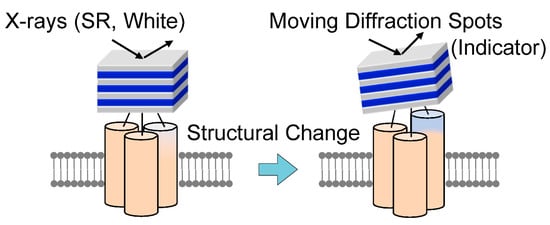
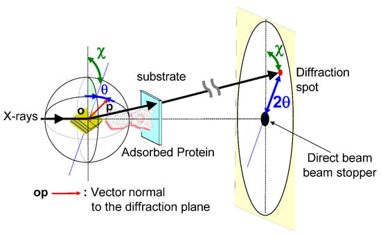
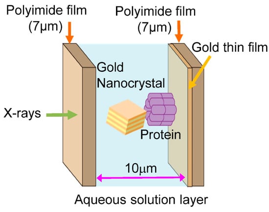
In 1998, the diffracted X-ray tracking (DXT) method pioneered the attainment of molecular dynamics measurements within individual molecules. This breakthrough revolutionized the field by enabling unprecedented insights into the complex workings of molecular systems. Similar to the single-molecule fluorescence labeling technique used in the visible range, DXT uses a labeling method and a pink beam to closely track the diffraction pattern emitted from the labeled gold nanocrystals. Moreover, by utilizing X-rays with extremely short wavelengths, DXT has achieved unparalleled accuracy and sensitivity, exceeding initial expectations. As a result, this remarkable advance has facilitated the search for internal dynamics within many protein molecules. 

diffracted X-ray tracking conformational protein dynamics single molecule observations using X-rays diffracted X-ray blinking

1. History and Concept of Single Molecule Dynamics Using X-rays

Single-molecule measurements using visible light, which started in the 1970s, have greatly advanced our understanding of the translational movement of single protein molecules moving in living cells and their real-time single-molecule interactions with other protein molecules [1][2]. In addition, the fluorescence resonance energy transfer (FRET) method using visible light was partially successful in monitoring the intramolecular motions of proteins [3]. However, this FRET method did not develop into a general-purpose method for measuring changes in the internal conformation of molecules. To measure the internal motions of protein molecules with high precision, it was necessary to observe the dynamics at atomic wavelengths; thus, expectations for X-rays, electron beams, and neutrons naturally increased [4][5][6][7][8][9].
Once the methodology for determining protein molecular structures using X-rays and electron beams was established, the next necessary information for understanding the mechanisms of protein molecular functions in more detail was the molecular conformational changes with atomic-level precision that accompany the functional expression of protein molecules. The actual observation of the internal motion of a single protein molecule started with the interpretation of multiple molecular structures attributed to single-particle analysis by cryo-electron microscopy [10][11][12]. However, this observation of internal molecular motion was a prediction and only a dynamic prediction from the static images of multiple molecular structures. The direction and speed of their protein motion, as well as their stability, cannot be rigorously determined. Continuous molecular structure and dynamic information need to be acquired. However, the quantum beam probes at this time were not sensitive enough to detect a single molecule.

2. Measuring Single Molecule Dynamics Using Diffracted X-ray Tracking 

A methodology for measuring the internal dynamics of single molecules using X-rays proposed in 1998 was the diffracted X-ray tracking (DXT) method; in DXT, gold nanocrystals were chemically labeled at sites of structural change, and diffraction spots from these nanocrystals were monitored over time [13][14][15][16][17][18][19][20]. This method was called the X-ray single-molecule tracking method. As shown in Figure 1, the principle is quite simple: one diffraction spot from one labeled nanocrystal is tracked on time-resolved diffraction images to determine whether the labeled nanocrystal and the labeled site of the protein molecule are in the same motion. DXT measurements on various systems have shown that in the microsecond to millisecond range, the real-space rotational motion of the nanocrystal diffraction spots coincides with the rotational motion of the domain containing the protein labeling sites. In some cases, this can also be determined from the correlation analysis between the size of the labeled nanocrystal and the motion of the diffraction spots, which are slightly slower due to the motion suppression effect of the nanocrystal labeling [20][21][22][23][24][25].
Figure 1. A principle diagram of single-molecule protein dynamic measurements using X-rays. A gold nanocrystal is labeled at the motion site of interest, and the positions of the X-ray diffraction spots diffracted by the labeled gold nanocrystal are tracked in a time-resolved manner. To track X-ray diffraction spots, the X-rays need to have a certain wavelength width.
The main dynamic information of the DXT is found on the two axes of the 2D diffraction image. These are the θ and χ directions shown in Figure 2. Regarding the two axes on the diffraction image, the region where diffraction points occur is limited by the X-ray energy width. However, because of this limited space, the motion of the diffraction points can be observed in the same direction as the internal motion of the molecule, as in real space. In particular, the rotational motion of protein molecules is directly related to their function. For example, considering that membrane proteins are usually formed in membrane polymers, the opening and closing of the channels, which are the most important functions of the membrane proteins, is not a translational movement but is achieved by rotation and twisting of each domain. The ability to directly confirm this rotation by zooming in on the X-ray diffraction image at high speed is extremely important information for the DXT method. Another advantage is that the rotational motion is reversed when the sample substrate is changed from upstream to downstream, as shown in Figure 3; this process confirms the direction of rotation, causing an easy check for reproducibility. Although tilting and twisting models are sometimes proposed for the internal dynamics of membrane protein molecules, as shown in Figure 4, the greatest advantage of DXT is that these two models can be easily distinguished as a combination of the θ and χ motions of DXT [26][27][28][29].
Figure 2. Schematic arrangement of the DXT instrumentation using the Laue method instrumentation for tracking X-ray diffraction. When tracking X-ray diffraction spots in time, the position of θ and χ at each measurement time is important.
Figure 3. Various methods exist for the adsorption of protein molecules onto DXT substrates. Since the molecules can be adsorbed to the substrate in an aligned orientation, the rotational motion (χ direction) of the molecules can be rotated in the desired direction, and the direction of rotation can be reversed for observation. This can also be effectively used for data evaluation. When the surface-adsorbed monolayer protein molecule is a two-dimensional crystalline film, the DXT data would pertain to the θ direction rather than the χ direction.
Figure 4. Channel molecules have two conformational dynamics; channel molecules are the most important and representative of membrane protein molecules. DXT can easily determine which of the two (the tilting model or the twisting model) is correct. In the tilting model, the motion in the θ direction significantly changes with the opening and closing motions of the channel. In the Twisting model, the motion in the θ direction in the opening and closing motions is slight; however, the motion in the χ direction is large.
The resolution of θ and χ, which are crucial for DXT measurements, is determined by the pixel size of the two-dimensional detector and the distance between the camera and the sample, known as the camera length. For instance [26], in DXT measurements, researchers recorded time-resolved diffraction images using an X-ray image intensifier (V5445P, Hamamatsu Photonics) and a CMOS camera (1024 pixels × 1024 pixels, Photron FASTCAM SA1.1). This FASTCAM SA1.1 high-speed camera offers exceptional speed, capturing up to 9000 frames per second, and delivers true 12-bit performance (dynamic range). The nominal entrance field of view for the X-ray image intensifier is 150 mm in diameter, with an effective pixel size of 0.1465 mm. With the incident X-ray’s peak energy set at 15.2 keV and a sample-to-detector distance of 100 mm in the DXT setup, a one-pixel movement of a diffraction spot in the tilting (θ) direction corresponds to 0.7 mrad/pixel (@15.2 keV). For most diffraction spots originating from gold nanocrystals situated 36.4 mm from the beam center, considering the d-spacing of Au (111) (d = 2.35 Å), this distance corresponds to 248.5 pixels in this configuration. The circle with a radius of 248.5 pixels corresponds to approximately 1560 pixels in circumference. Consequently, a one-pixel shift in the twisting (χ) direction corresponds to 4.0 mrad/pixel @15.2 keV.
The main dynamic information in the DXT is the two axes of the 2D diffraction image. These are the θ and χ directions shown in Figure 2. The environment around the sample is shown in Figure 5. Currently, DXT measurements are obtained in various places, as shown in Figure 6; for the two axes on the diffraction image obtained by DXT, as shown in Figure 2, the region where the diffraction points occur is limited by the X-ray energy width. However, because of this limited space, the motion of the diffraction points can be observed in the same direction as the internal motion of the molecule, as in real space.
Figure 5. Cross-section of a DXT sample. A protein molecule is adsorbed and immobilized on one side, and each protein molecule is chemically labeled with one gold nanocrystal. The aqueous solution layer is sandwiched between polyimide films and made as thin as 10 microns to minimize X-ray scattering from the aqueous solution layer. The polyimide film surface can be deposited with gold or chemically treated to react with protein molecules.
Figure 6. There are three main beamlines for DXT in Japan, with BL40XU at SPring-8 being the most suitable DXT beamline in terms of X-ray intensity and X-ray energy width (approximately 4%). Typically, time-resolved DXT of 10–100 microseconds can be performed. Recently, it has become possible to measure DXT while performing laser excitation.

References

  1. Michaelis, J.; Hettich, C.; Mlynek, J.; Sandoghdar, V. Optical microscopy using a single-molecule light source. Nature 2000, 405, 325–328.
  2. Celebrano, M.; Kukura, P.; Renn, A.; Sandoghdar, V. Single-molecule imaging by optical absorption. Nat. Photonics 2011, 5, 95–98.
  3. Weiss, S. Fluorescence Spectroscopy of Single Biomolecules. Science 1999, 283, 1676–1683.
  4. Fratalocchi, A.; Ruocco, G. Single-Molecule Imaging with X-Ray Free-Electron Lasers: Dream or Reality? Phys. Rev. Lett. 2011, 106, 105504.
  5. Sasaki, Y.C.; Suzuki, Y.; Ishibashi, T.; Satoh, I. Interference Effect of Electron-Capture X-Rays from an 125I-Labeled Protein Monolayer in an Aqueous Solution. Anal. Sci. 1995, 11, 545–548.
  6. Sasaki, Y.C.; Suzuki, Y.; Yasuda, K.; Tomioka, Y.; Ishibashi, T.; Satoh, I. Site determination of radioactive atoms from the interference effect of electron-capture rays: Structural change of 111In-labelled azobenzene derivative. Thin Solid Films 1996, 248–285, 456–458.
  7. Sasaki, Y.C.; Suzuki, Y.; Tomioka, Y.; Ishibashi, T.; Takahashi, M.; Satoh, I. Observation of Nanometer-Level Structural Changes by the Trans−Cis Transition of an Azobenzene Derivative Monolayer with a Radioactive Tracer. Langmuir 1996, 12, 4173–4175.
  8. Sasaki, Y.C.; Suzuki, Y.; Yamanashi, H.; Arai, A.; Yanagihara, M. Time-resolved fluorescent X-ray interference. J. Synchrotron Radiat. 1998, 5, 1075–1078.
  9. Sasaki, Y.C.; Yasuda, K.; Takahashi, M.; Satoh, I.; Ishiwata, S. Structural information from the interference effect of electron-capture X-rays. J. Radioanal. Nucl. Chem. 1999, 239, 341–344.
  10. Adrian, M.; Dubochet, J.; Lepault, J.; McDowall, A.W. Cryo-electron microscopy of viruses. Nature 1984, 308, 32–36.
  11. Frank, J. Single-Particle Imaging of Macromolecules by Cryo-Electron Microscopy. Annual Review of Biophysics and Biomolecular Structure. Annu. Rev. 2002, 31, 303–319.
  12. Assaiya, A.; Burada, A.P.; Dhingra, S.; Kumar, J. An overview of the recent advances in cryo-electron microscopy for life sciences. Bose, K., editor. Emerg. Top. Life Sci. 2021, 5, 151–168.
  13. Sasaki, Y.C.; Suzuki, Y.; Yagi, N.; Adachi, S.; Ishibashi, M.; Suda, H.; Toyota, K.; Yanagihara, M. Tracking of individual nanocrystals using diffracted x rays. Phys. Rev. E 2000, 62, 3843–3847.
  14. Sasaki, Y.C.; Okumura, Y.; Adachi, S.; Suzuki, Y.; Yagi, N. Diffracted X-ray tracking: New system for single molecular detection with X-rays. Nucl. Instrum. Methods Phys. Res. Sect. A Accel. Spectrometers Detect. Assoc. Equip. 2001, 467–468, 1049–1052.
  15. Sasaki, Y.C.; Okumura, Y.; Adachi, S.; Suda, H.; Taniguchi, Y.; Yagi, N. Picometer-Scale Dynamical X-Ray Imaging of Single DNA Molecules. Phys. Rev. Lett. 2001, 87, 248102.
  16. Sasaki, Y.C. Dynamical Observations of Soft Nanomaterials Using X-rays or High-Energy Probes. 69–107, Chapter 2, Soft Nanomaterial; Nalwa, H.S., Ed.; American Scientific Publishers: Valencia, CA, USA, 2009.
  17. Sasaki, Y.C. Dynamical Observations of Local Bio-molecular Sites Using Nanocrystals. AIP Conf. Proc. 2004, 705, 1023–1026.
  18. Sasaki, Y.C. Single protein molecular dynamics determined with ultra-high precision. Biochem. Soc. Trans. 2004, 32, 761–763.
  19. Ichiyanagi, K.; Sekiguchi, H.; Hoshino, M.; Kajiwara, K.; Hoshisashi, K.; Jae-won, C.; Tokue, M.; Matsushita, Y.; Nishijima, M.; Inoue, Y.; et al. Diffracted X-ray tracking for monitoring intramolecular motion in individual protein molecules using broad band X-ray. Rev. Sci. Instrum. 2013, 84, 103701.
  20. Sekiguchi, H.; Sasaki, Y.C. Dynamic 3D visualization of active protein’s motion using diffracted X-ray tracking. Jpn. J. Appl. Phys. 2019, 58, 120501.
  21. Okumura, Y.; Oka, T.; Kataoka, M.; Taniguchi, Y.; Sasaki, Y.C. Picometer-scale dynamical observations of individual membrane proteins: The case of bacteriorhodopsin. Phys. Rev. E 2004, 70, 021917.
  22. Okumura, Y.; Oka, T.; Taniguchi, Y.; Sasaki, Y.C. Dynamical Observations of Membrane Proteins: The Case of Bacteriorhodopsin. AIP Conf. Proc. 2004, 705, 1174–1177.
  23. Kawashima, Y.; Sasaki, Y.C.; Sugita, Y.; Yoda, T.; Okamoto, Y. Replica-exchange molecular dynamics simulation of diffracted X-ray tracking. Mol. Simul. 2007, 33, 97–102.
  24. Sagawa, T.; Azuma, T.; Sasaki, Y.C. Dynamical regulations of protein–ligand bindings at single molecular level. Biochem. Biophys. Res. Commun. 2007, 355, 770–775.
  25. Shimizu, H.; Iwamoto, M.; Konno, T.; Nihei, A.; Sasaki, Y.C.; Oiki, S. Global Twisting Motion of Single Molecular KcsA Potassium Channel upon Gating. Cell 2008, 132, 67–78.
  26. Sekiguchi, H.; Suzuki, Y.; Nishino, Y.; Kobayashi, S.; Shimoyama, Y.; Cai, W.; Nagata, K.; Okada, M.; Ichiyanagi, K.; Ohta, N.; et al. Real Time Ligand-Induced Motion Mappings of AChBP and nAChR Using X-ray Single Molecule Tracking. Sci. Rep. 2014, 4, 6384.
  27. Kozono, H.; Matsushita, Y.; Ogawa, N.; Kozono, Y.; Miyabe, T.; Sekiguchi, H.; Ichiyanagi, K.; Okimoto, N.; Taiji, M.; Kanagawa, O.; et al. Single-Molecule Motions of MHC Class II Rely on Bound Peptides. Biophys. J. 2015, 108, 350–359.
  28. Matsushita, Y.; Sekiguchi, H.; Ichiyanagi, K.; Ohta, N.; Ikezaki, K.; Goto, Y.; Sasaki, Y.C. Time-resolved X-ray Tracking of Expansion and Compression Dynamics in Supersaturating Ion-Networks. Sci. Rep. 2015, 5, 17647.
  29. Matsushita, Y.; Sekiguchi, H.; Wong, C.J.; Nishijima, M.; Ikezaki, K.; Hamada, D.; Goto, Y.; Sasaki, Y.C. Nanoscale Dynamics of Protein Assembly Networks in Supersaturated Solutions. Sci. Rep. 2017, 7, 13883.
More
Information
Subjects: Biophysics
Contributor MDPI registered users' name will be linked to their SciProfiles pages. To register with us, please refer to https://encyclopedia.pub/register :
View Times: 105
Revisions: 2 times (View History)
Update Date: 17 Nov 2023
1000/1000