Submitted Successfully!
To reward your contribution, here is a gift for you: A free trial for our video production service.
Thank you for your contribution! You can also upload a video entry or images related to this topic.
Version Summary Created by Modification Content Size Created at Operation
1 -- 3444 2023-11-15 10:14:56 |
2 Reference format revised. -11 word(s) 3433 2023-11-17 02:00:01 |

Video Upload Options

Do you have a full video?

Confirm

Are you sure to Delete?
Cite
If you have any further questions, please contact Encyclopedia Editorial Office.
Hardy, M.P.; Mansour, M.; Rowe, T.; Wymann, S. The Molecular Mechanisms of Complement Receptor 1. Encyclopedia. Available online: https://encyclopedia.pub/entry/51595 (accessed on 03 September 2024).
Hardy MP, Mansour M, Rowe T, Wymann S. The Molecular Mechanisms of Complement Receptor 1. Encyclopedia. Available at: https://encyclopedia.pub/entry/51595. Accessed September 03, 2024.
Hardy, Matthew P., Mariam Mansour, Tony Rowe, Sandra Wymann. "The Molecular Mechanisms of Complement Receptor 1" Encyclopedia, https://encyclopedia.pub/entry/51595 (accessed September 03, 2024).
Hardy, M.P., Mansour, M., Rowe, T., & Wymann, S. (2023, November 15). The Molecular Mechanisms of Complement Receptor 1. In Encyclopedia. https://encyclopedia.pub/entry/51595
Hardy, Matthew P., et al. "The Molecular Mechanisms of Complement Receptor 1." Encyclopedia. Web. 15 November, 2023.
The Molecular Mechanisms of Complement Receptor 1
Edit

Human complement receptor 1 (CR1) is a membrane-bound regulator of complement that has been the subject of attempts to generate soluble therapeutic compounds comprising different fragments of its extracellular domain. 

soluble complement receptor

1. Introduction

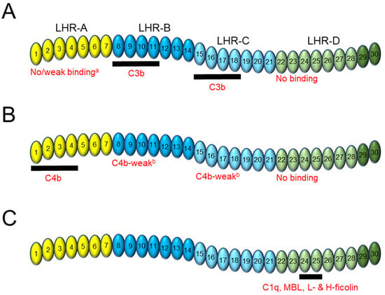
CR1 is a type I membrane glycoprotein expressed on the surface of erythrocytes (E-CR1) and immune cells that acts as a central regulator of the classical, lectin, and alternative pathways of the complement system [1][2][3][4]. The complement system is an integral part of the innate immune response and has been extensively reviewed [5][6][7][8][9][10]. The predominant allelic variant of CR1 (CR1*1) has a large, flexible extracellular domain comprised of 30 highly homologous domains called short consensus repeats (SCRs), followed by a transmembrane domain and a short, 43-amino acid cytoplasmic tail [11][12]. SCR domains 1–28 are arranged in groups of seven to form four larger units: long homologous repeat (LHR) domains -A to -D (Figure 1A) [11][13][14][15]. Including SCR domains 29–30 into an expanded definition of LHR-D has been performed routinely [16][17][18].

Figure 1. The contribution of CR1 domains to ligand binding. Shown schematically is the extracellular domain of CR1 made up of four long homologous repeat (LHR) domains, A to D. Each LHR domain (identified by a particular color) is itself comprised of 7 short consensus repeat (SCR) domains (numbered). LHR-B and LHR-C are highly homologous and so are shaded in similar colors to show their functional similarity. SCR29–30 (shaded in a darker green than SCR22–28) are not technically part of LHR-D. The extracellular domain of CR1 is also known as soluble CR1 (sCR1). Beneath each schema is shown the LHR and SCR contributions to (A) C3b binding, (B) C4b binding, and (C) C1q, mannose-binding lectin (MBL), L-ficolin, and H-ficolin binding. The thick horizontal black bar lies beneath the SCR domains responsible for the binding interaction. a Several studies show no binding of LHR-A to C3b; other experiments show weak binding (<10% relative to LHR-ABCD).b Binding observed in the absence of LHR-A.

2. CR1 Binding to C3b and C4b

2.1. General and Comparative

CR1 was first described as the receptor for C3b following a series of experiments that identified a 205,000 Dalton molecular weight glycoprotein from solubilized membrane fractions extracted from erythrocytes and immune cells with the ability to promote cleavage of C3b by complement factor I and to displace complement factor Bb from the alternative pathway C3 convertase [1][19]. Subsequent in vitro experiments using a variety of techniques such as radio-ligand binding assays and surface plasmon resonance (SPR) performed on both cell surface and soluble CR1 (sCR1) were able to estimate the affinity of the interaction to C3b in greater detail. This included both monomeric and dimeric forms of C3b.  Soluble CR1 (sCR1) refers to the entire extracellular domain of CR1 and usually describes a form derived recombinantly, since levels of endogenous sCR1 produced by proteolytic shedding from the cell surface are very low [20].
Despite some variation in the experimental results due to the nature of the techniques employed, it is evident that CR1 binds to dimeric C3b with low nanomolar affinity. This affinity is significantly stronger than the affinity of CR1 to monomeric C3b, suggesting a bivalent interaction of CR1 with dimeric C3b, leading to an increased avidity that has been noted by others [21][22]. Affinities generated to plasma-derived C3b [18][23] are generally weaker than earlier measurements using C3b fractions, possibly reflecting a preference for the interaction of CR1 with the predominantly monomeric C3b in the preparation, as the affinities are similar.
The first published reference to the binding specificity of CR1 to C4b was in 1980 [24]. Later studies confirmed this interaction [25][26], with one study also showing no difference in CR1 binding to C4b alone compared to C4bC2a (C4bC2b using current nomenclature) whereas a two-fold increase in affinity to C3bBb compared to C3b (despite very weak binding to factor Bb alone) was observed [26]. There is surprisingly little affinity data on the interaction between CR1 and C4b. However, from the available binding data.
In contrast to C3, C4 zymogen exists as two structurally similar isoforms, C4A and C4B [27]. While some early experiments showed increased binding of CR1 to activated C4Ab compared to C4Bb, a more recent experiment using SPR showed no quantitative difference in affinity between CR1 and either isoform of C4b [28][29][30]. With regards to the cleavage products of C4b, only a weak (~1.6 μM) affinity of CR1 to C4c has been demonstrated, with no binding observed to C4dg [28]. Finally, comparative competition experiments showed that approximately 10-fold more sCR1 was required to achieve 50% inhibition of the interaction of 125I-labelled C4b dimer (C4-ma, a C4b analog, was used) with E-CR1 compared to C3b dimer [21]. This further highlights the strength of the binding of CR1 to C3b relative to C4b.

2.2. Domain Contribution

Initial attempts to unravel the domain contribution of CR1 to C3b binding were relatively crude, utilizing proteolytic digestion of CR1 and immunoblotting to show the involvement of the N-terminal half of CR1 in C3b binding [31]. This was followed by experiments using single CR1 LHR domains or combinations thereof to build a more complete picture of each domain’s interaction with C3b, as shown in Figure 1A. Despite some experiments suggesting weak, qualitative binding of the LHR-A domain of CR1 to C3b (1–10% compared to LHR-ABCD) [25][32][33], other data showed no interaction [34][35][36].
There is strong evidence that LHR-B [16][25][31][32][33][34][36] and LHR-C [16][25][34][36][37][38] are the primary domains responsible for C3b binding. Klickstein et al. [25] were the first to demonstrate LHR-B and LHR-C binding to C3b, using LHR-BD and -CD truncation mutants to show similar C3b rosette formation (an in vitro assay where cellular clusters—“rosettes”—are formed by CR1-expressing cells bound to and surrounded by C3b-coated erythrocytes and visualized microscopically) compared to LHR-ABCD. However, subsequent experiments suggested that both LHR-B and -C are required to achieve full C3b binding, specifically the dimeric form. In one experiment, both LHR-BD and -CD mutants stably expressed on K562 cells and assayed for binding to C3b dimer showed dissociation constants of between 2.0 and 2.5 nM compared to only 1 nM for LHR-ABCD [16].
Further refinement of the C3b epitope on CR1 to individual SCR domains within both LHR-B and -C has been performed, and it has been suggested that SCR8–10 and SCR15–17 of LHR-B and -C, respectively, comprise the binding site for C3b [11][15].
In contrast to C3b, the interaction between CR1 and C4b takes place primarily in the LHR-A domain [25][32][33][35][36][39] with some contribution of the LHR-B and -C domains [25][32][33][36][38][39], and no contribution of the LHR-D domain [25][36][39] (Figure 1B).

3. CR1 Binding to Other Ligands

Far from being a useless appendage at the C-terminal end of the extracellular domain of CR1, the LHR-D domain is also involved in the binding of its own distinct ligands (Figure 1C). Like C3b and C4b, the complement classical pathway component C1q is an opsonin that binds to apoptotic cells and mediates their clearance [40][41]. C1q has also been shown to specifically bind CR1 with high affinity [42], and subsequent experiments using CR1 deletion mutants localized the interaction to LHR-D. Another study soon afterwards showed that the association of C1q with CR1 could occur independently of C3b and C4b binding [43].
Mannose-binding lectin (MBL), referred to as both an opsonin and a pattern recognition molecule, closely related to C1q and an initiating factor of the complement lectin pathway, was the second ligand discovered to specifically bind the CR1 LHR-D domain [44]. MBL and C1q have similar affinities to CR1 and compete for binding, suggesting an overlapping epitope [44].

4. Structural Data

Given the long, flexible, nature of the extracellular domain of CR1, which is analogous to a string of pearls, high-resolution structural information on the entire molecule and its interaction with ligand has not been able to be deduced. However, it has been possible to use those specific fragments of CR1 involved in ligand binding for structural studies, most notably for C3b but not C4b. Smith et al. [38] were the first to determine a medium-resolution structure of the C3b/C4b binding domains SCR15–17 (in LHR-C) by nuclear magnetic resonance techniques. The authors were then able to map onto the structure the various residues determined from previous mutagenesis to be involved in C3b and/or C4b binding [38].
A higher resolution crystal structure of C3b bound to CR1 domains SCR15–17 has more recently been described [45], providing detailed information on residues important for this interaction. Unfortunately, these studies only looked at receptor–ligand binding through the lens of a single interaction using a minimal binding fragment of CR1 and were not able to provide a holistic view of how CR1 interacts with multiple ligands (both monovalently and bivalently) across multiple sites. There has been one attempt to determine the structure of the entirety of soluble CR1 [12]

5. Decay Acceleration Activity of CR1

5.1. Classical/Lectin Pathway C3 and C5 Convertases

CR1 inhibits the formation of both the classical (and by inference the lectin) C3 convertase, consisting of a C4bC2b complex, and the classical C5 convertase, which consists of a C4bC2bC3b complex. The mechanism by which this decay acceleration activity (DAA) occurs is through the interaction of CR1 with C4b bound to cell surfaces, which displaces C2b from their shared binding site on C4b [46]. The classical C5 convertase can contain C3b bound covalently as a heterodimer to C4b [47].
The use of deletion mutants of CR1 has been critical to our understanding of how CR1 exerts DAA on the classical C3 and C5 convertases. For inhibition of the classical C3 convertase, both the LHR-A domain and one of either the LHR-B or -C domains are required, although the LHR-A domain appears to be the most important. Two studies [37][48] showed that LHR-AC and LHR-ACD deletion mutants had similar classical C3 convertase DAA as compared to LHR-ABCD, demonstrating that one of two C3b binding sites on CR1 as well as LHR-D are dispensable. Constructs encoding LHR-B, LHR-C, or combined LHR-BC domains exhibited residual DAA (8–12%) compared to LHR-A or LHR-ABCD [48][49], which correlates with the weak C4b binding described above for these domains and demonstrates the importance of the LHR-A domain. The CR1 LHR-A domain alone was found to have between 40 and 60% DAA compared experimentally to LHR-ABCD, which comprises the bulk of activity but is clearly insufficient for full DAA [48][49].
When considering the roles of the CR1 LHR domains in driving DAA of the classical C5 convertase, there are some key differences compared to the classical C3 convertase. It was demonstrated that a C3b binding site (LHR-B or -C) could be removed without affecting classical C5 convertase DAA [37][48]. However, constructs encoding LHR-A or LHR-BC showed almost no activity compared to LHR-ABCD or LHR-AC [48][49], suggesting that LHR-A with LHR-B or -C act synergistically to provide full DAA of the classical C5 convertase.

5.2. Alternative Pathway C3 and C5 Convertases

As with the classical C3 and C5 convertases, CR1 has been shown to mediate decay of the alternative pathway C3 convertase (C3bBb) and C5 convertase (C3bBbC3b) by competitive displacement of Factor Bb from the complexes [1][37][50]. Interestingly, and in line with its relative affinities to C3b and C4b, up to 10-fold more sCR1 is required for DAA of the classical convertases compared to the alternative convertases [37], suggesting a more potent effect on DAA of the alternative pathway.
The contribution of CR1 domains to the DAA of the alternative pathway C3 convertase is not well understood and confounded by spontaneous C3b dimer formation contaminating the experimental results previously used to examine C3 convertase DAA. Instead of C3b monomers, the presence of C3b dimers in the assay means that the results could be reflective of the output of an alternative C5 convertase DAA [48]. When an attempt to address this issue was performed, the bulk (95%) of the DAA of the C3 convertase appeared to be mediated by the LHR-A domain, with only minor (12–14%) contributions measured for the LHR-B and LHR-C domains [48]. This finding is hard to reconcile with the C3b ligand binding data for these domains described above, which show the reverse contributions of these same domains.
The domain contribution of CR1 to the DAA of the alternative pathway C5 convertase is better understood, although again the role of the LHR-A domain is unclear given the ambiguity of the available C3b binding data. What is clear is that LHR-A, -B, and -C domains are all required and act synergistically to provide full activity of sCR1. If either LHR-B or LHR-C is removed, there is a significant drop in alternative pathway C5 DAA. 

6. Co-Factor Activity of CR1

6.1. General and Comparative

Both C3b and C4b can be cleaved by complement Factor I in the presence of CR1, a process referred to as co-factor activity (CFA) and one that was first described for CR1 more than 40 years ago [19][46][51][52]. Subsequent experiments were able to confirm the CFA of CR1 by demonstrating its ability to promote the cleavage of C3b to iC3b and then to C3dg, as well as the cleavage of methylamine-treated C4 (C4-ma; a C4b analog) to C4c and C4d [16][21]. The CFA of CR1 is of particular interest when comparisons between its activity to C3b and C4b are made.

6.2. Domain Contribution

The contribution of CR1 LHR domains to C3b CFA appears reasonably well established but is by no means definitive. The uncertainty is driven primarily by the lack of discrimination in many experiments between the use of monomeric and dimeric ligand, and the somewhat qualitative nature of the assays used (e.g., visualization of cleavage products on protein gels), which could impact data interpretation. However, for C3b CFA, it was shown using both single domains and LHR domain deletion mutants that there is a clear contribution of the LHR-B and LHR-C domains and no contribution of the LHR-D domain [25][33][36][53]. This makes mechanistic sense since CR1 would need to bind ligand to allow Factor I-mediated cleavage to occur. LHR-C point mutants that reduce C3b binding also reduce CFA [53]. It appears that one of the two C3b binding sites can be deleted with minimal loss of C3b CFA, as demonstrated by comparing the relative CFA of an LHR-ACD mutant (8 nM IC50) with LHR-ABCD (5 nM IC50) [37]. It appears from the results of several studies that the LHR-A domain also contributes weak C3b CFA, although the degree varies from study to study [32][34][36][54].
The data pertaining to the role of the CR1 LHR domains in C4b CFA are harder to interpret in light of previously described ligand binding data (Figure 1B) and are somewhat conflicting for LHR-A (containing the main C4b binding site). For those studies that show C4b CFA for LHR-A [32][39][54], data suggest only modest activity with one study showing a threefold decrease in CFA of a construct encoding SCR1–4 of LHR-A compared to sCR1 [54] and another study showing that conversion of the C4b binding site in LHR-A to a C3b binding site by amino acid substitution produced stronger CFA [32]

7. Domain Contribution to CR1-Mediated Complement Pathway Inhibition

Until recently, the literature surrounding the role of LHR or SCR domains in mediating complement pathway inhibition as measured by hemolytic activity has been limited. Two studies examined individual and combinations of LHR domains for their ability to inhibit hemolytic activity, but no dose responses were generated since only single, high (1 uM) protein concentrations were used [17][22][36]. However, sufficient data were generated to determine that LHR-A, -B, -C, but not -D, contributed to the overall hemolytic inhibitory activity of CR1.

8. CSL040 and Its Mechanism of Action

CSL040 is a truncation variant of sCR1 containing the LHR-A, -B, and -C domains, and with the C-terminal LHR-D domain deleted [55]. The in vitro and in vivo functions of CSL040 have been described elsewhere [18][56][57].
When recombinant soluble versions of LHR-A, -B, and -C were assessed singly in vitro for their ability to inhibit classical pathway hemolytic activity, all three domains were separately shown to be weakly inhibitory [18]. LHR-A was more than 100-fold less active than CSL040, and LHR-B/-C was more than 1000-fold less active. This suggests that the greater affinity of C4b for LHR-A compared to LHR-B/-C (Figure 1B) is reflected in its greater contribution to classical pathway inhibitory activity, but also demonstrates that single LHR domains are not sufficient for full activity and a bivalent interaction with multiple domains is required. For alternative pathway hemolytic activity, LHR-A, -B, and -C each have more than 50-fold lower potency than CSL040 but have similar potency to each othe.
Adding the second, adjacent LHR domain (LHR-A to LHR-B, or LHR-B to LHR-C) to generate LHR-AB and LHR-BC provides a significant and synergistic increase in potency to both classical and alternative pathway hemolytic activity [18].
The final aspect of the molecular mechanism of CSL040 to be discussed is its significantly increased potency for an alternative pathway compared to the classical/lectin pathways. This phenomenon was first observed in vitro [18] but became more apparent following ex vivo pharmacodynamic assays conducted following the administration of CSL040 to either rats or non-human primates [57]. The increased duration of ex vivo alternative pathway inhibition by CSL040 compared to the classical/lectin pathway can be explained mechanistically from the DAA and CFA data described above showing that sCR1 has >10-fold greater affinity to C3b than to C4b [39], can block the binding of C3b to E-CR1 10-fold more strongly than C4b [21], has a 10-fold higher DAA of the alternative pathway convertases compared to those of the classical/lectin pathway [37], and is also much more effective in mediating C3b CFA than C4b CFA [54].

9. Conclusions

As shown in Figure 2, in order to inhibit complement activation and downstream lytic activity, CR1 must bind its ligands C3b and C4b. By doing so it competitively displaces C2b or Bb from the nascent convertases by decay acceleration activity. The interaction with monomeric or dimeric ligand will determine whether the DAA is against a C3 convertase (C4bC2b or C3bBb) or a C5 convertase (C4bC2bC3b or C3bBbC3b), respectively. CR1 binding to monomeric ligand appears to be monovalent and low(er) affinity, but bivalent and high(er) affinity to dimeric ligand, with different domains within a single CR1 molecule contributing to binding depending on the type of dimer: C3b-C3b, C3b-C4b or C4b-C4b. Spacing between ligand binding sites on CR1 is important—this allows the appropriate degree of SCR domain folding for contact to occur. The presence of a third C3b/C4b binding site in CR1 adds further complexity to this interaction, which remains to be elucidated. The binding of CR1 to C3b/C4b also provides an opportunity for CFA, with Factor I processing these CR1-bound activated complement fragments into their inactive forms by proteolytic cleavage. The relative strength of the interaction of CR1 with C3b compared to C4b determines the relative potency of this molecule to the classical, lectin, and alternative pathways, with inhibition of the latter being the most effective.
Figure 2. A model of the molecular mechanisms of soluble CR1. (A) Soluble CR1 (sCR1) binds to the CP/LP C3 convertase C4bC2b monovalently with low affinity, disrupting the interaction between membrane-associated C4b and C2b (decay acceleration activity). In this panel, LHR-A (yellow) of sCR1 binds C4b, allowing Factor I to cleave C4b into C4c and C4d (co-factor activity). For the AP C3 convertase (not shown), the mechanism is similar except that LHR-B or -C of sCR1 (blue) now interacts with C3b and displaces Bb from the C3bBb convertase. Factor I then cleaves C3b into iC3b and C3f. (B) Decay acceleration of the AP C5 convertase C3bBbC3b is mediated by the bivalent and high-affinity interaction of sCR1 with homodimeric C3b, displacing Bb from the convertase. This allows cleavage of C3b into iC3b and C3f by Factor I. For DAA of the CP/LP C5 convertase (not shown), this process is similar but with binding of sCR1 to heterodimeric C4bC3b and displacement of C2b, followed by cleavage of C3b/C4b by Factor I as described above. Following binding of sCR1 to C5 convertases, the unbound C3b or C4b binding site could potentially interact with additional convertases to further increase its potency.

References

  1. Fearon, D.T. Identification of the membrane glycoprotein that is the C3b receptor of the human erythrocyte, polymorphonuclear leukocyte, B lymphocyte, and monocyte. J. Exp. Med. 1980, 152, 20–30.
  2. Ross, G.D.; Lambris, J.D. Identification of a C3bi-specific membrane complement receptor that is expressed on lymphocytes, monocytes, neutrophils, and erythrocytes. J. Exp. Med. 1982, 155, 96–110.
  3. Ahearn, J.M.; Fearon, D.T. Structure and function of the complement receptors, CR1 (CD35) and CR2 (CD21). Adv. Immunol. 1989, 46, 183–219.
  4. Lublin, D.M.; Griffith, R.C.; Atkinson, J.P. Influence of glycosylation on allelic and cell-specific Mr variation, receptor processing, and ligand binding of the human complement C3b/C4b receptor. J. Biol. Chem. 1986, 261, 5736–5744.
  5. Lachmann, P.J. Biological functions of the complement system. Biochem. Soc. Trans. 1990, 18, 1143–1145.
  6. Ricklin, D.; Hajishengallis, G.; Yang, K.; Lambris, J.D. Complement: A key system for immune surveillance and homeostasis. Nat. Immunol. 2010, 11, 785–797.
  7. Kolev, M.; Le Friec, G.; Kemper, C. Complement—Tapping into new sites and effector systems. Nat. Rev. Immunol. 2014, 14, 811–820.
  8. Lachmann, P.J. The amplification loop of the complement pathways. Adv. Immunol. 2009, 104, 115–149.
  9. Pangburn, M.K. Initiation of the alternative pathway of complement and the history of “tickover”. Immunol. Rev. 2022, 313, 64–70.
  10. Wagner, E.; Frank, M.M. Therapeutic potential of complement modulation. Nat. Rev. Drug Discov. 2010, 9, 43–56.
  11. Krych-Goldberg, M.; Atkinson, J.P. Structure-function relationships of complement receptor type 1. Immunol. Rev. 2001, 180, 112–122.
  12. Furtado, P.B.; Huang, C.Y.; Ihyembe, D.; Hammond, R.A.; Marsh, H.C.; Perkins, S.J. The partly folded back solution structure arrangement of the 30 SCR domains in human complement receptor type 1 (CR1) permits access to its C3b and C4b ligands. J. Mol. Biol. 2008, 375, 102–118.
  13. Weis, J.H.; Morton, C.C.; Bruns, G.A.; Weis, J.J.; Klickstein, L.B.; Wong, W.W.; Fearon, D.T. A complement receptor locus: Genes encoding C3b/C4b receptor and C3d/Epstein-Barr virus receptor map to 1q32. J. Immunol. 1987, 138, 312–315.
  14. Klickstein, L.B.; Wong, W.W.; Smith, J.A.; Weis, J.H.; Wilson, J.G.; Fearon, D.T. Human C3b/C4b receptor (CR1). Demonstration of long homologous repeating domains that are composed of the short consensus repeats characteristics of C3/C4 binding proteins. J. Exp. Med. 1987, 165, 1095–1112.
  15. Liu, D.; Niu, Z.X. The structure, genetic polymorphisms, expression and biological functions of complement receptor type 1 (CR1/CD35). Immunopharmacol. Immunotoxicol. 2009, 31, 524–535.
  16. Kalli, K.R.; Hsu, P.H.; Bartow, T.J.; Ahearn, J.M.; Matsumoto, A.K.; Klickstein, L.B.; Fearon, D.T. Mapping of the C3b-binding site of CR1 and construction of a (CR1)2-F(ab’)2 chimeric complement inhibitor. J. Exp. Med. 1991, 174, 1451–1460.
  17. Mqadmi, A.; Abdullah, Y.; Yazdanbakhsh, K. Characterization of complement receptor 1 domains for prevention of complement-mediated red cell destruction. Transfusion 2005, 45, 234–244.
  18. Wymann, S.; Dai, Y.; Nair, A.G.; Cao, H.; Powers, G.A.; Schnell, A.; Martin-Roussety, G.; Leong, D.; Simmonds, J.; Lieu, K.G.; et al. A novel soluble complement receptor 1 fragment with enhanced therapeutic potential. J. Biol. Chem. 2021, 296, 100200.
  19. Fearon, D.T. Regulation of the amplification C3 convertase of human complement by an inhibitory protein isolated from human erythrocyte membrane. Proc. Natl. Acad. Sci. USA 1979, 76, 5867–5871.
  20. Karthikeyan, G.; Baalasubramanian, S.; Seth, S.; Das, N. Low levels of plasma soluble complement receptor type 1 in patients receiving thrombolytic therapy for acute myocardial infarction. J. Thromb. Thrombolysis 2007, 23, 115–120.
  21. Weisman, H.F.; Bartow, T.; Leppo, M.K.; Marsh, H.C., Jr.; Carson, G.R.; Concino, M.F.; Boyle, M.P.; Roux, K.H.; Weisfeldt, M.L.; Fearon, D.T. Soluble human complement receptor type 1: In vivo inhibitor of complement suppressing post-ischemic myocardial inflammation and necrosis. Science 1990, 249, 146–151.
  22. Makrides, S.C.; Scesney, S.M.; Ford, P.J.; Evans, K.S.; Carson, G.R.; Marsh, H.C., Jr. Cell surface expression of the C3b/C4b receptor (CR1) protects Chinese hamster ovary cells from lysis by human complement. J. Biol. Chem. 1992, 267, 24754–24761.
  23. Schramm, E.C.; Roumenina, L.T.; Rybkine, T.; Chauvet, S.; Vieira-Martins, P.; Hue, C.; Maga, T.; Valoti, E.; Wilson, V.; Jokiranta, S.; et al. Mapping interactions between complement C3 and regulators using mutations in atypical hemolytic uremic syndrome. Blood 2015, 125, 2359–2369.
  24. Ross, G.D. Analysis of the different types of leukocyte membrane complement receptors and their interaction with the complement system. J. Immunol. Methods 1980, 37, 197–211.
  25. Klickstein, L.B.; Bartow, T.J.; Miletic, V.; Rabson, L.D.; Smith, J.A.; Fearon, D.T. Identification of distinct C3b and C4b recognition sites in the human C3b/C4b receptor (CR1, CD35) by deletion mutagenesis. J. Exp. Med. 1988, 168, 1699–1717.
  26. Pangburn, M.K. Differences between the binding sites of the complement regulatory proteins DAF, CR1, and factor H on C3 convertases. J. Immunol. 1986, 136, 2216–2221.
  27. Belt, K.T.; Carroll, M.C.; Porter, R.R. The structural basis of the multiple forms of human complement component C4. Cell 1984, 36, 907–914.
  28. Clemenza, L.; Isenman, D.E. The C4A and C4B isotypic forms of human complement fragment C4b have the same intrinsic affinity for complement receptor 1 (CR1/CD35). J. Immunol. 2004, 172, 1670–1680.
  29. Gatenby, P.A.; Barbosa, J.E.; Lachmann, P.J. Differences between C4A and C4B in the handling of immune complexes: The enhancement of CR1 binding is more important than the inhibition of immunoprecipitation. Clin. Exp. Immunol. 1990, 79, 158–163.
  30. Reilly, B.D.; Mold, C. Quantitative analysis of C4Ab and C4Bb binding to the C3b/C4b receptor (CR1, CD35). Clin. Exp. Immunol. 1997, 110, 310–316.
  31. Prohaska, R.; Adolf, G.R. Characterization of the human erythrocyte complement receptor CR1 (C3b receptor) by epitope mapping. Immunobiology 1987, 174, 93–106.
  32. Krych, M.; Clemenza, L.; Howdeshell, D.; Hauhart, R.; Hourcade, D.; Atkinson, J.P. Analysis of the functional domains of complement receptor type 1 (C3b/C4b receptor; CD35) by substitution mutagenesis. J. Biol. Chem. 1994, 269, 13273–13278.
  33. Krych, M.; Hauhart, R.; Atkinson, J.P. Structure-function analysis of the active sites of complement receptor type 1. J. Biol. Chem. 1998, 273, 8623–8629.
  34. Scesney, S.M.; Makrides, S.C.; Gosselin, M.L.; Ford, P.J.; Andrews, B.M.; Hayman, E.G.; Marsh, H.C., Jr. A soluble deletion mutant of the human complement receptor type 1, which lacks the C4b binding site, is a selective inhibitor of the alternative complement pathway. Eur. J. Immunol. 1996, 26, 1729–1735.
  35. Krych, M.; Hourcade, D.; Atkinson, J.P. Sites within the complement C3b/C4b receptor important for the specificity of ligand binding. Proc. Natl. Acad. Sci. USA 1991, 88, 4353–4357.
  36. Yazdanbakhsh, K.; Kang, S.; Tamasauskas, D.; Sung, D.; Scaradavou, A. Complement receptor 1 inhibitors for prevention of immune-mediated red cell destruction: Potential use in transfusion therapy. Blood 2003, 101, 5046–5052.
  37. Wong, W.W.; Farrell, S.A. Proposed structure of the F’ allotype of human CR1. Loss of a C3b binding site may be associated with altered function. J. Immunol. 1991, 146, 656–662.
  38. Smith, B.O.; Mallin, R.L.; Krych-Goldberg, M.; Wang, X.; Hauhart, R.E.; Bromek, K.; Uhrin, D.; Atkinson, J.P.; Barlow, P.N. Structure of the C3b binding site of CR1 (CD35), the immune adherence receptor. Cell 2002, 108, 769–780.
  39. Reilly, B.D.; Makrides, S.C.; Ford, P.J.; Marsh, H.C., Jr.; Mold, C. Quantitative analysis of C4b dimer binding to distinct sites on the C3b/C4b receptor (CR1). J. Biol. Chem. 1994, 269, 7696–7701.
  40. Korb, L.C.; Ahearn, J.M. C1q binds directly and specifically to surface blebs of apoptotic human keratinocytes: Complement deficiency and systemic lupus erythematosus revisited. J. Immunol. 1997, 158, 4525–4528.
  41. Sontheimer, R.D.; Racila, E.; Racila, D.M. C1q: Its functions within the innate and adaptive immune responses and its role in lupus autoimmunity. J. Investig. Dermatol. 2005, 125, 14–23.
  42. Klickstein, L.B.; Barbashov, S.F.; Liu, T.; Jack, R.M.; Nicholson-Weller, A. Complement receptor type 1 (CR1, CD35) is a receptor for C1q. Immunity 1997, 7, 345–355.
  43. Tas, S.W.; Klickstein, L.B.; Barbashov, S.F.; Nicholson-Weller, A. C1q and C4b bind simultaneously to CR1 and additively support erythrocyte adhesion. J. Immunol. 1999, 163, 5056–5063.
  44. Ghiran, I.; Barbashov, S.F.; Klickstein, L.B.; Tas, S.W.; Jensenius, J.C.; Nicholson-Weller, A. Complement receptor 1/CD35 is a receptor for mannan-binding lectin. J. Exp. Med. 2000, 192, 1797–1808.
  45. Forneris, F.; Wu, J.; Xue, X.; Ricklin, D.; Lin, Z.; Sfyroera, G.; Tzekou, A.; Volokhina, E.; Granneman, J.C.; Hauhart, R.; et al. Regulators of complement activity mediate inhibitory mechanisms through a common C3b-binding mode. EMBO J. 2016, 35, 1133–1149.
  46. Iida, K.; Nussenzweig, V. Complement receptor is an inhibitor of the complement cascade. J. Exp. Med. 1981, 153, 1138–1150.
  47. Takata, Y.; Kinoshita, T.; Kozono, H.; Takeda, J.; Tanaka, E.; Hong, K.; Inoue, K. Covalent association of C3b with C4b within C5 convertase of the classical complement pathway. J. Exp. Med. 1987, 165, 1494–1507.
  48. Krych-Goldberg, M.; Hauhart, R.E.; Subramanian, V.B.; Yurcisin, B.M., 2nd; Crimmins, D.L.; Hourcade, D.E.; Atkinson, J.P. Decay accelerating activity of complement receptor type 1 (CD35). Two active sites are required for dissociating C5 convertases. J. Biol. Chem. 1999, 274, 31160–31168.
  49. Krych-Goldberg, M.; Hauhart, R.E.; Porzukowiak, T.; Atkinson, J.P. Synergy between two active sites of human complement receptor type 1 (CD35) in complement regulation: Implications for the structure of the classical pathway C3 convertase and generation of more potent inhibitors. J. Immunol. 2005, 175, 4528–4535.
  50. Kinoshita, T.; Takata, Y.; Kozono, H.; Takeda, J.; Hong, K.S.; Inoue, K. C5 convertase of the alternative complement pathway: Covalent linkage between two C3b molecules within the trimolecular complex enzyme. J. Immunol. 1988, 141, 3895–3901.
  51. Ross, G.D.; Lambris, J.D.; Cain, J.A.; Newman, S.L. Generation of three different fragments of bound C3 with purified factor I or serum. I. Requirements for factor H vs. CR1 cofactor activity. J. Immunol. 1982, 129, 2051–2060.
  52. Medof, M.E.; Iida, K.; Mold, C.; Nussenzweig, V. Unique role of the complement receptor CR1 in the degradation of C3b associated with immune complexes. J. Exp. Med. 1982, 156, 1739–1754.
  53. Smith, R.A. Targeting anticomplement agents. Biochem. Soc. Trans. 2002, 30, 1037–1041.
  54. Mossakowska, D.; Dodd, I.; Pindar, W.; Smith, R.A. Structure-activity relationships within the N-terminal short consensus repeats (SCR) of human CR1 (C3b/C4b receptor, CD35): SCR 3 plays a critical role in inhibition of the classical and alternative pathways of complement activation. Eur. J. Immunol. 1999, 29, 1955–1965.
  55. Hardy, M.P.; Rowe, T.; Wymann, S. Soluble Complement Receptor 1 Therapeutics. J. Immunol. Sci. 2022, 6, 1–17.
  56. Bongoni, A.K.; Vikstrom, I.B.; McRae, J.L.; Salvaris, E.J.; Fisicaro, N.; Pearse, M.J.; Wymann, S.; Rowe, T.; Morelli, A.B.; Hardy, M.P.; et al. A potent truncated form of human soluble CR1 is protective in a mouse model of renal ischemia-reperfusion injury. Sci. Rep. 2021, 11, 21873.
  57. Wymann, S.; Mischnik, M.; Leong, D.; Ghosh, S.; Tan, X.; Cao, H.; Kuehnemuth, B.; Powers, G.A.; Halder, P.; de Souza, M.J.; et al. Sialylation-dependent pharmacokinetics and differential complement pathway inhibition are hallmarks of CR1 activity in vivo. Biochem. J. 2022, 479, 1007–1030.
More
Information
Subjects: Immunology
Contributors MDPI registered users' name will be linked to their SciProfiles pages. To register with us, please refer to https://encyclopedia.pub/register : , , ,
View Times: 292
Revisions: 2 times (View History)
Update Date: 17 Nov 2023
1000/1000
ScholarVision Creations