Submitted Successfully!
To reward your contribution, here is a gift for you: A free trial for our video production service.
Thank you for your contribution! You can also upload a video entry or images related to this topic.
Version Summary Created by Modification Content Size Created at Operation
1 -- 6112 2023-11-03 19:30:22 |
2 format correct Meta information modification 6112 2023-11-14 01:53:16 | |
3 N/A Meta information modification 6112 2023-11-15 17:48:58 |

Video Upload Options

Do you have a full video?

Confirm

Are you sure to Delete?
Cite
If you have any further questions, please contact Encyclopedia Editorial Office.
Ingraldi, A.L.; Audet, R.G.; Tabor, A.J. Clinical Applications of Amnion-Derived Membranes. Encyclopedia. Available online: https://encyclopedia.pub/entry/51146 (accessed on 18 May 2024).
Ingraldi AL, Audet RG, Tabor AJ. Clinical Applications of Amnion-Derived Membranes. Encyclopedia. Available at: https://encyclopedia.pub/entry/51146. Accessed May 18, 2024.
Ingraldi, Alison L., Robert G. Audet, Aaron J. Tabor. "Clinical Applications of Amnion-Derived Membranes" Encyclopedia, https://encyclopedia.pub/entry/51146 (accessed May 18, 2024).
Ingraldi, A.L., Audet, R.G., & Tabor, A.J. (2023, November 03). Clinical Applications of Amnion-Derived Membranes. In Encyclopedia. https://encyclopedia.pub/entry/51146
Ingraldi, Alison L., et al. "Clinical Applications of Amnion-Derived Membranes." Encyclopedia. Web. 03 November, 2023.
Clinical Applications of Amnion-Derived Membranes
Edit

Biological tissues from various anatomical sources have been utilized for tissue transplantation and have developed into an important source of extracellular scaffolding material for regenerative medicine applications. Tissue scaffolds ideally integrate with host tissue and provide a homeostatic environment for cellular infiltration, growth, differentiation, and tissue resolution. The human amniotic membrane is considered an important source of scaffolding material due to its 3D structural architecture and function and as a source of growth factors and cytokines. This tissue source has been widely studied and used in various areas of tissue repair including intraoral reconstruction, corneal repair, tendon repair, microvascular reconstruction, nerve procedures, burns, and chronic wound treatment. 

human amniotic membrane amnion skin substitute chronic wound anti-inflammatory

1. Introduction

The unique, multilayered structure of the human amniotic membrane (AM), and its associated biological and physical characteristics, make it a highly biocompatible material suitable for use in a variety of regenerative medicine applications [1]. Human AM has been used for over a century in a wide variety of clinical applications including ophthalmologic, chronic wounds, burns, plastic reconstruction and periodontal procedures [2][3]. The positive outcomes from the historic uses of AM prompted further research and clinical investigations into additional human therapeutic applications. As research supported the use of AM for anti-inflammatory, angiogenic, anti-angiogenic and antimicrobial properties, and a source of diverse growth factors, more clinically relevant applications have been demonstrated [4][5][6].
Fetal membranes were first used in the 1900s to successfully treat acute and chronic traumatic wounds, burns, ulcers, and as a novel skin substitute for grafting [7][8][9][10]. Early uses in the ophthalmic field addressed various ocular pathologies, including plastic surgery of the conjunctiva [11] and ocular burns [12]. The amniotic membrane’s inherent elasticity allows it to conform to complex contoured surfaces, which led to its application in many reconstructive procedures, including the creation of artificial vaginas, and treating chronic complex wounds in diabetic patients [2][13][14]. Applications have expanded to support every tissue type within the human body, including ophthalmology; periodontal; skin and wound applications; burns; craniotomies presenting with dural defects; bladder; genital, and peritoneal reconstructive surgeries; prevention of surgical adhesions in abdominal surgeries; tendon and ligament repair; articular joint repair; nerve wrapping; chronic wounds; burns and plastics [2][3][14][15][16][17][18][19]. The growing fields of tissue engineering and regenerative medicine have brought greater attention to the varied human amniotic membrane as a tunable matrix for tissue engineered constructs.

2. Clinical Applications

2.1. Introduction

The intended outcome of using regenerative materials for clinical applications is the acceleration of healing and the restoration of the supporting tissues that have been damaged or lost from surgery, trauma, disease, infection, or other complications. Over the past century, the use of amniotic membranes to support healing in the clinical setting has expanded from skin transplantation [7] and burns (skin and ocular) [9][10] to additional ophthalmic applications [20][21][22], periodontal conditions [23][24], acute and chronic wounds including diabetic foot ulcers, venous leg ulcers and pressure ulcers [25][26][27][28], to plastic and reconstructive surgery including first to third degree burns [19][29][30], post-Mohs wounds [31][32], and surgical adhesions [18][33][34][35]. A variety of characteristics such as the promotion of re-epithelialization, reduction in inflammation and fibrosis, immunomodulation, the inhibition of foreign body reaction, and antimicrobial activity make AM useful in clinical therapies [3][17][36]. The clinical application of amniotic membrane not only maintains the structural and anatomical configuration of regenerated tissues, but also contributes to the enhancement of healing through the reduction of postoperative scarring and subsequent loss of function [15]. When used as a dressing or wound covering, AM acts as a structural barrier facilitating hemostasis, reducing water loss, providing a barrier to microbial colonization and infection, and reducing pain [29][37][38]. The diversity of AM applications includes combination with various cell types or synthetic matrices such as collagens or alginates to create broad application-based regenerative products for healing [3].
The specific applications for amnion, chorion, amnion–chorion, and multilayered AM allografts are broad and have significant overlap. There is variability in application and outcomes between studies using hAM, hCM, hACM or multilayered hAM allografts, in part related to the types of applications, the different sizes of membrane used, which membrane surface will be in contact with the wound (stromal or epithelial side), folding of the membrane, multilayered usage, graft re-application, graft resorption and if any additional protective coverage was implemented when treating defects. From a simple barrier for topical skin-wound applications to corneal repair, to more advanced forms such as membrane extracts and flowable micronized membrane injections, clinical application of amniotic membranes has expanded and continues to evolve.

2.2. Periodontal and Oral Surgery

Periodontal and oral surgery are common interventions for a variety of periodontal complications and diseases. Depending on the specific disease presentation, a variety of periodontal surgical techniques can be utilized including flap surgery, bone grafting and implantation or soft tissue autografting. During these procedures oral tissue damage does occur, and AM products sufficiently address this. Since the mid-1990s, the use of hAM allografts in periodontal and oral surgery to accelerate tissue regeneration has expanded, showing promising results in various specialties of dentistry [23][39][40][41][42].
Amnion (hAM), hCM and hACM allografts are popular for periodontal indications, acting as scaffolds that promote cell adhesion and specific protein synthesis to support the growth of bone and gingival tissue for regenerative procedures. The thinness of hAM, while beneficial for application in tight spaces, can be difficult to handle for some procedures [24][41]. Conversely the thickness of the hCM and hACM allografts make them easier to handle and contain a higher amount of growth factors [24][41][43][44]. While there are no standardized practices described for the application of AM in the oral cavity, two general clinical procedures have been described [39][41]. The implant procedure involves placing hAM, hCM or hACM beneath the gingiva for guided tissue regeneration of gingival recession with coronally advanced flap repair, and guided bone regeneration [41][45][46][47][48]. It is also used for soft tissue repair following dental implant placement [49], intrabony defects [50], Schneiderian membrane perforation repair, [24][41], furcation defects [39][41] wound management following surgical implant or periodontal surgery, and bisphosphonate-related osteonecrosis of the jaw [24][39][41][45][46][47][48]. The overlay procedure describes the use of graft material as a covering or barrier in mucosal defects such as mandibular vestibuloplasty [23][51].
The oral cavity is populated by many microbes which can impact the success of surgical procedures. During oral surgical procedures, implanted membranes are colonized by resident microbes, leading to biofilm formation. Ashraf et al. identified antimicrobial activity in hACM against three bacterial species, comparable to a tetracycline positive control, and suggested that this function may improve outcomes in oral surgical applications [52]. Dehydrated hACM and hCM have been used successfully in a variety of periodontal and alveolar regenerative procedures [24][41][48]. The laminin content of the hAM and hCM tissue layers promotes cell adhesion and proliferation, a function that facilitates healing and improves the outcomes of periodontal and surgical procedures [39][40]. Depending on the specific procedure, the self-adherence of dehydrated AM enhances epithelialization, reduces surgical time, and may eliminate the need for sutures [40].
The combination of anti-inflammatory, antifibrotic, antimicrobial, low immunogenicity, growth factor and cytokine content, plus the elasticity of hAM or relative stiffness of hCM and hACM enhance their regenerative potential, making these allografts suitable for a wide range of oral treatments or applications. These attributes altogether make this material relatively easy to use and offer unique future treatment options.

Differences between Membrane Types in Periodontal Application

Several studies support the use of hAM, hCM and hACM as effective additions to the current materials and techniques utilized for periodontal and oral regenerative procedures. However, further studies are necessary to demonstrate their effectiveness and identify the strengths and weaknesses of each allograft type for the application. One case study compared the efficacy of hAM versus hCM for treatment of gingival recession using a coronally advanced flap procedure and found a significant increase in the width of keratinized gingiva and gingival thickness within 6 months in both treatment groups, providing promising results for root coverage. The small patient pool and 6-month follow up window limited the ability of the authors to draw definitive conclusions about which membrane would be optimal, leading to a recommendation for future longitudinal studies [47]. Gulameabasse et al. performed a systematic review of recent clinical applications of hCM and hACM, including the treatment modalities in oral and periodontal surgery for regenerative purposes and concluded that additional studies are required to distinguish the benefits of these membranes compared to PTFE and other resorbable, conventional membranes [24].

2.3. Ophthalmology and Ocular Surgery

To date, ophthalmology is one of the most established fields for the application of hAM. De Rotth was the first to describe the use of fresh placental membranes to repair conjunctival defects as a replacement for the patient’s mucous membranes [11]. In 1946, dehydrated hAM, termed “amnioplastin” was used to treat acute ocular burns, resulting in rapid recovery with few complications or sequelae [12]. Even with these initial successes, it is speculated that the lack of AM preservation methods may be the reason for the unreported use of AM in the treatment of ocular surface disorders until the early 1990s [2]. Since 1995, AM transplantation has been successfully applied to ocular surface reconstruction in patients with a variety of ocular surface diseases.
When the limbus, the location of the corneal limbal stem cells responsible for renewing the transparent corneal epithelium, is damaged, hAM provides a substrate for expanding limbal stem cells to regenerate and form new and healthy tissue [22][53]. When appropriately processed and preserved, AM can be used in three unique types of application for ocular surface disorders: graft, patch, or stuffing/sandwiching. When used to cover an area of the ocular surface, it is either used as a patch, also known as the overlay technique, or a graft, also known as the inlay technique (see Figure 1) [22]. A patch or overlay denotes the application of the AM as a biological bandage with the epithelial side against the defect, and it is sutured into place. The membrane will eventually be removed or will slough off. Epithelialization of the ocular surface typically occurs underneath the membrane/bandage, with either the stromal or epithelial side of the AM facing the defect. Graft or inlay application indicates that the AM is trimmed to the size of the defect with the stromal side against the defect, allowing the epithelial side (facing out) to act as a scaffold for re-epithelialization. The graft is incorporated into the host’s tissue [14][20][22][54]. When clinicians encounter deep corneal or scleral ulcerations or small perforations resistant to conventional medical therapies, they can be treated using the filling-in, stuffing, or sandwich techniques by placing small pieces of hAM and layering them into the stromal defects. These may be followed up with grafting or patching over the area with additional membrane or materials [14][20][55].
Figure 1. hAM patch and graft ocular application: when used as a patch (overlay), hAM is sized to cover the defect and sutured into place with the stromal or epithelial in contact with the defect. When used as a graft (inlay), hAM is trimmed to the size of the defect and is placed epithelial side up (stromal side in contact with the cornea) to facilitate epithelialization. Created with www.biorender.com (accessed on 4 October 2023).
Amniotic membrane can be utilized for ocular surface reconstruction, including conjunctival repair and reconstruction, persistent corneal epithelial defects, corneal perforations, bullous keratopathy, limbal stem cell deficiency, deep corneal ulcers, neoplasia, and complications from Stevens–Johnson Syndrome [15][54][56][57][58]. The hAM can be clamped into a ring system. The fixation device application techniques have several advantages as they can be performed under topical anesthesia, so surgery time is shortened and there are no suture-related complications [21][59]. The orientation of the hAM is dependent on the application and clinical endpoint.
Ocular surface reconstruction applications have advanced considerably in the past two decades with the advent of amniotic membrane transplantation (AMT). In this tissue engineering technique, amniotic epithelial cells are harvested and expanded on denuded/decellularized amniotic membrane and transplanted to restore the structure and function of damaged ocular surfaces [20][22][53]. To restore the limbal cell population in limbal stem cell deficiencies caused by burns and other traumas using limbal cells from the healthy donor eye, simple limbal epithelial transplantation (SLET) or cultured (ex vivo) limbal epithelial transplantation (CLET) with hAM have been demonstrated to facilitate repopulation of the limbal stem cell niche [55][57][58][59][60][61][62][63].
When treating the ocular surface with hAM, suture fixation may be required to keep the membrane in place. If the ocular surface is heavily inflamed, the membrane disintegrates faster and may have to be reapplied several times or implanted with multiple layers, covered with a larger piece sutured in place [55][59]. Fixation with sutures adds additional surgical trauma for the patient, which can be avoided with suture-less applications [41][59]. Further improvements have been implemented to avoid hAM suturing and suture-removal entirely. The amnion can be fixed in place with a tissue adhesive such as fibrin glue or mounted on a plastic structure. For example, Prokera® and AmnioClip are commercially available medical devices that provide a suture-less biological bandage of cryopreserved hAM clipped to a thermoplastic ring set [21][59][63][64].
The ocular surface is a unique combination of two phenotypically distinct epithelial populations. The corneal epithelium is derived from stem cells in the limbus and the bulbar conjunctival epithelium is derived from stem cells in the fornix and/or bulbar conjunctiva [56][65]. The immune-privileged properties of hAM and the corneal epithelium and limbal stem cells are similar enough to have been called “parallel universe” tissues [66]. When the limbus is damaged, bulbar conjunctival epithelial cells can migrate into the cornea, resulting in neovascularization, inflammation, and loss of visual acuity. Restoration of the limbus with the use of hAM and autologous or allogeneic limbal cells as described above is critical to the restoration of vision [60][61][62]. In severe ocular afflictions the damage may not be limited to the corneal and conjunctival epithelia alone; additional tissues may require reconstruction. Applying hAM with the epithelial side up and the stromal side in direct contact with the affected tissue provides a substrate for the corneal or conjunctival epithelial cells to proliferate on, facilitating repair and regeneration [15][20].
Azuara-Blanco et al. evaluated amniotic membrane transplantation (AMT) for treating persistent corneal epithelial defects (PED) and corneal ulcers using cryopreserved hAM and observed that AMT was effective at re-epithelializing PED unless there was severe epithelial thinning, which subsequently required limbal or tectonic transplantation to restore the epithelium [67]. Koizumi et al. observed that cultured, explanted rabbit corneal, central and limbal epithelial cells expanded more rapidly on denuded versus intact amnion and have uniform leading edges of cell expansion. They also found that corneal cells from the limbus colonized the amnion surface more readily than central corneal cells [68]. Rama et al. identified that limbal corneal epithelial cells initially cultured on fibrin to maintain their stem cell expression (holoclone) retained holoclone properties when cultured on intact amnion, leading to long term recovery in patients receiving the cultured amnion graft [60]. These studies depict the ability of hAM to encourage cellular proliferation and remodeling.
In a randomized parallel-controlled clinical trial of severe chemical injury, Eslani et al. compared standard medical care to AMT plus standard medical care, assessing the primary outcome of time to complete epithelialization. They observed no difference in time to epithelial healing or secondary outcomes: visual acuity, central corneal neovascularization, or symblepharon formation. Comparing their data with published clinical studies of AMT for treating acute ocular injury, they summarized that AMT may provide the initial benefit of pain relief but may not be needed in addition to medical care and this may be beneficial only in moderate cases. For severe injuries, AMT is insufficient at preventing secondary long-term complications [69].
The reintroduction of AM in ophthalmic surgery has been demonstrated to be a viable alternative for many clinically challenging situations; however, it is not a panacea for all ocular ailments. One important drawback reported by Dua et al. is the loss of the membrane, either by degradation or by cheese-wiring of the sutures causing an element of tension in the immediate post-operative period [15]. Another undesirable effect is the possible residual subepithelial membrane persisting and impairing the vision of the patient. This may happen if the membrane used is from a relatively thicker portion of the amnion, nearest the umbilical cord [15]. An accumulation of blood (hematoma formation) under the membrane can occur during the immediate postoperative period or during suture removal after transplant; blood is usually absorbed but may need to be drained if excessive. Despite the widespread use of hAM in ocular surgery, very few complications have been reported [14][15][22].
Amniotic membrane allograft has been shown to be a beneficial biological dressing in ophthalmology and provides three basic functions. First, it acts as a physical barrier and covering to protect the underlying surface and facilitate healing. Second, it provides a substrate to support re-epithelialization. Third, it is a source of anti-inflammatory and antifibrotic growth factors and cytokines which also foster epithelialization [21][70][71]. The use of hAM for ocular surface defects resulted in reduced neuropathic corneal pain [72] and reduced pain associated with the friction of the eyelids over the healing ocular surface [15][73]. The allograft is used in ocular repair to treat epithelial defects or ulcers, or as a barrier-like bandage to cover the ocular surface to promote healing or replace damaged avascular eye tissue. Its thin, lightweight, elastic and nearly transparent qualities make it a suitable material for use on eye surfaces.

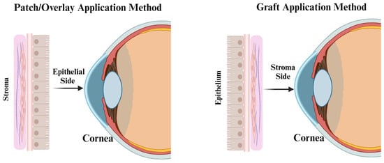
Differences between Membrane Types for Ophthalmic Applications

The inherent properties of the human amniotic membrane for ophthalmologic indications are evolving and its use is being investigated to assess how or if it promotes faster healing compared to standard treatment. The literature regarding AM in the ophthalmology field describes the application of hAM without the chorion layer. This is due to the inherent relative transparency of hAM compared to the thicker and less transparent hCM and hACM. Currently, two types of hAM are commercially available for ocular use: cryopreserved and dehydrated. Both come in a variety of thickness and sizes, with or without rings, depending on clinical need.

2.4. Chronic Wounds

Chronic nonhealing wounds are defined as wounds that fail to progress through the systematic and timely healing process seen in an acute wound closure [74]. More specific definitions include wounds that remain open and unresolved for >one month [75][76] or ≥three months [77][78]. Common chronic wounds include diabetic foot ulcers (DFU), venous leg ulcers (VLU), arterial ulcers, pressure ulcers, ischemic ulcers, and surgical and traumatic wounds. These complex wounds create a substantial personal and economic burden to affected patients and a challenge for providers seeking alternative treatment to improve care and ultimately achieve complete and lasting wound closure [76][79]. Additionally, chronic, nonhealing wounds, often associated with advanced age or underlying health conditions such as obesity, vascular insufficiency, diabetes, poor nutrition, alcoholism, etc., are susceptible to infections which can lead to additional physician visits, hospitalization, osteomyelitis, or amputation [80][81][82].
Wound healing is commonly conceptualized as four overlapping phases: hemostasis, inflammation, proliferation, and remodeling. In a chronic wound, healing is arrested in the inflammatory or proliferation phases and is often exacerbated by confounding factors such as diabetes, smoking, malnutrition, cardiovascular disease, aging, obesity, and more [81]. Granulation tissue is diminished, and re-epithelialization is halted, resulting in a persistent open wound, subject to infection and additional complications. Healing a chronic wound requires a shift in the cytokines that perpetuate the arrested phase, including coordination of fibroblast and keratinocyte proliferation and migration. The growth factors and ECM of AM contribute to the rescue of a chronic wound, facilitating progression back into the normal healing phases.
DFUs are the most common complication of diabetes with a pooled global prevalence of 6.3% [83] and it was estimated 15% to 25% of diabetic patients will develop a DFU in their lifetime, increasing the risk of life-threatening comorbidities and complications such as infection, cellulitis, osteomyelitis, myocardial infarction, increased hospitalization, or amputation [82][84].
The treatment process for chronic wounds follows a standard of care (SOC), which employs procedures such as debridement (sharps, enzymatic, biological, etc.), controlling infection, dressings to maintain a moisture-controlled environment, compression, negative pressure wound therapy (VAC therapy), skin grafts and physical offloading of pressure sites, but despite the careful attention, some wounds remain unresolved [85][86].
Although AM is used in the standard treatment options of various ocular and ophthalmologic indications, it had not become a common standard of care for chronic wounds. Historically this is due to issues related to tissue preparation, storage, and sterilization; however, current screening and processing methods have eliminated most of these concerns. Today, the practice of utilizing AM allografts for chronic wound treatment is usually applied when conventional treatment options fail, and it is essential to reassess and modify the patient’s treatment therapy.
In a nonblinded case study, four patients with refractory nonhealing wounds were treated with dhACM following sharp debridement. Reapplication was required in two patients when wound healing plateaued. All patients achieved 100% wound closure within 3, 7, 8, or 17 weeks from the initial dhACM application [87]. This result warranted further investigation into the broader application of AM. In an alternative case series, Regulski treated four elderly patients with comorbidities, an age range of 69–85 years, with DFU, VLU or traumatic wounds that remained open for a minimum of four weeks, with SOC, viable hAM, and secondary dressing or multilayer compression therapy as appropriate. All wounds healed with one to eight applications over two to eight weeks [25].
Multiple studies have reported superior wound healing following the application of AM to chronic diabetic foot ulcers (DFUs), venous leg ulcers (VLUs) and refractory non-healing wounds of varying etiologies. The Zelen group performed the first randomized controlled prospective trial comparing dhACM with SOC for treating DFUs and observed that the average wound area reduction rate was 3-times higher after amnion administration than or SOC alone at four weeks [88]. A meta-analysis of data from seven prospective randomized controlled trials (RCTs) treating DFUs with dhAM, acellular hAM, dhACM and cryo-hACM was conducted. Treatment groups were amniotic membrane plus SOC compared to SOC alone, with outcome assessments at 4-, 6- and 12-weeks. Amniotic membrane plus SOC resulted in faster DFU healing than SOC alone [27].
A similar performance in patients with chronic VLUs was also observed in several studies. In a pilot study by Mermet et al., 15 patients with chronic VLU were treated with AM. Ulcer evaluations were performed at the time of transplantation, with local pain assessed using a 0–100 visual analog scale (VAS). The data demonstrated 100% graft-take, re-epithelialization from the wound margins, a significant reduction of ulcer-related pain within the first seven days of application, and a baseline ulcer surface area reduction in 80% of patients, with 20% experiencing complete healing during a 3-month follow-up period [28]. A multicenter RCT assessed the percentage reduction in VLU wound area at the fourth week of care as a surrogate for complete wound closure at 12 or 24 weeks for patients receiving one or two dhACM applications with multilayer compression therapy (MLCT) or MLCT alone. After four weeks of treatment, 62% of those receiving one or two applications of dHACM plus MLCT achieved >40% wound closure compared to 32% receiving MLCT alone. The four-week surrogate could not be confirmed given the lack of long-term follow up [89]. In a retrospective review, the investigators analyzed deidentified data from 101 patients with non-healing lower extremity wounds that did not show a 50% reduction in size over four weeks of SOC. Following sharp debridement, wounds were treated with dhACM in sheet form (52.5%), micronized form (14.8%) or both sheet and micronized forms (32.7%) for up to 12 applications within the 12-week period. The wounds in 92 (91.1%) of the patients healed and nine remained unhealed [82].
The application of amnion allografts plus SOC has resulted in significantly faster rates of wound closure compared to SOC alone for refractory non-healing wounds from a variety of etiologies, such as DFUs and VLUs [82][88][89][90][91]. With recent efforts focusing on optimizing effective strategies for treating chronic wounds, AM is transitioning into a priority treatment option.
In a retrospective analysis of two randomized clinical trials utilizing dhACM and dhUC (dehydrated human umbilical cord), from two placenta-derived allografts (PDAs), Tettelbach et al. revealed a correlation between effective SOC debridement and DFU wound closure rates. When adequate debridement was performed, 86% of patients receiving a PDA attained complete wound closure compared to 30% with inadequate debridement. For the control group 60% attained complete wound closure with adequate debridement versus 0% with inadequate debridement [92].
Chronic wounds have a compromised microvasculature that can result in hypoxia and apoptosis which prevent healing; however, AM has been shown to stimulate angiogenesis. As previously mentioned, recent data demonstrate a sidedness effect of angiogenic response in hAM, where the epithelial side is more antiangiogenic, and the stromal side is proangiogenic [93]. This appears to be different with hACM since the chorion layer has a higher concentration of proangiogenic factors. including but not limited to VEGF, fibroblast growth factor-basic (bFGF), and PDGF-BB [43][44][94]. In addition to a hypoxic environment due to impaired vascularization, the bias towards inflammation is in part impacted and perpetuated by the presence of proinflammatory cytokines, infiltration of inflammatory cells, proteolytic enzymes, microbial presence, and necrotic tissue. The diverse array of growth factors and cytokines present in AM appears to act as a homeostatic reset, facilitating the progression of a stalled wound into the remaining healing phases.

Difference between Membrane Types for Chronic Wound Applications

Dehydrated hACM is the placental membrane type used in many controlled clinical trials and case reports to date, followed by dhAM and cryopreserved hAM. There is a dearth of applications of hCM in chronic wounds: it is mostly used in periodontal applications as described earlier. Characterization of hCM continues which may result in clinical applications for chronic wounds in the future [95][96].
The efficacy of AM allografts for treating chronic wounds is actively being studied. Overall, current evidence suggests the use of AM in chronic wounds (especially DFUs) can achieve relatively faster wound closure rates than conventional treatment methods alone, with an overall reduction in healing time. While AM has shown positive effects on healing in chronic wound care, numerous studies have shown that the viable cells produce the factors that act to reduce inflammation, improve cellular proliferation, migration, and angiogenesis [97]. More clinical research is needed to decipher which allograft type and process provides the optimal potential for wound healing.

2.5. Plastic Surgery

Plastic surgery involves the restoration, reconstruction, or alteration of physical defects of form or function in the human body and can be divided into two main categories: reconstructive and cosmetic. Plastic and reconstructive surgery requires a conformable scaffold and dressing materials that adhere to and support a moisture controlled wound environment, prevents the formation of surgical adhesions, and facilitates vascularization of skin grafts, flaps, and other tissue repairs. Human amnion and chorion allografts provide the plastic surgeon with a versatile material that meets these requirements, with added benefits including the promotion of re-epithelialization with its unique extracellular matrix, presence of growth factors to accelerate healing, semi-permeability facilitating the exchange of gases and liquids, a barrier against debris and microorganisms, and painless application and removal [17][19]. Plastic surgery is a diverse field within medicine that offers many clinical usages for AM application. Plastics specialties described below include reconstructive surgery applications which include tissue grafting, tissue engineering, post-surgery reconstructions, and burn treatments with the use of AM.
AM is a biocompatible, flexible, conformable, and mechanically durable scaffold providing a covering or barrier. The first plastics applications of amnion were for the treatment of traumatic wounds, burns, chronic skin ulcers, and for skin grafting [7][8][9][10]. Skin grafts are a widely practiced technique with multiple plastic surgery applications including surface wounds, burns, mucosal lining repairs, split-thickness skin grafts, and flap repairs, and are often associated with a variety of complications, including creating a donor site wound, graft rejection, mismatches in mechanical properties, texture and color, hair growth, wound contraction, and scar formation. When treating large wound beds, either from trauma, surgery or other complications, AM has been used as an overlay following standard grafting to replace conventional dressings such as cadaveric allografts or xenografts [19]. Amnion applied to undersurface skin flaps has been shown to significantly increase angiogenesis, reduce infiltrating neutrophils, and improve overall skin flap survival [19][98]. Amnion used as a dressing material for flap donor site wounds contours well with the wound bed, is easy to apply, provides a protective barrier from wound contamination, reduces pain, and its translucency allows holistic flap monitoring, while acting as a microbial barrier and providing pain reduction by covering nerve endings [17].
AM has been used successfully in cranial surgery procedures, such as duraplasty following craniectomy [99][100] and craniotomy [16], and myelomeningocele surgery in combination with a sensate perforator flap [98]. It has been proposed that AM supports underlying neurological tissue through the production of neurotrophic factors, such as nerve growth factor [19][98]. Additional applications of AM for post-surgical interventions include the application of AM as a wrap for the prevention of adhesions in tendon surgery [35], for the ulnar nerve during cubital tunnel surgery [18], and for prostatic nerve bundle repair [34].
The unique extracellular matrix in hAM makes it a suitable scaffold for supporting various cell cultures, while ensuring safety, minimizing morbidity, and maximizing quality of life for the recipient. In tissue engineering, AM is used as a delivery system for growth factors, cytokines, and ECM as well as viable multipotent cells present in fresh and cryopreserved viable membranes. The AM basement membrane, whether intact or decellularized, has been used as a substrate for ex vivo or in vitro culture and for the expansion of cells to facilitate wound healing, e.g., limbal stem cells for ocular damage and keratinocytes and fibroblasts for wounds [19][59][101]. An in vitro living skin equivalent of normal human keratinocytes cultured on de-epithelialized hAM demonstrated a more robust basement membrane and healthier epidermis, suggesting this would be a suitable skin substitute for skin defects including treating burns, wounds, and ulcers [102].
There are situations where the natural biodegradation of AM matches the time to heal; however, many clinical procedures require reapplication or stabilization of the AM to facilitate complete healing. In these instances, crosslinking AM with GA or EDC/NHS has been shown to stabilize the membrane and enhance its resistance to proteolytic degradation [103][104][105][106].
The use of AM for patients who have undergone Mohs micrographic surgery (MMS) provides an alternative healing option to healing by secondary intention, reducing infection, and tissue scarring [32][107]. Mohs surgery is a procedure used in treating skin cancer and involves removing thin layers of skin and examining them closely for signs of cancer until cancer-free margins are achieved with minimal damage to healthy tissue. The process results in surgical defects of varying sizes, depths, and locations such as the lower extremities, scalp, conchae, face, and hands. After surgery, repair options, including secondary intention, are limited and the healing process may be protracted [32]. Patients undergoing Mohs with full-thickness defects exposing bone are more susceptible to healing impediments such as infection, pinpoint bleeding, dehiscence, and scar formation [31]. In a case series, five elderly patients, age range 72–98, received MMS resulting in surgical defects to the underlying bone. Dehydrated hACM allografts were applied directly on the bone weekly, resulting in the absence of pain and granulation tissue formation, and healing in seven to twenty-one weeks for three patients. The two remaining patients experienced wound healing, yet resolution was incomplete due to the presence of comorbidity [31].
For surgical procedures that require the removal of tissue, whether for cosmetic purposes or medical (such as Mohs), tissue is lost and a surgical defect can remain. A preliminary study assessed the efficacy of amniotic tissue-derived allografts on wound closure time and cosmetic improvement in Mohs patients. However, the study encountered a variety of limitations; both in the location of the wound and the initial defect size, which affected the wound closure rate. Further investigation is warranted into the beneficial use of amniotic allografts in postoperative Mohs care [32]. In a recent retrospective case study, a plastics and reconstructive surgeon utilized dhAAM to treat post-Mohs surgical defects in geriatric patients. The amnion graft applications encouraged rapid tissue granulation and improved wound closure time in a variety of Mohs surgical defects. However, the study had limitations in sample size, wound variances, and patient demographics so statistically significant wound closure time was not captured [108]. In a larger-scale retrospective case-controlled study, 286 patients were assigned to control (autologous tissue flaps and full thickness skin grafts) or dhACM allografts and underwent MMS for basal or squamous cell carcinomas on the face, head, and neck. The patients who received the dhACM allograft had a five-fold lower incidence of infection and 97.9% had no postoperative complications compared to 71.3% in the autologous flaps and grafts group [109].
The application of hAM transplantation is effective both in the treatment of skin lesions and in patients suffering from toxic epidermal necrolysis (TEN) and Stevens–Johnson syndrome (SJS) [110]. TEN and SJS are classified as bullous diseases of the epidermis and manifest as severe cutaneous hypersensitivity reactions, with lesions affecting both the skin and mucous membranes and with poorly understood pathophysiology. Although TEN and SJS are rare, they are characterized by the high possibility of complications and a high mortality rate ranging from 3 to 10% for SJS and 25–50% for TEN [110][111]. More than half of patients being treated for SJS and TEN develop acute ocular involvement [57]. A 61-year-old patient with TEN covering 57% of her body, induced by treatment with an anti-epileptic, was treated with a two-fold treatment of intravenous immunoglobulin followed by amniotic membrane resulting in complete wound healing in 21 days [112].
The history of amnion usage in managing burn treatment is extensive, and compares its application with other treatment methods, with results indicating significantly reduced wound exudate, bacterial contamination, hypertrophic scarring, and healing time [113][114][115]. For many health care providers, the application of hAM has proven beneficial in the management and treatment of thermal wounds, from partial thickness burns to deeper and mixed burns [115]. The current treatment for burns involves early excision and grafting for deep partial-thickness and full-thickness burns. An RCT by Mohammadi et al. demonstrated a significant increase in the success rate of meshed split-thickness graft-take in burn wounds treated with fresh hAM dressings versus control [30]. In a controlled trial of dermal depth burns, Sawhney applied fresh hAM (up to 48 h from collection) or silver sulfadiazine cream control for three types of dermal burns: superficial, intermediate depth and deep. Both groups included bandage changes. In the hAM treatment groups, there was a significant decrease in time to heal for all three burn depth types with concomitant reductions in wound discharge and pain, and increased patient comfort during dressing changes [29]. A randomized pediatric partial-thickness facial burn treatment study compared frozen, glycerol-preserved amnion plus antimicrobial ointment versus antimicrobial ointment control on pediatric partial-thickness facial burns, and observed a significant reduction in dressing changes in the amnion group, but no significant changes in time to heal or the formation of hypertrophic scars at up to 12-months follow up, glycerol preserved amnion [113].
In a recent meta-analysis by Yang et al., eleven RCTs from 1985 to 2017 compared the efficacy of AM versus existing treatments, including SOC (three RCTs), silver sulfadiazine (three RCTs), polyurethane membrane (one RCT) and honey (one RCT) in acute burn wounds. AM was significantly more effective in reducing bacterial invasion, pain, scarring, and healing time compared to the existing treatments, except for honey, which was significantly more effective than AM. In skin-grafted burn wounds, AM was significantly more effective in reducing itching, scarring and time to graft-take compared with skin grafts affixed with staples [116].
A burn wound dressing needs to provide a basic physical and biological function, such as fluid handling, to promote the microenvironment conducive to wound healing. AM allografts used for temporary coverage of burn wounds exert both mechanical and physiological effects by protecting the wound, maintaining microbial control, covering exposed nerve endings, and hastening the healing process [29][37][38][113][114][115][117][118].
A common challenge for plastic surgeons in reconstruction surgery correcting facial and bodily defects from either birth, injury, disease, aging, or following surgery is maintaining cosmetic appearance. The human amniotic membrane provides an alternative treatment to improve the care for patients and achieve wound closure. Human amnion and chorion allografts provide the plastic surgeon with a versatile material that meets these requirements with some added benefits, such as promoting re-epithelialization with its unique protein matrix, allowing the elementary exchange of gases and liquids, acting as a barrier against debris and microorganisms, and painless application and removal. Its fluid handling capacity is a valuable function of a wound dressing, to cope with the exudates produced in vivo which are conducive to the microenvironment of a wound as it is healing [29][38][118]. As surgical techniques continue to advance, the effects of extensive scarring have diminished, but scarring is still possible.

Differences between Membrane Types for Plastic Applications

The unique properties of human amniotic membrane allografts are broadly used to heal and with the steadily evolving field of regenerative medicine, more applications are being investigated and implemented in plastics-based medicine. The two main AM types used for plastic and reconstructive surgery are hAM and hACM, fresh, dehydrated, and cryopreserved. Along with the intended uses and the subsequent clinical outcomes in plastics, continued controlled studies are needed to determine which AMs (hAM, hACM, fresh, dehydrated, cryopreserved) produce the most application-specific beneficial results for patients.

References

  1. Hu, Z.; Luo, Y.; Ni, R.; Hu, Y.; Yang, F.; Du, T.; Zhu, Y. Biological importance of human amniotic membrane in tissue engineer-ing and regenerative medicine. Mater. Today Bio. 2023, 22, 100790.
  2. Jay, R.M.; Huish, J.P.; Wray, J.H. Amniotic membrane in clinical medicine: History, current status, and future use. In Extracellu-lar Matrix-Derived Implants in Clinical Medicine, 1st ed.; Mooradian, D.L., Ed.; Elsevier Inc.: Amsterdam, The Netherlands, 2016; pp. 151–176.
  3. Fénelon, M.; Catros, S.; Meyer, C.; Fricain, J.C.; Obert, L.; Auber, F.; Louvrier, A.; Gindraux, F. Applications of human amni-otic membrane for tissue engineering. Membranes 2021, 11, 387.
  4. Tehrani, F.D.; Firouzeh, A.; Shabani, I.; Shabani, A. A review on modifications of amniotic membrane for biomedical applica-tions. Front. Bioeng. Biotechnol. 2021, 8, 606982.
  5. Elkhenany, H.; El-Derby, A.; Elkodous, M.A.; Salah, R.A.; Lotfy, A.; El-Badri, N. Applications of the amniotic membrane in tissue engineering and regeneration: The hundred-year challenge. Stem Cell Res. Ther. 2022, 13, 8.
  6. Munoz-Torres, J.R.; Martínez-González, S.B.; Lozano-Luján, A.D.; Martinez-Vázquez, M.C.; Velasco-Elizondo, P.; Garza-Veloz, I.; Martinez-Fierro, M.L. Biological properties and surgical applications of the human amniotic membrane. Front. Bioeng. Biotechnol. 2023, 10, 1067480.
  7. Davis, J.S. Skin grafting at the Johns Hopkins Hospital. Ann. Surg. 1909, 50, 542–549.
  8. Stern, M. The grafting of preserved amniotic membrane to burned and ulcerated surfaces, substituting skin grafts: A preliminary report. JAMA 1913, 60, 973–974.
  9. Sabella, N. Use of the fetal membranes in skin grafting. Med. Rec. 1913, 83, 478.
  10. Troensegaard-Hansen, E. Amniotic grafts in chronic skin ulceration. Lancet 1950, 1, 859–860.
  11. de Rötth, A. Plastic Repair of Conjunctival Defects with Fetal Membranes. Arch. Ophthalmol. 1940, 23, 522–525.
  12. Sorsby, A.; Symons, H.M. Amniotic membrane grafts in caustic burns of the eye: (Burns of the second degree). Br. J. Ophthalmol. 1946, 30, 337–345.
  13. John, T. Human amniotic membrane transplantation: Past, present, and future. Ophthalmol. Clin. N. Am. 2003, 16, 43–65.
  14. Mamede, A.C.; Carvalho, M.J.; Abrantes, A.M.; Laranjo, M.; Maia, C.J.; Botelho, M.F. Amniotic membrane: From structure and functions to clinical applications. Cell Tissue Res. 2012, 349, 447–458.
  15. Dua, H.S.; Gomes, J.A.P.; King, A.J.; Maharajan, V.S. The amniotic membrane of ophthalmology. Surv. Ophthalmol. 2004, 49, 51–77.
  16. Eichberg, D.G.; Ali, S.C.; Buttrick, S.S.; Komotar, R.J. The use of dehydrated amniotic membrane allograft for the augmenta-tion of dural repair in craniotomies. Cureus 2018, 10, e2586.
  17. Halim, A.S.; Ming, L.A.; Dorai, A.A.; Sulaiman, W.A.W. Use of amnion in plastic surgery. In Human Amniotic Membrane: Basic Science and Clinical Application, 1st ed.; Hilmy, N., Yusof, N., Nather, A., Eds.; World Scientific: Singapore, 2017; pp. 227–239.
  18. Mirzayan, R.; Russo, F.; Yang, S.-J.T.; Lowe, N.; Shean, C.J.; Harness, N.G. Human amniotic membrane wrapping of the ulnar nerve during cubital tunnel surgery reduces recurrence of symptoms. Arch. Bone Jt. Surg. 2022, 10, 969–975.
  19. Fairbairn, N.G.; Randolph, M.A.; Redmond, R.W. The clinical applications of human amnion in plastic surgery. J. Plast. Reconstr. Aesthetic Surg. 2014, 67, 662–675.
  20. Malhotra, C.; Jain, A.K. Human amniotic membrane transplantation: Different modalities of its use in ophthalmology. World J. Transpl. 2014, 4, 111–121.
  21. Tighe, S.; Mead, O.G.; Lee, A.; Tseng, S.C.G. Basic science review of birth tissue uses in ophthalmology. Taiwan. J. Ophthalmol. 2020, 10, 3–12.
  22. Burman, S.; Tejwani, S.; Vemuganti, G.K.; Gopinathan, U.; Sangwan, V.S. Ophthalmic applications of preserved human am-niotic membrane: A review of current indications. Cell Tissue Bank. 2004, 5, 161–175.
  23. Kesting, M.R.; Wolff, K.D.; Nobis, C.P.; Rohleder, N.H. Amniotic membrane in oral and maxillofacial surgery. Oral. Maxillofac. Surg. 2014, 18, 153–164.
  24. Gulameabasse, S.; Gindraux, F.; Catros, S.; Fricain, J.C.; Fénelon, M. Chorion and amnion/chorion membranes in oral and periodontal surgery: A systematic review. J. Biomed. Mater. Res. B Appl. Biomater. 2021, 109, 11216–11229.
  25. Regulski, M. Utilization of a viable human amnion membrane allograft in elderly patients with chronic lower extremity wounds of various etiologies. Wounds 2018, 30, E36–E40.
  26. Castellanos, G.; Bernabé-García, A.; Moraleda, J.M.; Nicolás, F.J. Amniotic membrane application for the healing of chronic wounds and ulcers. Placenta 2017, 59, 146–153.
  27. Laurent, I.; Astère, M.; Wang, K.R.; Cheng, Q.F.; Li, Q.F. Efficacy and time sensitivity of amniotic membrane treatment in patients with diabetic foot ulcers: A systematic review and meta-analysis. Diabetes Ther. 2017, 8, 967–979.
  28. Mermet, I.; Pottier, N.; Sainthillier, J.M.; Malugani, C.; Cairey-Remonnay, S.; Maddens, S.; Riethmuller, D.; Tiberghien, P.; Humbert, P.; Aubin, F. Use of amniotic membrane transplantation in the treatment of venous leg ulcers. Wound Repair. Regen. 2007, 15, 459–464.
  29. Sawhney, C.P. Amniotic membrane as a biological dressing in the management of burns. Burns 1989, 15, 339–342.
  30. Mohammadi, A.A.; Jafari, S.M.S.; Kiasat, M.; Tavakkolian, A.R.; Imani, M.T.; Ayaz, M.; Tolide-ie, H.R. Effect of fresh human amniotic membrane dressing on graft take in patients with chronic burn wounds compared with conventional methods. Burns 2013, 39, 349–353.
  31. Lyons, A.B.; Chipps, L.K.; Moy, R.L.; Herrmann, J.L. Dehydrated human amnion/chorion membrane allograft as an aid for wound healing in patients with full-thickness scalp defects after Mohs micrographic surgery. JAAD Case Rep. 2018, 4, 688–691.
  32. Seaton, K.; Mullens, D.; Barr, J.; Hull, E.; Averitte, R. Use of amniotic tissue-derived allografts post-mohs micrographic sur-gery: A preliminary study assessing wound closure rate. Wounds 2021, 33, 185–191.
  33. Chao, Y.C.; Humphreys, S.; Penfield, W. A new method of preventing adhesions: The use of amnioplastin after craniotomy. Br. Med. J. 1940, 1, 517–538.1.
  34. Patel, V.R.; Samavedi, S.; Bates, A.S.; Kumar, A.; Coelho, R.; Rocco, B.; Palmer, K. Dehydrated human amnion/chorion membrane allograft nerve wrap around the prostatic neurovascular bundle accelerates early return to continence and poten-cy following robot-assisted radical prostatectomy: Propensity score-matched analysis. Eur. Urol. 2015, 67, 977–980.
  35. Liu, C.; Bai, J.; Yu, K.; Liu, G.; Tian, S.; Tian, D. Biological amnion prevents flexor tendon adhesion in zone II: A controlled, multicenter clinical trial. Biomed. Res. Int. 2019, 2019, 2354325.
  36. Roy, A.; Mantay, M.; Brannan, C.; Griffiths, S. Placental tissues as biomaterials in regenerative medicine. BioMed Res. Int. 2022, 2022, 6751456.
  37. Gruss, J.S.; Jirsch, D.W. Human amniotic membrane: A versatile wound dressing. Can. Med. Assoc. J. 1978, 118, 1237–1246.
  38. Mohammadi, A.A.; Riazi, H.; Hasheminasab, M.J.; Sabet, B.; Mohammadi, M.K.; Abbasi, S.; Amini, M. Amniotic membrane dressing vs. conventional topical antibiotic dressing in hospitalized burn patients. Iran. Red. Crescent Med. J. 2009, 11, 66–70.
  39. Gupta, A.; Kedige, S.D.; Jain, K. Amnion and chorion membranes: Potential stem cell reservoir with wide applications in per-iodontics. Int. J. Biomater. 2015, 2015, 274082.
  40. Mohan, R.; Bajaj, A.; Gundappa, M. Human amnion membrane: Potential applications in oral and periodontal field. J. Int. Soc. Prev. Community Dent. 2017, 7, 15–21.
  41. Odet, S.; Louvrier, A.; Meyer, C.; Nicolas, F.J.; Hofman, N.; Chatelain, B.; Mauprivez, C.; Laurence, S.; Kerdjoudj, H.; Zwetyenga, N.; et al. Surgical appli-cation of human amniotic membrane and amnion-chorion membrane in the oral cavity and efficacy evaluation: Corollary with ophthalmological and wound healing experiences. Front. Bioeng. Biotechnol. 2021, 9, 685128.
  42. Fénelon, M.; Catros, S.; Fricain, J.C. What is the benefit of using amniotic membrane in oral surgery? A comprehensive re-view of clinical studies. Clin. Oral. Investig. 2018, 22, 1881–1891.
  43. McQuilling, J.P.; Kammer, M.; Kimmerling, K.A.; Mowry, K.C. Characterisation of dehydrated amnion chorion membranes and evaluation of fibroblast and keratinocyte responses in vitro. Int. Wound J. 2019, 16, 827–840.
  44. Koob, T.J.; Lim, J.J.; Massee, M.; Zabek, N.; Denozière, G. Properties of dehydrated human amnion/chorion composite grafts: Implications for wound repair and soft tissue regeneration. J. Biomed. Mater. Res. B Appl. Biomater. 2014, 102, 1353–1362.
  45. Jain, A.; Jaiswal, G.R.; Kumathalli, K.; Kumar, R.; Singh, A.; Sarwan, A. Comparative Evaluation of Platelet Rich Fibrin and Dehydrated Amniotic Membrane for the Treatment of Gingival Recession- A Clinical Study. J. Clin. Diagn. Res. 2017, 11, ZC24–ZC28.
  46. Esteves, J.; Bhat, K.M.; Thomas, B.; Varghese, J.M.; Jadhav, T. Efficacy of human chorion membrane allograft for recession coverage: A case series. J. Periodontal 2015, 86, 941–944.
  47. Pundir, A.J.; Agrawal, V.; Pundir, S.; Diwan, V.; Bodhi, S. Comparative evaluation of the efficacy of human chorion and amnion with coronally advanced flap for recession coverage: A case series. Clin. Adv. Periodontics 2016, 6, 118–126.
  48. Maity, S.; Priyadharshini, V. Comparison of chorion allograft and subepithelial connective tissue autograft in the treatment of gingival recession—A randomized controlled clinical trial. J. Oral. Biol. Craniofac Res. 2023, 13, 104–110.
  49. Velez, I.; Parker, W.B.; Siegel, M.A.; Hernandez, M. Cryopreserved amniotic membrane for modulation of periodontal soft tissue healing: A pilot study. J. Periodontol. 2010, 81, 1797–1804.
  50. Kiany, F.; Moloudi, F. Amnion membrane as a novel barrier in the treatment of intrabony defects: A controlled clinical trial. Int. J. Oral. Maxillofac. Implant. 2015, 30, 639–647.
  51. Samandari, M.H.; Yaghmaei, M.; Ejlali, M.; Moshref, M.; Saffar, A.S. Use of amnion as a graft material in vestibuloplasty: A preliminary report. Oral. Surg. Oral. Med. Oral. Pathol. Oral. Radiol. Endodontol. 2004, 97, 574–578.
  52. Ashraf, H.; Font, K.; Powell, C.; Schurr, M. Antimicrobial activity of an amnion-chorion membrane to oral microbes. Int. J. Dent. 2019, 2019, 1269534.
  53. Schwab, I.R.; Reyes, M.; Isseroff, R.R. Successful transplantation of bioengineered tissue replacements in patients with ocular surface disease. Cornea 2000, 19, 421–426.
  54. Letko, E.; Stechschulte, S.U.; Kenyon, K.R.; Sadeq, N.; Romero, T.R.; Samson, C.M.; Nguyen, Q.D.; Harper, S.L.; Primack, J.D.; Azar, D.T.; et al. Amniotic membrane inlay and overlay grafting for corneal epithelial defects and stromal ulcers. Arch. Ophthalmol. 2001, 119, 659–663.
  55. Hanada, K.; Shimazaki, J.; Shimmura, S.; Tsubota, K. Multilayered amniotic membrane transplantation for severe ulceration of the cornea and sclera. Am. J. Ophthalmol. 2001, 131, 323–331.
  56. Jirsova, K.; Jones, G.L.A. Amniotic membrane in ophthalmology: Properties, preparation, storage and indications for grafting—A review. Cell Tissue Bank. 2017, 18, 193–204.
  57. Nubile, M.; Dua, H.S.; Lanzini, M.; Ciancaglini, M.; Calienno, R.; Said, D.G.; Pocobelli, A.; Mastropasqua, R.; Carpineto, P. In vivo analysis of stromal integration of multilayer amniotic membrane transplantation in corneal ulcers. Am. J. Ophthalmol. 2011, 151, 809–822.
  58. Shay, E.; Kheirkhah, A.; Liang, L.; Sheha, H.; Gregory, D.G.; Tseng, S.C.G. Amniotic membrane transplantation as a new therapy for the acute ocular manifestations of Stevens-Johnson syndrome and toxic epidermal necrolysis. Surv. Ophthalmol. 2009, 54, 686–696.
  59. Kheirkhah, A.; Johnson, D.A.; Paranjpe, D.R.; Raju, V.K.; Casas, V.; Tseng, S.C.G. Temporary sutureless amniotic membrane patch for acute alkaline burns. Arch. Ophthalmol. 2008, 126, 1059–1066.
  60. Rama, P.; Matuska, S.; Paganoni, G.; Spinelli, A.; De Luca, M.; Pellegrini, G. Limbal stem-cell therapy and long-term corneal regeneration. N. Engl. J. Med. 2010, 363, 147–155.
  61. Datar, S.G.; Godse, G.N. Modified supportive simple limbal epithelial transplantation (M-SLET): A surgical technique modi-fied for limbal stem cell deficiency. Indian. J. Ophthalmol. 2022, 70, 4434–4437.
  62. Thokala, P.; Singh, A.; Singh, V.K.; Rathi, V.M.; Basu, S.; Singh, V.; MacNeil, S.; Sangwan, V.S. Economic, clinical and social impact of simple limbal epithelial transplantation for limbal stem cell deficiency. Br. J. Ophthalmol. 2022, 106, 923–928.
  63. Kotomin, I.; Valtink, M.; Hofmann, K.; Frenzel, A.; Morawietz, H.; Werner, C.; Funk, R.H.W.; Engelmann, K. Sutureless fixa-tion of amniotic membrane for therapy of ocular surface disorders. PLoS ONE 2015, 10, e0125035.
  64. Colatrella, N. Insertion and removal of sutureless amniotic membranes. J. Dry. Eye Dis. 2019, 2, 17–19.
  65. Ramos, T.; Scott, D.; Ahmad, S. An update on ocular surface epithelial stem cells: Cornea and conjunctiva. Stem Cells Int. 2015, 2015, 601731.
  66. Niederkorn, J.Y.; Wang, S. Immune privilege of the eye and fetus: Parallel universes? Transplantation 2005, 80, 1139–1144.
  67. Azuara-Blanco, A.; Pillai, C.T.; Dua, H.S. Amniotic membrane transplantation for ocular surface reconstruction. Br. J. Ophthalmol. 1999, 83, 399–402.
  68. Koizumi, N.; Fullwood, N.J.; Bairaktaris, G.; Inatomi, T.; Kinoshita, S.; Quantock, A.J. Cultivation of corneal epithelial cells on intact and denuded human amniotic membrane. Invest. Ophthalmol. Sci. 2000, 41, 2506–2513.
  69. Eslani, M.; Baradaran-Rafii, A.; Cheung, A.Y.; Kurji, K.H.; Hasani, H.; Djalilian, A.R.; Holland, E.J. Amniotic membrane transplantation in acute severe ocular chemical injury: A randomized clinical trial. Am. J. Ophthalmol. 2019, 199, 209–215.
  70. Hao, Y.; Ma, D.H.-K.; Hwang, D.G.; Kim, W.S.; Zhang, F. Identification of antiangiogenic and antiinflammatory proteins in human amniotic membrane. Cornea 2000, 19, 348–352.
  71. Gündüz, K.; Uçakhan, O.O.; Kanpolat, A.; Günlap, I. Nonpreserved human amniotic membrane transplantation for conjunc-tival reconstruction after excision of extensive ocular surface neoplasia. Eye 2006, 20, 351–357.
  72. Morkin, M.I.; Hamrah, P. Efficacy of self-retained cryopreserved amniotic membrane for treatment of neuropathic corneal pain. Ocul. Surf. 2018, 16, 132–138.
  73. Mejía, L.F.; Santamaría, J.P.; Acosta, C. Symptomatic management of postoperative bullous keratopathy with nonpreserved human amniotic membrane. Cornea 2002, 21, 342–345.
  74. Lazarus, G.S.; Cooper, D.M.; Knighton, D.R.; Percoraro, R.E.; Rodeheaver, G.; Robson, M.C. Definitions and guidelines for assessment of wounds and evaluation of healing. Wound Repair. Regen. 1994, 2, 165–170.
  75. Sen, C.K.; Gordillo, G.M.; Roy, S.; Kirsner, R.; Lambert, L.; Hunt, T.K.; Gottrup, F.; Gurtner, G.C.; Longaker, M.T. Human skin wounds: A major and snowballing threat to public health and the economy. Wound Repair. Regen. 2009, 17, 763–771.
  76. Dinh, T.; Lewis, C. Amnion applications in the foot and ankle. Clin. Podiatr. Med. Surg. 2019, 36, 563–576.
  77. Martin, P.; Nunan, R. Cellular and molecular mechanisms of repair in acute and chronic wound healing. Br. J. Dermatol. 2015, 173, 370–378.
  78. Pang, C.; Ibrahim, A.; Bulstrode, N.W.; Ferretti, P. An overview of the therapeutic potential of regenerative medicine in cu-taneous wound healing. Int. Wound J. 2017, 14, 450–459.
  79. Nussbaum, S.R.; Carter, M.J.; Fife, C.E.; DaVanzo, J.; Haught, R.; Nusgart, M.; Cartwright, D. An economic evaluation of the impact, cost, and Medicare policy implications of chronic nonhealing wounds. Value Health 2018, 21, 27–32.
  80. Fonder, M.A.; Lazarus, G.S.; Cowan, D.A.; Aronson-Cook, B.; Kohli, A.R.; Mamelak, A.J. Treating the chronic wound: A practical approach to the care of nonhealing wounds and wound care dressings. J. Am. Acad. Dermatol. 2008, 58, 185–206.
  81. Avishai, E.; Yeghiazaryan, K.; Golubnitschaja, O. Impaired wound healing: Facts and hypotheses for multi-professional con-siderations in predictive, preventive, and personalised medicine. EPMA J. 2017, 8, 23–33.
  82. Garoufalis, M.; Nagesh, D.; Sanchez, P.J.; Lenz, R.; Park, S.J.; Ruff, J.G.; Tien, A.; Goldsmith, J.; Seat, A. Use of dehydrated human amnion/chorion membrane allografts in more than 100 patients with six major types of refractory nonhealing wounds. J. Am. Podiatr. Med. Assoc. 2018, 108, 84–89.
  83. Zhang, P.; Lu, J.; Jing, Y.; Tang, S.; Zhu, D.; Bi, Y. Global epidemiology of diabetic foot ulceration: A systematic review and meta-analysis. Ann. Med. 2017, 49, 106–116.
  84. Singh, N.; Armstrong, D.G.; Lipsky, B.A. Preventing foot ulcers in patients with diabetes. JAMA 2005, 293, 217–228.
  85. Borda, L.J.; Macquhae, F.E.; Kirsner, R.S. Wound dressings: A comprehensive review. Curr. Dermatol. Rep. 2016, 5, 287–297.
  86. Falanga, V.; Isseroff, R.R.; Soulika, A.M.; Romanelli, M.; Margolis, D.; Kapp, S.; Granick, M.; Harding, K. Chronic wounds. Nat. Rev. Dis. Prim. 2022, 8, 50.
  87. Sheikh, E.S.; Sheikh, E.S.; Fetterolf, D.E. Use of dehydrated human amniotic membrane allografts to promote healing in pa-tients with refractory non-healing wounds. Int. Wound. J. 2014, 11, 711–717.
  88. Zelen, C.M.; Serena, T.E.; Denoziere, G.; Fetterolf, D.E. A prospective randomised comparative parallel study of amniotic membrane wound graft in the management of diabetic foot ulcers. Int. Wound. J. 2013, 10, 502–507.
  89. Serena, T.E.; Carter, M.J.; Le, L.T.; Sabo, M.J.; DiMarco, D.T. A multicenter, randomized, controlled clinical trial evaluating the use of dehydrated human amnion/chorion membrane allografts and multilayer compression therapy vs. multilayer com-pression therapy alone in the treatment of venous leg ulcers. Wound Repair Regen. 2014, 22, 688–693.
  90. Zelen, C.M. An evaluation of dehydrated human amniotic membrane allografts in patients with DFUs. J. Wound Care 2013, 22, 347–348, 350–351.
  91. Snyder, R.J.; Shimozaki, K.; Tallis, A.; Kerzner, M.; Reyzelman, A.; Lintzeris, D.; Bell, D.; Rutan, R.L.; Rosenblum, B. A pro-spective, randomized, multicenter, controlled evaluation of the use of dehydrated amniotic membrane allograft compared to standard of care for the closure of chronic diabetic foot ulcers. Wounds 2016, 28, 70–77.
  92. Tettelbach, W.H.; Cazzell, S.M.; Hubbs, B.; De Jong, J.L.; Forsyth, R.A.; Reyzelman, A.M. The influence of adequate debride-ment and placental-derived allografts on diabetic foot ulcers. J. Wound Care 2022, 31 (Suppl. S9), S16–S26.
  93. Niknejad, H.; Paeini-Vayghan, G.; Tehrani, F.A.; Khayat-Khoei, M.; Peirovi, H. Side dependent effects of the human amnion on angiogenesis. Placenta 2013, 34, 340–345.
  94. Duan-Arnold, Y.; Uveges, T.E.; Gyurdieva, A.; Johnson, A.; Danilkovich, A. Angiogenic potential of cryopreserved amniotic membrane is enhanced through retention of all tissue components in their native state. Adv. Wound Care 2015, 4, 513–522.
  95. Kjaergaard, N.; Hein, M.; Hyttel, L.; Helmig, R.B.; Schonheyder, H.C.; Uldbjerg, N.; Madsen, H. Antibacterial properties of human amnion and chorion in vitro. Eur. J. Obstet. Gynecol. Reprod. Biol. 2001, 94, 224–229.
  96. Frazão, L.P.; Viera de Castro, J.; Nogueira-Silva, C.; Neves, N.M. Decellularized human chorion membrane as a novel bio-material for tissue regeneration. Biomolecules 2020, 10, 1208.
  97. McQuilling, J.P.; Vines, J.B.; Kimmerling, K.A.; Mowry, K.C. Proteomic comparison of amnion and chorion and evaluation of the effects of processing on placental membranes. Wounds 2017, 29, E36–E40.
  98. de Weerd, L.; Weum, S.; Sjåvik, K.; Acharya, G.; Hennig, R.O. A new approach in the repair of a myelomeningocele using amnion and a sensate perforator flap. J. Plast. Reconstr. Aesthet. Surg. 2013, 66, 860–863.
  99. Turchan, A.; Rochman, T.F.; Ibrahim, A.; Fauziah, D.; Wahyuhadi, J.; Parenrengi, M.A.; Fauzi, A.A.; Sufarnap, E.; Bajamal, A.H.; Ferdiansyah; et al. Duraplasty using amniotic mem-brane versus temporal muscle fascia: A clinical comparative study. J. Clin. Neurosci. 2018, 50, 272–276.
  100. Marton, E.; Giordan, E.; Gallinaro, P.; Curzi, C.; Trojan, D.; Paolin, A.; Guerriero, A.; Rossi, S.; Bendini, M.; Longatti, P.; et al. Homologous amniotic membrane as a dural substitute in decompressive craniectomies. J. Clin. Neurosci. 2021, 89, 412–421.
  101. Niknejad, H.; Peirovi, H.; Jorjani, M.; Ahmadiani, A.; Ghanavi, J.; Seifalian, A.M. Properties of the amniotic membrane for potential use in tissue engineering. Eur. Cell Mater. 2008, 15, 88–99.
  102. Yang, L.; Shirakata, Y.; Shudou, M.; Dai, X.; Tokumaru, S.; Hirakawa, S.; Sayama, K.; Hamuro, J.; Hashimoto, K. New skin-equivalent model from de-epithelialized amnion membrane. Cell Tissue Res. 2006, 326, 69–77.
  103. Tanaka, Y.; Kubota, A.; Yokokura, S.; Uematsu, M.; Shi, D.; Yamato, M.; Okano, T.; Quantock, A.J.; Nishida, K. Optical me-chanical refinement of human amniotic membrane by dehydration and cross-linking. J. Tissue Eng. Regen. Med. 2012, 6, 731–737.
  104. Sarvari, R.; Keyhanvar, P.; Agbolaghi, S.; Roshangar, L.; Bahremani, E.; Keyhanvar, N.; Haghdoost, M.; Keshel, S.H.; Taghi-khani, A.; Firouzi, N.; et al. A comprehensive review on methods for promotion of mechani-cal features and biodegradation rate in amniotic membrane scaffolds. J. Mater. Sci. Mater. Med. 2022, 33, 32.
  105. Spoerl, E.; Wollensak, G.; Reber, F.; Pillunat, L. Cross-linking of human amniotic membrane by glutaraldehyde. Ophthalmic Res. 2004, 36, 71–77.
  106. Lai, J.Y.; Ma, D.-H.K. Glutaraldehyde cross-linking of amniotic membranes affects their nanofibrous structures and limbal epithelial cell culture characteristics. Int. J. Nanomed. 2013, 8, 4157–4168.
  107. Wilmas, K.M.; Patel, J.; Silapunt, S.; Doan, H.Q.; Migden, M.R. The use of a dehydrated complete human placental mem-brane allograft for Mohs surgical defects of the nose. Dermatol. Surg. 2023, 49, 343–347.
  108. Ingraldi, A.L.; Lee, D.; Tabor, A.J. Post-Mohs Surgical Defect Repair with Dehydrated Human Amnion-Amnion Membrane: A Retrospective Clinical Case Study. J. Med. Case Rep. Case Ser. 2023, 4.
  109. Toman, J.; Michael, G.M.; Wisco, O.J.; Adams, J.R.; Hubbs, B.S. Mohs defect repair with dehydrated human amnion/chorion membrane. Facial Plast. Surg. Aesthetic Med. 2022, 24, 48–53.
  110. Klama-Baryła, A.; Rojczyk, E.; Kitala, D.; Łabuś, W.; Smętek, W.; Wilemska-Kucharzewska, K.; Kucharzewski, M. Preparation of placental tissue transplants and their application in skin wound healing and chosen skin bullous diseases—Stevens-Johnson syndrome and toxic epidermal necrolysis treatment. Int. Wound J. 2020, 17, 491–507.
  111. Charlton, O.A.; Harris, V.; Phan, K.; Mewton, E.; Jackson, C.; Cooper, A. Toxic epidermal necrolysis and Steven-Johnson syndrome: A comprehensive review. Adv. Wound Care 2020, 9, 426–439.
  112. Aliabadi, S.B.; Dogahe, Z.H.; Feizkhah, A.; Mobayen, M.; Mirbolouk, B. Management of toxic epidermal necrolysis using early combination therapy of intravenous immunoglobulin and amniotic membrane grafting: A case report. J. Burn. Care Res. 2023, 44, 467–470.
  113. Branski, L.K.; Herndon, D.N.; Celis, M.M.; Norbury, W.B.; Masters, O.E.; Jeschke, M.G. Amnion in the treatment of pediatric partial-thickness facial burns. Burns 2008, 34, 393–399.
  114. Mostaque, A.K.; Rahman, K.B. Comparisons of the effects of biological membrane (amnion) and silver sulfadiazine in the management of burn wounds in children. J. Burn. Care Res. 2011, 32, 200–209.
  115. Tenenhaus, M. The use of dehydrated human amnion/chorion membranes in the treatment of burns and complex wounds. Ann. Plast. Surg. 2017, 78 (Suppl. S1), S11–S13.
  116. Yang, C.; Xiong, A.B.; He, X.C.; Ding, X.B.; Tian, X.L.; Li, Y.; Yan, H. Efficacy and feasibility of amniotic membrane for the treatment of burn wounds: A meta-analysis. J. Trauma. Acute Care Surg. 2021, 90, 744–755.
  117. Ravishankar, R.; Bath, A.S.; Roy, R. Amnion bank—The use of long-term glycerol preserved amniotic membranes in the man-agement of superficial and superficial partial thickness burns. Burns 2003, 29, 369–374.
  118. Kesting, M.R.; Wolff, K.D.; Hohlweg-Majert, B.; Steinstraesser, L. The role of allogenic amniotic membrane in burn treatment. J. Burn. Care Res. 2008, 29, 907–916.
More
Information
Contributors MDPI registered users' name will be linked to their SciProfiles pages. To register with us, please refer to https://encyclopedia.pub/register : , ,
View Times: 169
Revisions: 3 times (View History)
Update Date: 15 Nov 2023
1000/1000