Submitted Successfully!
To reward your contribution, here is a gift for you: A free trial for our video production service.
Thank you for your contribution! You can also upload a video entry or images related to this topic.
Version Summary Created by Modification Content Size Created at Operation
1 -- 3263 2023-11-02 09:42:10 |
2 format change Meta information modification 3263 2023-11-02 10:16:36 |

Video Upload Options

Do you have a full video?

Confirm

Are you sure to Delete?
Cite
If you have any further questions, please contact Encyclopedia Editorial Office.
Maniscalco, M.; Fuschillo, S.; Mormile, I.; Detoraki, A.; Sarnelli, G.; De Paulis, A.; Spadaro, G.; Cantone, E. Exhaled Nitric Oxide in Type 2 Diseases. Encyclopedia. Available online: https://encyclopedia.pub/entry/51076 (accessed on 29 April 2024).
Maniscalco M, Fuschillo S, Mormile I, Detoraki A, Sarnelli G, De Paulis A, et al. Exhaled Nitric Oxide in Type 2 Diseases. Encyclopedia. Available at: https://encyclopedia.pub/entry/51076. Accessed April 29, 2024.
Maniscalco, Mauro, Salvatore Fuschillo, Ilaria Mormile, Aikaterini Detoraki, Giovanni Sarnelli, Amato De Paulis, Giuseppe Spadaro, Elena Cantone. "Exhaled Nitric Oxide in Type 2 Diseases" Encyclopedia, https://encyclopedia.pub/entry/51076 (accessed April 29, 2024).
Maniscalco, M., Fuschillo, S., Mormile, I., Detoraki, A., Sarnelli, G., De Paulis, A., Spadaro, G., & Cantone, E. (2023, November 02). Exhaled Nitric Oxide in Type 2 Diseases. In Encyclopedia. https://encyclopedia.pub/entry/51076
Maniscalco, Mauro, et al. "Exhaled Nitric Oxide in Type 2 Diseases." Encyclopedia. Web. 02 November, 2023.
Exhaled Nitric Oxide in Type 2 Diseases
Edit

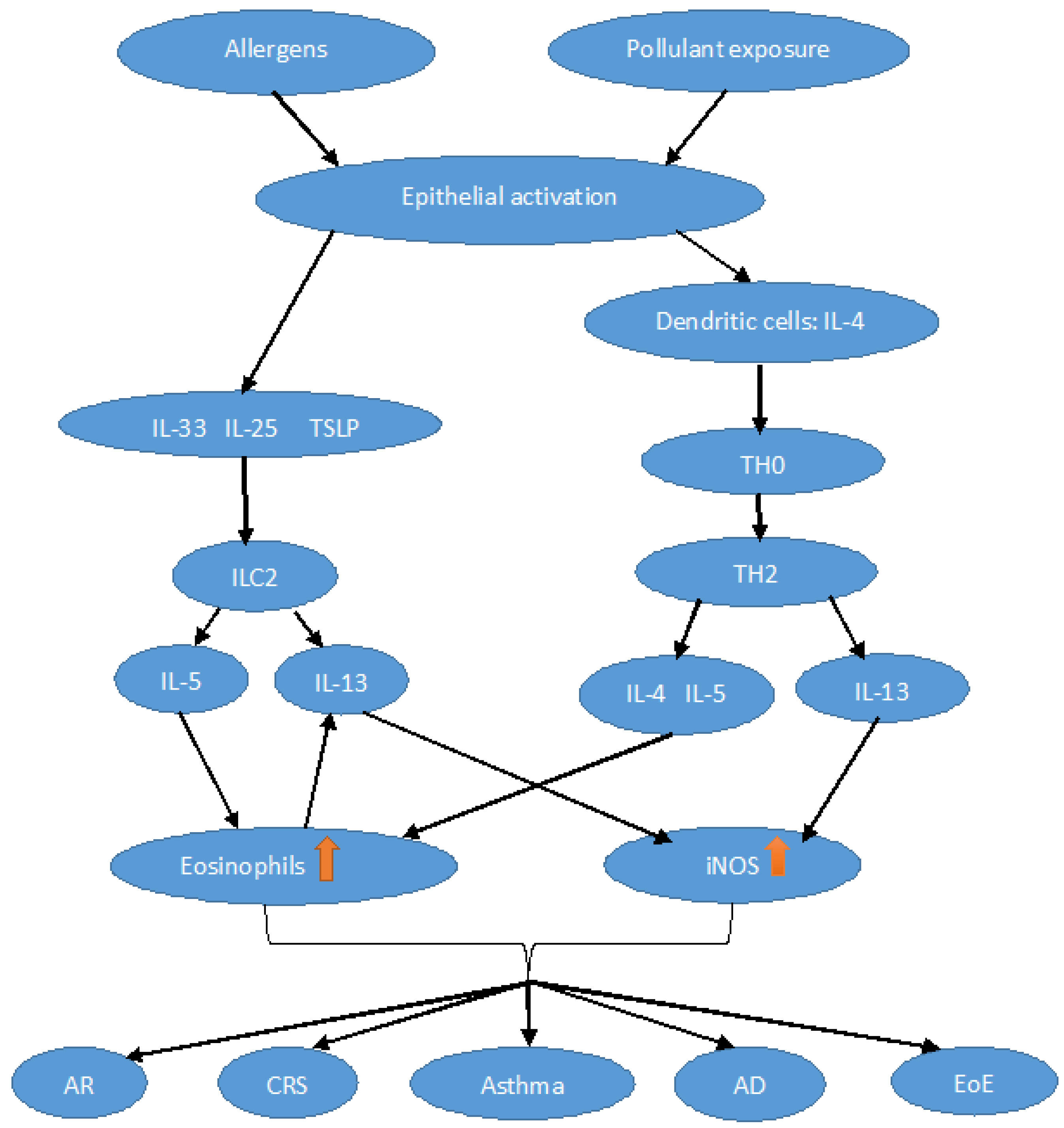
Nitric oxide (NO) is a short-lived gas molecule which has been studied for its role as a signaling molecule in the vasculature and later, in a broader view, as a cellular messenger in many other biological processes such as immunity and inflammation, cell survival, apoptosis, and aging. Fractional exhaled nitric oxide (FeNO) is a convenient, easy-to-obtain, and non-invasive method for assessing active, mainly Th2-driven, airway inflammation, which is sensitive to treatment with standard anti-inflammatory therapy. Consequently, FeNO serves as a valued tool to aid the diagnosis and monitoring of several asthma phenotypes. FeNO has been evaluated in several other respiratory and/or immunological conditions, including allergic rhinitis, chronic rhinosinusitis with/without nasal polyps, atopic dermatitis, eosinophilic esophagitis, and food allergy.

allergy asthma biomarkers COPD outcome mediators

1. Introduction

Nitric oxide (NO) is a short-lived gas molecule that has been studied since the late 80s for its role as a signaling molecule in the vasculature and later, in a broader view, as a cellular messenger in many other biological processes such as immunity and inflammation, cell survival, apoptosis, and aging [1][2]. NO is a Janus-faced molecule involved in both physiological and pathological pathways.
NO detected by a non-invasive method in exhaled air, fractional exhaled nitric oxide (FeNO), has gained increasing importance as a biomarker of type 2 inflammation [3], providing information about disease phenotype and the response to certain treatments such as steroids or biological drugs [4][5]. FeNO levels may increase in patients with acute or chronic airway inflammation, such as type 2 bronchial asthma [6], and some respiratory infections sustained by viruses through an interferon-gamma (IFNγ)-mediated pathway [7]. On the contrary, low FeNO levels are found in patients with neutrophilic asthma, chronic obstructive pulmonary disease (COPD), and cystic fibrosis [7]. Atopic subjects tend to present higher NO levels than non-atopic subjects due to an overexpression of iNOS in their airway epithelial cells [3][7].
The arrival of biological drugs has helped to gain further insight into NO physiopathology, and, in turn, the assessment of FeNO is a useful, non-invasive, and easy-to-obtain tool in monitoring conventional and biologic therapy and predicting the outcome of a wide spectrum of type 2-mediated diseases.

2. Physiopathological Aspects

NO is produced by the nitric oxide synthase (NOS) [1]. There are both constitutive and inducible isoforms of this enzyme. The constitutive ones (cNOS), which include nNOS and eNOS, are expressed, respectively, by neuronal tissue and endothelium, epithelia, platelets, and skeletal muscles [8][9][10]. The inducible form (iNOS), also known as NOS2, can be expressed by a wide variety of cells in response to exogenous stimuli (e.g., bacterial lipopolysaccharide) and inflammatory mediators (e.g., cytokines). Active NOS is a tetramer composed of two NOS proteins and two calmodulin molecules [11]. An increase in the intracellular calcium concentration stabilizes the bond between calmodulin and NOS monomers, stimulating the capability of the enzyme to produce NO. For the constitutive isoforms (nNOS and eNOS), a high calcium concentration is required to bind calmodulin, while iNOS can bind calmodulin with high affinity, irrespective of calcium levels [12]. For this reason, iNOS is called “calcium-independent NOS” and it can produce NO at nanomolar levels for prolonged periods, mainly contributing to the pathophysiological effects of NO and its detectable elevation in exhaled air [11].
iNOS is physiologically expressed in bronchial epithelial cells, ensuring a basal NO production mediated by IFNγ through JAK/STAT-1 signaling. In chronic airway inflammation, including asthmatic patients’ airways, iNOS is expressed by several cells, including macrophages, neutrophils, epithelial, endothelial, and vascular smooth muscle cells, which use L-arginine and oxygen as a substrate [13]. In paranasal sinuses, iNOS acts as a constitutive form, producing a large amount of NO under the stimulation of proinflammatory cytokines (i.e., TNF-α and IL-1β), possibly enhanced by pathways linked to the microbial films [14]. However, during Th2 inflammation in upper airway diseases, the primary sources of NO are epithelial cells and macrophages [15][16]. In COPD patients, an increase in iNOS expression in the peripheral lung and small airways has been described [17][18], together with an increase in nNOS expression and activity in the airway epithelial cells and type 1 pneumocytes as a result of oxidative stress [19], suggesting that in these patients, both isoforms contribute to an increased production of NO. Finally, several skin cell populations, including keratinocytes, endothelial cells, fibroblasts, melanocytes, adipocytes, Langerhans cells, neutrophils, and macrophages, can produce NO, participating in both skin homeostasis and pathological processes. Indeed, studies on AD patients have shown that the iNOS expressed in the skin allows NO to enter the lungs via the circulation, contributing to the NO production observed in these patients [20].
NO plays a role in both physiological and pathological conditions. For instance, in the nervous system, this molecule is involved in synaptic plasticity and the signaling within neurons [21], but it also exerts an action in many neurodegenerative conditions [22]. Indeed, excessive NO and the NO-mediated posttranslational modification of cysteine thiols can induce nitrosative stress in the nervous system, contributing to the neuropathology of many neurodegenerative diseases such as Alzheimer’s disease, Parkinson’s disease, amyotrophic lateral sclerosis, Huntington’s disease, and multiple sclerosis [23]. For example, in Huntington’s disease, the S-nitrosylation of key proteins involved in the disease progression and abnormal NO signaling in the peripheral blood tissues have been observed [24]. Of note, in multiple sclerosis, some studies have found an association between mutations in the iNOS gene and the disease progression [25]. In addition, in the central nervous system of patients affected by multiple sclerosis, higher levels of iNOS RNA have been found [26]. In addition, an increase in iNOS immunoreactivity has also been shown in the active lesions compared to the normal brain [27]. Finally, in both animal models and patients with Parkinson’s disease, NO plays a complex role, participating in both neuroprotective and neurodegenerative mechanisms [23]. Researchers found that the S-nitrosylation of some proteins involved in Parkinson’s disease pathogenesis, such as parkin, causes inhibition of its ubiquitination activity, leading to the formation of Lewy bodies and disease progression [28].
In inflammatory processes, NO production increases by participating in immune defense against infectious pathogens and causing noxious effects by increasing oxidative stress [29]. A basal production of a small amount of NO (i.e., femtomolar to picomolar) produced by the bronchial epithelium is fundamental for respiratory physiology [3]. Low constitutive NO levels are also involved in bronchodilatation via NO-activated guanylyl cyclase [30]. In addition, NO enhances lung development, promotes ciliary motility, and stimulates the production of surfactant [30]. On the contrary, augmented NO levels participate in airway inflammation, free radical production, bronchial hyperreactivity, mucus hypersecretion, increased vascular permeability, reduced ciliary heartbeat, and tissue damage [3].
NO plays a pivotal role in Th2 inflammation (Figure 1), stimulating the survival, activity, and recruitment of eosinophils, mast cells, basophils, and lymphocytes [31]. Other evidence comes from murine models; indeed, decreased eosinophilic infiltrates, bronchial thickening, and mucus secretion, together with lower concentrations of type 2 cytokines (i.e., interleukin (IL)-4, IL-5, and -13) and chemokines (i.e., eotaxin-1), were found in asthmatic mice “knock-out” for all NOS isoforms compared to the wild type [32]. The cytokine milieu typical of Th2-mediated disease induces iNOS expression by epithelial and inflammatory cells. Indeed, iNOS expression is observed under the stimulation of various type 2 cytokines, especially IL-4 and IL-13 [33][34]. On the contrary, IL-5 induces eosinophilia but is unrelated to iNOS activation and does not contribute to NO levels [35]. Indeed, IL-4 and Il-13 induce iNOS expression mainly through the STAT 6 pathway, while IL-5 acts through different pathways, primarily activating STAT 3 [36][37].
Figure 1. Schematic representation of the Th2 pathway. Dendritic cell activation by damaged epithelial cells secreting interleukin (IL)-4 prompts Th0 cells to differentiate into Th2 cells. Th2 cells secrete IL-5, among other cytokines, which represents the main stimulus for the production and recruitment of eosinophils (orange arrow) at the level of the target organ. Infections or exposure to pollutants induce epithelial cells to release the so-called alarmins (IL-25, IL-33, thymic stromal lymphopoietin (TSLP)) which activate innate lymphoid cells 2 (ILC2), which produce Th2 cytokines. IL-13, mainly secreted by eosinophils, activates the expression (orange arrow) of inducible nitric oxide synthase (iNOS) and increases the production of NO. AR, allergic rhinitis; CRS, chronic rhinosinusitis; EoE, eosinophilic esophagitis; AD, atopic dermatitis.

3. Exhaled NO in Type 2 Lower Airway Diseases

3.1. FeNO in T2 High Asthma

Based on the prevalent type of airway inflammation, asthma has been broadly categorized into two endotypes: T2-high and T2-low. T2 airway inflammation in patients with asthma, although occurring along a continuum, is characterized by a specific set of cytokines, including IL-4, IL-5, IL-13, and the so-called alarmins, IL-25 and IL-33, and thymic stromal lymphopoietin (TSLP), secreted by activated epithelial cells. The T2 inflammatory response is frequently associated with atopy (i.e., the genetic tendency to develop allergic diseases, such as allergic rhinitis, asthma, and atopic dermatitis, associated with heightened immune responses to diverse antigens/allergens, leading to CD4+ Th2 differentiation and overproduction of immunoglobulin E (IgE)) [38], eosinophilic disorders, and parasitic infections [39].
In approximately 50–70% of asthmatic patients, a T2 inflammatory response represents the pathogenetic mechanism responsible for the disease [40].
Both innate and adaptive immune responses are involved in the inflammation of type 2 asthma. Innate lymphoid cells (ILC2s) and Th2 cells are critical in orchestrating the type 2 inflammatory response. When exposed to pollutants or viral and fungal infections, ILC2s are stimulated by alarmins secreted by epithelial cells. Allergens, on the other hand, are the activators of Th2 cells. Once stimulated, ILC2 and Th2 produce type 2 cytokines, including IL-4, IL-5, and IL-13, responsible for the main alterations present in asthma. IL-4 and IL-13 cause class switching of B cells to produce IgE. IL-13 is responsible for hyperplasia of both globular and smooth cells. IL-5 is the key cytokine involved in the differentiation, maturation, and survival of eosinophils [40]. Eosinophils, mast cells, and basophils are the main effector cells of the T2 inflammation pathway [39][41].
T2-high asthma phenotypes include both early- and late-onset asthma, frequent exacerbations, atopy, eosinophilia, and increased levels of FeNO.
FeNO levels, compared to healthy controls, are elevated in T2 high asthma patients [42] and other respiratory diseases such as AR and CRSwNP [3].
In asthma, FeNO originates predominantly from the lower airways [43], and a relationship between FeNO and airway eosinophilic inflammation has been reported [44]. FeNO, eosinophils, and IgE are considered the most relevant biomarkers of the T2 inflammatory pathway.

3.2. FeNO in Cough Variant Asthma

CVA is a specific and atypical asthma phenotype and the most common cause of chronic cough. About 33.3% of patients with chronic cough are caused by CVA [45]. CVA is associated with airway hyperresponsiveness and chronic eosinophilic inflammation, in both central and peripheral airways, and manifests itself as a non-productive cough without wheezing or shortness of breath [46]. A positive response to the bronchial provocation test and the efficacy of bronchodilator therapy in preventing cough are the most important diagnostic criterions for CVA.
A subset of subjects with CVA (approximately 30%), especially those with higher airway responsiveness, higher sputum eosinophils, and atopy, can progress into classic asthma in the absence of appropriate treatment for asthma [47].
Small airways dysfunction (SAD) is a clinically relevant characteristic of CVA [48]. Feng-Jia et al. retrospectively analysed 150 patients with CVA and found that small airway function was significantly reduced when compared with patients without CVA [49]. In a prospective study, FeNO waa measured at exhalation flow rates of 200 mL/s (reflecting the inflammation of peripheral small airway) [50]; FeNO > 11 ppb had high diagnostic value for CVA especially in SAD patients [51]. Combining FeNO and small airway function indexes might increase the diagnostic value for differentiating CVA from typical asthma [52].
FeNO levels in patients with CVA are lower than those observed in patients with classic asthma, indicating greater chronic eosinophilic airway inflammation in the latter than in the former [52].
Compared with other small airway pulmonary function tests such as MMEF/MEF50, FeNO shows increased sensitivity and specificity in the diagnosis of CVA, and it is negatively correlated with both MMEF and MEF50 values. In addition, combining FeNO with MMEF/MEF50 increases the diagnostic accuracy of CVA in children [53], resulting in a rapid and accurate diagnosis, thus avoiding unnecessary treatments.
The diagnostic accuracy of FeNO in detecting CVA in chronic cough patients with AR was higher than in those with chronic cough without AR [54].
FeNO can be used as a predictor for airway eosinophilic inflammation but also as a predictor for steroid responsiveness in patients with chronic cough. The value of 34.5 ppb of FeNO has been suggested to distinguish between patients with chronic cough who respond to corticosteroid treatment and non-responders (with 85% sensitivity and about 90% specificity) [55].

3.3. FeNO in COPD

Airway inflammation in COPD is driven by type 1 immune response; however, in a subset of COPD patients, a T2 inflammatory response plays a significant role both in the stable state and during exacerbation of the disease [56]. Eosinophilic airway inflammation has been reported in up to 20–40% of COPD patients [57]. Blood eosinophils persistently above 2% can be found in 15–37% of patients with COPD [58], suggesting a pathogenetic role of T2 inflammatory immunity in a subset of patients.
FeNO levels in COPD patients are higher than those observed in healthy non-smokers; however, they are not as high as those observed in untreated asthma [59].
In COPD subjects, high FeNO values, as compared to those with low values, may suggest eosinophilic inflammation and the presence of some asthmatic features [60], airway eosinophilic inflammation [61], and increased spirometric response to inhaled corticosteroids [62].
Increased FeNO levels have been reported during the exacerbated phases of COPD [63], and in clinically stable COPD outpatients, FeNO levels persistently above 20 ppb are associated with a significantly higher risk of exacerbation [64].
FeNO monitoring in COPD has a less defined role as compared to asthma. However, FeNO might also reflect in COPD the presence of airway eosinophilia and predict the response to corticosteroids. In addition, FeNO could play a role as a potential prognostic biomarker in COPD. Indeed, its level increased in patients with greater disease severity and during acute exacerbations [65] and in those at higher risk of exacerbation [64].
As compared to healthy subjects, a greater intra-individual FeNO variation in COPD patients during stable clinical conditions has been reported [66]. In addition, FeNO variability is influenced by COPD exacerbations, with FeNO increasing at the onset of exacerbation and FeNO value variability being associated with future risk of exacerbations [67].
In conclusion, in COPD, although FeNO values show greater variability, its use, particularly when combined with other T2-type inflammatory biomarkers, could improve COPD phenotypic discrimination or help to select candidates for personalized therapies, particularly in the context of biological therapy.

4. Exhaled NO in Type 2 Upper Airway Diseases

4.1. FeNO in Allergic Rhinitis

Upper and lower airways represent a continuous anatomo-physiological entity, and a high percentage (up to 38%) of patients with AR also have asthma [68][69]. Indeed, asthma and AR share a common etiopathogenesis, characterized by a T2 inflammatory substrate with the release of different biomarkers, among which NO is one of the most studied. In addition, the early and proper therapeutic management of both diseases seems to achieve mutual benefits. Likely, the measurement of NO in the upper respiratory tract provides information on the degree of eosinophilic inflammation, completing the clinical and non-invasive management of AR [70].
These results taken together suggest that FeNO is rather a measure of allergy than of the severity of the disease in AR, but it could gain a value in evaluating the therapeutic effect of certain drugs in these patients.

4.2. FeNO in Chronic Rhino-Sinusitis

CRS is a chronic, heterogeneous, and multifactorial inflammatory disease of the upper airways, whose prevalence in the general adult population is about 10–15% [71]. In CRS, as well as in asthma, the activation of ILC-2 cells leads to the release of cytokines (Il-9, Il-4, Il-5, and IL-13) that promote a Th-2 inflammatory response [6].
CRS, like asthma, is a phenotypically heterogeneous disease. Depending on the presence or absence of nasal polyps, CRS can be classified into two types: CRS with (CRSwNP) and without nasal polyps (CRSsNP). In addition, from the histological point of view of CRSwNP, depending on the presence or absence of eosinophils infiltrating the nasal mucosa, two distinct histo-phenotypes have been reported: the eosinophilic-CRSwNP (ECRSwNP) and the non-eosinophilic-CRSwNP (NECRSwNP) [6]. The inflammatory CRS phenotypes present a substantial variability in their geographic distribution. Indeed, in Caucasian populations, CRSwNP more frequently presents with a type 2 (eosinophilic) inflammatory profile, while in Asian populations, mixed inflammatory profiles (type 1 and 3) have been reported [72].
Lower airway morbidity is highly prevalent in patients with CRSwNP [73], being present with asthma in up to 30–70% of them [74]. Accordingly, about 25% of patients with severe eosinophilic asthma have CRSwNP as a comorbidity [75].
In patients with CRSwNP, type 2 inflammation affects not only the nasal mucosa but also the bronchi and alveoli, even in the absence of asthmatic comorbidity [76]. CRSwNP with and without asthma is the most severe form of T2 inflammation in the upper airway, and CRSwNP and asthma are linked through the underlying T2 inflammatory pathway, emphasizing the continuum between the upper and lower airways [72].

5. FeNO in Eosinophilic Esophagitis

Among eosinophilic gastrointestinal disorders (EGIDs), EoE is the most frequent and the most prevalent cause of chronic esophagitis, after gastroesophageal reflux disease (GERD), which is often a cause of misdiagnosis [77]. EoE is a chronic, allergen-driven eosinophilic inflammatory disease of the esophagus affecting both children and adults. Its incidence and prevalence are both rising [78].
Symptoms of EoE are related to esophageal dysfunction [79]. The gold standard diagnostic method is endoscopic biopsy with confirmed diagnosis in the presence of several eosinophils/high power field (hpf) ≥ 15 [80].
The pathogenic pathway underlying EoE is an antigen-driven Th2 immune response. Ingested allergens, especially food allergens, interacting with esophageal epithelial cells stimulate them to release alarmins (IL-25, IL-33, TSLP), thereby activating a T2-mediated immune response [81]. The relevant pathogenetic role of food allergens in sustaining the eosinophilic inflammation in EoE is suggested by the observation that dietary therapy is effective in up to 70% of patients with EoE [81].

6. Exhaled NO in Other Type 2 Diseases

6.1. Atopic Dermatitis

With a prevalence of 3% in adults and 20% in children, AD is one of the most frequent allergic conditions affecting the skin [82]. Pathogenetical mechanisms leading to this condition possibly involve environmental, genetic, and immunologic factors which cause inflammation and dysfunction of the skin barrier [83]. The presence of AD could influence FeNO levels [84][85]. Indeed, among the others, FeNO is a biomarker for the presence of a type 2 immune pattern, with increased levels of different cytokines such as IL-4 and IL-13, which are involved iNOS induction and thus NO production [86].
All of this evidence taken together indicates that currently, FeNO assessment is not a reliable tool in patients with concomitant mild or moderate AD and respiratory disease. [20][87].

6.2. Food Allergy

Food allergy likely affects nearly 5% of adults and 8% of children, with growing evidence of an increase in prevalence [88].
Among food allergies, a peanut allergy can be a life threatening event and accounts for approximately two-thirds of all fatal food-induced anaphylaxis [89]. Clinical peanut allergy resolves in up to 20% of children [90].
FeNO has been shown to be elevated in children with a peanut allergy who have “outgrown” their asthma [91]. In addition, FeNO may improve the ability to predict allergic reaction during a peanut challenge [92][93]. In particular, a change in FeNO during a peanut challenge was related to the severity of reaction. FeNO decreased more significantly in those who subsequently developed anaphylaxis than in those with a clinical allergy, not anaphylaxis, or who had a negative peanut challenge (tolerance) [94].
FeNO was increased also in patients with asthma and sensitization to other food allergens, with or without sensitization to airborne allergens [95]. In this research, the increase in FeNO was related to the increase in other local and systemic type 2 inflammation markers, such as serum eosinophil cationic protein (S-ECP) and periostin [95].
As a bedside test that can be used in children, it has potential for further research into mechanisms of anaphylaxis in food allergies and potentially assists in predicting an imminent anaphylactic reaction in some patients.

References

  1. Kiss, H.; Orlos, Z.; Gellert, A.; Megyesfalvi, Z.; Mikaczo, A.; Sarkozi, A.; Vasko, A.; Miklos, Z.; Horvath, I. Exhaled Biomarkers for Point-of-Care Diagnosis: Recent Advances and New Challenges in Breathomics. Micromachines 2023, 14, 391.
  2. Moncada, S.; Palmer, R.M.; Higgs, E.A. Nitric oxide: Physiology, pathophysiology, and pharmacology. Pharmacol. Rev. 1991, 43, 109–142.
  3. Escamilla-Gil, J.M.; Fernandez-Nieto, M.; Acevedo, N. Understanding the Cellular Sources of the Fractional Exhaled Nitric Oxide (FeNO) and Its Role as a Biomarker of Type 2 Inflammation in Asthma. Biomed. Res. Int. 2022, 2022, 5753524.
  4. American Thoracic Society; European Respiratory Society. ATS/ERS recommendations for standardized procedures for the online and offline measurement of exhaled lower respiratory nitric oxide and nasal nitric oxide, 2005. Am. J. Respir. Crit. Care Med. 2005, 171, 912–930.
  5. Medrek, S.K.; Parulekar, A.D.; Hanania, N.A. Predictive Biomarkers for Asthma Therapy. Curr. Allergy Asthma Rep. 2017, 17, 69.
  6. Mormile, M.; Mormile, I.; Fuschillo, S.; Rossi, F.W.; Lamagna, L.; Ambrosino, P.; de Paulis, A.; Maniscalco, M. Eosinophilic Airway Diseases: From Pathophysiological Mechanisms to Clinical Practice. Int. J. Mol. Sci. 2023, 24, 7254.
  7. Alving, K.; Malinovschi, A. Basic aspects of exhaled nitric oxide. Eur. Respir. Monogr. 2010, 49, 1–31.
  8. Bahadoran, Z.; Carlstrom, M.; Mirmiran, P.; Ghasemi, A. Nitric oxide: To be or not to be an endocrine hormone? Acta. Physiol. 2020, 229, e13443.
  9. Hoiland, R.L.; Caldwell, H.G.; Howe, C.A.; Nowak-Fluck, D.; Stacey, B.S.; Bailey, D.M.; Paton, J.F.R.; Green, D.J.; Sekhon, M.S.; Macleod, D.B.; et al. Nitric oxide is fundamental to neurovascular coupling in humans. J. Physiol. 2020, 598, 4927–4939.
  10. Horvath, I.; Sandor, N.T.; Ruttner, Z.; McLaughlin, A.C. Role of nitric oxide in regulating cerebrocortical oxygen consumption and blood flow during hypercapnia. J. Cereb. Blood Flow Metab. 1994, 14, 503–509.
  11. Korhonen, R.; Lahti, A.; Kankaanranta, H.; Moilanen, E. Nitric oxide production and signaling in inflammation. Curr. Drug Targets Inflamm. Allergy 2005, 4, 471–479.
  12. Forstermann, U.; Sessa, W.C. Nitric oxide synthases: Regulation and function. Eur. Heart J. 2012, 33, 829–837.
  13. Marcos, M.C.; Cisneros Serrano, C. What is the added value of FeNO as T2 biomarker? Front. Allergy 2022, 3, 957106.
  14. Spector, B.M.; Shusterman, D.J.; Zhao, K. Nasal nitric oxide flux from the paranasal sinuses. Curr. Opin. Allergy Clin. Immunol. 2023, 23, 22–28.
  15. Van der Veen, R.C. Nitric oxide and T helper cell immunity. Int. Immunopharmacol. 2001, 1, 1491–1500.
  16. Marcuccio, G.; Ambrosino, P.; Merola, C.; Manzo, F.; Motta, A.; Rea, G.; Cantone, E.; Maniscalco, M. Clinical Applications of Nasal Nitric Oxide in Allergic Rhinitis: A Review of the Literature. J. Clin. Med. 2023, 12, 81.
  17. Brindicci, C.; Kharitonov, S.A.; Ito, M.; Elliott, M.W.; Hogg, J.C.; Barnes, P.J.; Ito, K. Nitric oxide synthase isoenzyme expression and activity in peripheral lung tissue of patients with chronic obstructive pulmonary disease. Am. J. Respir. Crit. Care Med. 2010, 181, 21–30.
  18. Ricciardolo, F.L.; Caramori, G.; Ito, K.; Capelli, A.; Brun, P.; Abatangelo, G.; Papi, A.; Chung, K.F.; Adcock, I.; Barnes, P.J.; et al. Nitrosative stress in the bronchial mucosa of severe chronic obstructive pulmonary disease. J. Allergy Clin. Immunol. 2005, 116, 1028–1035.
  19. Maarsingh, H.; Leusink, J.; Bos, I.S.; Zaagsma, J.; Meurs, H. Arginase strongly impairs neuronal nitric oxide-mediated airway smooth muscle relaxation in allergic asthma. Respir. Res. 2006, 7, 6.
  20. Adler, B.L.; Friedman, A.J. Nitric oxide therapy for dermatologic disease. Future. Sci. OA 2015, 1, FSO37.
  21. Bradley, S.A.; Steinert, J.R. Nitric Oxide-Mediated Posttranslational Modifications: Impacts at the Synapse. Oxidative Med. Cell. Longev. 2016, 2016, 5681036.
  22. Nakamura, T.; Lipton, S.A. Emerging roles of S-nitrosylation in protein misfolding and neurodegenerative diseases. Antioxid. Redox Signal. 2008, 10, 87–101.
  23. Tripathi, M.K.; Kartawy, M.; Amal, H. The role of nitric oxide in brain disorders: Autism spectrum disorder and other psychiatric, neurological, and neurodegenerative disorders. Redox Biol. 2020, 34, 101567.
  24. Haun, F.; Nakamura, T.; Shiu, A.D.; Cho, D.H.; Tsunemi, T.; Holland, E.A.; La Spada, A.R.; Lipton, S.A. S-nitrosylation of dynamin-related protein 1 mediates mutant huntingtin-induced mitochondrial fragmentation and neuronal injury in Huntington’s disease. Antioxid. Redox Signal. 2013, 19, 1173–1184.
  25. Barcellos, L.F.; Begovich, A.B.; Reynolds, R.L.; Caillier, S.J.; Brassat, D.; Schmidt, S.; Grams, S.E.; Walker, K.; Steiner, L.L.; Cree, B.A.; et al. Linkage and association with the NOS2A locus on chromosome 17q11 in multiple sclerosis. Ann. Neurol. 2004, 55, 793–800.
  26. Kanwar, J.R.; Kanwar, R.K.; Krissansen, G.W. Simultaneous neuroprotection and blockade of inflammation reverses autoimmune encephalomyelitis. Brain 2004, 127, 1313–1331.
  27. Smith, K.J.; Lassmann, H. The role of nitric oxide in multiple sclerosis. Lancet Neurol. 2002, 1, 232–241.
  28. Yao, D.; Gu, Z.; Nakamura, T.; Shi, Z.Q.; Ma, Y.; Gaston, B.; Palmer, L.A.; Rockenstein, E.M.; Zhang, Z.; Masliah, E.; et al. Nitrosative stress linked to sporadic Parkinson’s disease: S-nitrosylation of parkin regulates its E3 ubiquitin ligase activity. Proc. Natl. Acad. Sci. USA 2004, 101, 10810–10814.
  29. Cinelli, M.A.; Do, H.T.; Miley, G.P.; Silverman, R.B. Inducible nitric oxide synthase: Regulation, structure, and inhibition. Med. Res. Rev. 2020, 40, 158–189.
  30. Antosova, M.; Mokra, D.; Pepucha, L.; Plevkova, J.; Buday, T.; Sterusky, M.; Bencova, A. Physiology of nitric oxide in the respiratory system. Physiol. Res. 2017, 66, S159–S172.
  31. Yatera, K.; Mukae, H. Possible pathogenic roles of nitric oxide in asthma. Respir. Investig. 2019, 57, 295–297.
  32. Akata, K.; Yatera, K.; Wang, K.Y.; Naito, K.; Ogoshi, T.; Noguchi, S.; Kido, T.; Toyohira, Y.; Shimokawa, H.; Yanagihara, N.; et al. Decreased Bronchial Eosinophilic Inflammation and Mucus Hypersecretion in Asthmatic Mice Lacking All Nitric Oxide Synthase Isoforms. Lung 2016, 194, 121–124.
  33. Wenzel, S.; Castro, M.; Corren, J.; Maspero, J.; Wang, L.; Zhang, B.; Pirozzi, G.; Sutherland, E.R.; Evans, R.R.; Joish, V.N.; et al. Dupilumab efficacy and safety in adults with uncontrolled persistent asthma despite use of medium-to-high-dose inhaled corticosteroids plus a long-acting beta2 agonist: A randomised double-blind placebo-controlled pivotal phase 2b dose-ranging trial. Lancet 2016, 388, 31–44.
  34. Corren, J.; Lemanske, R.F.; Hanania, N.A.; Korenblat, P.E.; Parsey, M.V.; Arron, J.R.; Harris, J.M.; Scheerens, H.; Wu, L.C.; Su, Z.; et al. Lebrikizumab treatment in adults with asthma. N. Engl. J. Med. 2011, 365, 1088–1098.
  35. Pavord, I.D.; Korn, S.; Howarth, P.; Bleecker, E.R.; Buhl, R.; Keene, O.N.; Ortega, H.; Chanez, P. Mepolizumab for severe eosinophilic asthma (DREAM): A multicentre, double-blind, placebo-controlled trial. Lancet 2012, 380, 651–659.
  36. Chibana, K.; Trudeau, J.B.; Mustovich, A.T.; Hu, H.; Zhao, J.; Balzar, S.; Chu, H.W.; Wenzel, S.E. IL-13 induced increases in nitrite levels are primarily driven by increases in inducible nitric oxide synthase as compared with effects on arginases in human primary bronchial epithelial cells. Clin. Exp. Allergy 2008, 38, 936–946.
  37. Suresh, V.; Mih, J.D.; George, S.C. Measurement of IL-13-induced iNOS-derived gas phase nitric oxide in human bronchial epithelial cells. Am. J. Respir. Cell Mol. Biol. 2007, 37, 97–104.
  38. Justiz Vaillant, A.A.; Modi, P.; Jan, A. Atopy. In StatPearls; StatPearls Publishing: Treasure Island, FL, USA, 2023.
  39. Ahmad, J.G.; Marino, M.J.; Luong, A.U. Unified Airway Disease: Future Directions. Otolaryngol. Clin. N. Am. 2023, 56, 181–195.
  40. Maspero, J.; Adir, Y.; Al-Ahmad, M.; Celis-Preciado, C.A.; Colodenco, F.D.; Giavina-Bianchi, P.; Lababidi, H.; Ledanois, O.; Mahoub, B.; Perng, D.W.; et al. Type 2 inflammation in asthma and other airway diseases. ERJ Open Res. 2022, 8, 00576–2021.
  41. Popovic-Grle, S.; Stajduhar, A.; Lampalo, M.; Rnjak, D. Biomarkers in Different Asthma Phenotypes. Genes 2021, 12, 801.
  42. Kharitonov, S.A.; Yates, D.; Robbins, R.A.; Logan-Sinclair, R.; Shinebourne, E.A.; Barnes, P.J. Increased nitric oxide in exhaled air of asthmatic patients. Lancet 1994, 343, 133–135.
  43. Kharitonov, S.A.; Chung, K.F.; Evans, D.; O’Connor, B.J.; Barnes, P.J. Increased exhaled nitric oxide in asthma is mainly derived from the lower respiratory tract. Am. J. Respir. Crit. Care Med. 1996, 153, 1773–1780.
  44. Jatakanon, A.; Lim, S.; Kharitonov, S.A.; Chung, K.F.; Barnes, P.J. Correlation between exhaled nitric oxide, sputum eosinophils, and methacholine responsiveness in patients with mild asthma. Thorax 1998, 53, 91–95.
  45. Chen, J.; Lin, W.; Gu, H.; Ying, K.; Li, T.; Shao, H. Study on the Relationship Between Bronchoalveolar Lavage Fluid Cell Count, Th1/Th2 Cytokines and Pulmonary Function in Patients with Cough Variant Asthma. J. Asthma Allergy 2022, 15, 1713–1720.
  46. Lai, K.; Shen, H.; Zhou, X.; Qiu, Z.; Cai, S.; Huang, K.; Wang, Q.; Wang, C.; Lin, J.; Hao, C.; et al. Clinical Practice Guidelines for Diagnosis and Management of Cough-Chinese Thoracic Society (CTS) Asthma Consortium. J. Thorac. Dis. 2018, 10, 6314–6351.
  47. Kim, C.K.; Kim, J.T.; Kang, H.; Yoo, Y.; Koh, Y.Y. Sputum eosinophilia in cough-variant asthma as a predictor of the subsequent development of classic asthma. Clin. Exp. Allergy 2003, 33, 1409–1414.
  48. Lougheed, M.D.; Turcotte, S.E.; Fisher, T. Cough variant asthma: Lessons learned from deep inspirations. Lung 2012, 190, 17–22.
  49. Chen, F.-j.; Huang, X.-y.; Lin, G.-p.; Liu, Y.-l.; Xie, C.-m. Validity of fractional exhaled nitric oxide and small airway function indices in diagnosis of cough-variant asthma. J. Asthma 2018, 55, 750–755.
  50. Paredi, P.; Kharitonov, S.A.; Meah, S.; Barnes, P.J.; Usmani, O.S. A novel approach to partition central and peripheral airway nitric oxide. Chest 2014, 145, 113–119.
  51. Bai, H.; Shi, C.; Yu, S.; Wen, S.; Sha, B.; Xu, X.; Yu, L. A comparative study on the value of lower airway exhaled nitric oxide combined with small airway parameters for diagnosing cough-variant asthma. Ther. Adv. Respir. Dis. 2023, 17, 17534666231181259.
  52. Wang, Y.; Zhao, L.; Chen, F.; Guo, Y.; Ma, H.; Han, B.; Yi, J.; Kong, X. Diagnostic Value of Fractional Exhaled Nitric Oxide and Small Airway Function in Differentiating Cough-Variant Asthma from Typical Asthma. Can. Respir. J. 2021, 2021, 9954411.
  53. Zhu, H.; Zhang, R.; Hao, C.; Yu, X.; Tian, Z.; Yuan, Y. Fractional Exhaled Nitric Oxide (FeNO) Combined with Pulmonary Function Parameters Shows Increased Sensitivity and Specificity for the Diagnosis of Cough Variant Asthma in Children. Med. Sci. Monit. 2019, 25, 3832–3838.
  54. Chen, L.C.; Zeng, G.S.; Wu, L.L.; Zi, M.; Fang, Z.K.; Fan, H.Z.; Yu, H.P. Diagnostic value of FeNO and MMEF for predicting cough variant asthma in chronic cough patients with or without allergic rhinitis. J. Asthma 2021, 58, 326–333.
  55. Shebl, E.; Abdel-moety, H. Assessment of the role of fractional exhaled nitric oxide as a predictor of airway eosinophilia and corticosteroid responsiveness in patients with chronic cough. Egypt. J. Bronchol. 2020, 14, 15.
  56. Barnes, P.J. Inflammatory mechanisms in patients with chronic obstructive pulmonary disease. J. Allergy Clin. Immunol. 2016, 138, 16–27.
  57. Fernandes, L.; Rane, S.; Mandrekar, S.; Mesquita, A.M. Eosinophilic Airway Inflammation in Patients with Stable Biomass Smoke-versus Tobacco Smoke-Associated Chronic Obstructive Pulmonary Disease. J. Health Pollut. 2019, 9, 191209.
  58. Singh, D.; Kolsum, U.; Brightling, C.E.; Locantore, N.; Agusti, A.; Tal-Singer, R.; ECLIPSE Investigators. Eosinophilic inflammation in COPD: Prevalence and clinical characteristics. Eur. Respir. J. 2014, 44, 1697–1700.
  59. Ansarin, K.; Chatkin, J.M.; Ferreira, I.M.; Gutierrez, C.A.; Zamel, N.; Chapman, K.R. Exhaled nitric oxide in chronic obstructive pulmonary disease: Relationship to pulmonary function. Eur. Respir. J. 2001, 17, 934–938.
  60. Chen, F.J.; Huang, X.Y.; Liu, Y.L.; Lin, G.P.; Xie, C.M. Importance of fractional exhaled nitric oxide in the differentiation of asthma-COPD overlap syndrome, asthma, and COPD. Int. J. Chronic Obstr. Pulm. Dis. 2016, 11, 2385–2390.
  61. Malerba, M.; Radaeli, A.; Olivini, A.; Damiani, G.; Ragnoli, B.; Montuschi, P.; Ricciardolo, F.L. Exhaled nitric oxide as a biomarker in COPD and related comorbidities. Biomed. Res. Int. 2014, 2014, 271918.
  62. Zietkowski, Z.; Kucharewicz, I.; Bodzenta-Lukaszyk, A. The influence of inhaled corticosteroids on exhaled nitric oxide in stable chronic obstructive pulmonary disease. Respir. Med. 2005, 99, 816–824.
  63. Agusti, A.G.; Villaverde, J.M.; Togores, B.; Bosch, M. Serial measurements of exhaled nitric oxide during exacerbations of chronic obstructive pulmonary disease. Eur. Respir. J. 1999, 14, 523–528.
  64. Alcazar-Navarrete, B.; Ruiz Rodriguez, O.; Conde Baena, P.; Romero Palacios, P.J.; Agusti, A. Persistently elevated exhaled nitric oxide fraction is associated with increased risk of exacerbation in COPD. Eur. Respir. J. 2018, 51, 1701457.
  65. Maziak, W.; Loukides, S.; Culpitt, S.; Sullivan, P.; Kharitonov, S.A.; Barnes, P.J. Exhaled nitric oxide in chronic obstructive pulmonary disease. Am. J. Respir. Crit. Care Med. 1998, 157, 998–1002.
  66. De Laurentiis, G.; Maniscalco, M.; Cianciulli, F.; Stanziola, A.; Marsico, S.; Lundberg, J.O.; Weitzberg, E.; Sofia, M. Exhaled nitric oxide monitoring in COPD using a portable analyzer. Pulm. Pharmacol. Ther. 2008, 21, 689–693.
  67. Schumann, D.M.; Papakonstantinou, E.; Kostikas, K.; Grize, L.; Tamm, M.; Stolz, D. Variability of fractional exhaled nitric oxide is associated with the risk and aetiology of COPD exacerbations. Respirology 2023, 28, 445–454.
  68. Mormile, I.; Granata, F.; Detoraki, A.; Pacella, D.; Della Casa, F.; De Rosa, F.; Romano, A.; de Paulis, A.; Rossi, F.W. Predictive Response to Immunotherapy Score: A Useful Tool for Identifying Eligible Patients for Allergen Immunotherapy. Biomedicines 2022, 10, 971.
  69. Di Spigna, G.; Ladogana, P.; Covelli, B.; Ricciardone, M.; Salzano, S.; Spalletti Cernia, D.; Mormile, I.; Varriale, G.; Catapano, O.; Spadaro, G.; et al. Component resolved diagnosis by recombinant allergens in patients with allergies to inhalants. J. Biol. Regul. Homeost. Agents 2020, 34, 1729–1737.
  70. Brindisi, G.; De Vittori, V.; De Nola, R.; Di Mauro, A.; De Castro, G.; Baldassarre, M.E.; Cicinelli, E.; Cinicola, B.; Duse, M.; Zicari, A.M. The Role of Nasal Nitric Oxide and Anterior Active Rhinomanometry in the Diagnosis of Allergic Rhinitis and Asthma: A Message for Pediatric Clinical Practice. J. Asthma Allergy 2021, 14, 265–274.
  71. Striz, I.; Golebski, K.; Strizova, Z.; Loukides, S.; Bakakos, P.; Hanania, N.A.; Jesenak, M.; Diamant, Z. New insights into the pathophysiology and therapeutic targets of asthma and comorbid chronic rhinosinusitis with or without nasal polyposis. Clin. Sci. 2023, 137, 727–753.
  72. Fokkens, W.J.; Lund, V.J.; Hopkins, C.; Hellings, P.W.; Kern, R.; Reitsma, S.; Toppila-Salmi, S.; Bernal-Sprekelsen, M.; Mullol, J.; Alobid, I.; et al. European Position Paper on Rhinosinusitis and Nasal Polyps 2020. Rhinology 2020, 58, 1–464.
  73. Bachert, C.; Bhattacharyya, N.; Desrosiers, M.; Khan, A.H. Burden of Disease in Chronic Rhinosinusitis with Nasal Polyps. J. Asthma Allergy 2021, 14, 127–134.
  74. Laidlaw, T.M.; Mullol, J.; Woessner, K.M.; Amin, N.; Mannent, L.P. Chronic Rhinosinusitis with Nasal Polyps and Asthma. J. Allergy Clin. Immunol. Pract. 2021, 9, 1133–1141.
  75. Harrison, T.W.; Chanez, P.; Menzella, F.; Canonica, G.W.; Louis, R.; Cosio, B.G.; Lugogo, N.L.; Mohan, A.; Burden, A.; McDermott, L.; et al. Onset of effect and impact on health-related quality of life, exacerbation rate, lung function, and nasal polyposis symptoms for patients with severe eosinophilic asthma treated with benralizumab (ANDHI): A randomised, controlled, phase 3b trial. Lancet Respir. Med. 2021, 9, 260–274.
  76. Paoletti, G.; Melone, G.; Guida, G.; Pirola, F.; Malvezzi, L.; Pelaia, C.; Mariani, A.; Racca, F.; Malipiero, G.; Ferri, S.; et al. Extended nitric oxide analysis in patients with chronic rhinosinusitis with nasal polyps, with or without associated asthma. J. Breath Res. 2020, 15, 016007.
  77. Straumann, A.; Katzka, D.A. Diagnosis and Treatment of Eosinophilic Esophagitis. Gastroenterology 2018, 154, 346–359.
  78. Steinbach, E.C.; Hernandez, M.; Dellon, E.S. Eosinophilic Esophagitis and the Eosinophilic Gastrointestinal Diseases: Approach to Diagnosis and Management. J. Allergy Clin. Immunol. Pract. 2018, 6, 1483–1495.
  79. Leung, J.; Nguyen-Traxler, A.; Lee, E.M.; Yip, J.S.; Weinstock, J.V.; Chan, W.W.; Ngo, P.; Weinstein, B.J.; Bonis, P.A. Assessment of fractionated exhaled nitric oxide as a biomarker for the treatment of eosinophilic esophagitis. Allergy Asthma Proc. 2012, 33, 519–524.
  80. Furuta, G.T.; Liacouras, C.A.; Collins, M.H.; Gupta, S.K.; Justinich, C.; Putnam, P.E.; Bonis, P.; Hassall, E.; Straumann, A.; Rothenberg, M.E.; et al. Eosinophilic esophagitis in children and adults: A systematic review and consensus recommendations for diagnosis and treatment. Gastroenterology 2007, 133, 1342–1363.
  81. Fernandez-Becker, N.Q.; Raja, S.; Scarpignato, C.; Lynch, K.L.; Ahuja, N.K.; Horsley-Silva, J.L. Eosinophilic esophagitis: Updates on key unanswered questions. Ann. N. Y. Acad. Sci. 2020, 1481, 30–42.
  82. Salava, A.; Rieppo, R.; Lauerma, A.; Salo, V. Age-dependent Distribution of Atopic Dermatitis in Primary Care: A Nationwide Population-based Study from Finland. Acta Derm. Venereol. 2022, 102, adv00738.
  83. Tsakok, T.; Woolf, R.; Smith, C.H.; Weidinger, S.; Flohr, C. Atopic dermatitis: The skin barrier and beyond. Br. J. Dermatol. 2019, 180, 464–474.
  84. Czubaj-Kowal, M.; Nowicki, G.J.; Kurzawa, R.; Polak, M.; Slusarska, B. Factors Influencing the Concentration of Exhaled Nitric Oxide (FeNO) in School Children Aged 8–9-Years-Old in Krakow, with High FeNO Values ≥ 20 ppb. Medicina 2022, 58, 146.
  85. Kumar, R.; Gupta, N. Exhaled nitric oxide atopy, and spirometry in asthma and rhinitis patients in India. Adv. Respir. Med. 2017, 85, 186–192.
  86. Akdis, C.A.; Arkwright, P.D.; Bruggen, M.C.; Busse, W.; Gadina, M.; Guttman-Yassky, E.; Kabashima, K.; Mitamura, Y.; Vian, L.; Wu, J.; et al. Type 2 immunity in the skin and lungs. Allergy 2020, 75, 1582–1605.
  87. Hanusch, B.; Sinningen, K.; Brinkmann, F.; Dillenhofer, S.; Frank, M.; Jockel, K.H.; Koerner-Rettberg, C.; Holtmann, M.; Legenbauer, T.; Langrock, C.; et al. Characterization of the L-Arginine/Nitric Oxide Pathway and Oxidative Stress in Pediatric Patients with Atopic Diseases. Int. J. Mol. Sci. 2022, 23, 2136.
  88. Sicherer, S.H.; Sampson, H.A. Food allergy: Epidemiology, pathogenesis, diagnosis, and treatment. J. Allergy Clin. Immunol. 2014, 133, 291–307.
  89. Bock, S.A.; Munoz-Furlong, A.; Sampson, H.A. Fatalities due to anaphylactic reactions to foods. J. Allergy Clin. Immunol. 2001, 107, 191–193.
  90. Hourihane, J.O. Peanut allergy. Pediatr. Clin. North Am. 2011, 58, 445–458.
  91. Hughes, J.L.; Brown, T.; Edgar, J.D.; Shields, M.D. Peanut allergy and allergic airways inflammation. Pediatr. Allergy Immunol. 2010, 21, 1107–1113.
  92. Preece, K.; Bhatia, R.; Belcher, J.; Patchett, K.; McElduff, P.; Collison, A.; Mattes, J. The fraction of exhaled nitric oxide improves prediction of clinical allergic reaction to peanut challenge in children. Clin. Exp. Allergy 2014, 44, 371–380.
  93. Percival, E.; Bhatia, R.; Preece, K.; McElduff, P.; McEvoy, M.; Collison, A.; Mattes, J. Reproducibility of serum IgE, Ara h2 skin prick testing and fraction of exhaled nitric oxide for predicting clinical peanut allergy in children. Allergy Asthma Clin. Immunol. 2016, 12, 35.
  94. Percival, E.; Bhatia, R.; Preece, K.; McEvoy, M.; Collison, A.; Mattes, J. Change in exhaled nitric oxide during peanut challenge is related to severity of reaction. Allergy Asthma Clin. Immunol. 2020, 16, 64.
  95. Patelis, A.; Alving, K.; Middelveld, R.; James, A.; Ono, J.; Ohta, S.; Izuhara, K.; Borres, M.P.; Forsberg, B.; Janson, C.; et al. IgE sensitization to food allergens and airborne allergens in relation to biomarkers of type 2 inflammation in asthma. Clin. Exp. Allergy 2018, 48, 1147–1154.
More
Information
Subjects: Allergy
Contributors MDPI registered users' name will be linked to their SciProfiles pages. To register with us, please refer to https://encyclopedia.pub/register : , , , , , , ,
View Times: 154
Revisions: 2 times (View History)
Update Date: 02 Nov 2023
1000/1000