Submitted Successfully!
To reward your contribution, here is a gift for you: A free trial for our video production service.
Thank you for your contribution! You can also upload a video entry or images related to this topic.
Version Summary Created by Modification Content Size Created at Operation
1 -- 3128 2023-11-01 19:53:13 |
2 format change Meta information modification 3128 2023-11-02 02:22:19 |

Video Upload Options

Do you have a full video?

Confirm

Are you sure to Delete?
Cite
If you have any further questions, please contact Encyclopedia Editorial Office.
Ghassemi, K.; Inouye, K.; Takhmazyan, T.; Bonavida, V.; Yang, J.; De Barros, N.R.; Thankam, F.G. Extracellular Vesicles in Myocardial Injury and Healing Response. Encyclopedia. Available online: https://encyclopedia.pub/entry/51052 (accessed on 18 May 2024).
Ghassemi K, Inouye K, Takhmazyan T, Bonavida V, Yang J, De Barros NR, et al. Extracellular Vesicles in Myocardial Injury and Healing Response. Encyclopedia. Available at: https://encyclopedia.pub/entry/51052. Accessed May 18, 2024.
Ghassemi, Kaitlyn, Keiko Inouye, Tatevik Takhmazyan, Victor Bonavida, Jia-Wei Yang, Natan Roberto De Barros, Finosh G. Thankam. "Extracellular Vesicles in Myocardial Injury and Healing Response" Encyclopedia, https://encyclopedia.pub/entry/51052 (accessed May 18, 2024).
Ghassemi, K., Inouye, K., Takhmazyan, T., Bonavida, V., Yang, J., De Barros, N.R., & Thankam, F.G. (2023, November 01). Extracellular Vesicles in Myocardial Injury and Healing Response. In Encyclopedia. https://encyclopedia.pub/entry/51052
Ghassemi, Kaitlyn, et al. "Extracellular Vesicles in Myocardial Injury and Healing Response." Encyclopedia. Web. 01 November, 2023.
Extracellular Vesicles in Myocardial Injury and Healing Response
Edit

Increased prevalence of cardiovascular disease and potentially life-threatening complications of myocardial infarction (MI) has led to emerging therapeutic approaches focusing on myocardial regeneration and restoration of physiologic function following infarction. Extracellular vesicle (EV) technology has gained attention owing to the biological potential to modulate cellular immune responses and promote the repair of damaged tissue. Also, EVs are involved in local and distant cellular communication following damage and play an important role in initiating the repair process. Vesicles derived from stem cells and cardiomyocytes (CM) are of particular interest due to their ability to promote cell growth, proliferation, and angiogenesis following MI. 

engineered vesicles myocardial regeneration regenerative cardiology

1. Introduction

Myocardial Infarction (MI) is highly prevalent and is the leading cause of global mortality. Diverse mechanisms have been proposed utilizing a wide array of molecular signaling pathways and genetic modification aiming at cardiomyocyte (CM) replenishment, as CM loss is the major pathological event associated with MI. MI pathology impairs the physiologic function, CM proliferation, signaling, and phenotypic transformation. Also, MI is associated with inflammatory responses, signaling by microRNAs (miRNAs), long non-coding RNAs (LncRNAs), and fibrotic changes affecting the functioning of CMs. Cell-based therapies have been successfully attempted for cardiac regeneration [1][2].
Interestingly, the regenerative potential of cells largely depends on secretory vesicles or EVs’ cargo (RNAs, proteins, surface receptors, proteins, and transcription factors) carriers distinct from one another. Translationally, EVs can be modified to alter their targets, carrying molecules, and trafficking [3][4]. EV-based communication between neighboring cells is crucial in maintaining integrity and morphology, increasing their potential therapeutic effects. Recent studies unveiled the healing responses of EV-derived communication facilitated by decreased fibrosis, improved cardiac function, reduced oxidative stress, and accelerated myocardial regeneration [4][5][6]

2. Vesicle-Mediated Cardiac Regeneration

EVs play a key role in cellular communication by delivering these messengers for modulating the effect of functional molecules with physiological effects [7]. Based on etiology and size, EVs are classified into exosomes, apoptotic bodies, and microvesicles. Exosomes are regarded as a ‘natural drug delivery system (DDS)’ because they can encapsulate and transport discrete molecules and messengers distinct from their tissue or fluid of origin. EVs have been the target of extensive studies into their potential for restorative therapies [8]. Generally, the EVs have been implicated in mediating cardioprotection through reduced apoptosis and fibrosis and enhanced angiogenesis [7].
Multiple cell and tissue types secrete EVs, which have been detected in bodily fluids such as blood, cerebrospinal fluid (CSF), saliva, and ascites. EVs are involved in both local and distant communication in cardiac tissue following an MI, through which a damaged heart communicates with other tissues and with itself to initiate the repair process [7]. Owing to their endosomal origin, EVs contain membrane transport and fusion proteins (GTPases, annexins, flotillin), tetraspanins (CD9, CD63, CD81, CD82 [FT1]), heat shock proteins (HSC70, HSP90), proteins involved in multivesicular body biogenesis (Alix, TSG101), as well as lipid-related proteins and phospholipases [7][9]. Once released, EVs communicate/interact with surrounding cells or are released into the systemic circulation [10]. This section examines EVs of different cellular and proliferative origins in the context of cardiac regeneration.
Circulatory EVs serve multiple purposes in cardiac regeneration. Firstly, the release of EVs is increased under situations of cellular stress and CM damage, and these vesicles are released into the systemic circulation. Functionally, EVs elicit beneficial responses in self-healing and multidirectional differentiation and regeneration; however, their rapid clearance by the circulatory and cardiac systems with their high level of hemodynamics is challenging [11]. Translationally, the retention of EVs in target tissues has been attributed to the advancements in hydrogel technology, which improved the sustained bioavailability of EVs in the target site. EVs, especially stem cell-derived EVs and other cell types, are considered a promising strategy for improving myocardial function following MI through reducing CM apoptosis, promoting angiogenesis, reducing scar tissue formation and infarct size, and reversing inflammation-induced injury [12].

3. Immune Cell-Derived EVs

Generally, the inflammatory response in cardiac tissue following MI occurs in two phases. The first phase, the early inflammatory phase, is mediated by M1 macrophages that secrete cytokines and chemokines to remove dead or injured cells through an inflammatory response [4][13][14][15][16]. The second regenerative phase is mediated by M2 macrophages that secrete pro-fibrotic and anti-inflammatory cytokines, including IL-10 and TGF-β [17]. Also, the M2 macrophages promote angiogenesis and extracellular matrix (ECM) deposition through the secretion of VEGF. Dendritic cells (DCs) maintain macrophage homeostasis, and T cells migrate to damaged areas to mediate the inflammatory response [18][19]. Importantly, these cell types secrete EVs to facilitate intercellular and extracellular communication [18][20].
DC-secreted EVs (DC-EVs) bear immune stimulatory molecules. They are composed of complexes of major histocompatibility (MHC)-peptides, T cell co-stimulatory molecules, and molecular elements such as CC-chemokine receptor 7 (CCR7), which guides mature DCs to lymphoid organs such as the spleen to regulate the systemic immune response. These EVs directly activate CD4+ cells in the spleen, which are crucial in improving myocardial healing post-MI [18]. Once activated by DC-EVs, regulatory T cells (Tregs) secrete their own subset of EVs that induce polarization of macrophages into the M2 phenotype to arrest the pro-inflammatory mediators and activate anti-inflammatory mediators. This ultimately results in the suppression of apoptosis of myocardial cells and a reduction in infarct size. The role of B cell-secreted EVs in the context of cardiac ischemia is less clear. However, it is believed to be mediated primarily through activating CD169+ macrophages and MHC-II peptide complexes that induce T cell responses and alter antigen-presenting capacities [21]. Importantly, self-antigens become unintended targets of maladaptive pro-inflammatory immune responses in settings such as cardiac ischemia or coronary artery disease [22]. Macrophage modulation has been known to provide a vital regulatory effect upon damaged myocardium by inducing a pro- or anti-inflammatory state. M1-secreted EVs pro-inflammatory miR-155 into endothelial cells and reduce angiogenesis, exacerbating myocardial injury. In contrast, M2-EVs transport high levels of miR-148 into CM, thus activating the thioredoxin-interacting protein (TXNIP) pathway and inactivating the TLR4/NF-κB/NLRP3 inflammasome signaling pathway, improving the viability of injured CM and a reduction in the size of infarct [18].

4. Mesenchymal Stem Cells (MSCs)-Derived EVs

MSCs are mesoderm-derived somatic multipotent stem cells originating from bone marrow, umbilical cord, pulp, and fat. In addition to their ability to differentiate into multiple lineages, MSCs have secretory effects that regulate immunosuppressive, anti-inflammatory, pro-angiogenic, or anti-fibrotic responses [23]. These responses are relevant in cardiac regeneration owing to the inherent non-proliferative nature of adult CM being replaced by fibrotic scar tissue, which ultimately accelerates heart failure [24]. The tendency of CMs to undergo apoptosis in response to ischemic stress of damage is inhibited by these MSC-derived EVs, further demonstrating their cardioprotective effects [25].
Several miRNAs have been implicated in the anti-inflammatory processes that MSC-derived EVs promote in cardiomyocytes. For instance, miR-182 has been shown to promote switching M1 macrophages to M2 phenotypes in the peri-infarcted heart tissue. M1 macrophages are recruited to the infarct zone and participate in the inflammation and healing of the myocardium [4][15][16][22]. Importantly, the injection of EVs into damaged myocardium has been shown to reduce the number of CD68+ macrophages, improving the polarization state of macrophages and reducing inflammation [13]. Similarly, miR-233 has been shown to downregulate both SEMA3A and STAT3 expression, reducing the inflammatory response in macrophages [26]. Also, miR-181c has been a key miRNA in EV-mediated T cell regulation, blunting the pro-inflammatory Toll-like receptor 4 (TLR4) pathway [13]. Additionally, miR-19a, miR-22, miR-199a, and miR-214 are involved in EV-mediated anti-apoptotic and anti-oxidative effects of reactive inflammatory endogenous cardiomyocytes post-injury, as seen through the administration of MSC-derived EVs into intramyocardial, intravenous, intracoronary, and intra-pericardiac sac tissues [27]. The pro-angiogenic effects of MSC-derived EVs are mediated by miR-126, miR-210, miR-20a, and VEGF by promoting pro-angiogenic mRNA expression in ischemic tissues [28]. Lastly, the anti-fibrotic effects of MSC-derived EVs are mediated by miR-19a, miR-29, and miR-133 [10][13][26]. Cardiac regeneration and collagen deposition are parallel processes during the repair phase following the ischemic episodes. Scar formation is beneficial for cardiac repair in the short term, as it provides strength and lessens the chances of ventricular rupture; however, it is inversely correlated with cardiac regeneration in the long term [18]. MSC-derived EVs carrying specific miRNAs mediate several ECM proteins and collagen deposition in damaged myocardium [18]. MSC-derived EVs represent a promising therapeutic approach for ameliorating cardiac regeneration and minimizing maladaptive changes in the heart following ischemia.

5. EVs from Cardiomyocytes (CMs) and Cardiac Fibroblasts (CF)

Healthy CMs have been shown to release EVs containing tropomyosin, myosin-bound protein c, and glyceraldehyde 3-phosphate dehydrogenase (GAPDH). In contrast, CMs exposed to hypoxic conditions secrete various heat shock proteins (HSP27 and HSP90 [10]. On the other hand, the metabolic profile of CFs is altered by EV’s contents, which reduce inflammation and promote angiogenesis [7][8]. There is extensive crosstalk between CMs and the surrounding non-CMs, mostly CFs. Together, these cell types form a structural and functional syncytium. Bidirectional crosstalk mediates mechanical and electrical function in both normal and diseased hearts. The crosstalk mechanism between CMs and CFs has been thought to be due to a combination of paracrine factors, direct cell-cell communication, and interactions in the ECM [28]. Also, the crosstalk between CMs and CFs in ischemic injury has been owed to EVs, mainly due to their ability to transport miRNAs. For instance, miRNAs within EVs change based on systemic and localized disease states, as in the case of CM-derived EVs [29]. Under normal conditions, CMs release EVs containing miRNAs involved in cell growth and survival, such as miR-17, miR-20a, and miR-23b. However, under oxidative and metabolic stress, CMs release EVs containing metabolic regulators such as miR-16, miR-19a, miR-19b, miR-23a, and miR-23b, enhancing angiogenesis and decreasing the uncontrolled collagen deposition in damaged myocardium [30]. Additionally, CM-derived EVs transport miRNAs that contribute to furthering disease states, such as miR-208a [29]. Interestingly, EVs containing miR-208a contributed to fibroblast proliferation and differentiation into myofibroblasts. These cumulative changes increase cardiac stiffness and remodeling, contributing to heart disease progression following ischemic injury [29].

6. EVs from Cardiosphere-Derived Cells (CDCs)

Several studies have proposed three main sources of new CMs: circulating progenitors from bone marrow that reach the myocardium through systemic circulation, facilitating the differentiation into CMs; pre-existing CMs that divide and multiply through mitosis; and resident/endogenous myocardial multipotent stem cells that proliferate and differentiate into the three main cell types of the heart: CMs, vascular endothelial cells, and smooth muscle cells [31]. Cardiospheres are multicellular clusters that contain several cell types, including CMs, cardiac stem cells (CSCs), cardiac progenitor cells (CPCs), endothelial cells, and smooth muscle cells [32]. Although resident CPCs persist within adult mammalian myocardium, the lack of postnatal mitotic ability limits myocardial regeneration [33]. However, EVs secreted from CDCs promote CM proliferation, enhance angiogenesis, and prevent apoptosis. EVs released by non-injured myocardium in the tissue surrounding the infarct zone contain miRNAs, small molecules, and peptides released at target areas and exert paracrine effects that reprogram CMs and rescue the peri-infarcted region. CDC-derived EVs alter the secretory profile of fibroblasts to anti-fibrotic, anti-apoptotic, and pro-angiogenic and up-regulate the expression of VEGF and stromal-derived factor-1 (SDF-1) and alter miRNA profiles. In addition, CDC-derived EVs share similar mi-RNA profiles to MSC-derived EVs, and increased levels of miR-210, miR-132, and miR-146a-3p promote pro-angiogenic and anti-apoptotic effects in damaged myocardium post-MI [23].

7. Cardiac Endothelial Cell (CEC)-Derived EVs

Increasing evidence indicated CEC as a key pathologic determinant in cardiac remodeling through interactions with different cells in the myocardium and via secreting autocrine, paracrine, and juxtacrine factors [34]. Certain CEC-derived miRNAs are either up-regulated or downregulated, depending on the type of damage sustained to the myocardium. For example, certain miRNAs such as miRNA-21, miRNA-29a, miRNA-133a, and miRNA-155 were up-regulated in the systemic circulation of patients or experimental models with MI or pressure overload, whereas miRNA-150 was downregulated. In further detail, miRNA-29a was increased in the blood of patients with hypertrophic cardiomyopathy [35]. Serum levels of miRNA-133a, however, were increased in patients with acute MI, unstable angina pectoris, and takotsubo cardiomyopathy [36]. CECs, in contrast to CMs and CFs, have direct contact with circulating blood. Therefore, miRNAs secreted by CECs can act as serum biomarkers for cardiovascular disease [34].
Also, CECs secrete EVs that have a downstream effect on regulating specific B cells. CEC- EVs contain integrin avβ6, which stimulates B cells to release TGF-β. Naïve B cells fail to produce TGF-β without first being stimulated by lipopolysaccharide (LPS). Hence, exposure to EV-secreted avβ6 and LPS generate new immune regulatory cells [37]. Tregs and Bregs are examples of these regulatory immune cells that exert their effects by modulating the effects of specific cells, such as CD4+ effector T cells, that further induce immune inflammation in the body. The downstream is the suppression of effector T cell proliferation, reducing the skewed immune response following ischemic injury to prevent further damage and accelerate healing [18]. In addition, treatment with Tregs reduces inflammation and prevents chronic rejection of heart allografts [37]. The ability of CECs to secrete molecules that regulate immune cells opens new doors to the therapeutic potential of EVs in the context of heart failure and heart transplantation.

8. Adipose-Derived Stem Cells (ADSC)-Derived EVs

Reperfusion therapy is the typical standard therapy for MI. However, myocardial ischemia/reperfusion (I/R) causes fibrosis and apoptosis, leading to downstream CM injury [6]. Adipose-derived stem cells (ADSCs) have become a preferred cell type for the treatment of I/R injuries as opposed to other MSCs for several reasons: they are relatively easy to harvest, have multilineage differentiation and superior proliferation rate [6][9][38] Luo et al., demonstrated that ADSCs overexpressing miR-126 decreased hypoxic myocardial injury by reducing the expression of inflammation factors. This suggests that ADSC-derived EVs protect myocardial cells from apoptosis, inflammation, and fibrosis, thus preventing myocardial damage and favoring angiogenesis and myocardial repair [39]. Therefore, administering miR-126-enriched EV treatment is a potential therapeutic alternative where stem cell therapy fails to reduce myocardial injury or promote the regeneration process after MI [23].
Lee et al. reported that ADSC-derived conditioned medium (ADSC-CM) was collected and injected into injured cardiac tissue, and cardiac function was examined via echocardiography. In essence, the expression of apoptosis-related proteins, such as p53 up-regulated modulator of apoptosis (PUMA), p-p53, collagen 3, fibronectin, fibrosis-related proteins (ETS-1), and B cell lymphoma 2 (BCL2) was significantly downregulated by ADSC-CM. The mechanism is believed to be due to the presence of miR-221/222, which is present in large amounts in ADSC-CM that target and regulate PUMA and ETS-1 protein levels. The knockdown of PUMA and ETS-1 decreased induction of apoptosis and fibrosis, respectively, through the phosphorylation of p38 and NF-κB mediating apoptosis through the PUMA/p53/BCL2 pathway. The ETS-1/fibronectin/collagen 3 pathway mediated the fibrosis pathway [6]. Overall results showed a protective effect of ADSC-CM from cardiac apoptosis and fibrosis after injury [23][39].

9. Bone Marrow-Derived Stem Cells (BMSC)-Derived EVs

Bone marrow-derived stem cells (BMSC) repopulate hematopoietic and nonhematopoietic tissues, including endothelium, hepatocytes, neuroectodermal cells, lungs, gut, epithelia, and CMs [40]. Through their angiogenic and anti-inflammatory properties, BMSCs possess regenerative and therapeutic potential for treating cardiac disease [41]. BMSCs differentiate into endothelial progenitor cells (EPCs), angioblasts, or CD34+ cells. These cell types are transplantable into the ischemic myocardium, where they aggregate into foci of neovascularization. This has been demonstrated by two processes: the generation of new blood vessels from vascular endothelial progenitor cells (vasculogenesis) and the formation of new vessels from pre-existing vessels (angiogenesis) [40][42]. Injection of BMSCs directly into damaged tissue promotes the secretion of angiogenic factors such as VEGF, FGF, and Ang-1, further promoting angiogenesis post-injury [42]. Neovascularization benefits cardiac function in the context of previously damaged ischemic myocardium, both anatomically and functionally, and shows great promise for cell therapy in the future [40][41]. Bone marrow-derived hematopoietic stem cells (HSCs) have been studied for their participation in de novo vasculogenesis. However, it is unclear whether their effects are due to paracrine stimulation by surrounding MSCs [43].
Additionally, BMSCs have proven effective in reducing inflammation during the healing responses [42]. The inflammatory process that assists in healing post-MI can have detrimental effects on the tissues if left unchecked. Xu et al. have shown that BMSCs that were pre-conditioned with LPS could mediate post-MI inflammation via the polarization of macrophages toward an anti-inflammatory phenotype [44]. Also, the pre-treated BMSCs secreted diverse cardioprotective growth factors [44]. Myeloid-derived growth factor (Mydgf), secreted by bone marrow-derived monocytes and macrophages, plays an important role in heart regeneration by reducing scar size and improving function after an MI [45]. In a trial by Wang et al., Mydgf was shown to promote CM proliferation post-injury [46].

10. Epicardial Adipose-Derived Stem Cells-Derived EVs
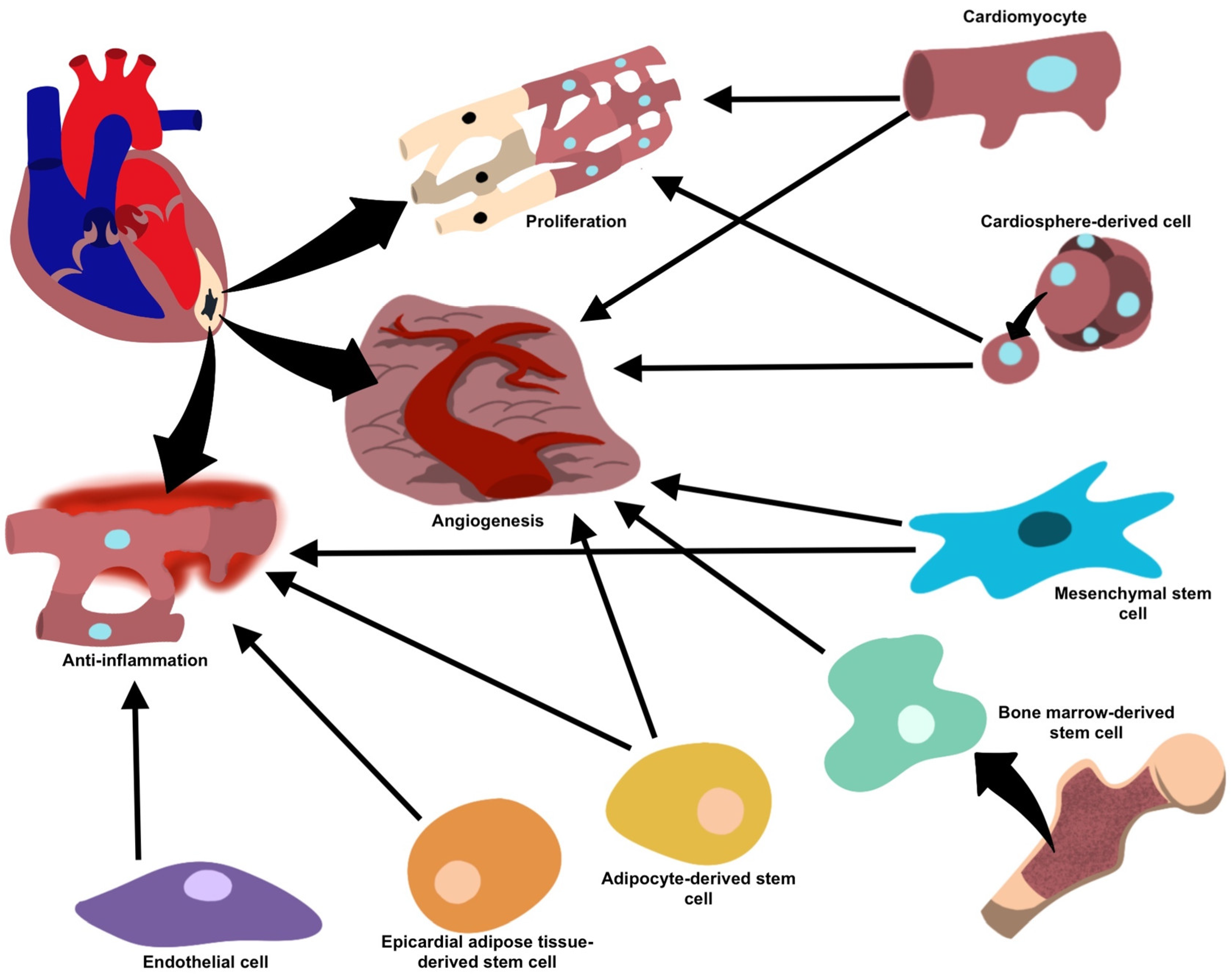
Epicardial adipose tissue-derived stem cells (EATDS) are a potential mesenchymal stem cell source for cardiac regeneration. The proximity of these cells to the epicardium and their similarities in their vascular supply with cardiac muscle makes them candidates to serve in cardiac regeneration pathways. Studies have shown that EATDS possess a greater capacity to differentiate into cardiomyocytes [47]. While there is much to be understood in the mechanisms and pathways of epicardial fat (EF) and its connections to cardiac-related disease, promising preliminary results elucidated phenotypic changes resulting in cardiac regeneration potential [48].
One study by Lambert et al. showed that EVs secreted by epicardial adipose cells showed higher levels of VEGF and reduced COL18A1, demonstrating the angiogenic potential of these cells [49]. Another study by Ozkaynak et al. observing the myocardial regenerative capacity of EATDS found that these cells demonstrated an increase in vascular density and a clinically significant increase in ejection fraction [47]. Additionally, EATDS appear to play a role in cell differentiation [29]. Yang et al. showed that EVs derived from these cells demonstrated adipogenic capabilities [47]. Exosomal–ribosomal proteins have been investigated for their effectiveness in modulating EATDS, revealing their pro-inflammatory, anti-inflammatory, proliferative, and non-proliferative properties, suggesting their cardiac regeneration potential [48]. Similar to BMSCs, it is unclear whether the reparative properties of these cells are due to the paracrine signaling of neighboring cells [47]. A summary of all the previously discussed cell types and their relationships can be found in Table 1 and Figure 1. Their promise as potential therapeutic targets warrants further investigation.
Figure 1. Target cells and their proposed mechanisms for vesicle-mediated cardiac repair.
Table 1. Cell sources and EV contents for cardiac regeneration.

Target Cell

Mechanism Of Repair/Regeneration and Mediators

References

Mesenchymal stem cells

Anti-inflammatory (miR-182, miR-233, miR-181c, miR-19a, miR-22, miR-199a, miR-214), anti-fibrotic (miR-19a, miR-29, miR-133) and pro-angiogenic (miR-126, miR-210, miR-20a, VEGF) processes facilitating repair and regeneration and inhibits the formation of fibrotic scar tissue.

[24]

Cardiomyocytes

Cell growth (miR-17, miR-20a, miR-23b) under normal conditions, and enhanced angiogenesis and decreased collagen deposition (miR-16, miR-19a, miR-19b, miR-23a, miR-23b) under stress.

[30]

Cardiosphere-derived cells

Pro-angiogenic and pro-apoptotic properties (miR-210, miR-132, miR-146a-3p).

[23]

Endothelial cells

Reduce inflammation and facilitate healing (integrin avβ6).

[37]

Adipose-derived stem cells

Anti-inflammatory properties (miR-126) prevent fibrosis and favor angiogenesis, facilitating repair.

[39]

Bone marrow-derived stem cells

Neovascularization and vasculogenesis, once implanted into ischemic cardiac tissue.

[40][41]

Epicardial adipose tissue-derived stem cells

Upregulation in regenerative properties and proliferative/anti-inflammatory proteins during periods of cellular stress or ischemia, as well as differentiation of cell types.

[48]

References

  1. Tzahor, E.; Poss, K.D. Cardiac Regeneration Strategies: Staying Young at Heart. Science 2017, 356, 1035–1039.
  2. Sheng, K.; Nie, Y.; Gao, B. Recent Advances in Myocardial Regeneration Strategy. J. Int. Med. Res. 2019, 47, 5453–5464.
  3. Kalluri, R.; LeBleu, V.S. The Biology, Function, and Biomedical Applications of Exosomes. Science 2020, 367, eaau6977.
  4. Joladarashi, D.; Kishore, R. Mesenchymal Stromal Cell Exosomes in Cardiac Repair. Curr. Cardiol. Rep. 2022, 24, 405–417.
  5. Shen, M.; Chen, T. Mesenchymal Stem Cell-Derived Exosomes and Their Potential Agents in Hematological Diseases. Oxid. Med. Cell Longev. 2021, 2021, 4539453.
  6. Lee, T.L.; Lai, T.C.; Lin, S.R.; Lin, S.W.; Chen, Y.C.; Pu, C.M.; Lee, I.T.; Tsai, J.S.; Lee, C.W.; Chen, Y.L. Conditioned Medium from Adipose-Derived Stem Cells Attenuates Ischemia/Reperfusion-Induced Cardiac Injury through the MicroRNA-221/222/PUMA/ETS-1 Pathway. Theranostics 2021, 11, 3131–3149.
  7. Sahoo, S.; Losordo, D. Cardiac Repair and Regeneration after MI: What Is Known? Circ. Res. 2014, 114, 333–344.
  8. Ong, S.G.; Wu, J.C. Exosomes as Potential Alternatives to Stem Cell Therapy in Mediating Cardiac Regeneration. Circ. Res. 2015, 117, 7–9.
  9. Kolios, G.; Moodley, Y. Introduction to Stem Cells and Regenerative Medicine. Respiration 2012, 85, 3–10.
  10. Matsuzaka, Y.; Yashiro, R. Therapeutic Strategy of Mesenchymal-Stem-Cell-Derived Extracellular Vesicles as Regenerative Medicine. Int. J. Mol. Sci. 2022, 23, 6480.
  11. Hegyesi, H.; Pallinger, É.; Mecsei, S.; Hornyák, B.; Kovácsházi, C.; Brenner, G.B.; Giricz, Z.; Pálóczi, K.; Kittel, Á.; Tóvári, J.; et al. Circulating Cardiomyocyte-Derived Extracellular Vesicles Reflect Cardiac Injury during Systemic Inflammatory Response Syndrome in Mice. Cell. Mol. Life Sci. 2022, 79, 84.
  12. Ju, Y.; Hu, Y.; Yang, P.; Xie, X.; Fang, B. Extracellular Vesicle-Loaded Hydrogels for Tissue Repair and Regeneration. Mater. Today Bio 2023, 18, 100522.
  13. Wang, X.; Gu, H.; Qin, D.; Yang, L.; Huang, W.; Essandoh, K.; Wang, Y.; Caldwell, C.C.; Peng, T.; Zingarelli, B.; et al. Exosomal MIR-223 Contributes to Mesenchymal Stem Cell-Elicited Cardioprotection in Polymicrobial Sepsis. Sci. Rep. 2015, 5, 13721.
  14. Wang, X.; Chen, Y.; Zhao, Z.; Meng, Q.; Yu, Y.; Sun, J.; Yang, Z.; Chen, Y.; Li, J.; Ma, T.; et al. Engineered Exosomes with Ischemic Myocardium-Targeting Peptide for Targeted Therapy in Myocardial Infarction. J. Am. Heart Assoc. 2018, 7, e008737.
  15. Wang, H.; Xie, Y.; Salvador, A.M.; Zhang, Z.; Chen, K.; Li, G.; Xiao, J. Exosomes: Multifaceted Messengers in Atherosclerosis. Curr. Atheroscler. Rep. 2020, 22, 57.
  16. Wang, X.; Bai, L.; Liu, X.; Shen, W.; Tian, H.; Liu, W.; Yu, B. Cardiac Microvascular Functions Improved by MSC-Derived Exosomes Attenuate Cardiac Fibrosis after Ischemia–Reperfusion via PDGFR-β Modulation. Int. J. Cardiol. 2021, 344, 13–24.
  17. Dong, J.; Wu, B.; Tian, W. Exosomes Derived from Hypoxia-Preconditioned Mesenchymal Stem Cells (HypoMSCs-Exo): Advantages in Disease Treatment. Cell Tissue Res. 2023, 392, 621–629.
  18. Wen, H.; Peng, L.; Chen, Y. The Effect of Immune Cell-Derived Exosomes in the Cardiac Tissue Repair after Myocardial Infarction: Molecular Mechanisms and Pre-Clinical Evidence. J. Cell. Mol. Med. 2021, 25, 6500–6510.
  19. Wu, Y.; Peng, W.; Fang, M.; Wu, M.; Wu, M. MSCs-Derived Extracellular Vesicles Carrying MiR-212-5p Alleviate Myocardial Infarction-Induced Cardiac Fibrosis via NLRC5/VEGF/TGF-Β1/SMAD Axis. J. Cardiovasc. Transl. Res. 2022, 15, 302–316.
  20. Fang, W.H.; Agrawal, D.K.; Thankam, F.G. Smart Exosomes: A Smart Approach for Tendon Regeneration. Tissue Eng. Part. B Rev. 2022, 28, 613–625.
  21. Wu, R.; Gao, W.; Yao, K.; Ge, J. Roles of Exosomes Derived from Immune Cells in Cardiovascular Diseases. Front. Immunol. 2019, 10, 648.
  22. Mihailovic, P.M.; Lio, W.M.; Herscovici, R.; Chyu, K.Y.; Yano, J.; Zhao, X.; Zhou, J.; Zhou, B.; Freeman, M.R.; Yang, W.; et al. Keratin 8 Is a Potential Self-Antigen in the Coronary Artery Disease Immunopeptidome: A Translational Approach. PLoS ONE 2019, 14, e0213025.
  23. Lazar, E.; Benedek, T.; Korodi, S.; Rat, N.; Lo, J.; Benedek, I. Stem Cell-Derived Exosomes—An Emerging Tool for Myocardial Regeneration. World J. Stem Cells 2018, 10, 106–115.
  24. Shafei, S.; Khanmohammadi, M.; Ghanbari, H.; Nooshabadi, V.T.; Tafti, S.H.A.; Rabbani, S.; Kasaiyan, M.; Basiri, M.; Tavoosidana, G. Effectiveness of Exosome Mediated MiR-126 and MiR-146a Delivery on Cardiac Tissue Regeneration. Cell Tissue Res. 2022, 390, 71–92.
  25. Nasser, M.I.; Masood, M.; Adlat, S.; Gang, D.; Zhu, S.; Li, G.; Li, N.; Chen, J.; Zhu, P. Mesenchymal Stem Cell-Derived Exosome MicroRNA as Therapy for Cardiac Ischemic Injury. Biomed. Pharmacother. 2021, 143, 112118.
  26. Sun, S.J.; Wei, R.; Li, F.; Liao, S.Y.; Tse, H.F. Mesenchymal Stromal Cell-Derived Exosomes in Cardiac Regeneration and Repair. Stem Cell Rep. 2021, 16, 1662–1673.
  27. He, C.; Zheng, S.; Luo, Y.; Wang, B. Exosome Theranostics: Biology and Translational Medicine. Theranostics 2018, 8, 237–255.
  28. Zhang, P.; Su, J.; Mende, U. Cross Talk between Cardiac Myocytes and Fibroblasts: From Multiscale Investigative Approaches to Mechanisms and Functional Consequences. Am. J. Physiol. Heart Circ. Physiol. 2012, 303, H1385–H1396.
  29. Yang, W.; Tu, H.; Tang, K.; Huang, H.; Ou, S.; Wu, J. MiR-3064 in Epicardial Adipose-Derived Exosomes Targets Neuronatin to Regulate Adipogenic Differentiation of Epicardial Adipose Stem Cells. Front. Cardiovasc. Med. 2021, 8, 709079.
  30. Wojciechowska, A.; Braniewska, A.; Kozar-Kamińska, K. MicroRNA in Cardiovascular Biology and Disease. Adv. Clin. Exp. Med. 2017, 26, 865–874.
  31. Mancuso, T.; Barone, A.; Salatino, A.; Molinaro, C.; Marino, F.; Scalise, M.; Torella, M.; De Angelis, A.; Urbanek, K.; Torella, D.; et al. Unravelling the Biology of Adult Cardiac Stem Cell-Derived Exosomes to Foster Endogenous Cardiac Regeneration and Repair. Int. J. Mol. Sci. 2020, 21, 3725.
  32. Smyth, T.; Kullberg, M.; Malik, N.; Smith-Jones, P.; Graner, M.W.; Anchordoquy, T.J. Biodistribution and Delivery Efficiency of Unmodified Tumor-Derived Exosomes. J. Control. Release 2015, 199, 145–155.
  33. Maring, J.A.; Beez, C.M.; Falk, V.; Seifert, M.; Stamm, C. Myocardial Regeneration via Progenitor Cell-Derived Exosomes. Stem Cells Int. 2017, 2017, 7849851.
  34. Wang, Y.; Wang, C.; Ma, J. Role of Cardiac Endothelial Cells-Derived MicroRNAs in Cardiac Remodeling. Discov. Med. 2019, 28, 95–105.
  35. Roncarati, R.; Viviani Anselmi, C.; Losi, M.A.; Papa, L.; Cavarretta, E.; Da Costa Martins, P.; Contaldi, C.; Saccani Jotti, G.; Franzone, A.; Galastri, L.; et al. Circulating MiR-29a, among Other up-Regulated MicroRNAs, Is the Only Biomarker for Both Hypertrophy and Fibrosis in Patients with Hypertrophic Cardiomyopathy. J. Am. Coll. Cardiol. 2014, 63, 920–927.
  36. Ono, K.; Kuwabara, Y.; Han, J. MicroRNAs and Cardiovascular Diseases. FEBS J. 2011, 278, 1619–1633.
  37. Song, J.; Chen, X.; Wang, M.; Xing, Y.; Zheng, Z.; Hu, S. Cardiac Endothelial Cell-Derived Exosomes Induce Specific Regulatory B Cells. Sci. Rep. 2014, 4, 7583.
  38. Lee, C.; Mitsialis, S.A.; Aslam, M.; Vitali, S.H.; Vergadi, E.; Konstantinou, G.; Sdrimas, K.; Fernandez-Gonzalez, A.; Kourembanas, S. Exosomes Mediate the Cytoprotective Action of Mesenchymal Stromal Cells on Hypoxia-Induced Pulmonary Hypertension. Circulation 2012, 126, 2601–2611.
  39. Luo, Q.; Guo, D.; Liu, G.; Chen, G.; Hang, M.; Jin, M. Exosomes from MiR-126-Overexpressing Adscs Are Therapeutic in Relieving Acute Myocardial Ischaemic Injury. Cell. Physiol. Biochem. 2017, 44, 2105–2116.
  40. Lee, N.; Thorne, T.; Losordo, D.W.; Yoon, Y.S. Repair of Ischemic Heart Disease with Novel Bone Marrow-Derived Multipotent Stem Cells. Cell Cycle 2005, 4, 861–864.
  41. Müller, P.; Lemcke, H.; David, R. Stem Cell Therapy in Heart Diseases-Cell Types, Mechanisms and Improvement Strategies. Cell. Physiol. Biochem. 2018, 48, 2607–2655.
  42. Azari, Z.; Nazarnezhad, S.; Webster, T.J.; Hoseini, S.J.; Brouki Milan, P.; Baino, F.; Kargozar, S. Stem cell-mediated Angiogenesis in Skin Tissue Engineering and Wound Healing. Wound Repair. Regen. 2022, 30, 421–435.
  43. Mangialardi, G.; Madeddu, P. Bone Marrow-Derived Stem Cells: A Mixed Blessing in the Multifaceted World of Diabetic Complications. Curr. Diabetes Rep. 2016, 16, 43.
  44. Xu, M.; Shaw, G.; Murphy, M.; Barry, F. Induced Pluripotent Stem Cell-Derived Mesenchymal Stromal Cells Are Functionally and Genetically Different From Bone Marrow-Derived Mesenchymal Stromal Cells. Stem Cells 2019, 37, 754–765.
  45. Korf-Klingebiel, M.; Reboll, M.R.; Klede, S.; Brod, T.; Pich, A.; Polten, F.; Napp, L.C.; Bauersachs, J.; Ganser, A.; Brinkmann, E.; et al. Myeloid-Derived Growth Factor (C19orf10) Mediates Cardiac Repair Following Myocardial Infarction. Nat. Med. 2015, 21, 140–149.
  46. Wang, Y.; Li, Y.; Feng, J.; Liu, W.; Li, Y.; Liu, J.; Yin, Q.; Lian, H.; Liu, L.; Nie, Y. Mydgf Promotes Cardiomyocyte Proliferation and Neonatal Heart Regeneration. Theranostics 2020, 10, 9100–9112.
  47. Özkaynak, B.; Şahin, I.; Özenc, E.; Subaşı, C.; Oran, D.S.; Totoz, T.; Tetikkurt, Ü.S.; Mert, B.; Polat, A.; Okuyan, E.; et al. Mesenchymal Stem Cells Derived from Epicardial Adipose Tissue Reverse Cardiac Remodeling in a Rabbit Model of Myocardial Infarction. Eur. Rev. Med. Pharmacol. Sci. 2021, 25, 4372–4384.
  48. Thankam, F.G.; Agrawal, D.K. Single Cell Genomics Identifies Unique Cardioprotective Phenotype of Stem Cells Derived from Epicardial Adipose Tissue under Ischemia. Stem Cell Rev. Rep. 2022, 18, 294–335.
  49. Lambert, C.; Arderiu, G.; Bejar, M.T.; Crespo, J.; Baldellou, M.; Juan-Babot, O.; Badimon, L. Stem Cells from Human Cardiac Adipose Tissue Depots Show Different Gene Expression and Functional Capacities. Stem Cell Res. Ther. 2019, 10, 361.
More
Information
Contributors MDPI registered users' name will be linked to their SciProfiles pages. To register with us, please refer to https://encyclopedia.pub/register : , , , , , ,
View Times: 150
Revisions: 2 times (View History)
Update Date: 02 Nov 2023
1000/1000