Submitted Successfully!
To reward your contribution, here is a gift for you: A free trial for our video production service.
Thank you for your contribution! You can also upload a video entry or images related to this topic.
Version Summary Created by Modification Content Size Created at Operation
1 -- 1757 2023-10-31 16:29:32 |
2 references update Meta information modification 1757 2023-11-01 04:23:18 |

Video Upload Options

Do you have a full video?

Confirm

Are you sure to Delete?
Cite
If you have any further questions, please contact Encyclopedia Editorial Office.
Nunes, L.G.A.; Cain, A.; Comyns, C.; Hoffmann, P.R.; Krahn, N. Elucidating Selenoprotein M Function from Structure. Encyclopedia. Available online: https://encyclopedia.pub/entry/51004 (accessed on 15 May 2024).
Nunes LGA, Cain A, Comyns C, Hoffmann PR, Krahn N. Elucidating Selenoprotein M Function from Structure. Encyclopedia. Available at: https://encyclopedia.pub/entry/51004. Accessed May 15, 2024.
Nunes, Lance G. A., Antavius Cain, Cody Comyns, Peter R. Hoffmann, Natalie Krahn. "Elucidating Selenoprotein M Function from Structure" Encyclopedia, https://encyclopedia.pub/entry/51004 (accessed May 15, 2024).
Nunes, L.G.A., Cain, A., Comyns, C., Hoffmann, P.R., & Krahn, N. (2023, October 31). Elucidating Selenoprotein M Function from Structure. In Encyclopedia. https://encyclopedia.pub/entry/51004
Nunes, Lance G. A., et al. "Elucidating Selenoprotein M Function from Structure." Encyclopedia. Web. 31 October, 2023.
Elucidating Selenoprotein M Function from Structure
Edit

Selenocysteine (Sec), the 21st amino acid, is structurally similar to cysteine but with a sulfur to selenium replacement. This single change retains many of the chemical properties of cysteine but often with enhanced catalytic and redox activity. Incorporation of Sec into proteins is unique, requiring additional translation factors and multiple steps to insert Sec at stop (UGA) codons. These Sec-containing proteins (selenoproteins) are found in all three domains of life where they often are involved in cellular homeostasis (e.g., reducing reactive oxygen species). The essential role of selenoproteins in humans requires us to maintain appropriate levels of selenium, the precursor for Sec, in our diet. Too much selenium is also problematic due to its toxic effects. Deciphering the role of Sec in selenoproteins is challenging for many reasons, one of which is due to their complicated biosynthesis pathway. However, clever strategies are surfacing to overcome this and facilitate production of selenoproteins.

selenocysteine SELENOM selenoprotein oxidoreductase

1. Introduction

Selenium acts as a double-edged sword: an essential micronutrient for humans that becomes toxic in excess [1]. Selenium is found in both organic (selenomethionine, selenocysteine (Sec)) and inorganic (selenite, selenate) forms, of which the latter is found to be more toxic to humans [2][3]. Sec, the 21st natural amino acid, is biosynthesized on its tRNA to convert inorganic selenium to an organic form, readily used by cells for protein translation [3]. Humans have 25 selenoproteins (proteins containing Sec) that are responsible for cellular function (e.g., redox reactions, immune response, thyroid hormone metabolism). The inability to express these selenoproteins (due to selenium deficiency) have been associated with diseases including cancer, neurodegenerative diseases, Keshan disease, inflammatory bowel diseases, and diabetes [1][4][5]. Selenium supplementation has been proposed to prevent and treat some of these diseases; however, there are also data that show that excess selenium or overexpression of selenoproteins is connected to disease (e.g., diabetes, neurodegenerative diseases) [6][7][8][9][10]. These conflicting results suggest that maximizing selenoprotein production through selenium supplementation is not always a solution and researchers are missing key information to properly prescribe selenium [11]. Part of this gap is due to lack of understanding on the role that Sec has in the cellular function of selenoproteins [12].
The enhanced chemical reactivity of Sec has been an advantage when studying selenoproteins in vivo. Probes have been designed to specifically recognize Sec over cysteine, and radioactive selenium can be fed to cultures to visualize incorporation [13]. Studying selenoproteins in vitro, however, is more challenging. This is because they follow a translation path that is unique to all other proteins. Sec is encoded by a nonsense codon (UGA), but only in the presence of specific regulatory elements to designate Sec insertion instead of termination. Downstream of the specific UGA codon, in the 3′-untranslated region (UTR) of eukaryotes, is an mRNA hairpin known as the Sec insertion sequence (SECIS) element. This hairpin structure is highly conserved [14] for interaction with SECIS-binding protein 2 (SBP2) and a specialized elongation factor (eEFSec). eEFSec can discriminate the unique structure of tRNASec from tRNAs for the other 20 amino acids, promoting elongation instead of termination [15][16]. Details of the translation mechanism involving these additional factors: SECIS element, SBP2, and eEFSec, are still not fully understood [17]. Moreover, the mechanism differs in each domain of life, adding to the complexity of feasibly overexpressing selenoproteins for detailed analysis of their cellular function [18].

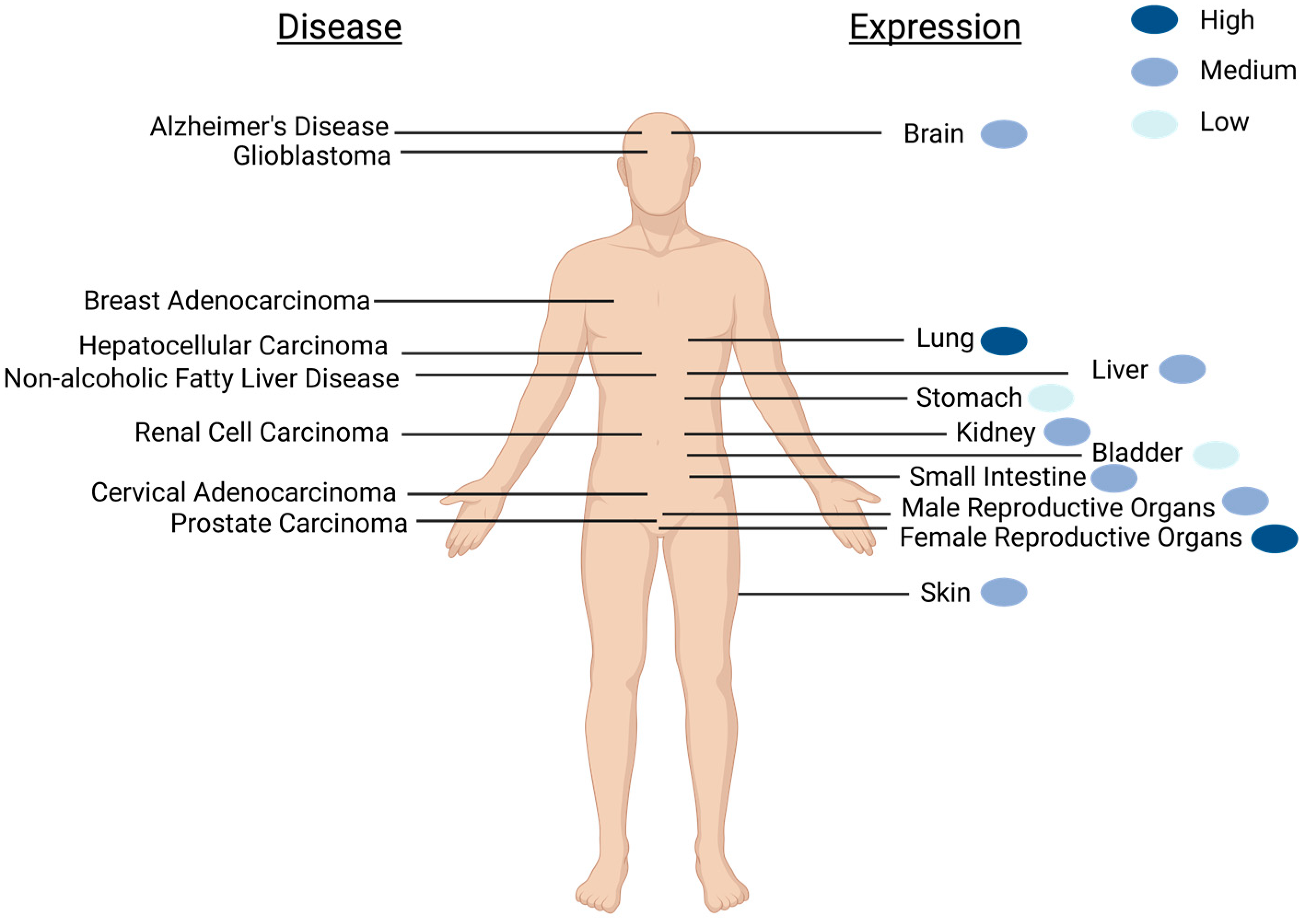
2. Expression of SELENOM Is Widespread

SELENOM is widely expressed throughout the body (e.g., heart, lung, kidney, stomach, small intestine, skin, testis, uterus, ovary, and brain), but not expressed in all tissue types (e.g., muscle and thymus) [19][20] (Figure 1). In the brain, SELENOM expression is extensive, detectable in multiple brain regions including the olfactory bulb, cortex, hippocampus, hypothalamus, brain stem, cerebellum, and cerebellar cortex lysates [20][21]. Immunohistochemistry staining for SELENOM distribution in mice coronal brain sections revealed SELENOM localization in multiple brain regions including the periventricular and arcuate nuclei of the hypothalamus; the ventral tegmental area; red nucleus; the CA1, CA2, and CA3 regions of the hippocampus; the medial septum; and the granular, Purkinje, and molecular layers of the cerebellum [21]. This high expression level of SELENOM in multiple brain regions suggests an important role in brain function.
Figure 1. Schematic of SELENOM expression and SELENOM-related diseases as found in humans. Diseases that correlate with aberrant SELENOM expression are shown on the left, while the location of major organs with detectable SELENOM expression is on the right. Relative expression levels of healthy individuals are portrayed with blue-colored dots. The widespread expression of SELENOM is not limited to what is depicted. Created with BioRender.com.
A global knockout of SELENOM in mice did not result in adverse cognitive or motor defects as one may have expected. Instead, metabolic dysregulation caused by diminished hypothalamic leptin signaling was observed [21][22]. Given that most of SELENOM is found in GABAergic cells, this observation made sense. However, one thing to consider when studying SELENOM in mice is that human expression levels are much lower and the distribution of selenoproteins differ between the organisms [23]. For example, mice have high expression levels of GPx1, GPx4, SELENOF, SELENOK, SELENOM, SELENOS, and SELENOW, while SELENOW and SELENOF are the highest expressed in humans [23]. Since the mechanisms by which many selenoproteins function are still not clear, researchers cannot rule out whether another selenoprotein in the brain compensates for cognitive and motor function in the absence of SELENOM.
Beyond the brain and examining the entirety of the human body, the Human Protein Atlas (https://www.proteinatlas.org/ENSG00000198832-SELENOM, accessed 14 March 2023) shows that generally SELENOM is expressed in the cytoplasm but localizes to the perinuclear region and nucleoplasm. SELENOM is also highly expressed in the thyroid gland, lungs, and female reproductive organs. The glandular system is most prominent for mRNA expression, with exocrine glandular cells expressing the highest levels. There is low cancer specificity for SELENOM; however, its expression in renal cell carcinoma (RCC) is unfavorable and used as a prognostic marker.

3. Elucidating SELENOM Function from Structure

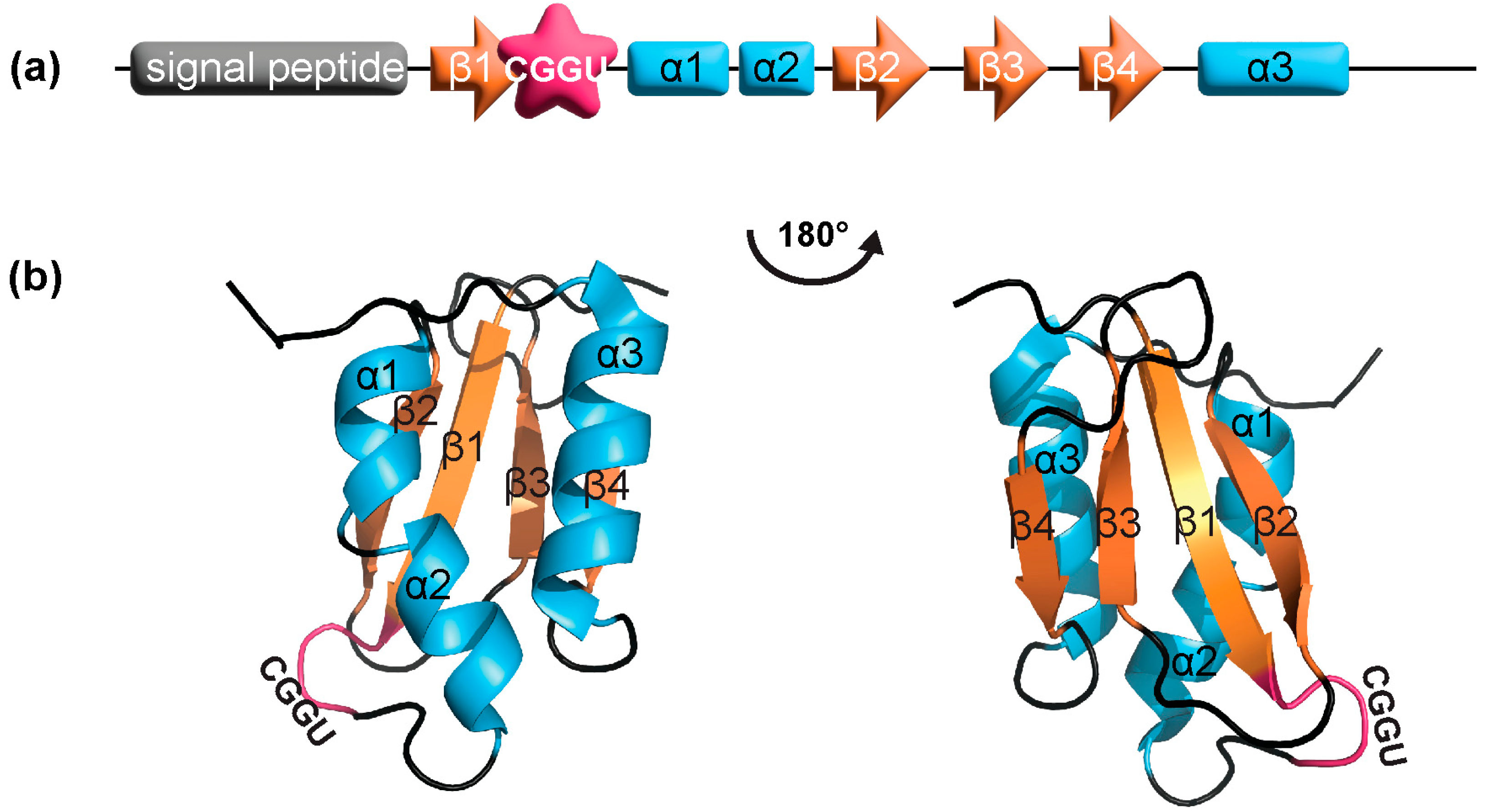
3.1. SELENOM Is Structurally Close to Thioredoxin

As with elucidating the function of a newly discovered protein, when studying a new selenoprotein, an initial step is to scan the structure space for similar proteins that have been previously characterized. Structurally, the closest relative to SELENOM is thioredoxin [24]. Thioredoxins are oxidoreductases with a defined thioredoxin (TXN)-fold identified by a two-layer α/β/α sandwich with a βαβββα secondary structure. Moreover, they harbor a CXXC active-site motif, where X can be any amino acid [25]. Through this CXXC motif, TXNs catalyze the reduction of disulfide bonds as part of a catalytic cycle involving thioredoxin reductase (TXNRD) or through activation by reaction oxygen species (ROS) [26][27] (Figure 2a). In SELENOM, the CXXC motif is found as CXXU, where U refers to Sec [19]. The similar chemistry between C and U suggests that this motif also serves as a redox center and SELENOM participates as an oxidoreductase.
Figure 2. Oxidoreductase pathways. (a) The thioredoxin (TXN) cycle illustrates how TXNRD, an essential selenoprotein, catalyzes the reduction of oxidized thioredoxin (TXN-S2) using NADPH as an electron donor. Reduced thioredoxin (TXN-(SH)2) plays a critical role in oxidizing proteins involved in cellular redox homeostasis, influencing processes like DNA synthesis, antioxidant defense, and apoptosis. (b) In the GPx pathway, GPx catalyzes the reduction of peroxides, including hydrogen peroxide (H2O2), using reduced glutathione (GSH) as an electron donor. The resulting oxidized glutathione (GSSG) can be converted back to GSH through the action of glutathione reductase (GR), ensuring a continuous supply for cellular antioxidant defense. From activity assays, SELENOM could play a role in reducing peroxides like GPx.
The redox active amino acids (C or U) in the CXXC/U motif are surface-accessible in SELENOM. This differs from TXN, but is seen in protein disulfide isomerases (PDIs). PDIs are also oxidoreductases that typically consist of two catalytic TXN-like domains (containing the CXXC motif), separated by two non-catalytic TXN-like domains. However, some PDIs only contain a single catalytic domain [28][29], which is analogous to what is observed in the structure of SELENOM. Furthermore, the position of the CXXU motif is located between the C-terminus of strand β1 and N-terminus of helix α1, which compares to what is found in both TXNs and PDIs [24].

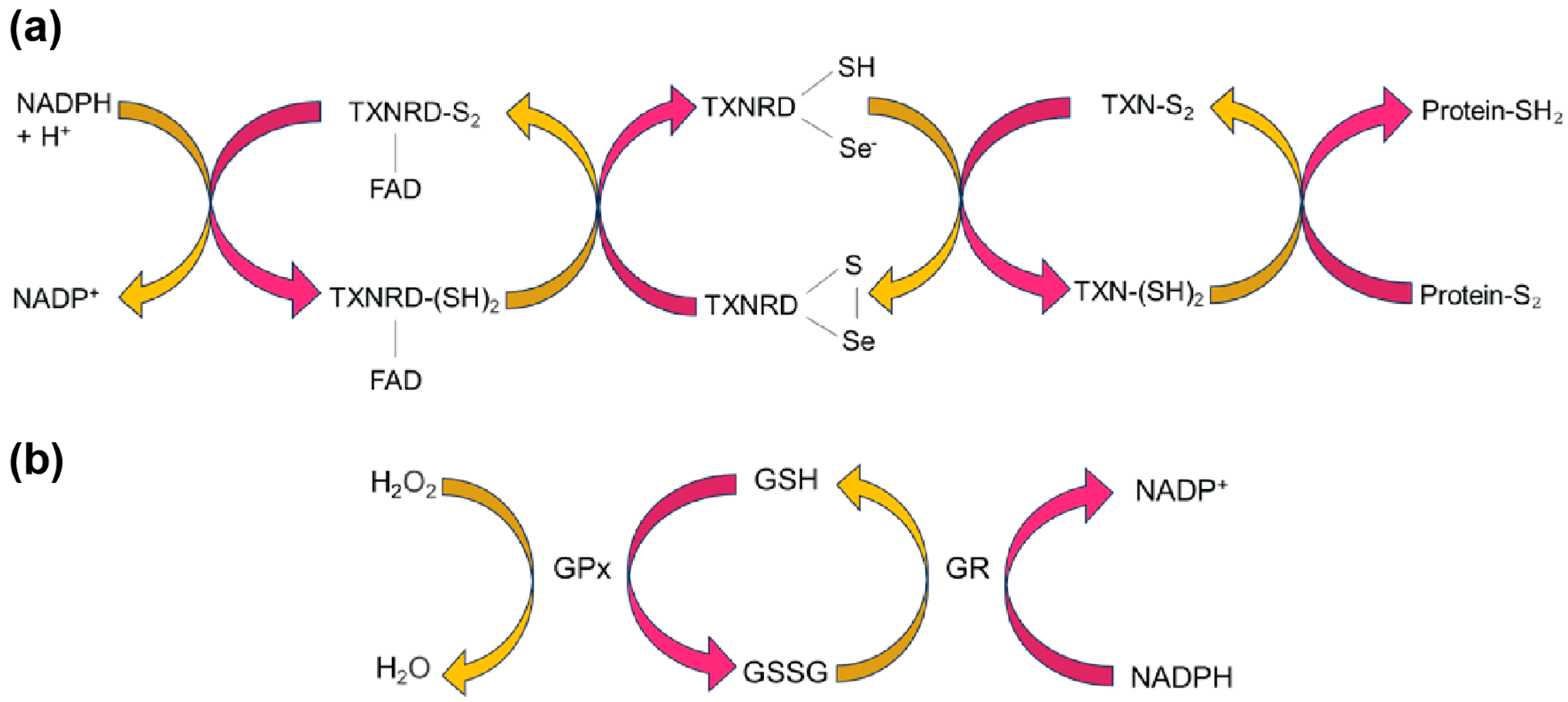
3.2. SELENOM Defines a New Thioredoxin Family

Another structural homolog found for SELENOM is SELENOF (previously named Sep15) [19][30]. Although their sequence identity is only 31%, SELENOF shares multiple regions of significant sequence identity to SELENOM [31]. The major similarity that distinguishes SELENOM and SELENOF from other TXN families is its unique TXN-like fold. Its central α/β domain, composed of three α-helices (α1-α3) and a mixed parallel/anti-parallel four-stranded β-sheet (β1-β4), represents the most basic TXN-like fold [32] (Figure 3). The CXXU motif located within this unique TXN-like fold is also unlike other oxidoreductases. While X refers to any amino acid, only certain amino acids are typically found in nature. These include GP, GH, and PH, found in TXNs, PDIs, and disulfide oxidases, respectively [24]. SELENOM has the sequence motif CGGU, which has only been observed thus far in SELENOF as CGU [24].
Figure 3. SELENOM (a) domain structure and (b) NMR structure (PDB: 2A2P). The first 25 residues are the endoplasmic reticulum signal peptide, which was removed for protein expression. Residues 25–34 and 121–145 are not shown in the structure due to high flexibility. The CXXU motif is shown in magenta, α-helices in cyan, and β-strands in orange.
In addition to a defining TXN-like fold, both SELENOM and SELENOF have other structural features that group them into a separate subfamily of TXNs. The conserved proline at the N-terminus of strand β3 is typically found in the cis-conformation [26], while in SELENOM and SELENOF, it is in the trans-conformation [25]. Furthermore, these proteins are missing a charge pair that in TXNs and PDIs are involved in proton transfer [27]. The functional importance of these structural differences remains unclear and is still under study.

3.3. SELENOM Does Not Bind UGGT

Among the many similarities between SELENOF and SELENOM, there are differences that potentially separate their functions. These differences lie at the termini. Both selenoproteins have an N-terminus that contains an ER-signaling sequence, which is subsequently cleaved during protein maturation. In SELENOF, an elongated cysteine-rich extension follows the signaling sequence prior to strand β1, while in SELENOM, this is a short extension. Furthermore, the C-terminus of SELENOM is a flexible extension, but it is short and unstructured in SELENOF [24]. The cysteine-rich extension at the N-terminus of SELENOF is known to mediate a high-affinity complex with the folding sensor of the calnexin cycle-UDP-glucose:glycoprotein glucosyltransferase (UGGT) [31][33]. The function of this binding interaction is not fully investigated, though it is suggested to be a PDI co-factor, assisting UGGT in assessing misfolded glycoproteins [24]. The absence of this cysteine-rich region in SELENOM confirmed that UGGT is not an interacting partner for SELENOM [31].

References

  1. Rayman, M.P. Selenium intake, status, and health: A complex relationship. Hormones 2020, 19, 9–14.
  2. Marschall, T.A.; Bornhorst, J.; Kuehnelt, D.; Schwerdtle, T. Differing cytotoxicity and bioavailability of selenite, methylselenocysteine, selenomethionine, selenosugar 1 and trimethylselenonium ion and their underlying metabolic transformations in human cells. Mol. Nutr. Food Res. 2016, 60, 2622–2632.
  3. Lazard, M.; Dauplais, M.; Blanquet, S.; Plateau, P. Recent advances in the mechanism of selenoamino acids toxicity in eukaryotic cells. Biomol. Concepts 2017, 8, 93–104.
  4. Speckmann, B.; Steinbrenner, H. Selenium and selenoproteins in inflammatory bowel diseases and experimental colitis. Inflamm. Bowel Dis. 2014, 20, 1110–1119.
  5. Avery, J.C.; Hoffmann, P.R. Selenium, selenoproteins, and immunity. Nutrients 2018, 10, 1203.
  6. Fairweather-Tait, S.J.; Filippini, T.; Vinceti, M. Selenium status and immunity. Proc. Nutr. Soc. 2022, 82, 32–38.
  7. Vinceti, M.; Filippini, T.; Jablonska, E.; Saito, Y.; Wise, L.A. Safety of selenium exposure and limitations of selenoprotein maximization: Molecular and epidemiologic perspectives. Environ. Res. 2022, 211, 113092.
  8. Urbano, T.; Filippini, T.; Lasagni, D.; De Luca, T.; Sucato, S.; Polledri, E.; Bruzziches, F.; Malavolti, M.; Baraldi, C.; Santachiara, A.; et al. Associations between urinary and dietary selenium and blood metabolic parameters in a healthy northern italy population. Antioxidants 2021, 10, 1193.
  9. Urbano, T.; Vinceti, M.; Mandrioli, J.; Chiari, A.; Filippini, T.; Bedin, R.; Tondelli, M.; Simonini, C.; Zamboni, G.; Shimizu, M.; et al. Selenoprotein P concentrations in the cerebrospinal fluid and serum of individuals affected by amyotrophic lateral sclerosis, mild cognitive impairment and Alzheimer’s Dementia. Int. J. Mol. Sci. 2022, 23, 9865.
  10. Huang, Y.C.; Combs, G.F., Jr.; Wu, T.L.; Zeng, H.; Cheng, W.H. Selenium status and type 2 diabetes risk. Arch. Biochem. Biophys. 2022, 730, 109400.
  11. Steinbrenner, H.; Duntas, L.H.; Rayman, M.P. The role of selenium in type-2 diabetes mellitus and its metabolic comorbidities. Redox Biol. 2022, 50, 102236.
  12. Rayman, M.P. Selenium and human health. Lancet 2012, 379, 1256–1268.
  13. Chung, C.Z.; Krahn, N. The selenocysteine toolbox: A guide to studying the 21st amino acid. Arch. Biochem. Biophys. 2022, 730, 109421.
  14. Labunskyy, V.M.; Hatfield, D.L.; Gladyshev, V.N. Selenoproteins: Molecular pathways and physiological roles. Physiol. Rev. 2014, 94, 739–777.
  15. Krahn, N.; Fischer, J.T.; Söll, D. Naturally occurring tRNAs with non-canonical structures. Front. Microbiol. 2020, 11, 596914.
  16. Serrão, V.H.B.; Silva, I.R.; da Silva, M.T.A.; Scortecci, J.F.; de Freitas Fernandes, A.; Thiemann, O.H. The unique tRNASec and its role in selenocysteine biosynthesis. Amino Acids 2018, 50, 1145–1167.
  17. Hilal, T.; Killam, B.Y.; Grozdanović, M.; Dobosz-Bartoszek, M.; Loerke, J.; Bürger, J.; Mielke, T.; Copeland, P.R.; Simonović, M.; Spahn, C.M.T. Structure of the mammalian ribosome as it decodes the selenocysteine UGA codon. Science 2022, 376, 1338–1343.
  18. Peng, J.J.; Yue, S.Y.; Fang, Y.H.; Liu, X.L.; Wang, C.H. Mechanisms affecting the biosynthesis and incorporation rate of selenocysteine. Molecules 2021, 26, 7120.
  19. Korotkov, K.V.; Novoselov, S.V.; Hatfield, D.L.; Gladyshev, V.N. Mammalian selenoprotein in which selenocysteine (Sec) incorporation is supported by a new form of Sec insertion sequence element. Mol. Cell. Biol. 2002, 22, 1402–1411.
  20. Zhang, Y.; Zhou, Y.; Schweizer, U.; Savaskan, N.E.; Hua, D.; Kipnis, J.; Hatfield, D.L.; Gladyshev, V.N. Comparative analysis of selenocysteine machinery and selenoproteome gene expression in mouse brain identifies neurons as key functional sites of selenium in mammals. J. Biol. Chem. 2008, 283, 2427–2438.
  21. Pitts, M.W.; Reeves, M.A.; Hashimoto, A.C.; Ogawa, A.; Kremer, P.; Seale, L.A.; Berry, M.J. Deletion of selenoprotein M leads to obesity without cognitive deficits. J. Biol. Chem. 2013, 288, 26121–26134.
  22. Gong, T.; Hashimoto, A.C.; Sasuclark, A.R.; Khadka, V.S.; Gurary, A.; Pitts, M.W. Selenoprotein M promotes hypothalamic leptin signaling and thioredoxin antioxidant activity. Antioxid. Redox Signal. 2021, 35, 775–787.
  23. Sasuclark, A.R.; Khadka, V.S.; Pitts, M.W. Cell-type specific analysis of selenium-related genes in brain. Antioxidants 2019, 8, 120.
  24. Ferguson, A.D.; Labunskyy, V.M.; Fomenko, D.E.; Araç, D.; Chelliah, Y.; Amezcua, C.A.; Rizo, J.; Gladyshev, V.N.; Deisenhofer, J. NMR structures of the selenoproteins Sep15 and SelM reveal redox activity of a new thioredoxin-like family. J. Biol. Chem. 2006, 281, 3536–3543.
  25. Oberacker, T.; Kraft, L.; Schanz, M.; Latus, J.; Schricker, S. The importance of thioredoxin-1 in health and disease. Antioxidants 2023, 12, 1078.
  26. Lu, J.; Holmgren, A. The thioredoxin antioxidant system. Free Radic. Biol. Med. 2014, 66, 75–87.
  27. Muri, J.; Kopf, M. The thioredoxin system: Balancing redox responses in immune cells and tumors. Eur. J. Immunol. 2023, 53, e2249948.
  28. Alanen, H.I.; Williamson, R.A.; Howard, M.J.; Lappi, A.K.; Jantti, H.P.; Rautio, S.M.; Kellokumpu, S.; Ruddock, L.W. Functional characterization of ERp18, a new endoplasmic reticulum-located thioredoxin superfamily member. J. Biol. Chem. 2003, 278, 28912–28920.
  29. Anelli, T.; Alessio, M.; Mezghrani, A.; Simmen, T.; Talamo, F.; Bachi, A.; Sitia, R. ERp44, a novel endoplasmic reticulum folding assistant of the thioredoxin family. EMBO J. 2002, 21, 835–844.
  30. Flowers, B.; Bochnacka, O.; Poles, A.; Diamond, A.M.; Kastrati, I. Distinct roles of SELENOF in different human cancers. Biomolecules 2023, 13, 486.
  31. Korotkov, K.V.; Kumaraswamy, E.; Zhou, Y.; Hatfield, D.L.; Gladyshev, V.N. Association between the 15-kDa selenoprotein and UDP-glucose:glycoprotein glucosyltransferase in the endoplasmic reticulum of mammalian cells. J. Biol. Chem. 2001, 276, 15330–15336.
  32. Qi, Y.; Grishin, N.V. Structural classification of thioredoxin-like fold proteins. Proteins 2005, 58, 376–388.
  33. Labunskyy, V.M.; Ferguson, A.D.; Fomenko, D.E.; Chelliah, Y.; Hatfield, D.L.; Gladyshev, V.N. A novel cysteine-rich domain of Sep15 mediates the interaction with UDP-glucose:glycoprotein glucosyltransferase. J. Biol. Chem. 2005, 280, 37839–37845.
More
Information
Contributors MDPI registered users' name will be linked to their SciProfiles pages. To register with us, please refer to https://encyclopedia.pub/register : , , , ,
View Times: 252
Revisions: 2 times (View History)
Update Date: 01 Nov 2023
1000/1000