Submitted Successfully!
To reward your contribution, here is a gift for you: A free trial for our video production service.
Thank you for your contribution! You can also upload a video entry or images related to this topic.
Version Summary Created by Modification Content Size Created at Operation
1 -- 3542 2023-10-19 13:24:06 |
2 The references in the sections 3-4 and 4 are corrected. Meta information modification 3542 2023-10-19 13:46:14 | |
3 Reference format revised. + 6 word(s) 3548 2023-10-24 10:17:46 | |
4 Ref 88 is replaced with Imakawa et al. and ref 104 is replaced with ref 88. Meta information modification 3548 2023-10-24 15:15:09 | |
5 Reference format revised. Meta information modification 3548 2023-10-25 10:51:40 |

Video Upload Options

Do you have a full video?

Confirm

Are you sure to Delete?
Cite
If you have any further questions, please contact Encyclopedia Editorial Office.
Kaneko-Ishino, T.; Ishino, F. Retrovirus-Derived RTL/SIRH Genes in Eutherian Developmental System. Encyclopedia. Available online: https://encyclopedia.pub/entry/50551 (accessed on 14 May 2024).
Kaneko-Ishino T, Ishino F. Retrovirus-Derived RTL/SIRH Genes in Eutherian Developmental System. Encyclopedia. Available at: https://encyclopedia.pub/entry/50551. Accessed May 14, 2024.
Kaneko-Ishino, Tomoko, Fumitoshi Ishino. "Retrovirus-Derived RTL/SIRH Genes in Eutherian Developmental System" Encyclopedia, https://encyclopedia.pub/entry/50551 (accessed May 14, 2024).
Kaneko-Ishino, T., & Ishino, F. (2023, October 19). Retrovirus-Derived RTL/SIRH Genes in Eutherian Developmental System. In Encyclopedia. https://encyclopedia.pub/entry/50551
Kaneko-Ishino, Tomoko and Fumitoshi Ishino. "Retrovirus-Derived RTL/SIRH Genes in Eutherian Developmental System." Encyclopedia. Web. 19 October, 2023.
Retrovirus-Derived RTL/SIRH Genes in Eutherian Developmental System
Edit

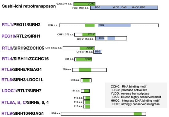
Eutherians have 11 retrotransposon Gag-like (RTL)/sushi-ichi retrotransposon homolog (SIRH) genes presumably derived from a certain retrovirus. Accumulating evidence indicates that the RTL/SIRH genes play a variety of roles in the mammalian developmental system, such as in the placenta, brain, and innate immune system, in a eutherian-specific manner. It has been shown that the functional role of Paternally Expressed 10 (PEG10) in placental formation is unique to the therian mammals, as are the eutherian-specific roles of PEG10 and PEG11/RTL1 in maintaining the fetal capillary network and the endocrine regulation of RTL7/SIRH7 (aka Leucine Zipper Down-Regulated in Cancer 1 (LDOCK1)) in the placenta. In the brain, PEG11/RTL1 is expressed in the corticospinal tract and hippocampal commissure, mammalian-specific structures, and in the corpus callosum, a eutherian-specific structure. Unexpectedly, at least three RTL/SIRH genes, RTL5/SIRH8, RTL6/SIRH3, and RTL9/SIRH10, play important roles in combating a variety of pathogens, namely viruses, bacteria, and fungi, respectively, suggesting that the innate immunity system of the brain in eutherians has been enhanced by the emergence of these new components. 

PEG10 PEG11/RTL1 RTL/SIRH genes placenta brain innate immunity

1. Introduction

Paternally expressed 10 (PEG10) [1] and PEG11/Retrotransposon Gag-like 1 (RTL1) [2], together with syncytin [3][4], opened the door to a new field of research on retrovirus-derived genes in mammalian development and evolution. The fact that these are all essential endogenous genes despite their retroviral origin [3][4][5][6][7][8] has had a huge impact not only on genome biology but also on developmental and evolutionary biology, because retrotransposons, including endogenous retroviruses (ERVs), had long been construed to be “junk” in the mammalian genome.
The discovery of retroviral envelop (ENV)-derived syncytin in primates stimulated the search for similar genes in primates as well as other lineages of eutherians and marsupials [9][10][11][12][13][14][15][16][17][18]. The discovery of essential functions for the retroviral GAG and POL-derived PEG10 and PEG11/RTL1 [5][6][19] led to the further screening of RTL/sushi-ichi retrotransposon homolog (SIRH) genes in eutherians (Figure 1) [5][20][21][22].
Figure 1. RTL/SIRH proteins in mice. There are 11 retrotransposon Gag-like/sushi-ichi retrotransposon homologs (RTL/SIRH) genes in eutherians that encode proteins with homology to the sushi-ichi retrotransposon GAG (green) and POL (light blue). The mouse RTL/SIRH proteins are shown as representative examples. The CCHC RNA binding motif and/or the DSG viral protease motif are conserved in certain RTL/SIRH genes. Purple: formal name, black: aliases. Sushi-ich was originally isolated from Takifugu rubripes [23][24] and belongs to the vertebrate-like Ty3/Gypsy, the V-clade in the chromovirus lineage [25].

2. Discovery of PEG10 and PEG11/RTL1 from Genomic Imprinting Research

Genomic imprinting, a term which describes the functional differences between the paternally and maternally derived genomes, was discovered by pronuclear transplantation experiments in mice in 1984. Three groups independently demonstrated that parthenogenetic embryos with two maternally derived genomes exhibit early embryonic lethality due to severe placental defects, while androgenetic embryos with two paternally derived genomes exhibit severe embryonic growth retardation associated with an overgrown placenta [26][27][28]. Extensive genetic analysis of mice with Robertsonian translocations of specific chromosomal loci also revealed functional differences between the parental chromosomes associated with early to late embryonic and postanal lethality as well as growth abnormalities [29][30]. The presence of imprinted genes with monoallelic paternal or maternal expression causes these genomic imprinting phenotypes [31][32][33][34][35].
PEG10 and PEG11/RTL1 have been identified as paternally expressed imprinted genes on human chromosome 7q21 and its orthologous mouse proximal chromosome 6 [1][36], and on the distal end of sheep chromosome 18 [2], respectively. It is known that maternal duplication of proximal chromosome 6 results in early embryonic lethality [30]. Peg10 is responsible for this early embryonic lethal phenotype as well as parthenogenetic death due to severe placental dysplasia [5]. An inheritable form of muscular hypertrophy, called the callipyge phenotype, is mapped to the distal end of sheep chromosome 18 [37]. In humans and mice, paternal and maternal duplication of its orthologous imprinted region, human chromosome 14 and mouse distal chromosome 12, cause Kagami–Ogata (KOS14) and Temple syndromes (TS14), two genomic imprinting disorders, and late embryonic/neonatal lethal phenotypes associated with growth abnormalities, respectively [30][38][39][40][41][42]. PEG11/RTL1, together with Delta-like 1 homologue (DLK1), are the major genes responsible for KOS14 and TS14 as well as the abnormal phenotypes in mice caused by paternal and maternal duplication of distal chromosome 12 [6][18][43][44][45][46][47].
Both PEG10 and PEG11/RTL1 have homology to GAG and POL of the sushi-ichi long terminal repeat (LTR) retrotransposon [1][2][23][24][48]. Therefore, they were originally thought to be derived from the sushi-ichi retrotransposon, and, thus, were named RTL and/or SIRH. However, it is reasonable to assume that they were originally derived from the GAG and POL of a certain extinct retrovirus having a high degree of homology to the suchi-ichi retrotransposon [49][50], since PEG10 arose in a common therian ancestor and PEG11/RTL1 and the other RTL/SIRH genes also arose in a common eutherian ancestor [22][51][52], while the gypsy retrotransposon, which includes the sushi-ichi retrotransposon, is an infectious retrovirus in Drosophila melanogaster [53][54].

2.1. Roles of PEG10 and PEG11/RTL1 in Placental Evolution in Mammals

The PEG10 protein is expressed in all of the trophoblast cell lines in the placenta. Paternal transmission of the Peg10 KO allele (hereafter referred to as Peg10 KO) causes early embryonic lethality due to poor placental growth associated with a complete lack of the labyrinth and spongiotrophoblast layers, because only the paternal allele of Peg10 is active, while its maternal allele is repressed by the genomic imprinting mechanism [5]. As the labyrinth layer is an essential part of the placenta, where nutrient and gas exchange occur between fetal and maternal blood cells, Peg10 KO embryos cannot grow beyond 9.5 days post coitus (dpc).
Paternal transmission of the Peg11/RTL1 KO allele (hereafter referred to as Peg11/Rtl1 Pat-KO) causes late fetal/neonatal lethality associated with late fetal growth retardation, while maternal transmission of the Peg11/RTL1 KO allele (hereafter referred to as Peg11/Rtl1 Mat-KO) causes neonatal lethality associated with abnormal fetal growth due to the overexpression of Peg11/Rtl1 [6][45] This is because of the presence of maternally expressed antiPeg11/antiRtl1, a non-coding RNA encoding 7 microRNAs (miRNAs) that target Peg11/Rtl1 mRNA via an RNAi mechanism [55][56][57]. The PEG11/RTL1 protein is restricted to expression in the endothelial cells of the fetal capillaries in the labyrinth layer of the placenta. Severe abnormalities of the fetal capillaries were observed in both the Peg11/Rtl1 Pat- and Mat-KO placenta.

2.2. Roles of PEG11/RTL1 in Muscle Development

PEG11/RTL1 is one of the major causative genes for the imprinting diseases KOS14 and TS14, which are caused by abnormal regulation of the imprinting region, that is, paternal and maternal disomy of human chromosome 14, respectively [19][38][39][40][41][42]. The former is characterized by neonatal lethality with respiratory failure, placentomegaly, polyhydramnios, developmental delay and/or intellectual disability, as well as feeding difficulties [39][41], whereas the latter is characterized by prenatal and postnatal growth retardation, feeding difficulties, muscle hypotonia, motor delay, early onset of puberty, and mild intellectual disability [40][42]. Their phenotypes are quite different, but their sites of damage are quite similar, i.e., in the muscle and brain as well as the placenta.
Importantly, Peg11/Rtl1 Mat- and Pat-KO mice that, respectively, overexpress and lack PEG11/RTL1 expression, are very good models for KOS14 and TS14, not only in the placenta [6][19] but also in the muscle and brain as well (see next section) [43][44]. In skeletal muscle, expression of the PEG11/RTL1 protein is restricted to the late fetal and neonatal stages, and is, therefore, not detectable after 2 weeks of age, even in adults [43]. In the neonatal stage, Peg11/Rtl1 Mat-KO mice had a significantly larger muscle fiber size, while Peg11/Rtl1 Pat-KO mice had significantly thinner muscle fibers. However, after fixation, the muscle fibers of the Peg11/Rtl1 Mat-KO mice exhibited severe shrinkage and detachment from the extracellular matrix (ECM) muscle, indicating that it is immature and more fragile than normal muscle.
In myocytes, PEG11/RTL1 partially colocalizes with DESMIN, a component of the sarcomere cytoskeleton that connects the sarcomere to membranes of the sarcolemma and the nucleus at the Z-disc [43], thus, acting as the force-generating machinery in muscle. This suggests that PEG11/RTL1 plays some role in stabilizing the muscle contractile apparatus and/or regulating muscle contraction in the fetal/neonatal muscle fibers.
It should be noted that PEG11/RTL1 and DLK1 are the major genes that cause the sheep callipyge phenotype because PEG11/RTL1 was first identified in the course of the sheep callipyge study [2]. Both PEG11/RTL1 and DLK1 are critically involved in muscle development, and studies in transgenic mice also support this conclusion [58][59]. In sheep, PEG11/RTL1 expression is relatively high until the late fetal stage, declines from just before birth, and is barely expressed after birth. The callipyge mutation recapitulates the normal fetal-like PEG11/RTL1 expression program during postnatal development, and this may contribute to the emergence of the muscle hypertrophy phenotype [60].

2.3. PEG10 and PEG11/RTL1 in Neurological Disorders

KOS14 and TS14 patients exhibit certain neurodevelopmental disorders, such as developmental delay and/or intellectual disability and feeding difficulties in the former [39][41], and feeding difficulties, motor delay, early onset of puberty and mild intellectual disability in the latter [40][42]. DLK1 is critically involved in the early onset of puberty in TS14 [46][47], while PEG11/RTL1 is responsible for the other neurodevelopmental phenotypes in these patients, as Peg11/Rtl1 Mat- and Pat KO mice, which overexpress and lack PEG11/RTL1 expression, provide strong evidence for this conclusion [44].
As in the case with muscle, Peg11/Rtl1 mRNA expression in the central nervous system is restricted during the fetal to neonatal period, but at lower levels, and is barely detectable in adults. The PEG11/RTL1 protein is detected in the descending tracts, commissural fibers including the hippocampal commissure and corpus callosum, as well as the limbic system, i.e., the hippocampal fimbria, fornix, and medial amygdala nucleus [44]. The corticospinal tract, one of the descending tracts, and the hippocampal commissure are mammalian-specific brain structures, whereas the corpus callosum is a eutherian-specific brain structure [61][62][63]. The corticospinal tract runs from layer V of the neocortex to the brainstem and spinal cord and is responsible for fine voluntary skilled muscle movements of the limbs, while the hippocampal commissure is involved in hippocampal-dependent memory output [44][61][64]. The corpus callosum is responsible for communication between the two hemispheres enabling faster transmission and integration of information from both sides [61][62][63]. These results suggest that PEG11/RTL1 is deeply involved in the functional evolution of the eutherian brain. 
Peg11/Rtl1 Mat- and Pat-KO mice exhibit neurodevelopmental abnormalities corresponding to these expression sites, such as decreased spontaneous movement, increased anxiety-like behavior, and learning and memory impairments [44]. These symptoms suggest impairment of the corticospinal tract involved in trunk and limb movement, and/or the corpus callosum, hippocampal commissure, and medial amygdala nucleus. It is likely that they are also associated with the developmental delay and intellectual disability observed in KOS14 and TS14 patients [44].
PEG10 has also been implicated in another neurological disorder, amyotrophic lateral sclerosis (ALS), a fatal neurodegenerative disease characterized by progressive loss of motor function, typically in middle age [65]. UBQLN2, a member of the ubiquilin family involved in proteasomal degradation, is the gene responsible for familial ALS [66]. Whiteley and colleagues demonstrated that UBQLN2 facilitates the proteasome-dependent degradation of the PEG10-ORF1/2 protein, and that PEG10-ORF1/2 is specifically upregulated in the spinal cord of ALS patients compared to healthy controls [67][68].

3. Retrovirus-Derived RTL/SIRH Genes as Eutherian-Specific Genes

3.1. RTL7/SIRH7 in the Placenta

In addition to maternal–fetal exchange and maternal tolerance of feto-paternal antigens, the placenta is a major endocrine organ during pregnancy [69]. RTL7/SIRH7 (formal name: Leucine Zipper Down-Regulated in Cancer 1 (LDOC1)) is another essential placental gene, like PEG10 and PEG11/RTL1. It is expressed in trophoblast lineages in early placental development and regulates trophoblast differentiation, and, thus, is deeply involved in several types of hormone production in trophoblast cells during pregnancy [70][71]. Rtl7/Sirh7 KO placentas have an irregular boundary between the spongiotrophoblast and labyrinth layers, as well as a decreased number of SpTs, although the fetuses appear normal. Female Rtl7/Sirh7 KO exhibit delayed parturition due to residual progesterone (P4) in the serum on 18.5 dpc, one day before the parturition, and pups die due to inadequate maternal care [72]. Since all pups are viable when foster mothers are used, it is indicated that the problems are due to the KO mothers.
As Rtl7/Sirh7 is an X-linked gene, maternal transmission of the Rtl7/Sirh7 KO allele results in a null phenotype in the placenta due to imprinted X-inactivation in the mouse placenta [73][74]. In Rtl7/Sirh7 KO conceptuses, their placentas overproduce placental P4, leading to a delayed transition from placental lactogen (PL)1 to PL2 in the giant trophoblast cells in the KO placenta, which presumably in turn leads to delayed downregulation of maternal ovarian P4 production in the late pregnancy, resulting in delayed parturition.
Although the ovary was thought to be the major P4 producing organ in rodents throughout gestation [69], P4 production is observed in rodent placenta during 9.5 to 11.5 dpc when a temporal reduction in serum P4 level occurs due to a shift from the corpus luteum of pseudopregnancy to pregnancy [75][76], strongly indicating that placental P4 plays an important role in the maintenance of gestation at this critical stage and that Rtl7/Sirh7 plays an important role in placental P4 production [72].

3.2. RTL4/SIRH11 in the Brain

RTL4/SIRH11 (aka Zinc Finger CCHC Domain-Containing Protein 16 (ZCCHC16) is a causative gene in autism spectrum disorders (ASD) [77]. Lim et al. performed a comprehensive screening of patients with ASD and identified a family with a rare nonsense ZCCHC16 mutation leading to ASD in a male proband and his male sibling, as it is an X-linked gene [77].
In mice, Sirh11/Zcchc16 KO mice exhibit increased impulsivity and decreased spatial memory, presumably due to low recovery of noradrenaline in the frontal cortex although they do not exhibit lethality or growth abnormalities [78]. They do not adapt to routine processes and tend to exhibit extreme behavior when transferred to a new environment. They display agitated movements in their cages when staff personnel enter the breeding room, and sometimes jump out when the cages are changed, even after a long breeding period. In particular, they remained hyperactive for 5 consecutive nights in the home cage activity test, while normal control mice gradually settled to lower levels of activity. In the light/dark transition test, the latency before entering into the light chamber was significantly decreased, while the number of transitions was significantly increased, suggesting a reduced attention and/or enhanced impulsivity. In the Y-maze test, KO mice exhibited a lower success rate, suggesting that they have a poor working memory. This is likely due to low noradrenaline (NA) recovery in the frontal cortex, because the locus coeruleus (LC) NA neurons have been reported to play important roles in attention, behavioral flexibility, and modulation of cognition [79][80][81] and their activation occurs in concert with the cognitive shifts that facilitate dynamic reorganization of target neural networks, allowing rapid behavioral adaptation to the demands of changing environmental demands [82], indicating that all of the behavioral defects of the Sirh11/Zcchc16 KO mice are somehow related to a dysregulation of the noradrenergic system in the brain [78].
RTL4/SIRH11 is a very important gene in neurodevelopment, and it is likely that it confers a critically important advantage both in the competition that occurs in daily life and in the evolution of the eutherian brain. However, because RTL4/SIRH11 expression is very low in the brain in both humans and mice, it remains unclear exactly where the RTL4/SIRH11 protein is expressed and what its function is.

3.3. RTL8A, 8B, 8C/SIRH5, 6, 4 in the Brain

RTL8A, 8B, 8C/SIRH5, 6, 4 are triplet genes that encode almost identical proteins of 112 to 113 amino acids (aa). Their number (2–4, mostly 3, excluding pseudogenes) and aa sequence are well conserved in eutherians, suggesting they confer an evolutionary advantage. Therefore, it is of interest to know why they exist as multiple genes and what their function is. However, in most cases the RTL8A, 8B, 8C/SIRH5, 6, 4 genes within the same species exhibit higher homology to each other than other species, suggesting that they are not in a precise orthologous relationship in eutherians, presumably due to independent gene conversion events in each species.
It was recently reported that the RTL8A, 8B, 8C/SIRH5, 6, 4 proteins accumulated together with the PEG10 protein in the neuronal cells differentiated from iPS cells of AS patients [83]. AS is a neurodevelopmental genomic imprinting disorder which is characterized by delayed development, intellectual disability, severe speech impairment, ataxia, and other symptoms [84][85]. It is caused by paternal uniparental disomy of chromosome 15 and/or mutations of a maternally expressed UBE3A gene. This implies that the RTL8A, B, C/SIRH5, 6, 4, and PEG10 proteins are directly targeted by UBE3A in neuronal cells.

3.4. RTL6/SIRH3, RTL5/SIRH8 and RTL9/SIRH10 Are Microglial Genes in the Brain

RTL6/SIRH3 (aka LDOCKL), which encodes an extremely basic protein (pI = 11.15), is the most conserved gene among the RTL/SIRH genes, with a non-synonymous/synonymous (dn/ds) rate of less than 0.1. Despite its evolutionary importance, it has been very difficult to identify the RTL6/SIRH3 protein because of the lack of effective antibodies. Analysis of Rtl6-CV KI mice, in which a Venus ORF is integrated into the endogenous Rtl6/Sirh3 locus immediately after the C-terminus, demonstrated that the RTL6/SIRH3 protein is expressed in the central nervous system during development and that it is secreted by microglia and responds to lipopolysaccharide (LPS). It was subsequently demonstrated in Rtl6/Sirh3 KO mice that the RTL6/SIRH3 protein functionally protects against bacteria by removing  LPS [49]. Thus, the Rtl6/Sirh3 gene plays an important role in the innate immune system in the brain.
RTL5/SIRH8 (aka Retrotransposon Gag Domain-Containing Protein (RGAG4)) is phylogenetically related to RTL6/SIRH3 and well conserved in eutherians, despite there being some exceptions. It encodes a larger protein which covers the entire RTL6/SIRH3 and is strongly acidic (pI = 4.39), and functions as another microglial gene in the innate immune system against viruses by removing double-stranded RNA from the brain [49]. Analysis of Rtl5-CmC KI mice in which an mCherry ORF is integrated into the endogenous Rtl5/Sirh8 locus immediately after the C-terminus demonstrated that the RTL5/SIRH8 protein is also expressed in the brain and that the RTL5/SIRH8 protein is likewise secreted by microglia and responds to double-stranded (ds) RNA.

Using the same approach of combining Venus KI mice and KO mice, RTL9/SIRH10 (aka RGAG1) was demonstrated to be another microglial gene that is actively protective against fungi by reacting to zymosan, the cell wall of fungi [86]. It encodes a large protein comprising two herpes virus-derived domains in addition to the GAG-like domain. The role of the first two regions remains unknown, but the latter is essential for zymosan removal. Unlike RTL6/SIRH3 and RTL5/SIRH8, RTL9/SIRH10 is restricted to the lysosomes of microglia, where the zymosan is ultimately taken up and degraded. Thus, at least three RLT/SIRH genes are involved in the clearance of bacterial, viral, and fungal pathogens from the brain, suggesting that these genes must have critically contributed to the evolution of the innate immune system in eutherians [87][86].

3.5. Relationship between RTL/SIRH Genes and Extraembryonic Tissues

All of these studies clearly demonstrate that domestication of retrovirus-derived genes made important contributions to the generation of eutherian-specific features in the placenta and brain. Microglia originate from the yolk sac during early development and eventually become permanently resident in the brain. Therefore, it is of interest to notice that the placenta and yolk sac, the extraembryonic tissues in intrauterine development, evidently serve as origin sites for the incubation of such retrovirus-derived genes, including both PEG10 and PEG11/RTL1, in the course of mammalian evolution [22][34][49][88]. It is known that the extraembryonic tissues have lower levels of DNA methylation than the embryos; therefore, endogenous retroviruses and retrotransposons can express there even at a low level. This may have promoted the domestication (exaptation) of retrovirus-derived genes in eutherians.

4. Two Types of Exapted Genes: Acquired RTL/SIRH Genes and Captured Syncytin Genes

PEG10, PEG11/RTL1, and other RTL/SIRH genes are thought to be derived from GAG and POL of an extinct retrovirus. PEG10 emerged between 164 and 148 MYA, after the diversification from monotremes and before the split between eutherians and marsupials [51], while PEG11/RTL1 and the other RTL/SIRH genes emerged between 148 and 120 MYA, after the diversification from marsupials and before the emergence of common eutherian ancestor [22][52]. They completely lost LTR sequences at both ends and also an ENV gene, which were present at the time of the original retroviral insertions. Their encoded proteins share 20 to 30% homology to the sushi-ichi retrotransposon but are completely different proteins from the original GAG and POL, each with its own unique aa sequence and novel function. Hence, they are referred to as genes acquired from a retrovirus [22].
In contrast, ENV-derived syncytins were sequentially domesticated in a lineage-specific manner after the establishment of the eutherians. For example, syncytin-1 and syncytin-2 emerged 40 and 20 MYA in primates from different retroviruses, respectively [3][4][9]. They retain almost all of the ENV sequences of the original retrovirus and, therefore, have fusion activity, which is important for syncytiotrophoblast cell fusion in the placenta. The LTRs, GAG, and POL sequences often persist in a remnant form due to severe mutations. Therefore, it is hypothesized that the syncytin gene was replaced several times in each eutherian lineage by a newly integrated ENV gene having superior fusion activity [88][89]. Therefore, they are called captured genes [89]. Thus, there are two distinct ways to domesticate retrovirus-derived genes.

References

  1. Ono, R.; Kobayashi, S.; Wagatsuma, H.; Aisaka, K.; Kohda, T.; Kaneko-Ishino, T.; Ishino, F. A retrotransposon-derived gene, PEG10, is a novel imprinted gene located on human chromosome 7q21. Genomics 2001, 73, 232–237.
  2. Charlier, C.; Segers, K.; Wagenaar, D.; Karim, L.; Berghmans, S.; Jaillon, O.; Shay, T.; Weissenbach, J.; Cockett, N.; Gyapay, G.; et al. Human-ovine comparative sequencing of a 250-kb imprinted domain encompassing the callipyge (clpg) locus and identification of six imprinted transcripts: DLK1, DAT, GTL2, PEG11, antiPEG11, and MEG8. Genome Res. 2001, 11, 850–862.
  3. Blond, J.-L.; Lavillette, D.; Cheynet, V.; Bouton, O.; Oriol, G.; Chapel-Fernandes, S.; Mandrand, B.; Mallet, F.; Cosset, F.-L. An envelope glycoprotein of the human endogenous retrovirus HERV-W is expressed in the human placenta and fuses cells expressing the type D mammalian retrovirus receptor. J. Virol. 2000, 74, 3321–3329.
  4. Mi, S.; Lee, X.; Li, X.-P.; Veldman, G.M.; Finnerty, H.; Racie, L.; LaVallie, E.; Tang, X.-Y.; Edouard, P.; Howes, S.; et al. Syncytin is a captive retroviral envelope protein involved in human placental morphogenesis. Nature 2000, 403, 785–789.
  5. Ono, R.; Nakamura, K.; Inoue, K.; Naruse, M.; Usami, T.; Wakisaka-Saito, N.; Hino, T.; Suzuki-Migishima, R.; Ogonuki, N.; Miki, H.; et al. Deletion of Peg10, an imprinted gene acquired from a retrotransposon, causes early embryonic lethality. Nat. Genet. 2006, 38, 101–106.
  6. Sekita, Y.; Wagatsuma, H.; Nakamura, K.; Ono, R.; Kagami, M.; Wakisaka, N.; Hino, T.; Suzuki-Migishima, R.; Kohda, T.; Ogura, A.; et al. Role of retrotransposon-derived imprinted gene, Rtl1, in the feto-maternal interface of mouse placenta. Nat. Genet. 2008, 40, 243–248.
  7. Dupressoir, A.; Vernochet, C.; Bawa, O.; Harper, F.; Pierron, G.; Opolon, P.; Heidmann, T. Syncytin-A knockout mice demonstrate the critical role in placentation of a fusogenic, endogenous retrovirus-derived, envelope gene. Proc. Natl. Acad. Sci. USA 2009, 106, 12127–12132.
  8. Dupressoir, A.; Vernochet, C.; Harper, F.; Guegan, J.; Dessen, P.; Pierron, G.; Heidmann, T. A pair of co-opted retroviral envelope syncytin genes is required for formation of the two-layered murine placental syncytiotrophoblast. Proc. Natl. Acad. Sci. USA 2011, 108, 1164–1173.
  9. Blaise, S.; de Parseval, N.; Bénit, L.; Heidmann, T. Genomewide screening for fusogenic human endogenous retrovirus envelopes identifies syncytin 2, a gene conserved on primate evolution. Proc. Natl. Acad. Sci. USA 2003, 100, 13013–13018.
  10. Mallet, F.; Bouton, O.; Prudhomme, S.; Cheynet, V.; Oriol, G.; Bonnaud, B.; Lucotte, G.; Duret, L.; Mandrand, B. The endogenous retroviral locus ERVWE1 is a bona fide gene involved in hominoid placental physiology. Proc. Natl. Acad. Sci. USA 2004, 101, 1731–1736.
  11. Heidmann, O.; Vernochet, C.; Dupressoir, A.; Heidemann, T. Identification of an endogenous retroviral envelope gene with fusogenic activity and placenta-specific expression in the rabbit: A new “syncytin” in a third order of mammals. Retrovirology 2009, 6, 107.
  12. Cornelis, G.; Heidmann, O.; Degrelle, S.A.; Vernochet, C.; Lavialle, C.; Letzelter, C.; Bernard-Stoecklin, S.; Hassanin, A.; Mulot, B.; Guillomot, M.; et al. Captured retroviral envelope syncytin gene associated with the unique placental structure of higher ruminants. Proc. Natl. Acad. Sci. USA 2013, 110, E828–E837.
  13. Cornelis, G.; Vernochet, C.; Malicorne, S.; Souquere, S.; Tzika, A.C.; Goodman, S.M.; Catzeflis, F.; Robinson, T.J.; Milinkovitch, M.C.; Pierron, G.; et al. Retroviral envelope syncytin capture in an ancestrally diverged mammalian clade for placentation in the primitive Afrotherian tenrecs. Proc. Natl. Acad. Sci. USA 2014, 111, E4332–E4341.
  14. Cornelis, G.; Vernochet, C.; Carradec, Q.; Souquere, S.; Mulot, B.; Catzeflis, F.; Nilsson, M.A.; Menzies, B.R.; Renfree, M.B.; Pierron, G.; et al. Retroviral envelope gene captures and syncytin exaptation for placentation in marsupials. Proc. Natl. Acad. Sci. USA 2015, 112, E487–E496.
  15. Nakaya, Y.; Koshi, K.; Nakagawa, S.; Hashizume, K.; Miyazawa, T. Fematrin-1 is involved in fetomaternal cell-to-cell fusion in Bovinae placenta and has contributed to diversity of ruminant placentation. J. Virol. 2013, 87, 10563–10572.
  16. Sugimoto, J.; Sugimoto, M.; Bernstein, H.; Jinno, Y.; Schust, D. A novel human endogenous retroviral protein inhibits cell-cell fusion. Sci. Rep. 2013, 3, 1462.
  17. Sugimoto, J.; Schust, D.J.; Kinjo, T.; Aoki, Y.; Jinno, Y.; Kudo, Y. Suppressyn localization and dynamic expression patterns in primary human tissues support a physiologic role in human placentation. Sci. Rep. 2019, 9, 19502.
  18. Kitao, K.; Shoji, H.; Miyazawa, T.; Nakagawa, S. Dynamic Evolution of Retroviral Envelope Genes in Egg-Laying Mammalian Genomes. Mol. Biol. Evol. 2023, 40, msad090.
  19. Kagami, M.; Sekita, Y.; Nishimura, G.; Irie, M.; Kato, F.; Okada, M.; Yamamori, S.; Kishimoto, H.; Nakayama, M.; Tanaka, Y.; et al. Deletions and epimutations affecting the human chromosome 14q32.2 imprinted region in individuals with paternal and maternal upd(14)-like phenotypes. Nat. Genet. 2008, 40, 237–242.
  20. Brandt, J.; Schrauth, S.; Veith, A.-M.; Froschauer, A.; Haneke, T.; Schultheis, C.; Gessler, M.; Leimeister, C.; Volff, J.-N. Transposable elements as a source of genetic innovation: Expression and evolution of a family of retrotransposon-derived neogenes in mammals. Gene 2005, 345, 101–111.
  21. Youngson, N.A.; Kocialkowski, S.; Peel, N.; Ferguson-Smith, A.C. A small family of sushi-class retrotransposon-derived genes in mammals and their relation to genomic imprinting. J. Mol. Evol. 2005, 61, 481–490.
  22. Kaneko-Ishino, T.; Ishino, F. The role of genes domesticated from LTR retrotransposons and retroviruses in mammals. Front. Microbiol. 2012, 3, 262.
  23. Poulter, R.; Butler, M. A retrotransposon family from the pufferfish (fugu) Fugu rubripes. Gene 1998, 215, 241–249.
  24. Butler, M.; Goodwin, T.; Simpson, M.; Singh, M.; Poulter, R. Vertebrate LTR retrotransposons of the Tf1/Sushi Group. J. Mol. Evol. 2001, 52, 260–274.
  25. Llorens, C.; Muñoz-Pomer, A.; Bernad, L.; Botella, H.; Moyam, A. Network dynamics of eukaryotic LTR retroelements beyond phylogenetic trees. Biol. Direct 2009, 4, 41.
  26. Surani, M.A.; Barton, S.C.; Norris, M.L. Development of reconstituted mouse eggs suggests imprinting of the genome during gametogenesis. Nature 1984, 308, 548–550.
  27. McGrath, J.; Solter, D. Completion of mouse embryogenesis requires both the maternal and paternal genomes. Cell 1984, 37, 179–183.
  28. Mann, J.R.; Lovell-Badge, R.H. Inviability of parthenogenones is determined by pronuclei, not egg cytoplasm. Nature 1984, 310, 66–67.
  29. Cattanach, B.M.; Kirk, M. Differential activity of maternally and paternally derived chromosome regions in mice. Nature 1985, 315, 496–498.
  30. Cattanach, B.M.; Beechey, C.V. Autosomal and X-chromosome imprinting. Development 1990, 108, 63–72.
  31. Kaneko-Ishino, T.; Kohda, T.; Ishino, F. The regulation and biological significance of genomic imprinting in mammals. J. Biochem. 2003, 133, 699–711.
  32. Bartolomei, M.S.; Ferguson-Smith, A.C. Mammalian genomic imprinting. Cold Spring Harb. Perspect. Biol. 2011, 3, a002592.
  33. Barlow, D.P.; Bartolomei, M.S. Genomic imprinting in mammals. Cold Spring Harb. Perspect. Biol. 2014, 6, a018382.
  34. Kaneko-Ishino, T.; Ishino, F. Mammalian-specific genomic functions: Newly acquired traits generated by genomic imprinting and LTR retrotransposon-derived genes in mammals. Proc. Jpn. Acad. Ser. B Phys. Biol. Sci. 2015, 91, 511–538.
  35. Kaneko-Ishino, T.; Ishino, F. Evolution of viviparaity in mammals: What genomic imprinting tells us about mammalian placental evolution. Reprod. Fert. Dev. 2019, 31, 1219–1227.
  36. Ono, R.; Shiura, H.; Abratani, Y.; Kohda, T.; Kaneko-Ishino, T.; Ishino, F. Identification of a large novel imprinted gene cluster on mouse proximal chromosome 6. Genome Res. 2003, 13, 1696–1705.
  37. Cockett, N.E.; Jackson, S.P.; Shay, T.L.; Nielsen, D.; Moore, S.S.; Steele, M.R.; Barendse, W.; Green, R.D.; Georges, M. Chromosomal localization of the callipyge gene in sheep (Ovis aries) using bovine DNA markers. Proc. Natl. Acad. Sci. USA 1994, 91, 3019–3023.
  38. Kotzot, D. Maternal uniparental disomy 14 dissection of the phenotype with respect to rare autosomal recessively inherited traits, trisomy mosaicism, and genomic imprinting. Ann. Genet. 2004, 47, 251–260.
  39. Kagami, M.; Nishimura, G.; Okuyama, T.; Hayashidani, M.; Takeuchi, T.; Tanaka, S.; Ishino, F.; Kurosawa, K.; Ogata, T. Segmental and full paternal isodisomy for chromosome 14 in three patients: Narrowing the critical region and implication for the clinical features. Am. J. Med. Genet. 2005, 138A, 127–132.
  40. Ioannides, Y.; Lokulo-Sodipe, K.; Mackay, D.J.; Davies, J.H.; Temple, I.K. Temple syndrome: Improving the recognition of an underdiagnosed chromosome 14 imprinting disorder: An analysis of 51 published cases. J. Med. Genet. 2014, 51, 495–501.
  41. Kagami, M.; Kurosawa, K.; Miyazaki, O.; Ishino, F.; Matsuoka, K.; Ogata, T. Comprehensive clinical studies in 34 patients with molecularly defined UPD (14)pat and related conditions (Kagami Ogata syndrome). Eur. J. Hum. Genet. 2015, 23, 1488–1498.
  42. Kagami, M.; Nagasaki, K.; Kosaki, R.; Horikawa, R.; Naiki, Y.; Saitoh, S.; Tajima, T.; Yorifuji, T.; Numakura, C.; Mizuno, S.; et al. Temple syndrome: Comprehensive molecular and clinical findings in 32 Japanese patients. Genet. Med. 2017, 19, 1356–1366.
  43. Kitazawa, M.; Hayashi, S.; Imamura, M.; Takeda, S.; Oishi, Y.; Kaneko-Ishino, T.; Ishino, F. Deficiency and overexpression of Rtl1 in the mouse cause distinct muscle abnormalities related to Temple and Kagami-Ogata syndromes. Development 2020, 147, dev185918.
  44. Kitazawa, M.; Sutani, A.; Kaneko-Ishino, T.; Ishino, F. The role of eutherian-specific RTL1 in the nervous system and its implications for the Kagami-Ogata and Temple syndromes. Genes Cells 2021, 26, 165–179.
  45. Kitazawa, M.; Tamura, M.; Kaneko-Ishino, T.; Ishino, F. Severe damage to the placental fetal capillary network causes mid- to late fetal lethality and reductionin placental size in Peg11/Rtl1 KO mice. Genes Cells 2017, 22, 174–188.
  46. Dauber, A.; Cunha-Silva, M.; Macedo, D.B.; Brito, V.N.; Abreu, A.P.; Roberts, S.A.; Montenegro, L.R.; Andrew, M.; Kirby, A.; Weirauch, M.T.; et al. Paternally inherited DLK1 deletion associated with familial central precocious puberty. J. Clin. Endocrinol. Metab. 2017, 102, 1557–1567.
  47. Macedo, D.B.; Kaiser, U.B. DLK1, Notch signaling and the timing of puberty. Semin. Reprod. Med. 2019, 37, 174–181.
  48. Volff, J.; Körting, C.; Schartl, M. Ty3/Gypsy retrotransposon fossils in mammalian genomes: Did they evolve into new cellular functions? Mol. Biol. Evol. 2001, 18, 266–270.
  49. Irie, M.; Itoh, J.; Matsuzawa, A.; Ikawa, M.; Kiyonari, H.; Kihara, M.; Suzuki, T.; Hiraoka, Y.; Ishino, F.; Kaneko-Ishino, T. Retrovirus-derived acquired genes, RTL5 and RTL6, are novel constituents of the innate immune system in the eutherian brain. Development 2022, 149, dev200976.
  50. Imakawa, K.; Kusama, K.; Kaneko-Ishino, T.; Nakagawa, S.; Kitao, K.; Miyazawa, T.; Ishino, F. Endogenous retroviruses and placental evolution, development, and diversity. Cells 2022, 11, 2458.
  51. Suzuki, S.; Ono, R.; Narita, T.; Pask, A.J.; Shaw, G.; Wang, C.; Kohda, T.; Alsop, A.E.; Graves, J.A.M.; Kohara, Y.; et al. Retrotransposon Silencing by DNA Methylation Can Drive Mammalian Genomic Imprinting. PLoS Genet. 2007, 3, e55.
  52. Edwards, C.A.; Mungall, A.J.; Matthews, L.; Ryder, E.; Gray, D.J.; Pask, A.J.; Shaw, G.; Graves, J.A.; Rogers, J.; Dunham, I.; et al. The evolution of the DLK1-DIO3 imprinted domain in mammals. PLoS Biol. 2008, 6, e135.
  53. Kim, A.; Terzian, C.; Santamaria, P.; Pelisson, A.; Purd’homme, N.; Bucheton, A. Retroviruses in invertebrates: The gypsy retrotransposon is apparently an infectious retrovirus of Drosophila melanogaster. Proc. Natl. Acad. Sci. USA 1994, 91, 1285–1289.
  54. Song, S.U.; Gerasimova, T.; Kurkulos, M.; Boeke, J.D.; Corces, V.G. An env-like protein encoded by a Drosophila retroelement: Evidence that gypsy is an infectious retrovirus. Genes Dev. 1994, 8, 2046–2057.
  55. Seitz, H.; Youngson, N.; Lin, S.-P.; Dalbert, S.; Paulsen, M.; Bachellerie, J.-P.; Ferguson-Smith, A.C.; Cavaillé, J. Imprinted microRNA genes transcribed antisense to a reciprocally imprinted retrotransposon-like gene. Nat. Genet. 2003, 34, 261–262.
  56. Davis, E.; Caiment, F.; Tordoir, X.; Cavaillé, J.; Ferguson-Smith, A.; Cockett, N.; Georges, M.; Charlier, C. RNAi-mediated allelic trans-interaction at the imprinted Rtl1/Peg11 locus. Curr. Biol. 2005, 15, 743–749.
  57. Ito, M.; Sferruzzi-Perri, A.N.; Edwards, C.A.; Adalsteinsson, B.T.; Allen, S.E.; Loo, T.-H.; Kitazawa, M.; Kaneko-Ishino, T.; Ishino, F.; Stewart, C.L.; et al. A trans-homologue interaction between reciprocally imprinted miR-127 and Rtl1 regulates placenta development. Development 2015, 142, 2425–2430.
  58. Xu, X.; Ectors, F.; Davis, E.E.; Pirottin, D.; Cheng, H.; Farnir, F.; Hadfield, T.; Cockett, N.; Charlier, C.; Georges, M.; et al. Ectopic expression of retrotransposon-derived PEG11/RTL1 contributes to the callipyge muscular hypertrophy. PLoS ONE 2015, 10, e0140594.
  59. Abdallah, B.M.; Ditzel, N.; Mahmood, A.; Isa, A.; Traustadottir, G.A.; Schilling, A.F.; Ruiz-Hidalgo, M.-J.; Laborda, J.; Amling, M.; Kassem, M. DLK1 is a novel regulator of bone mass that mediates estrogen deficiency–induced bone loss in mice. J. Bone Miner. Res. 2011, 26, 1457–1471.
  60. Byrne, K.; Colgrave, M.L.; Vuocolo, T.; Pearson, R.; Bidwell, C.A.; Cockett, N.E.; Lynn, D.J.; Fleming-Waddell, J.N.; Tellam, R.L. The imprinted retrotransposon-like gene PEG11 (RTL1) is expressed as a full-length protein in skeletal muscle from Callipyge sheep. PLoS ONE 2010, 5, e8638.
  61. Mihrshahi, R. The corpus callosum as an evolutionary innovation. J. Exp. Zool. 2006, 306B, 8–17.
  62. Suárez, R.; Gobius, I.; Richards, L.J. Evolution and development of interhemispheric connections in the vertebrate forebrain. Front. Hum. Neurosci. 2014, 14, 497.
  63. Fame, R.M.; MacDonald, J.L.; Macklis, J.D. Development, specification, and diversity of callosal projection neurons. Trends Neurosci. 2011, 34, 41–50.
  64. Aboitiz, F.; Montiel, J. One hundred million years of interhemispheric communication: The history of the corpus callosum. Braz. J. Med. Biol. Res. 2003, 36, 409–420.
  65. Brown, R.H.; Al-Chalabi, A. Amyotrophic Lateral Sclerosis. N. Engl. J. Med. 2017, 377, 162–172.
  66. Deng, H.X.; Chen, W.; Hong, S.T.; Boycott, K.M.; Gorrie, G.H.; Siddique, N.; Yang, Y.; Fecto, F.; Shi, Y.; Zhai, H.; et al. Mutations in UBQLN2 cause dominant X-linked juvenile and adult-onset ALS and ALS/dementia. Nature 2011, 477, 211–215.
  67. Whiteley, A.M.; Prado, M.A.; de Poot, S.A.; Paulo, J.A.; Ashton, M.; Dominguez, S.; Weber, M.; Ngu, H.; Szpyt, J.; Jedrychowski, M.P.; et al. Global proteomics of Ubqln2-based murine models of ALS. J. Biol. Chem. 2021, 296, 100153.
  68. Black, H.H.; Hanson, J.L.; E Roberts, J.; Leslie, S.N.; Campodonico, W.; Ebmeier, C.C.; Holling, G.A.; Tay, J.W.; Matthews, A.M.; Ung, E.; et al. UBQLN2 restrains the domesticated retrotransposon PEG10 to maintain neuronal health in ALS. Elife 2023, 12, e79452.
  69. Malassine, A.; Frendo, J.-L.; Evain-Brion, D. A comparison of placental development and endocrine functions between the human and mouse model. Hum. Reprod. Update 2003, 9, 531–539.
  70. Arensburg, J.; Payne, A.H.; Orly, J. Expression of steroidogenic genes in maternal and extraembryonic cells during early pregnancy in mice. Endocrinology 1999, 140, 5220–5232.
  71. Simmons, D.G.; Rawn, S.; Davies, A.; Hughes, M.; Cross, J.C. Spatial and temporal expression of the 23 murine Prolactin/Placental Lactogen-related genes is not associated with their position in the locus. BMC Genom. 2008, 9, 352.
  72. Naruse, M.; Ono, R.; Irie, M.; Nakamura, K.; Furuse, T.; Hino, T.; Oda, K.; Kashimura, M.; Yamada, I.; Wakana, S.; et al. Sirh7/Ldoc1 knockout mice exhibit placental P4 overproduction and delayed parturition. Development 2014, 141, 4763–4771.
  73. Lyon, M.F. Gene action in the X-chromosome of the mouse (Mus musculus L.). Nature 1961, 190, 372–373.
  74. Takagi, N.; Sasaki, M. Preferential inactivation of the paternally derived X chromosome in the extraembryonic membranes of the mouse. Nature 1975, 256, 640–642.
  75. Murr, S.M.; Stabenfeldt, G.H.; Bradford, G.E.; Geschwind, I.I. Plasma progesterone during pregnancy in the mouse. Endocrinology 1974, 94, 1209–1211.
  76. Virgo, B.B.; Bellward, G.D. Serum progesterone levels in the pregnant and postpartum laboratory mouse. Endocrinology 1974, 95, 1486–1490.
  77. Lim, E.T.; Raychaudhuri, S.; Sanders, S.J.; Stevens, C.; Sabo, A.; MacArthur, D.G.; Neale, B.M.; Kirby, A.; Ruderfer, D.M.; Fromer, M.; et al. Rare complete knockouts in humans: Population distribution and significant role in autism spectrum disorders. Neuron 2013, 77, 235–242.
  78. Irie, M.; Yoshikawa, M.; Ono, R.; Iwafune, H.; Furuse, T.; Yamada, I.; Wakana, S.; Yamashita, Y.; Abe, T.; Ishino, F.; et al. Cognitive function related to the Sirh11/Zcchc16 gene acquired from an LTR retrotransposon in eutherians. PLoS Genet. 2015, 11, e1005521.
  79. Aston-Jones, G.; Rajkowski, J.; Cohen, J. Role of locus coeruleus in attention and behavioral flexibility. Biol. Psychiatry 1999, 46, 1309–1320.
  80. Berridge, C.W.; Waterhouse, B.D. The locus coeruleus-noradrenergic system: Modulation of behavioral state and state-dependent cognitive processes. Brain Res. Brain Res. Rev. 2003, 42, 33–84.
  81. Sara, S.J. The locus coeruleus and noradrenergic modulation of cognition. Nat. Rev. Neurosci. 2009, 10, 211–223.
  82. Bouret, S.; Sara, S.J. Network reset: A simplified overarching theory of locus coeruleus noradrenaline function. Trends Neurosci. 2005, 28, 574–582.
  83. Pandya, N.J.; Wang, C.; Costa, V.; Lopatta, P.; Meier, S.; Zampeta, F.I.; Punt, A.M.; Mientjes, E.; Grossen, P.; Distler, T.; et al. Secreted retrovirus-like GAG-domain-containing protein PEG10 is regulated by UBE3A and is involved in Angelman syndrome pathophysiology. Cell Rep. Med. 2021, 2, 100360.
  84. Nicholls, R.D.; Knepper, J.L. Genome organization, function, and imprinting in Prader-Willi and Angelman syndromes. Annu. Rev. Genom. Hum. Genet. 2001, 2, 153–175.
  85. Buiting, K.; Williams, C.; Horsthemke, B. Angelman syndrome—Insights into a rare neurogenetic disorder. Nat. Rev. Neurol. 2016, 12, 584–593.
  86. Ishino, F.; Itoh, J.; Irie, M.; Matsuzawa, A.; Naruse, M.; Suzuki, T.; Hiraoka, Y.; Kaneko-Ishino, T. Retrovirus-derived RTL9 plays an important role in innate antifungal immunity in the eutherian brain. bioRxiv 2023, 2023-06.
  87. Irie, M.; Itoh, J.; Matsuzawa, A.; Ikawa, M.; Kiyonari, H.; Kihara, M.; Suzuki, T.; Hiraoka, Y.; Ishino, F.; Kaneko-Ishino, T. Retrovirus-derived acquired genes, RTL5 and RTL6, are novel constituents of the innate immune system in the eutherian brain. Development 2022, 149, dev200976.
  88. Imakawa, K.; Nakagawa, S.; Miyazawa, T. Baton pass hypothesis: Successive incorporation of unconserved endogenous retroviral genes for placentation during mammalian evolution. Genes Cells 2005, 20, 771–788.
  89. Lavialle, C.; Cornelis, G.; Dupressoir, A.; Esnault, C.; Heidmann, O.; Vernochet, C.; Heidmann, T. Paleovirology of ‘syncytins’, retroviral env genes exapted for a role in placentation. Phil. Trans. R. Soc. B 2013, 368, 20120507.
More
Information
Contributors MDPI registered users' name will be linked to their SciProfiles pages. To register with us, please refer to https://encyclopedia.pub/register : ,
View Times: 162
Revisions: 5 times (View History)
Update Date: 25 Oct 2023
1000/1000