Submitted Successfully!
To reward your contribution, here is a gift for you: A free trial for our video production service.
Thank you for your contribution! You can also upload a video entry or images related to this topic.
Version Summary Created by Modification Content Size Created at Operation
1 -- 4456 2023-10-18 18:18:48 |
2 format change + 3 word(s) 4459 2023-10-19 04:29:15 |

Video Upload Options

Do you have a full video?

Confirm

Are you sure to Delete?
Cite
If you have any further questions, please contact Encyclopedia Editorial Office.
Nguyen, N.T.; Jaramillo-Martinez, V.; Mathew, M.; Suresh, V.V.; Sivaprakasam, S.; Bhutia, Y.D.; Ganapathy, V. Sigma Receptors in Iron/Heme Homeostasis and Ferroptosis. Encyclopedia. Available online: https://encyclopedia.pub/entry/50483 (accessed on 05 September 2024).
Nguyen NT, Jaramillo-Martinez V, Mathew M, Suresh VV, Sivaprakasam S, Bhutia YD, et al. Sigma Receptors in Iron/Heme Homeostasis and Ferroptosis. Encyclopedia. Available at: https://encyclopedia.pub/entry/50483. Accessed September 05, 2024.
Nguyen, Nhi T., Valeria Jaramillo-Martinez, Marilyn Mathew, Varshini V. Suresh, Sathish Sivaprakasam, Yangzom D. Bhutia, Vadivel Ganapathy. "Sigma Receptors in Iron/Heme Homeostasis and Ferroptosis" Encyclopedia, https://encyclopedia.pub/entry/50483 (accessed September 05, 2024).
Nguyen, N.T., Jaramillo-Martinez, V., Mathew, M., Suresh, V.V., Sivaprakasam, S., Bhutia, Y.D., & Ganapathy, V. (2023, October 18). Sigma Receptors in Iron/Heme Homeostasis and Ferroptosis. In Encyclopedia. https://encyclopedia.pub/entry/50483
Nguyen, Nhi T., et al. "Sigma Receptors in Iron/Heme Homeostasis and Ferroptosis." Encyclopedia. Web. 18 October, 2023.
Sigma Receptors in Iron/Heme Homeostasis and Ferroptosis
Edit

Sigma receptors are non-opiate/non-phencyclidine receptors that bind progesterone and/or heme and also several unrelated xenobiotics/chemicals. They reside in the plasma membrane and in the membranes of the endoplasmic reticulum, mitochondria, and nucleus. The biology/pharmacology of these proteins focused primarily on their role in neuronal functions in the brain/retina. However, there have been developments in the field with the discovery of unexpected roles for these proteins in iron/heme homeostasis. Sigma receptor 1 (S1R) regulates the oxidative stress-related transcription factor NRF2 and protects against ferroptosis, an iron-induced cell death process. Sigma receptor 2 (S2R), which is structurally unrelated to S1R, complexes with progesterone receptor membrane components PGRMC1 and PGRMC2. S2R, PGRMC1, and PGRMC2, either independently or as protein–protein complexes, elicit a multitude of effects with a profound influence on iron/heme homeostasis. This includes the regulation of the secretion of the iron-regulatory hormone hepcidin, the modulation of the activity of mitochondrial ferrochelatase, which catalyzes iron incorporation into protoporphyrin IX to form heme, chaperoning heme to specific hemoproteins thereby influencing their biological activity and stability, and protection against ferroptosis.

sigma receptors progesterone receptor membrane components labile iron pool ferroptosis ferrochelatase hepcidin heme chaperone cytochrome P450 hemochromatosis cancer

1. Introduction

Sigma receptors are non-traditional receptors that are not directly coupled to second messengers, like many of the G-protein-coupled receptors, or to gene transcription, like many of the nuclear receptors. They are also not like the growth factor receptors that are associated with tyrosine phosphorylation either. The term “receptor” was assigned to these proteins simply because they bind to a variety of endogenous metabolites and exogenous chemicals with high affinity, often with Kd values in the nanomolar-to-micromolar range. The term “sigma” was assigned to the member first identified in this class of proteins because the ligand SKF-10,047 that bound to that protein was a morphine congener whose pharmacological actions could be differentiated from those of the other known morphine (opiate) receptors—mu (μ), kappa (κ), and delta (δ) [1]. Based on the already existing Greek names for the opiate receptors, the new protein that bound SKF-10,047 was called the sigma (σ) receptor simply because of the first letter S in the name of the ligand. Subsequent studies showed, however, that the pharmacological effects of sigma receptor ligands could not be blocked by classical opiate receptor antagonists, such as naloxone [2]. It became clear then that the sigma receptor is not an opiate receptor. Since the features of the binding site in the sigma receptor were found to have some similarities to an already known binding site for phencyclidine, the idea that the sigma receptor could be the same as the phencyclidine binding site was entertained for some time. Even this notion was dispelled subsequently [3]. This led to the definition of the sigma receptor as a non-opiate, non-phencyclidine binding site. Continued research in the area of this newly discovered sigma receptor indicated the existence of two distinct classes of binding sites with overlapping ligand specificities, thus leading to the classification of two different sigma receptors, sigma receptor 1 (S1R) and sigma receptor 2 (S2R) (for reviews, Refs. [4][5][6][7]). Traditionally, the most widely used ligands to differentiate between the two subtypes were (+)-pentazocine for S1R and 1,3-di(2-tolyl)guanidine (DTG) for S2R. As such, (+)-pentazocine binding measured in the presence of DTG is referred to as S1R, and DTG binding measured in the presence of (+)-pentazocine is referred to as S2R. While this definition seems to be fairly correct for S2R, it might not be true for S1R because of the significant overlapping affinity of DTG for both subtypes, which could lead to an underestimation of the S1R binding site. With continued interest in these receptors, several new ligands have now been identified with differential selectivity toward S1R and S2R. In particular, N-(4-(6,7-dimethoxy-3,4-dihydroisoquinolin-2(1H)-yl)butyl)-2-(2-fluoroethoxy)-5-iodo-3-methoxybenzamide (RHM-4) has been shown to be far superior to DTG as a selective ligand for S2R in binding studies [8]. Therefore, (+)-pentazocine binding in the presence of RHM-4 rather than in the presence of DTG might be a better strategy for monitoring the S1R binding site. (+)-Pentazocine and RHM-4 are both available in a radiolabeled form to monitor the binding sites selective for S1R and S2R, respectively.
Interestingly, the similarity between S1R and S2R exists only in the sharing of several ligands with overlapping affinities. Successful cloning and the resultant molecular identification of the two receptors led to a surprising revelation—there is no similarity in the primary structure (i.e., amino acid sequence) between the two proteins (Table 1) (reviewed in Refs. [9][10][11]). However, both are integral membrane proteins with one (S1R) or four (S2R) membrane-spanning transmembrane domains. Subsequently, two other proteins were identified, primarily based on ligand-binding features, including the binding of steroids, such as progesterone, that seemed to be related to S1R and S2R, at least at the pharmacological level. These are progesterone receptor membrane component 1 (PGRMC1) and PGRMC2 (reviewed in Refs. [12][13][14][15]). Again, despite the significant overlap in ligands, cloning and the molecular characterization of PGRMC1 and PGRMC2 revealed that the latter two proteins have no structural relationship whatsoever with S1R and S2R (Table 1). However, PGRMC1 and PGRMC2 exhibit a significant similarity between themselves in the amino acid sequence (Table 1). But, S2R has been found to form a complex with PGRMC1, and some of the pharmacological actions assigned to S2R might actually be mediated by this complex. This functional connection and the substantial sharing of the ligands form the basis to group all four proteins under the umbrella term “sigma receptors”. There are several outstanding in-depth reviews on the historical, pharmacological, biological, and structural aspects of these four proteins, authored by experts in this field [9][11][16][17][18][19][20][21].
Table 1. Amino acid sequence identity among S1R, S2R, PGRMC1, and PGRMC2 determined using the multiple sequence alignment program Clustal Omega.
 

S1R (%)

S2R (%)

PGRMC1 (%)

PGRMC2 (%)

S1R

100

21

24

25

S2R

21

100

21

21

PGRMC1

24

21

100

58

PGRMC2

25

21

58

100

2. Sigma Receptor 1 (S1R)

2.1. Amino Acid Sequence and Structure of S1R

S1R was first identified at the molecular level in guinea pig liver [22]. Subsequently, it was cloned from a human placental choriocarcinoma cell line [23], rat brain [24], and mouse [25]. The organization of the human gene coding for S1R has been elucidated [26]. The gene, located in chromosome 9p13, is about 7 kb long and the coding region consists of four exons. The promoter region contains binding sites for the cytokine-responsive transcription factors and also for the xenobiotic-responsive transcription factor (aryl hydrocarbon receptor AhR). The organization of the murine gene has also been elucidated [25]. The human S1R protein consists of 223 amino acids (Figure 1). An analysis of the amino acid sequence using the MINNOU protein transmembrane prediction server [27] predicts the presence of two transmembrane domains in the protein (highlighted in yellow in Figure 1A), five α-helices (identified in red below the sequence in Figure 1A), and nine β-strands (identified in green below the sequence in Figure 1A). At the level of primary structure, S1R has no similarity to S2R, PGRMC1, or PGRMC2, with the identity in the amino acid sequence below 25% (Table 1). Recently, the crystal structures of human S1R [28][29] and X. laevis S1R have been determined [30]. Human S1R adopts a homotrimeric configuration (Figure 1B) with each monomer possessing a membrane-spanning transmembrane domain at the N-terminus, followed by a β-barrel body containing the ligand-binding site. The second theoretically predicted transmembrane domain does not traverse the lipid bilayer but lies within the internal leaflet of the lipid bilayer. Each monomer also contains a cupin-like barrel, which houses the ligand-binding site.
Figure 1. (A) Amino acid sequence and structure of human S1R. The predicted transmembrane domains (shaded in yellow), α-helices (indicated in red below the amino acid sequence), and β-strands (indicated in green below the amino acid sequence) according to the analysis of the amino acid sequence of human S1R [23]. (B) The homotrimeric structure of human S1R (PDB: 5HK1), each monomer with a membrane-spanning transmembrane domain at the N-terminus, and a second predicted transmembrane domain at the C-terminus on the membrane interface with the cytosolic side.
S1R binds a wide variety of ligands [31][32], but the researchers focused in this present research on the ability of this receptor to bind heme and progesterone because of the pharmacological relationship of this receptor to S2R and the two progesterone-binding proteins, PGRMC1 and PGRMC2, and also because of the emphasis in this present research on the role of S1R in iron/heme homeostasis. Based on the molecular docking analysis using the AutoDock Vina program, the researchers deduced the theoretical binding energies for progesterone and heme to interact with S1R. The values are −10.1 kcal/mole for progesterone and −9 kcal/mole for heme. This corresponds to Kd values of 39 nM for progesterone and 250 nM for heme. This theoretical analysis predicts high-affinity binding of both ligands to S1R. However, to date, only the binding of progesterone to S1R has been demonstrated and studied [33][34][35]. Progesterone binds to S1R with a Kd value of 200–400 nM. These values were, however, obtained by indirect means from the dose-dependent competitive inhibition of the binding of S1R ligands by progesterone. When determined directly from the binding of progesterone as the ligand to S1R, the value was 95 nM [36]. It is noteworthy that the experimentally determined Kd values for progesterone in different studies are in a similar range to the theoretically derived value. Since S1R is expressed in neural tissues at high levels, it is conceivable that progesterone and other steroids identified as neurosteroids may elicit at least some of their effects in the brain via this receptor [37][38].
The molecular docking analysis indicates a high-affinity binding of heme to S1R, but this feature has not yet been validated experimentally. The theoretically derived Kd value for the interaction of S1R with heme (250 nM) strengthens the possibility that S1R could be a heme-binding protein. Progesterone, dehydroepiandrosterone, sphingosine, N,N-dimethylsphingosine, and N-dimethyltryptamine have been proposed as the endogenous ligands for S1R.

2.2. Role of S1R in Protection against Neurodegeneration

It has been well established in several studies that S1R plays a protective role in the brain against various forms of neurodegeneration. This includes Alzheimer’s disease, Parkinson’s disease, age-related macular degeneration, diabetic retinopathy, glaucoma, and many other forms of retinal degeneration. Several detailed reviews are available in the literature on this topic [18][39][40][41][42]; readers are referred to these reviews for this topic.

2.3. Functional Relationship of S1R to Transcription Factor NRF2

NRF2 is an important transcription factor that regulates gene expression in response to oxidative stress. The ability of this protein to control gene expression is regulated by changes in protein levels, as well as cellular localization. The levels of NRF2 protein can be influenced by changes in expression at the transcriptional level and also by binding to its cytoplasmic partner Keap1 and the resultant ubiquitination and proteasomal degradation. When not bound to Keap1, NRF2 translocates to the nucleus and mediates its effects on the transcription of specific genes by binding to cis-elements known as antioxidant-responsive elements present in the promoters of these target genes [43]. Here, the researchers highlight four genes whose transcription is induced by nuclear NRF2; these are glucose-6-phosphate dehydrogenase (G6PD), glutamate-cysteine ligase catalytic subunit (GCLC), glutamate-cysteine ligase modifier subunit (GCLM), and the cystine transporter SLC7A11, as these gene products are directly related to the antioxidant machinery in cells that protects against lipid peroxidation and iron-induced ferroptotic cell death. GCLC and GCLM are involved in the first step in glutathione synthesis, namely the ligation of cysteine to glutamate to form γ-glutamylcysteine, which then is ligated to glycine resulting in glutathione. Cysteine availability in cells is rate-limiting for glutathione synthesis; SLC7A11 provides the cells with this rate-limiting amino acid in the form of cystine, the most prevalent form of cysteine in circulation [44][45][46]. Glutathione is obligatory for the removal of lipid peroxides and hydrogen peroxide via glutathione peroxidases (GPXs). During this step, glutathione (GSH) is converted into oxidized glutathione (GSSG), which needs to be reduced back to GSH to continue the cycle. This reductive step, catalyzed by glutathione reductase, requires NADPH as the electron donor. Hexose monophosphate shunt is the primary metabolic pathway that generates this electron donor, and G6PD catalyzes the first and the rate-limiting step in this pathway. Free iron in ferrous form (Fe2+), also known as labile iron, generates hydroxyl radicals (OH) from hydrogen peroxide via the Fenton reaction (Fe2+ + H2O2 → Fe3+ + OH + OH). These hydroxyl radicals oxidize polyunsaturated fatty acids in biological membranes and produce lipid hydroperoxides (ROOH), a process known as lipid peroxidation. The lipid hydroperoxides also undergo the Fenton reaction to generate lipid alkoxyl radical (Fe2+ + ROOH → Fe3+ + RO + OH), which perpetuates lipid peroxidation. H2O2 and ROOH are detoxified with glutathione. As such, under the conditions of excess free iron and deficiency of glutathione, membrane lipids are oxidized to result in a form of cell death called ferroptosis. Since NRF2 maintains cellular levels of glutathione, this transcription factor is directly related to iron biology as a protector of iron-induced ferroptosis.
The activation of S1R with specific ligands, such as (+)-pentazocine, increases the levels of NRF2 protein and NRF2 mRNA in a retinal cone photoreceptor cell line, accompanied by an increase in NRF2-ARE (antioxidant-responsive element) transcriptional activity [47][48]. In a genetic photoreceptor degeneration model in mice, the activation of S1R rescues photoreceptor function, and the effect is obligatorily dependent on the presence of NRF2 [48]. The deletion of S1R results in decreased NRF2 transcriptional activity in retinal Muller cells [49]. In liver cancer cells, oxidative stress induced by inhibitors (erastin, sorafenib) of the cystine transporter SLC7A11 increases S1R protein but without any change in S1R mRNA [50]. These results show a functional crosstalk between S1R and oxidative stress; the activation of S1R prevents oxidative stress by inducing antioxidant response via NRF2 and, at the same time, the induction of oxidative stress increases S1R protein levels. It is important to note that the S1R-dependent NRF2-ARE transcription activity increases the expression of SLC7A11 and GPX4, both proteins being critical for the glutathione-mediated removal of lipid peroxides. Interestingly, the expression of S1R is negatively controlled by NRF2; a decrease in cellular levels of NRF2, or the inhibition of NRF2 with pharmacological agents, is associated with an increase in S1R protein levels [50]. This suggests an effective feedback regulation between S1R and NRF2; the activation of S1R positively controls NRF2 expression and, conversely, NRF2 negatively controls S1R expression.

2.4. Protection against Ferroptosis by S1R and Its Relationship to Hemochromatosis and Cancer

The functional interaction between S1R and NRF2-ARE transcriptional activity is directly related to iron homeostasis and ferroptosis. Oxidative stress increases the levels of the labile iron pool and decreases the levels of glutathione in cells with a resultant induction of ferroptosis; this iron-induced cell death process is accelerated by the knockdown of S1R [50]. This shows that S1R protects against ferroptosis, which is supported further by the findings that the knockdown of S1R increases the labile iron pool and the lipid-peroxidation marker MDA (malondialdehyde) [50]. Results similar to the knockdown of S1R are also seen when cells are treated with pharmacological agents that function as antagonists of S1R, such as haloperidol [51].
Excess iron and iron-induced ferroptosis have a connection to several diseases, particularly hemochromatosis and cancer. Hemochromatosis is a genetic disorder of iron overload [52][53], the most prevalent single-gene disease among Caucasians and Hispanics [54]. This disorder is associated with an age-dependent accumulation of iron in multiple systemic organs. Even though hemochromatosis is a genetic disease, clinical symptoms resulting from the excessive accumulation of iron appear only after decades of life. It is surprising that cellular damage does not occur in this disease at a much earlier stage. How do tissues that accumulate excess iron in this disease escape ferroptosis? This biological conundrum is also apparent in cancer. Iron is critical for various cellular functions that are obligatory for cell proliferation, and, accordingly, cancer cells find ways to accumulate iron to support their growth [55][56]. How do cancer cells manage to increase iron levels without being subjected to ferroptosis? It is obvious that hemochromatosis and cancer must be associated with an increase in antioxidant machinery to prevent iron-induced lipid peroxidation and ferroptosis. It is already known that the expression and activity of the cystine transporter SLC7A11 are increased in hemochromatosis and cancer [57][58], which is expected to increase cellular levels of glutathione and provide protection against lipid peroxidation and ferroptosis. These findings highlight the potential role of S1R in these diseases. Several studies have demonstrated a tumor-promoting role for S1R [59][60]. If S1R protects cells from ferroptosis, the tumor-promoting effect of this receptor makes sense. It is important to point out here, however, that the S1R-ferroptosis axis is not likely to be the sole basis for the ability of this receptor to support tumor growth. This receptor is known to regulate a plethora of cellular functions, including mitochondrial function, unfolded protein response, autophagy, and cholesterol metabolism, among others, all of which play a dynamic role in cancer cells. Protection against ferroptosis is yet another important function of S1R that might be critical for the survival of cancer cells, particularly in light of the fact that cancer cells are obligated to accumulate iron to support their rapid proliferation and growth. Given these findings in the field of S1R, it is intriguing to note that there have been no studies reported in the literature on the status of S1R expression and activity in hemochromatosis, the prototypical iron overload disorder.

3. Sigma Receptor 2 (S2R)

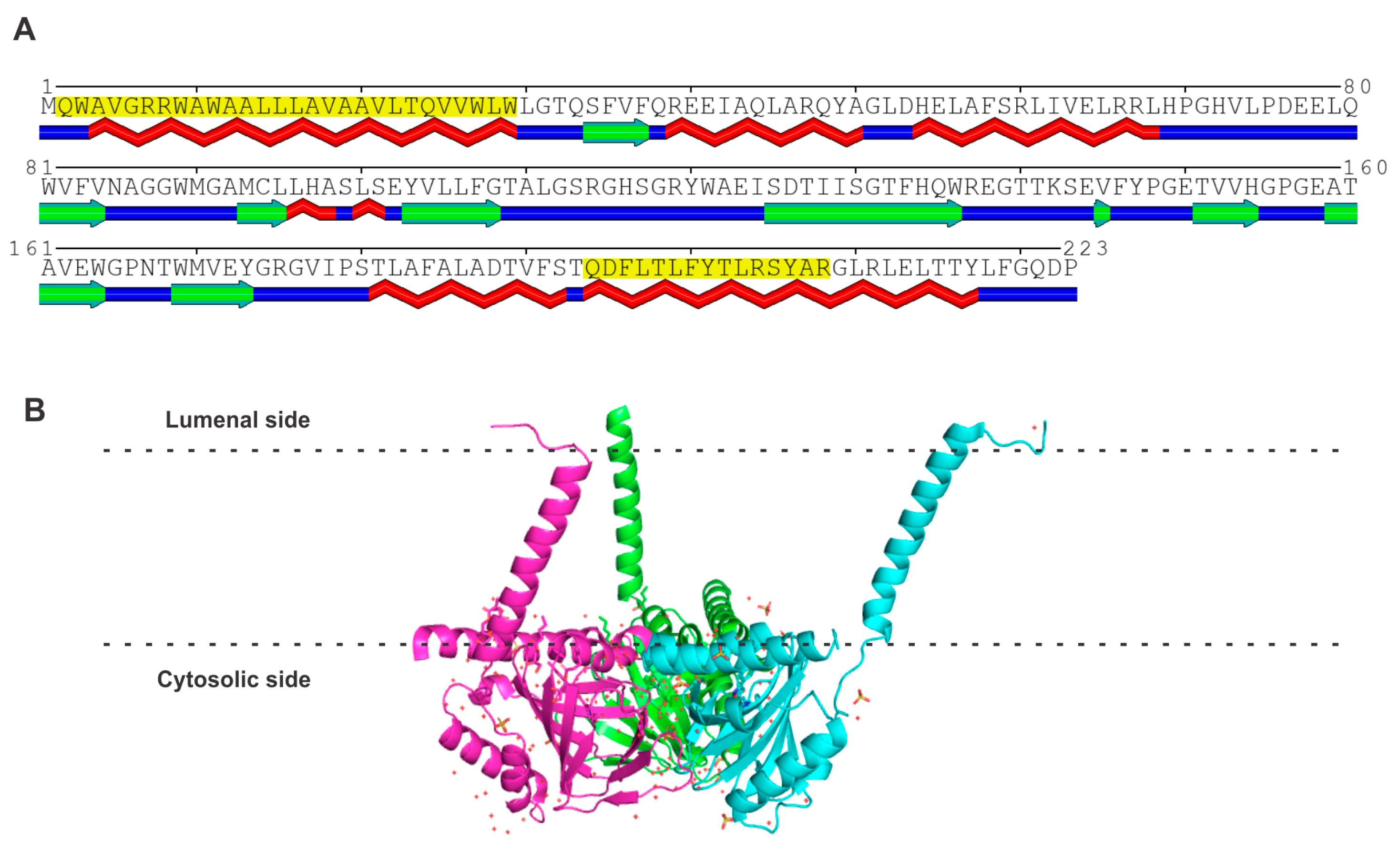
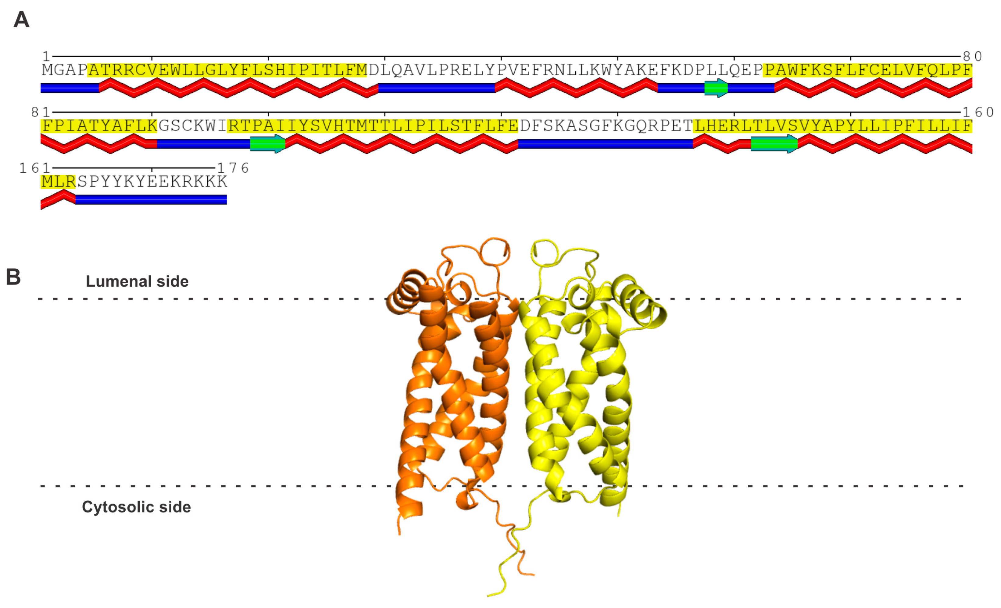
3.1. Amino Acid Sequence and Structure of S2R

It is important to begin this section with the statement at the onset that sigma receptor 2 (S2R) is not the same as progesterone receptor membrane component 1 (PGRMC1) [reviewed in 9–11,14,15]. This is necessary because of several publications in the literature that claimed PGRMC1 to be S2R [61][62][63]. There is no doubt that a functional relationship exists between the two proteins, but these two proteins are distinct at the molecular level. The actual molecular identity of S2R was not known until 2017, more than 20 years after the cloning of S1R. It was Alon et al. [64] who were successful in cloning S2R and showed that S2R is not PGRMC1 but is instead identical to an already known protein called TMEM97 (transmembrane-protein 97) or MAC30 (meningioma-associated protein 30). S2R consists of 176 amino acids; it belongs to a family of proteins in which the prototypical member is the emopamil-binding protein (EBP). However, unlike EBP, which possesses steroid isomerase activity, S2R does not possess any enzymatic activity. S2R has four transmembrane domains and three small stretches of β-strands (Figure 2A). The POLYVIEW-2D protein structure visualization server [65] was used to predict the transmembrane domains. The AlphaFold model of the amino acid sequence, as per analysis using the Robetta server, yielded a monomer with four transmembrane domains. However, a recent report on the crystal structure of bovine S2R has shown the protein to exist as a homodimer (PDB: 7MFI) [66]. Therefore, the AlphaFold model of human S2R was superimposed onto the structure of bovine S2R to generate the homodimer model for human S2R (Figure 2B). The membrane boundaries were predicted with the OPM (Orientations of Proteins in Membranes) server [67]. S2R is an integral protein of the endoplasmic reticulum, but it translocates to other sites in the cell to form protein–protein complexes in the plasma membrane, as well as in the lysosomal membrane. The gene coding for this protein is 9.5 kb long and is located in chromosome 17q11.2. A review of the molecular, pharmacological, and biological aspects of S2R was recently published by Izzo et al. [68] as the proceedings of an international symposium on this receptor. The biology of S2R is connected to a broad spectrum of cellular functions, including cholesterol transport and metabolism, progesterone signaling, autophagy, and membrane-bound protein trafficking. A notable feature of S2R is that it bears no similarity in amino acid sequence to S1R (Table 1) despite the fact that both proteins are identified as the two subtypes of sigma receptor. As already mentioned earlier in this research, the subtype classification into S1R and S2R was completed solely based on ligand binding long before the molecular identities of the two proteins were established.
Figure 2. (A) Amino acid sequence and structure of human S2R. The predicted transmembrane domains (shaded in yellow) and β-strands (indicated in green below the amino acid sequence) according to the analysis of the amino acid sequence of human S2R using the POLYVIEW program [65]. (B) The AlphaFold model of human S2R is a monomer, but this structure was superimposed onto the recently described homodimeric structure of bovine S2R to generate the model for human S2R.
A wide variety of pharmacological agents bind to S2R [31][69]. As for the endogenous ligands for this protein, the most likely candidate is the oxysterol known as 20(S)-hydroxycholesterol [70]. As discussed below, one of the well-established biological functions of S2R is its involvement in cholesterol homeostasis. Therefore, it makes sense that one of the metabolites of cholesterol functions as an endogenous ligand for this receptor. In addition, the expression of S2R appears to be under the control of the sterol-dependent transcription factor SREBP-2 (sterol regulatory element binding protein-2) [71].
All known biological functions of S2R seem to be mediated by protein–protein interactions with other proteins (see below). Since S2R is expressed in the brain, and some of its functions are related to the clearance of amyloid-β, there is a growing interest in the potential of this receptor and its ligands in the treatment of Alzheimer’s disease (reviewed in Ref. [21]). For this current research, however, the researchers focused on the ligands heme and progesterone. Surprisingly, the researchers found no published reports in the literature on the interaction of either of these ligands with the cloned S2R. Therefore, the researchers used the molecular docking approach to evaluate theoretically the binding of heme and progesterone with the S2R protein. This analysis yielded a value of −7.1 kcal/mole for the binding energy for heme, which translates to a Kd value of 6.2 μM, indicating a low affinity for the interaction. However, it might be appropriate to have this relatively low affinity if S2R functions as a heme chaperone, like PGRMC1/2 (see below). The value for the binding energy for the interaction of progesterone is −7.9 kcal/mole, which corresponds to a Kd value of 1.6 μM. This theoretically derived dissociation constant for progesterone binding is significantly higher than the corresponding value for S1R, which has been shown to bind progesterone experimentally. These values predict a lower affinity for progesterone binding to S2R than to S1R. Therefore, it would be of interest to determine if the cloned S2R actually binds progesterone.

4. Progesterone Receptor Membrane Components 1 and 2 (PGRMC1 and PGRMC2)

4.1. Amino Acid Sequences and Structures of PGRMC1 and PGRMC2

PGRMC1 and PGRMC2 are closely related proteins in the amino acid sequence, with approximately 60% identity (Table 1). But, they do not bear any significant sequence similarity to either S1R or S2R. PGRMC1 contains 195 amino acids and PGRMC2 contains 247 amino acids. Both proteins possess a single membrane-spanning transmembrane domain, highlighted in yellow in Figure 3A. PGRMC1 is an integral membrane protein present in the plasma membrane, mitochondrial membrane, and the membrane of the endoplasmic reticulum. PGRMC2 is also an integral membrane protein and is found in the nuclear membrane and in the membrane of the endoplasmic reticulum. The gene coding for PGRMC1 is located in the X chromosome (Xq24). PGRMC1 is a hemoprotein; the heme in PGRMC1 is penta-coordinated, and Tyr113 serves as the fifth axial ligand for iron in heme (iron in heme is already coordinated to nitrogen; one each in the four pyrroles of protoporphyrin IX). This leaves the sixth coordination surface of heme open, which allows the heme–heme hydrophobic stacking of two heme-containing monomers (Figure 3B) [62]. The resultant homodimer also forms a disulfide link with Cys129, but this covalent linking is not obligatory for dimer formation. The dimerization of heme-bound PGRMC1 has been authenticated with the deduction of its crystal structure [62]. The heme-dimerized PGRMC1 interacts with the EGF receptor [62]. Recent studies by Kabe et al. [72] have identified certain naturally occurring compounds (e.g., glycyrrhizin) that specifically bind to heme-dimerized PGRMC1 and interfere with the interaction of the PGRMC1 dimer with an EGF receptor, with functional consequences in terms of chemoresistance in colon cancer cells. PGRMC2 also binds heme; theoretical modeling, according to the AlphaFold program, suggests a monomeric structure (Figure 3B). In both proteins, the region that is not associated with the membrane contains α-helices and β-strands. The gene coding for PGRMC2 is located in chromosome 4q28.2. The binding of heme, as well as progesterone, to PGRMC1 and PGRMC2, has been established experimentally.
Figure 3. Amino acid sequence and structure for PGRMC1 and PGRMC2. (A) Transmembrane and secondary structure prediction of PGRMC1 and PGRMC2. The region highlighted in yellow in each protein represents the membrane-spanning transmembrane domain. Predicted α-helices are identified in red below the amino acid sequence, and β-strands are identified in green below the amino acid sequence. The POLYVIEW program [65] was used for these predictions. (B) Robetta model for PGRMC1 homodimer based on the crystal structure (PDB: 4X8Y). The heme ligand bound to each monomer is shown in yellow. (C) Robetta model for PGRMC2 monomer. Membrane boundaries were predicted with OPM (Orientations of Proteins in Membranes) server [67].

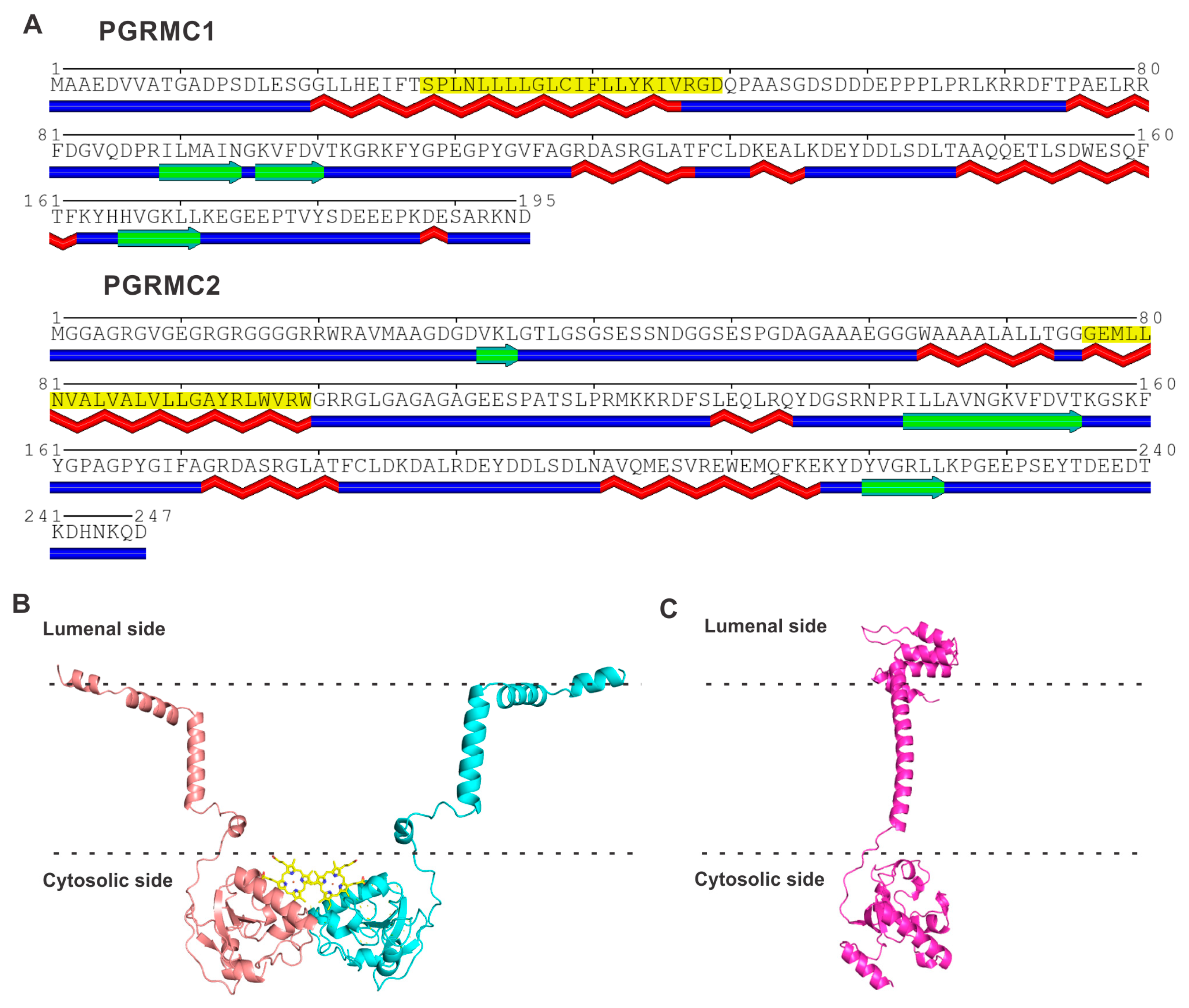
4.2. Common Structural Features in PGRMC1 and PGRMC2

Among the four proteins that form the focus of this present research, only PGRMC1 and PGRMC2 are structurally similar. Both bind heme and progesterone. These two proteins are not only similar in amino acid sequence but also share a homologous cytochrome b5-like heme/steroid binding domain [73][74]. There are two other proteins that possess this domain: neudesin and neuferricin. However, unlike PGRMC1 and PGRMC2, which are integral membrane proteins, neudesin and neuferricin are secreted proteins. Because of their ability to bind progesterone, and their feature as integral membrane proteins, PGRMC1 and PGRMC2 are called membrane-associated progesterone receptors to distinguish them from the classical progesterone receptors that function as transcription factors and are not associated with membranes. Even though S1R binds progesterone, may even interact with heme, and is an integral membrane protein, it does not possess the cytochrome b5-like domain. The same is true with S2R. Therefore, S1R and S2R are not members of the membrane-associated progesterone receptor family.

References

  1. Martin, W.R.; Eades, C.G.; Thompson, J.A.; Huppler, R.E.; Gilbert, P.E. The effects of morphine and nalorphine-like drugs in the nondependent and morphine-dependent chronic spinal dog. J. Pharmacol. Exp. Ther. 1976, 197, 517–532.
  2. Vaupel, D.B. Naltrexone fails to antagonize the sigma effects of PCP and SKF10,047 in the dog. Eur. J. Pharmacol. 1983, 92, 269–274.
  3. Largent, B.L.; Gundlach, A.L.; Snyder, S.H. Pharmacological and autoradiographic discrimination of sigma and phencyclidine receptor binding sites in brain with (+)-SKF 10,047, (+)--3--N-(1-propyl)piperidine and -1-piperidine. J. Pharmacol. Exp. Ther. 1986, 238, 739–748.
  4. Su, T.P. Pharmacologic characterizations of sigma receptors. NIDA Res. Monogr. 1993, 133, 41–53.
  5. Quirion, R.; Bowen, W.D.; Itzhak, Y.; Junien, J.L.; Musacchio, J.M.; Rothman, R.B.; Su, T.P.; Tam, S.W.; Taylor, D.P. A proposal for the classification of sigma binding sites. Trends Pharmacol. Sci. 1992, 13, 85–86.
  6. Bowen, W.D. Sigma receptors: Recent advances and new clinical potentials. Pharm. Acta Helv. 2000, 74, 211–218.
  7. Fishback, J.A.; Robsen, M.J.; Xu, Y.T.; Matsumoto, R.R. Sigma receptors: Potential targets for a new class of antidepressant drug. Pharmacol. Ther. 2010, 127, 271–282.
  8. Weng, C.C.; Riad, A.; Lieberman, B.P.; Xu, K.; Peng, X.; Mikitsh, J.L.; Mach, R.H. Characterization of sigma-2 receptor-specific binding sites using DTG and RHM-4. Pharmaceuticals 2022, 15, 1564.
  9. Schmidt, H.R.; Kruse, A.C. The molecular function of σ receptors: Past, present, and future. Trends Pharmacol. Sci. 2019, 40, 636–654.
  10. Pergolizzi, J.; Varrassi, G.; Coleman, M.; Breve, F.; Christo, D.K.; Christo, P.J.; Moussa, C. The sigma enigma: A narrative review of sigma receptors. Cureus 2023, 15, e35756.
  11. Lizama, B.N.; Kahle, J.; Catalano, S.M.; Caggiano, A.O.; Grundman, M.; Hamby, M.E. Sigma-2 receptors—From basic biology to therapeutic target: A focus on age-related degenerative diseases. Int. J. Mol. Sci. 2023, 24, 6251.
  12. Hasegawa, S.; Kasubuchi, M.; Terasawa, K.; Kimura, I. Perspectives on membrane-associated progesterone receptors as prospective therapeutic targets. Curr. Drug Targets 2016, 17, 1189–1197.
  13. Ryu, C.S.; Klein, K.; Zanger, U.M. Membrane associated progesterone receptors: Promiscuous proteins with pleiotropic functions—Focus on interactions with cytochrome P450. Front. Pharmacol. 2017, 8, 159.
  14. Cahill, M.A. Unde venisti PGRMC? Grand-scale biology from early eukaryotes and eumetazoan animal origins. Front. Biosci. (Landmark Ed.) 2022, 27, 317.
  15. Cahill, M.A. Quo vadis PGRMC? Grand-scale biology in human health and disease. Front. Biosci. (Landmark Ed.) 2022, 27, 318.
  16. Cahill, M.A. Progesterone receptor membrane component 1: An integrative review. J. Steroid Biochem. Mol. Biol. 2007, 105, 16–36.
  17. Pru, J.K.; Clark, N.C. PGRMC1 and PGRMC2 in uterine physiology and disease. Front. Neurosci. 2013, 7, 168.
  18. Smith, S.B.; Wang, J.; Cui, X.; Mysona, B.A.; Zhao, J.; Bollinger, K.E. Sigma 1 receptor: A novel therapeutic target in retinal disease. Prog. Retin. Eye Res. 2018, 67, 130–149.
  19. Malar, D.S.; Thitilertdecha, P.; Ruckvongacheep, K.S.; Brimson, S.; Tencomnao, T.; Brimson, J.M. Targeting sigma receptors for the treatment of neurodegenerative and neurodevelopmental disorders. CNS Drugs 2023, 37, 399–440.
  20. Wang, T.; Jia, H. The sigma receptors in Alzheimer’s disease: New potential targets for diagnosis and therapy. Int. J. Mol. Sci. 2023, 24, 12025.
  21. Yang, K.; Zeng, C.; Wang, C.; Sun, M.; Yin, D.; Sun, T. Sigma-2 receptor—A potential target for cancer/Alzheimer’s disease treatment via its regulation of cholesterol homeostasis. Molecules 2020, 25, 5439.
  22. Hanner, M.; Moebius, F.F.; Flandorfer, A.; Knaus, H.G.; Striessnig, J.; Kempner, R.; Glossmann, H. Purification, molecular cloning, and expression of the mammalian sigma1-binding site. Proc. Natl. Acad. Sci. USA 1996, 93, 8072–8077.
  23. Kekuda, R.; Prasad, P.D.; Fei, Y.J.; Leibach, F.H.; Ganapathy, V. Cloning and functional expression of the human type 1 sigma receptor (hSigmaR1). Biochem. Biophys. Res. Commun. 1996, 229, 553–558.
  24. Seth, P.; Fei, Y.J.; Li, H.W.; Huang, W.; Leibach, F.H.; Ganapathy, V. Cloning and functional characterization of a sigma receptor from rat brain. J. Neurochem. 1998, 70, 922–931.
  25. Seth, P.; Leibach, F.H.; Ganapathy, V. Cloning and structural analysis of the cDNA and the gene encoding the murine type 1 sigma receptor. Biochem. Biophys. Res. Commun. 1997, 241, 535–540.
  26. Prasad, P.D.; Li, H.W.; Fei, Y.J.; Ganapathy, M.E.; Fujita, T.; Plumley, L.H.; Yang-Feng, T.L.; Leibach, F.H.; Ganapathy, V. Exon-intron structure, analysis of promoter region, and chromosomal localization of the human type 1 sigma receptor gene. J. Neurochem. 1998, 70, 443–451.
  27. Cao, B.; Porollo, A.; Adamczak, R.; Jarrell, M.; Meller, J. Enhanced recognition of protein transmembrane domains with prediction-based structural profiles. Bioinformatics 2006, 22, 303–309.
  28. Schmidt, H.R.; Zheng, S.; Gurpinar, E.; Koehl, A.; Manglik, A.; Kruse, A.C. Crystal structure of the human σ1 receptor. Nature 2016, 532, 527–530.
  29. Schmidt, H.R.; Betz, R.M.; Dror, R.O.; Kruse, A.C. Structural basis for σ1 receptor ligand recognition. Nat. Struct. Mol. Biol. 2018, 25, 981–987.
  30. Meng, F.; Xiao, Y.; Ji, Y.; Sun, Z.; Zhou, X. An open-like conformation of the sigma-1 receptor reveals its ligand entry pathway. Nat. Commun. 2022, 13, 1267.
  31. Georgiadis, M.O.; Karoutzou, O.; Foscolos, A.S.; Papanastasiou, I. Sigma receptor (σR) ligands with antiproliferative and anticancer activity. Molecules 2017, 22, 1408.
  32. Ye, N.; Qin, W.; Tian, S.; Xu, Q.; Wold, E.A.; Zhou, J.; Zhen, X.C. Small molecules selectively targeting sigma-1 receptor for the treatment of neurological diseases. J. Med. Chem. 2020, 63, 15187–15217.
  33. Su, T.P.; London, E.D.; Jaffe, J.H. Steroid binding at sigma receptors suggests a link between endocrine, nervous, and immune systems. Science 1988, 240, 219–221.
  34. Yamada, M.; Nishigami, T.; Nakasho, K.; Nishimoto, Y.; Miyaji, H. Relationship between sigma-like site and progesterone-binding site of adult male rat liver microsomes. Hepatology 1994, 20, 1271–1280.
  35. Ramamoorthy, J.D.; Ramamoorthy, S.; Mahesh, V.B.; Leibach, F.H.; Ganapathy, V. Cocaine-sensitive sigma receptor and its interaction with steroid hormones in the human placental syncytiotrophoblast and in choriocarcinoma cells. Endocrinology 1995, 136, 924–932.
  36. Ganapathy, M.E.; Prasad, P.D.; Huang, W.; Seth, P.; Leibach, F.H.; Ganapathy, V. Molecular and ligand-binding characterization of the sigma receptor in the Jurkat human T lymphocyte cell line. J. Pharmacol. Exp. Ther. 1999, 289, 251–260.
  37. Maurice, T.; Urani, A.; Phan, V.L.; Romieu, P. The interaction between neuroactive steroids and the sigma1 receptor function: Behavioral consequences and therapeutic opportunities. Brain Res. Brain Res. Rev. 2001, 37, 116–132.
  38. Maurice, T. Neurosteroids and sigma1 receptors, biochemical and behavioral relevance. Pharmacopsychiatry 2004, 37 (Suppl. 3), S171–S182.
  39. Mysona, B.A.; Kansara, N.; Zhao, J.; Bollinger, K. The role of sigma 1 receptor as a neuroprotective target in glaucoma. Adv. Exp. Med. Biol. 2017, 964, 299–307.
  40. Ryskamp, D.A.; Korban, S.; Zhemkov, V.; Kraskovskaya, N.; Bezprozvanny, I. Neuronal sigma-1 receptors: Signaling functions and protective roles in neurodegenerative diseases. Front. Neurosci. 2019, 13, 862.
  41. Wu, N.H.; Ye, Y.; Wan, B.B.; Yu, Y.D.; Liu, C.; Chen, Q.J. Emerging benefits: Pathophysiological functions and target drugs of the sigma-1 receptor in neurodegenerative diseases. Mol. Neurobiol. 2021, 58, 5649–5666.
  42. Couly, S.; Yasui, Y.; Su, T.P. SIGMAR1 confers innate resilience against neurodegeneration. Int. J. Mol. Sci. 2023, 24, 7767.
  43. Ma, Q. Role of Nrf2 in oxidative stress and toxicity. Annu. Rev. Pharmacol. Toxicol. 2013, 53, 401–426.
  44. Ganapathy, V.; Thangaraju, M.; Prasad, P.D. Nutrient transporters in cancer: Relevance to Warburg hypothesis and beyond. Pharmacol. Ther. 2009, 121, 29–40.
  45. Lewerenz, J.; Hewett, S.J.; Huang, Y.; Lambros, M.; Gout, P.W.; Kalivas, P.W.; Massie, A.; Smolders, I.; Methner, A.; Pergande, M.; et al. The cystine/glutamate antiporter system xc− in health and disease: From molecular mechanisms to novel therapeutic opportunities. Antioxid. Redox. Signal. 2013, 18, 522–555.
  46. Bhutia, Y.D.; Babu, E.; Ramachandran, S.; Ganapathy, V. Amino acid transporters in cancer and their relevance to “glutamine addiction”: Novel targets for the design of a new class of anticancer drugs. Cancer Res. 2015, 75, 1782–1788.
  47. Wang, J.; Zhao, J.; Cui, X.; Mysona, B.A.; Navneet, S.; Saul, A.; Ahuja, M.; Lambert, N.; Gazaryan, I.G.; Thomas, B.; et al. The molecular chaperone sigma 1 receptor mediates rescue of retinal cone photoreceptor cells via modulation of NRF2. Free Radic. Biol. Med. 2019, 134, 604–616.
  48. Barwick, S.R.; Siddiq, M.S.; Wang, J.; Xiao, H.; Marshall, B.; Perry, E.; Smith, S.B. Sigma 1 receptor co-localizes with NRF2 in retinal photoreceptor cells. Antioxidants 2021, 10, 981.
  49. Wang, J.; Shanmugam, A.; Markand, S.; Zorrilla, E.; Ganapathy, V.; Smith, S.B. Sigma 1 receptor regulates the oxidative stress response in primary retinal Muller glial cells via NRF2 signaling and system xc−, the Na+-independent glutamate-cystine exchanger. Free Radic. Biol. Med. 2015, 86, 25–36.
  50. Bai, T.; Lei, P.; Zhou, H.; Liang, R.; Zhu, R.; Wang, W.; Zhou, L.; Sun, Y. Sigma-1 receptor protects against ferroptosis in hepatocellular carcinoma cells. J. Cell. Mol. Med. 2019, 23, 7349–7359.
  51. Bai, T.; Wang, S.; Zhao, Y.; Zhu, R.; Wang, W.; Sun, Y. Haloperidol, a sigma receptor 1 antagonist, promotes ferroptosis in hepatocellular carcinoma cells. Biochem. Biophys. Res. Commun. 2017, 491, 919–925.
  52. Babitt, J.L.; Lin, H.Y. The molecular pathogenesis of hereditary hemochromatosis. Semin. Liver Dis. 2011, 31, 280–292.
  53. Adams, P.C.; Jeffrey, G.; Ryan, J. Haemochromatosis. Lancet 2023, 401, 1811–1821.
  54. Merryweather-Clarke, A.T.; Pointon, J.J.; Jouanolle, A.M.; Rochette, J.; Robson, K.J.H. Geography of HFE C282Y and H63D mutations. Genet. Test. 2000, 4, 183–198.
  55. Torti, S.V.; Torti, F.M. Iron and cancer: More ore to be mined. Nat. Rev. Cancer 2013, 13, 342–355.
  56. Rodriguez, R.; Schreiber, S.L.; Conrad, M. Persister cancer cells: Iron addiction and vulnerability to ferroptosis. Mol. Cell 2022, 82, 728–740.
  57. Gnanaprakasam, J.P.; Thangaraju, M.; Liu, K.; Ha, Y.; Martin, P.M.; Smith, S.B.; Ganapathy, V. Absence of iron-regulatory protein Hfe results in hyperproliferation of retinal pigment epithelium. Role of cystine-glutamate exchanger. Biochem. J. 2009, 424, 243–252.
  58. Bhutia, Y.D.; Ogura, J.; Grippo, P.J.; Torres, C.; Sato, T.; Wachtel, M.; Ramachandran, S.; Babu, E.; Sivaprakasam, S.; Rajasekaran, D.; et al. Chronic exposure to excess iron promotes EMT and cancer via p53 loss in pancreatic cancer. Asian J. Pharm. Sci. 2020, 15, 237–251.
  59. Kim, F.J.; Maher, C.M. Sigma1 pharmacology in the context of cancer. Handb. Exp. Pharmacol. 2017, 244, 237–308.
  60. Robinson, T.S.; Osman, M.A. An emerging role for sigma receptor 1 in personalized treatment of breast cancer. Cancers 2023, 15, 3464.
  61. Ahmed, I.S.A.; Chamberlain, C.; Craven, R.J. S2R(PGRMC1): The cytochrome-related sigma-2 receptor that regulates lipid and drug metabolism and hormone signaling. Expert Opin. Drug Metab. Toxicol. 2012, 8, 361–370.
  62. Kabe, Y.; Nakane, T.; Koike, I.; Yamamoto, T.; Sugiura, Y.; Harada, E.; Sugase, K.; Shimamura, T.; Ohmura, M.; Muraoka, K.; et al. Haem-dependent dimerization of PGRMC1/sigma-2 receptor facilitates cancer proliferation and chemoresistance. Nat. Commun. 2016, 7, 11030.
  63. Xu, J.; Zeng, C.; Chu, W.; Pan, F.; Rothfuss, J.M.; Zhang, F.; Tu, Z.; Zhou, D.; Zeng, D.; Vangveravong, S.; et al. Identification of the PGRMC1 protein complex as the putative sigma-2 receptor binding site. Nat. Commun. 2011, 2, 380.
  64. Alon, A.; Schmidt, H.; Wood, M.D.; Sahn, J.J.; Martin, S.F.; Kruse, A.C. Identification of the gene that codes for the σ2 receptor. Proc. Natl. Acad. Sci. USA 2017, 114, 7160–7165.
  65. Porollo, A.A.; Adamczak, R.; Meller, J. POLYVIEW: A flexible visualization tool for structural and functional annotations of proteins. Bioinformatics 2004, 20, 2460–2462.
  66. Alon, A.; Lyu, J.; Braz, J.M.; Tummino, T.A.; Craik, V.; O’Meara, M.J.; Webb, C.M.; Radchenko, D.S.; Moroz, Y.S.; Huang, X.P.; et al. Structures of the σ2 receptor enable docking for bioactive ligand discovery. Nature 2021, 600, 759–764.
  67. Lomize, M.A.; Pogozheva, I.D.; Joo, H.; Mosberg, H.I.; Lomize, A.L. OPM database and PPM web server: Resources for positioning of proteins in membranes. Nucleic Acids Res. 2012, 40, D370–D376.
  68. Izzo, N.J.; Colom-Cadena, M.; Riad, A.A.; Xu, J.; Singh, M.; Abate, C.; Cahill, M.A.; Spires-Jones, T.L.; Bowen, W.D.; Mach, R.H.; et al. Proceedings from the fourth international symposium on σ-2 receptors: Role in health and disease. eNeuro 2020, 7, ENEURO.0317-20.2020.
  69. Guo, L.; Zhen, X. Sigma-2 receptor ligands: Neurobiological effects. Curr. Med.Chem. 2015, 22, 989–1003.
  70. Cheng, Y.S.; Zhang, T.; Ma, X.; Pratuangtham, S.; Zhang, G.C.; Ondrus, A.A.; Mafi, A.; Lomenick, B.; Jones, J.J.; Ondrus, A.E. A proteome-wide map of 20(S)hydroxycholesterol interactors in cell membranes. Nat. Chem. Biol. 2021, 17, 1271–1280.
  71. Shen, H.; Li, J.; Xie, X.; Yang, H.; Zhang, M.; Wang, B.; Kent, K.C.; Plutzky, J.; Guo, L.W. BRD2 regulation of sigma-2 receptor upon cholesterol deprivation. Life Sci. Alliance 2020, 4, e201900540.
  72. Kabe, Y.; Koike, I.; Yamamoto, T.; Hirai, M.; Kanai, A.; Furuhata, R.; Tsugawa, H.; Harada, E.; Sugase, K.; Hanadate, K.; et al. Glycyrrhizin derivatives suppress cancer chemoresistance by inhibiting progesterone receptor membrane component 1. Cancers 2021, 13, 3265.
  73. Kimura, I.; Nakayama, Y.; Konishi, M.; Terasawa, K.; Ohta, M.; Itoh, N.; Fujimoto, M. Functions of MAPR (membrane-associated progesterone receptor) family members as heme/steroid-binding proteins. Curr. Protein Pept. Sci. 2012, 13, 687–696.
  74. Hehenberger, E.; Eitel, M.; Fortunato, S.A.V.; Miller, D.J.; Keeling, P.J.; Cahill, M.A. Early eukaryotic origins and metazoan elaboration of MAPR family proteins. Mol. Phylogenet. Evol. 2020, 148, 106814.
More
Information
Contributors MDPI registered users' name will be linked to their SciProfiles pages. To register with us, please refer to https://encyclopedia.pub/register : , , , , , ,
View Times: 188
Revisions: 2 times (View History)
Update Date: 19 Oct 2023
1000/1000
ScholarVision Creations