Submitted Successfully!
To reward your contribution, here is a gift for you: A free trial for our video production service.
Thank you for your contribution! You can also upload a video entry or images related to this topic.
Version Summary Created by Modification Content Size Created at Operation
1 -- 2464 2023-10-11 20:31:38 |
2 update references and layout -4 word(s) 2460 2023-10-12 04:15:18 |

Video Upload Options

Do you have a full video?

Confirm

Are you sure to Delete?
Cite
If you have any further questions, please contact Encyclopedia Editorial Office.
Ford, H.; Liu, Q.; Fu, X.; Strieder-Barboza, C. White Adipose Tissue Heterogeneity in the Single-Cell Era. Encyclopedia. Available online: https://encyclopedia.pub/entry/50152 (accessed on 16 May 2024).
Ford H, Liu Q, Fu X, Strieder-Barboza C. White Adipose Tissue Heterogeneity in the Single-Cell Era. Encyclopedia. Available at: https://encyclopedia.pub/entry/50152. Accessed May 16, 2024.
Ford, Hunter, Qianglin Liu, Xing Fu, Clarissa Strieder-Barboza. "White Adipose Tissue Heterogeneity in the Single-Cell Era" Encyclopedia, https://encyclopedia.pub/entry/50152 (accessed May 16, 2024).
Ford, H., Liu, Q., Fu, X., & Strieder-Barboza, C. (2023, October 11). White Adipose Tissue Heterogeneity in the Single-Cell Era. In Encyclopedia. https://encyclopedia.pub/entry/50152
Ford, Hunter, et al. "White Adipose Tissue Heterogeneity in the Single-Cell Era." Encyclopedia. Web. 11 October, 2023.
White Adipose Tissue Heterogeneity in the Single-Cell Era
Edit

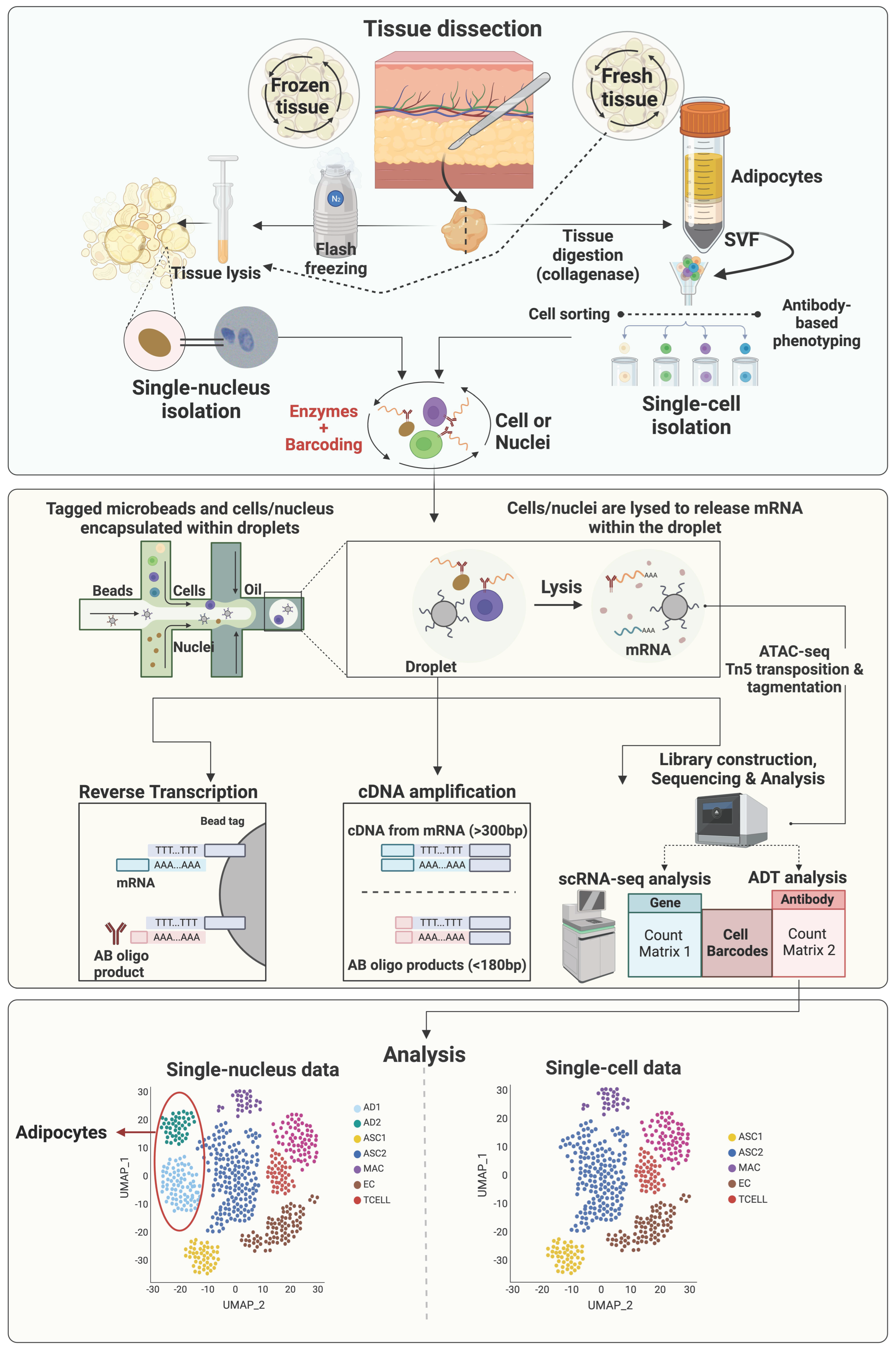
Adipose tissue is a major modulator of metabolic function by regulating energy storage and by acting as an endocrine organ through the secretion of adipokines. With the advantage of next-generation sequencing-based single-cell technologies, adipose tissue has been studied at single-cell resolution, thus providing unbiased insight into its molecular composition. Recent single-cell RNA sequencing studies in human and mouse models have dissected the transcriptional cellular heterogeneity of subcutaneous (SAT), visceral (VAT), and intramuscular (IMAT) white adipose tissue depots and revealed unique populations of adipose tissue progenitor cells, mature adipocytes, immune cell, vascular cells, and mesothelial cells that play direct roles on adipose tissue function and the development of metabolic disorders.

adipose single-cell sequencing cattle

1. Introduction

Advances in research have revealed white adipose tissue as a complex organ that modulates metabolism and health via endocrine and other signaling mechanisms [1]. As such, considerable efforts have been made to further the understanding of this tissue, particularly as it relates to metabolic dysfunction and disease. These efforts have primarily focused on adipose-related disorders in humans, such as obesity and type 2 diabetes, with limited studies in cattle and other livestock.
Investigations in recent decades have demonstrated that not all white adipose tissue depots are the same, with distinct functional differences between subcutaneous, visceral, and intramuscular sites [2]. Furthermore, these depot-specific functional differences are driven by significant cellular heterogeneity [3], resulting in distinct metabolic, immune, and inflammatory profiles. Early studies using antibodies to identify and isolate unique cell populations in adipose tissue depots were critical for beginning to understand the cellular diversity within these tissues [4]; however, the advent of single-cell/nucleus RNA sequencing (sc/snRNAseq) techniques has revolutionized the field and opened up new avenues of exploration. As evidenced by research endeavors in cancer biology [5], the application of sc/snRNAseq technologies in the field of adipose tissue biology has great potential to further enhance the understanding of how adipose tissue contributes to metabolic function and the pathogenesis of disease in both humans and livestock.

2. The Single-Cell Era: Applicability of Single-Cell/Nucleus RNA Sequencing to Decode Adipose Tissue Heterogeneity

Studies preceding the use of single-cell RNA sequencing (scRNAseq) focused on identifying cell types within adipose tissue utilizing fluorescent-activated cell sorting (FACS) based on the presence or absence of specific cell surface markers. Larger, clonally identical populations of cells were then propagated and characterized in downstream analyses. These studies were important for the initial identification of different cell types relating to adipogenesis in adipose tissue, including Lin-/Cd34+/Cd29+/Sca1+/Cd24+ ASPCs [6], Pdgfrα+ mesenchymal cells [7], Pdgfrb+ and Myh11+ mural cells [8][9][10][11][12][13][14], and mature adipocytes (ASC-1 for white; PAT2 and P2RX for brown and beige) [15]. As reviewed by Ferrero et al. [16], the use of FACS has been important in deciphering the heterogeneity of ASPCs, particularly in recent years with the development and utilization of scRNAseq applications. In addition, the work of Astori et al. [4] helped to further define the components of the stromal vascular fraction (SVF) including monocytes, granulocytes, hematopoietic stem cells, and endothelial cells. Lineage tracing via Cre-recombinase mouse models has been utilized to identify adipocyte precursors [7][11][17], and characterize mature adipocytes [18] in white adipose tissue of mice [7][11][17]. However, one limitation of the FACS- and lineage tracing-based strategies is the lack of single markers, which makes it very challenging to identify and validate ASPC heterogeneity.
With the advantage of developing next-generation sequencing (NGS)-based single-cell technologies, researchers can now profile adipose tissue at single-cell resolution to provide unbiased insight into its molecular composition [19]. scRNAseq was first developed by Tang et al. in 2009 to sequence the individual cell from a four-cell stage embryo [20]. Since then, significant efforts have been made on advancing various scRNAseq techniques. Among those, the droplet-based microfluidic technology is the most widely used scRNA-seq platform due to its increased throughput and more automated protocols [21][22]. In 2016, 10× Genomics first launched a commercial droplet-microfluidics-based scRNA-seq platform, the Chromium. In Chromium, cells and beads, which contain primers, cellular barcodes, and a unique molecular identifier (UMI), in separate channels are mixed in a microfluidic device and partitioned by nanoliter-size oil droplets for further single-cell cell lysis, reverse transcription, and cDNA library construction [22][23][24][25][26].
Studies have demonstrated scRNAseq as an extremely powerful tool in studying adipose tissue stromal cellular composition and plasticity. The high throughput of scRNAseq provides detailed information of the cell identity, gene expression profiles, and changes in cellular states that may arise under normal physiological and pathological conditions. Various scRNAseq studies have demonstrated the heterogeneities of the mesenchymal stem cells (MSCs), immune cells, mesothelial cells, and endothelial cells in white adipose tissues [27]. Due to their high abundancy, heterogeneity, and functional importance, many scRNAseq studies focused on ASPCs defined as cells expressing the common mesenchymal markers, Pdgfra and Pdgfrb. Moreover, owing to the power of single-cell profiling, distinct ASPC populations expressing adipogenic markers, like Pparg, Lpl, and Cd36, and those enriched for genes related to ECM remodeling and inflammation have been identified [27].
Most early scRNAseq studies of adipose tissue used freshly isolated intact cells, however, with some limitations. First, this strategy requires prolonged stressful enzymatic digestion, which might alter gene expression profiles of sensitive cell types. In addition, due to the prolonged sampling process of human and large animal tissues, it is very difficult to finish cell isolation immediately after sample collection on the same day. Moreover, mature adipocytes, the parenchymal cells of adipose tissue, are too large for most scRNAseq platforms. Alternatively, single-nuclei RNA sequencing (snRNAseq), which uses single nuclei, has overcome these obstacles [28][29][30]. The general workflow of sc/snRNAseq and their differences are illustrated in Figure 1. Single-nuclei RNA sequencing does not require tissue digestion (e.g., collagenase) to release individual viable cells and therefore can avoid the enzyme-induced cell-type biases, which may result in artificial transcriptional differences [31][32]. Moreover, the source of nuclei can be either fresh or frozen tissue samples, providing increased flexibility [33]. Due to the smaller size of nuclei, nuclei of all types of cells can be identified via snRNAseq, making it possible to study adipocytes and other larger cells, such as neurons. However, snRNAseq also has its limitations. One major issue is the loss of most cytosolic content, which contains most mature mRNA, leading to low numbers of genes detected and failure to detect low abundant transcripts. Another concern is the high level of ambient RNA, which must be carefully removed via additional sample and sequencing data processing [31][34]. Without the protection of cytoplasm, mRNA is more susceptible to RNase-mediated degradation, which can be an issue for samples with cells expressing high levels of RNases and must be addressed by adding sufficient RNase inhibitors [31][34].
Figure 1. General workflow of single-cell and single-nucleus RNA sequencing. This figure was prepared in Biorender and is licensed to be published (Agreement number: LA25UR522V).
Integrating the sc/snRNAseq data with other omics data is becoming a popular strategy to gain more comprehensive and multidimensional understanding of tissue heterogeneity. The assay for transposase-accessible chromatin using sequencing (ATACseq; Figure 1) is a powerful method for determining chromatin accessibility across the genome, an important component of the epigenome [35], and identifying potential transcription factors regulating differential gene expression [36]. In 2015, ATACseq was optimized for single-cell applications [37][38]. Single-cell and single-nuclei ATACseq facilitates the single-cell open chromatin landscaping of heterogeneous tissues like adipose tissue. It is expected that more and more studies integrating sc/snRNAseq and sc/snATACseq data to uncover the contribution of epigenetic regulation of gene expression to the heterogeneities of cells residing in adipose tissue will be published in the near future.
Researchers describe recent studies utilizing sc/snRNASeq to characterize the heterogeneity of adipocyte progenitor cells, mature adipocytes, mesothelial cells, immune cells, and vascular cells in white adipose tissue. While SAT and VAT have garnered most of the research attention for their implication in metabolic diseases in both humans and livestock, recent investigations into IMAT have provided valuable insight into the mechanisms contributing to enhanced marbling in pigs and beef cattle, as well as the pathogenesis of human diseases, including muscular dystrophies and sarcopenia. Studies using sc/snRNASeq to elucidate the cellular heterogeneity of intermuscular adipose tissue are lacking but represent a novel area of study for future research endeavors in this field.

3. Transcriptional Diversity of Adipose Tissue Immune Cells

The network of immune cells in white adipose tissue represents an important link between metabolic and immune function and these cells are implicated both in the prevention and pathogenesis of metabolic disease and inflammation. Recent sc/snRNASeq approaches have provided critical insight into the specific types of immune cells present in adipose tissue as well as transcriptional changes that occur in these cells during different metabolic conditions.

3.1. Mouse Models

In their study on β3-adrenergic stimulation in mice, Burl et al. [39] identified populations of macrophages (C1qa, Trem2, Adgre1, Spp1) and natural killer and T cells (NKT; Nkg7), as well as a population of mixed macrophages and dendritic cells (Cd74) in epididymal VAT. Notably, β3 stimulation resulted in an increase in the abundance of macrophages and a decrease in the abundance of NKT cells in adipose tissue. Additionally, the authors described a subpopulation of lipid-associated macrophages characterized by a high expression of Spp1, Cd36, Fabp5, Lpl, and Lipa and suggested to be involved in the clearance of dead adipocytes and stimulation of adipogenesis by new adipocytes.
Nine distinct immune cell subpopulations were identified in VAT (epididymal adipose tissue) from obese and lean mice by Sárvári et al. [30], six of which were classified as macrophages (Adgre1, Lyz2, Ccl6), with one subpopulation of dendritic cells (Cd244a, Cd209a), one subpopulation of T cells (Skap1), and one subpopulation of B cells (Ms4a1, Cd79a, Cd79b). Among macrophages, the authors identified subpopulations of perivascular macrophages (Mrc1, Lyve1, Cd163), lipid-associated macrophages (Lpl, Trem2, Cd9), non-perivascular macrophages (Fcrls, Ear2), collagen-expressing macrophages (Col5a2, Tgfbr3, Col3a1), proliferating lipid-associated macrophages (Pola1, Kif11, Kif15) and regulatory macrophages (Prg4, Tgfb2, Ltbp1). Notably, the authors found a dramatic increase in lipid-associated and proliferating lipid-associated macrophages in obese mice, and a shift in the transcriptional profile of perivascular and non-perivascular macrophages towards increased expression of lipid metabolism genes (Pparg, Lpl) compared to lean mice.

3.2. Human Models

Strieder-Barboza identified macrophage (MAC; MERTK, CD163), dendritic cell (DC; CD1D, FLT3), T cell, and NK cell (SKAP1) populations that were similarly distributed across human abdominal SAT and omental VAT [40]. Four major MAC subtypes were identified including lipid-associated macrophages (LAM) expressing high ITGAX, TREM2, CD9, and CD52, and two MRC1/CD206+ MAC subtypes expressing marker genes of resident macrophages such as F13A1, PDGFC, and LYVE1, as previously reported [41]. Another resident macrophage was identified by the lower MRC1 expression and the unique TIMD4 expression [42]. Emont et al. [28] also identified a range of immune cells in human SAT and VAT samples including macrophages (MAFB, CYBB), monocytes (CYBB, CSF3R), dendritic cells (FLT3), mast cells (CPA3), neutrophils (CSF3R), B cells (MS4A1), NK cells (KLRD1), and T cells (IL7R). While Emont et al. [28] included mouse adipose tissue samples in their analysis, they did not identify major differences between mouse and human immune cells.
Analyzing SAT and VAT samples from human patients undergoing bariatric surgery, Vijay et al. [43] found a similar panel of immune cells including naïve T cells (IL7R), activated T cells (CCL5, IL32), NK cells (NKG7), as well as a subpopulation of dysfunctional T cells characterized by high expression of the metallothioneins MT1F and MT1G. Macrophages (CD68) were also identified in this study, including a subpopulation of lipid-associated macrophages characterized by a high expression of lipid metabolism-related genes (LIPA, LPL, CD36, FABP4), as well as subpopulations of pro-inflammatory (CXCL3, CXCL8, IL1B) and anti-inflammatory (FOLR2, KLF4) macrophages. Dendritic cells, characterized by a high expression of HLA genes, and B cells (IGKC, CD79A, CD37) were also identified in these adipose tissue samples [43]. Resident (CD14, CD8, CD163) and pro-inflammatory (ITGAX, CD86, TREM2) macrophages were also described by Whytock et al. [44] in human SAT. Immune cells were identified in the integrated dataset of human white adipose tissue samples described by Massier et al. [45], including B cells (MS4A1), mast cells (CPA3), T, NK, and NKT cells (CD3D), and a large group of cells including macrophages, dendritic cells, and monocytes (MRC1, CD11c, MRC1, FABP4). Innate lymphoid cells (ILCs) have only recently been characterized as important components of the innate immune system and are typically associated with their roles at mucosal barriers; however, Hildreth et al. [46] identified a subpopulation of ILCs in human SAT, characterized by high expression of KLRB1, KIT, CD200R1, and CCR6. Supporting the findings of Sárvári et al. [30] and Vijay et al. [43], Hildreth et al. [46] also identified a subpopulation of lipid-associated macrophages (TREM2, CD9, LPL), that were in greater abundance in SAT samples from obese patients compared to lean patients.
Analyzing fatty infiltration in human skeletal muscle, Fitzgerald et al. [47] identified a general population of macrophages (MRC1, C1QA) as well as a subpopulation of inflammatory macrophages (CD68, PECAM1), but did not describe the presence of additional immune cell populations.

3.3. Livestock Models

The recent snRNAseq data on dairy cows’ abdominal SAT and omental VAT revealed populations of macrophages, and natural killer and T cells, and a significant increase in the abundance of these immune cell populations in VAT vs. SAT, specifically on the proportion of lipid-associated macrophages (LAM) [48]. Macrophages (MAC) expressing MRC1 and MSR1 corresponded to nearly 10% of all adipose tissue nuclei and were the most heterogenous cell type in the adipose tissue of dairy cows. Researchers identified five distinct MAC subtypes, including two subtypes of LAMs (FABP4, LPL, CD36, FASN, CD9), perivascular MACs, M2-like MACs (CD163, CD206), and a MAC subtype enriched for complement genes (C3, CFI, CFB) and S100A12 and S100A8, suggesting these cells were likely monocytes or differentiating macrophages [43]. Dairy cow adipose tissue MAC subpopulations were similar to the ones identified via snRNAseq across human SAT and VAT [40]. Notably, researchers also identified a SAT-specific MAC subtype characterized by ABL1, SPTBN1, ZBTB16, and ASAMTSK3. In addition to MAC, the snRNAseq data also revealed a cluster of cells that were enriched for gene markers of T cells (CCL5, CD3E, CD2, CD247, CD52) and natural killer cells (NKG7, CTSW) comprising nearly 3% and 1.3% of total nuclei in VAT and SAT, respectively. The data on the immune cell abundance and heterogeneity in the SAT and VAT of dairy cows highlighted depot specificities and potential distinct functional roles for immune cell subtypes, particularly macrophages. However, the specific roles of MAC and NKT populations in the SAT and VAT of dairy cows and how they underly health and disease statuses are yet to be elucidated.
In other animal models, Li et al. [49] did not detect any immune cells in the breast muscle of chicken at either 5 and 100 days old, whereas a distinct population of myeloid-derived cells (MRC1) and a population of general immune cells (CD3E) were identified in the IMAT of pigs [50].

References

  1. Harvey, I.; Boudreau, A.; Stephens, J.M. Adipose tissue in health and disease. Open Biol. 2020, 10, 200291.
  2. Komolka, K.; Albrecht, E.; Wimmers, K.; Michal, J.J.; Maak, S. Molecular heterogeneities of adipose depots-potential effects on adipose-muscle cross-talk in humans, mice and farm animals. J. Genom. 2014, 2, 31.
  3. Corvera, S. Cellular Heterogeneity in Adipose Tissues. Annu. Rev. Physiol. 2021, 83, 257–278.
  4. Astori, G.; Vignati, F.; Bardelli, S.; Tubio, M.; Gola, M.; Albertini, V.; Bambi, F.; Scali, G.; Castelli, D.; Rasini, V.; et al. “In vitro” and multicolor phenotypic characterization of cell subpopulations identified in fresh human adipose tissue stromal vascular fraction and in the derived mesenchymal stem cells. J. Transl. Med. 2007, 5, 55.
  5. Lei, Y.; Tang, R.; Xu, J.; Wang, W.; Zhang, B.; Liu, J.; Yu, X.; Shi, S. Applications of single-cell sequencing in cancer research: Progress and perspectives. J. Hematol. Oncol. 2021, 14, 91.
  6. Rodeheffer, M.S.; Birsoy, K.; Friedman, J.M. Identification of white adipocyte progenitor cells in vivo. Cell 2008, 135, 240–249.
  7. Lee, Y.-H.; Emilio, A.; Granneman, J.G. In Vivo Identification of Bipotential Adipocyte Progenitors Recruited by β3-Adrenoceptor Activation and High-Fat Feeding. Cell Metab. 2012, 15, 480–491.
  8. Long, J.Z.; Svensson, K.J.; Tsai, L.; Zeng, X.; Roh, H.C.; Kong, X.; Rao, R.R.; Lou, J.; Lokurkar, I.; Baur, W.; et al. A Smooth Muscle-Like Origin for Beige Adipocytes. Cell Metab. 2014, 19, 810–820.
  9. Shao, M.; Vishvanath, L.; Busbuso, N.C.; Hepler, C.; Shan, B.; Sharma, A.X.; Chen, S.; Yu, X.; An, Y.A.; Zhu, Y.; et al. De novo adipocyte differentiation from Pdgfrbeta(+) preadipocytes protects against pathologic visceral adipose expansion in obesity. Nat. Commun. 2018, 9, 890.
  10. Vishvanath, L.; MacPherson, K.A.; Hepler, C.; Wang, Q.A.; Shao, M.; Spurgin, S.B.; Wang, M.Y.; Kusminski, C.M.; Morley, T.S.; Gupta, R.K. Pdgfrbeta+ Mural Preadipocytes Contribute to Adipocyte Hyperplasia Induced by High-Fat-Diet Feeding and Prolonged Cold Exposure in Adult Mice. Cell Metab. 2016, 23, 350–359.
  11. Berry, R.; Rodeheffer, M.S. Characterization of the adipocyte cellular lineage in vivo. Nat. Cell Biol. 2013, 15, 302–308.
  12. Tran, K.-V.; Gealekman, O.; Frontini, A.; Zingaretti, M.C.; Morroni, M.; Giordano, A.; Smorlesi, A.; Perugini, J.; De Matteis, R.; Sbarbati, A.; et al. The Vascular Endothelium of the Adipose Tissue Gives Rise to Both White and Brown Fat Cells. Cell Metab. 2012, 15, 222–229.
  13. Tang, W.; Zeve, D.; Suh, J.M.; Bosnakovski, D.; Kyba, M.; Hammer, R.E.; Tallquist, M.D.; Graff, J.M. White fat progenitor cells reside in the adipose vasculature. Science 2008, 322, 583–586.
  14. Gupta, R.K.; Mepani, R.J.; Kleiner, S.; Lo, J.C.; Khandekar, M.J.; Cohen, P.; Frontini, A.; Bhowmick, D.C.; Ye, L.; Cinti, S.; et al. Zfp423 expression identifies committed preadipocytes and localizes to adipose endothelial and perivascular cells. Cell Metab. 2012, 15, 230–239.
  15. Ussar, S.; Lee, K.Y.; Dankel, S.N.; Boucher, J.; Haering, M.F.; Kleinridders, A.; Thomou, T.; Xue, R.; Macotela, Y.; Cypess, A.M.; et al. ASC-1, PAT2, and P2RX5 are cell surface markers for white, beige, and brown adipocytes. Sci. Transl. Med. 2014, 6, 247ra103.
  16. Ferrero, R.; Rainer, P.; Deplancke, B. Toward a Consensus View of Mammalian Adipocyte Stem and Progenitor Cell Heterogeneity. Trends Cell Biol. 2020, 30, 937–950.
  17. Krueger, K.C.; Costa, M.J.; Du, H.; Feldman, B.J. Characterization of Cre recombinase activity for in vivo targeting of adipocyte precursor cells. Stem Cell Rep. 2014, 3, 1147–1158.
  18. Eguchi, J.; Wang, X.; Yu, S.; Kershaw, E.E.; Chiu, P.C.; Dushay, J.; Estall, J.L.; Klein, U.; Maratos-Flier, E.; Rosen, E.D. Transcriptional control of adipose lipid handling by IRF4. Cell Metab. 2011, 13, 249–259.
  19. Hwang, B.; Lee, J.H.; Bang, D. Single-cell RNA sequencing technologies and bioinformatics pipelines. Exp. Mol. Med. 2018, 50, 96.
  20. Tang, F.; Barbacioru, C.; Wang, Y.; Nordman, E.; Lee, C.; Xu, N.; Wang, X.; Bodeau, J.; Tuch, B.B.; Siddiqui, A.; et al. mRNA-Seq whole-transcriptome analysis of a single cell. Nat. Methods 2009, 6, 377–382.
  21. Chen, G.; Ning, B.; Shi, T. Single-Cell RNA-Seq Technologies and Related Computational Data Analysis. Front. Genet. 2019, 10, 317.
  22. Zhang, X.; Li, T.; Liu, F.; Chen, Y.; Yao, J.; Li, Z.; Huang, Y.; Wang, J. Comparative Analysis of Droplet-Based Ultra-High-Throughput Single-Cell RNA-Seq Systems. Mol. Cell 2019, 73, 130–142.e135.
  23. Klein, A.M.; Mazutis, L.; Akartuna, I.; Tallapragada, N.; Veres, A.; Li, V.; Peshkin, L.; Weitz, D.A.; Kirschner, M.W. Droplet barcoding for single-cell transcriptomics applied to embryonic stem cells. Cell 2015, 161, 1187–1201.
  24. Zilionis, R.; Nainys, J.; Veres, A.; Savova, V.; Zemmour, D.; Klein, A.M.; Mazutis, L. Single-cell barcoding and sequencing using droplet microfluidics. Nat. Protoc. 2017, 12, 44–73.
  25. Macosko, E.Z.; Basu, A.; Satija, R.; Nemesh, J.; Shekhar, K.; Goldman, M.; Tirosh, I.; Bialas, A.R.; Kamitaki, N.; Martersteck, E.M.; et al. Highly Parallel Genome-wide Expression Profiling of Individual Cells Using Nanoliter Droplets. Cell 2015, 161, 1202–1214.
  26. Zheng, G.X.; Terry, J.M.; Belgrader, P.; Ryvkin, P.; Bent, Z.W.; Wilson, R.; Ziraldo, S.B.; Wheeler, T.D.; McDermott, G.P.; Zhu, J.; et al. Massively parallel digital transcriptional profiling of single cells. Nat. Commun. 2017, 8, 14049.
  27. Maniyadath, B.; Zhang, Q.; Gupta, R.K.; Mandrup, S. Adipose tissue at single-cell resolution. Cell Metab. 2023, 35, 386–413.
  28. Emont, M.P.; Jacobs, C.; Essene, A.L.; Pant, D.; Tenen, D.; Colleluori, G.; Di Vincenzo, A.; Jorgensen, A.M.; Dashti, H.; Stefek, A.; et al. A single-cell atlas of human and mouse white adipose tissue. Nature 2022, 603, 926–933.
  29. Sun, W.; Dong, H.; Balaz, M.; Slyper, M.; Drokhlyansky, E.; Colleluori, G.; Giordano, A.; Kovanicova, Z.; Stefanicka, P.; Balazova, L.; et al. snRNA-seq reveals a subpopulation of adipocytes that regulates thermogenesis. Nature 2020, 587, 98–102.
  30. Sárvári, A.K.; Van Hauwaert, E.L.; Markussen, L.K.; Gammelmark, E.; Marcher, A.B.; Ebbesen, M.F.; Nielsen, R.; Brewer, J.R.; Madsen, J.G.S.; Mandrup, S. Plasticity of Epididymal Adipose Tissue in Response to Diet-Induced Obesity at Single-Nucleus Resolution. Cell Metab. 2021, 33, 437–453.e435.
  31. Denisenko, E.; Guo, B.B.; Jones, M.; Hou, R.; de Kock, L.; Lassmann, T.; Poppe, D.; Clement, O.; Simmons, R.K.; Lister, R.; et al. Systematic assessment of tissue dissociation and storage biases in single-cell and single-nucleus RNA-seq workflows. Genome Biol. 2020, 21, 130.
  32. van den Brink, S.C.; Sage, F.; Vertesy, A.; Spanjaard, B.; Peterson-Maduro, J.; Baron, C.S.; Robin, C.; van Oudenaarden, A. Single-cell sequencing reveals dissociation-induced gene expression in tissue subpopulations. Nat. Methods 2017, 14, 935–936.
  33. Van Hauwaert, E.L.; Gammelmark, E.; Sarvari, A.K.; Larsen, L.; Nielsen, R.; Madsen, J.G.S.; Mandrup, S. Isolation of nuclei from mouse white adipose tissues for single-nucleus genomics. STAR Protoc. 2021, 2, 100612.
  34. Alvarez, M.; Rahmani, E.; Jew, B.; Garske, K.M.; Miao, Z.; Benhammou, J.N.; Ye, C.J.; Pisegna, J.R.; Pietilainen, K.H.; Halperin, E.; et al. Enhancing droplet-based single-nucleus RNA-seq resolution using the semi-supervised machine learning classifier DIEM. Sci. Rep. 2020, 10, 11019.
  35. Buenrostro, J.D.; Giresi, P.G.; Zaba, L.C.; Chang, H.Y.; Greenleaf, W.J. Transposition of native chromatin for fast and sensitive epigenomic profiling of open chromatin, DNA-binding proteins and nucleosome position. Nat. Methods 2013, 10, 1213–1218.
  36. Liu, Q.; Li, C.; Deng, B.; Gao, P.; Wang, L.; Li, Y.; Shiri, M.; Alkaifi, F.; Zhao, J.; Stephens, J.M.; et al. Tcf21 marks visceral adipose mesenchymal progenitors and functions as a rate-limiting factor during visceral adipose tissue development. Cell Rep. 2023, 42, 112166.
  37. Buenrostro, J.D.; Wu, B.; Litzenburger, U.M.; Ruff, D.; Gonzales, M.L.; Snyder, M.P.; Chang, H.Y.; Greenleaf, W.J. Single-cell chromatin accessibility reveals principles of regulatory variation. Nature 2015, 523, 486–490.
  38. Cusanovich, D.A.; Daza, R.; Adey, A.; Pliner, H.A.; Christiansen, L.; Gunderson, K.L.; Steemers, F.J.; Trapnell, C.; Shendure, J. Multiplex single-cell profiling of chromatin accessibility by combinatorial cellular indexing. Science 2015, 348, 910–914.
  39. Burl, R.B.; Ramseyer, V.D.; Rondini, E.A.; Pique-Regi, R.; Lee, Y.-H.; Granneman, J.G. Deconstructing adipogenesis induced by β3-adrenergic receptor activation with single-cell expression profiling. Cell Metab. 2018, 28, 300–309.e304.
  40. Barboza, C.S.; Flesher, C.; Geletka, L.; Delproposto, J.; Eichler, T.; Akinleye, O.; Ky, A.; Ehlers, A.; O’Rourke, R.; Lumeng, C.N. Single-nuclei transcriptome of human adipose tissue reveals metabolically distinct depot-specific adipose progenitor subpopulations. bioRxiv 2022, arXiv:2022.2006.2029.496888.
  41. Muir, L.A.; Cho, K.W.; Geletka, L.M.; Baker, N.A.; Flesher, C.G.; Ehlers, A.P.; Kaciroti, N.; Lindsly, S.; Ronquist, S.; Rajapakse, I.; et al. Human CD206+ macrophages associate with diabetes and adipose tissue lymphoid clusters. JCI Insight 2022, 7, e146563.
  42. Dick, S.A.; Wong, A.; Hamidzada, H.; Nejat, S.; Nechanitzky, R.; Vohra, S.; Mueller, B.; Zaman, R.; Kantores, C.; Aronoff, L. Three tissue resident macrophage subsets coexist across organs with conserved origins and life cycles. Sci. Immunol. 2022, 7, eabf7777.
  43. Vijay, J.; Gauthier, M.-F.; Biswell, R.L.; Louiselle, D.A.; Johnston, J.J.; Cheung, W.A.; Belden, B.; Pramatarova, A.; Biertho, L.; Gibson, M.J.N.M. Single-cell analysis of human adipose tissue identifies depot- and disease-specific cell types. Nat. Metab. 2019, 2, 97–109.
  44. Whytock, K.L.; Sun, Y.; Divoux, A.; Yu, G.; Smith, S.R.; Walsh, M.J.; Sparks, L.M. Single cell full-length transcriptome of human subcutaneous adipose tissue reveals unique and heterogeneous cell populations. Iscience 2022, 25, 104772.
  45. Massier, L.; Jalkanen, J.; Elmastas, M.; Zhong, J.; Wang, T.; Nono Nankam, P.A.; Frendo-Cumbo, S.; Bäckdahl, J.; Subramanian, N.; Sekine, T. An integrated single cell and spatial transcriptomic map of human white adipose tissue. Nat. Commun. 2023, 14, 1438.
  46. Hildreth, A.D.; Ma, F.; Wong, Y.Y.; Sun, R.; Pellegrini, M.; O’Sullivan, T.E. Single-cell sequencing of human white adipose tissue identifies new cell states in health and obesity. Nat. Immunol. 2021, 22, 639–653.
  47. Fitzgerald, G.; Turiel, G.; Gorski, T.; Soro-Arnaiz, I.; Zhang, J.; Casartelli, N.C.; Masschelein, E.; Maffiuletti, N.A.; Sutter, R.; Leunig, M.; et al. MME(+) fibro-adipogenic progenitors are the dominant adipogenic population during fatty infiltration in human skeletal muscle. Commun. Biol. 2023, 6, 111.
  48. Michelotti, T.C.; Kisby, B.R.; Flores, L.S.; Tegeler, A.P.; Fokar, M.; Crasto, C.; Menarim, B.C.; Loux, S.C.; Strieder-Barboza, C. Single-nuclei analysis reveals depot-specific transcriptional heterogeneity and depot-specific cell types in adipose tissue of dairy cows. Front. Cell Dev. Biol. 2022, 10, 1025240.
  49. Li, J.; Xing, S.; Zhao, G.; Zheng, M.; Yang, X.; Sun, J.; Wen, J.; Liu, R. Identification of diverse cell populations in skeletal muscles and biomarkers for intramuscular fat of chicken by single-cell RNA sequencing. BMC Genom. 2020, 21, 752.
  50. Wang, L.; Zhao, X.; Liu, S.; You, W.; Huang, Y.; Zhou, Y.; Chen, W.; Zhang, S.; Wang, J.; Zheng, Q.; et al. Single-nucleus and bulk RNA sequencing reveal cellular and transcriptional mechanisms underlying lipid dynamics in high marbled pork. NPJ Sci. Food 2023, 7, 23.
More
Information
Contributors MDPI registered users' name will be linked to their SciProfiles pages. To register with us, please refer to https://encyclopedia.pub/register : , , ,
View Times: 257
Revisions: 2 times (View History)
Update Date: 12 Oct 2023
1000/1000