Submitted Successfully!
To reward your contribution, here is a gift for you: A free trial for our video production service.
Thank you for your contribution! You can also upload a video entry or images related to this topic.
Version Summary Created by Modification Content Size Created at Operation
1 -- 2323 2023-09-27 10:09:23 |
2 Reference format revised. Meta information modification 2323 2023-09-28 07:13:28 |

Video Upload Options

Do you have a full video?

Confirm

Are you sure to Delete?
Cite
If you have any further questions, please contact Encyclopedia Editorial Office.
Rodríguez-Cendal, A.I.; Gómez-Seoane, I.; De Toro-Santos, F.J.; Fuentes-Boquete, I.M.; Señarís-Rodríguez, J.; Díaz-Prado, S.M. Biomedical Applications of the Biopolymer Poly(3-hydroxybutyrate-co-3-hydroxyvalerate). Encyclopedia. Available online: https://encyclopedia.pub/entry/49703 (accessed on 03 August 2024).
Rodríguez-Cendal AI, Gómez-Seoane I, De Toro-Santos FJ, Fuentes-Boquete IM, Señarís-Rodríguez J, Díaz-Prado SM. Biomedical Applications of the Biopolymer Poly(3-hydroxybutyrate-co-3-hydroxyvalerate). Encyclopedia. Available at: https://encyclopedia.pub/entry/49703. Accessed August 03, 2024.
Rodríguez-Cendal, Ana Isabel, Iván Gómez-Seoane, Francisco Javier De Toro-Santos, Isaac Manuel Fuentes-Boquete, José Señarís-Rodríguez, Silvia María Díaz-Prado. "Biomedical Applications of the Biopolymer Poly(3-hydroxybutyrate-co-3-hydroxyvalerate)" Encyclopedia, https://encyclopedia.pub/entry/49703 (accessed August 03, 2024).
Rodríguez-Cendal, A.I., Gómez-Seoane, I., De Toro-Santos, F.J., Fuentes-Boquete, I.M., Señarís-Rodríguez, J., & Díaz-Prado, S.M. (2023, September 27). Biomedical Applications of the Biopolymer Poly(3-hydroxybutyrate-co-3-hydroxyvalerate). In Encyclopedia. https://encyclopedia.pub/entry/49703
Rodríguez-Cendal, Ana Isabel, et al. "Biomedical Applications of the Biopolymer Poly(3-hydroxybutyrate-co-3-hydroxyvalerate)." Encyclopedia. Web. 27 September, 2023.
Biomedical Applications of the Biopolymer Poly(3-hydroxybutyrate-co-3-hydroxyvalerate)
Edit

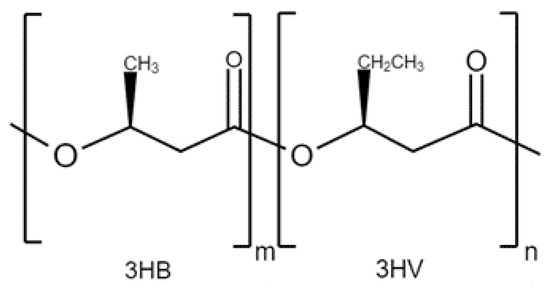
Poly(3-hydroxybutyrate-co-3-hydroxyvalerate) (PHBV) is a biodegradable and biocompatible biopolymer that has gained popularity in the field of biomedicine. PHBV has shown to be a versatile platform for drug delivery, offering controlled release, enhanced therapeutic efficacy, and reduced side effects. The encapsulation of various drugs, such as anticancer agents, antibiotics, and anti-inflammatory drugs, in PHBV nanoparticles or microspheres has been extensively investigated, demonstrating enhanced drug stability, prolonged release kinetics, and increased bioavailability. Additionally, PHBV has been used as a scaffold material for tissue engineering applications, such as bone, cartilage, and skin regeneration. The incorporation of PHBV into scaffolds has been shown to improve mechanical properties, biocompatibility, and cellular interactions, making them suitable for tissue engineering constructs. 

polyhydroxyalkanoates (PHA) Poly(3-hydroxybutyrate-co-3-hydroxyvalerate) (PHBV) regeneration drug delivery nanoparticle

1. Introduction

PHBV is a copolymer originated from the incorporation of (3HV) monomers in P(3HB) biopolymer. The structure is shown in Figure 1. The incorporation of these monomers makes P(3HB) less fragile, which is the principal limitation to replace polypropylene, making it more flexible and resistant [1]. The amount of (3HV) in the polymer also affects its structure, as higher concentrations result in a more amorphous particle. Note that the release of the encapsulated drug or factor is not based on matrix degradation, but on diffusion through the amorphous regions of the sphere, so its surface modification plays a very important role in its biodegradability [2].
Figure 1. The general structure of PHBV.
In addition, PHBV has high immunotolerance, greater chemical inactivity, and has already been developed on an industrial scale. Some limitations of this material include its marked hydrophobicity, high fragility, low impact resistance, and poor thermal stability. However, these problems can be addressed by reinforcing PHBV [3] with other materials, such as other polymers, natural fibers [4], nanometals [5], nano-cellulose [6], and carbon nanotubes [7][8] among others. A concrete example is the use of bacterial cellulose (BC) which is a sustainable polysaccharide produced by bacteria that can be mixed with PHBV. Toxicological studies have shown that BC is non-toxic and does not provoke inflammatory responses or oxidative stress at the cellular level. In vitro and in vivo tests confirm the safety of this biopolymer in medical applications. Both BC and PHBV meet the requirements for clinical use [9].

2. Applications

2.1. PHBV Composites for Drug Delivery Applications

Drug encapsulation and delivery systems provide significant pharmacological advantages, such as enhancing the physical and chemical stability of encapsulated active ingredients, increasing bioavailability, acting as controlled-release systems, reducing fluctuations in drug concentrations in the blood, penetrating specific barriers and tissues, reducing adverse effects, and protecting the encapsulated molecule [10].
To achieve better control over drug release, some strategies have been developed to vary particle size distributions and degradability. One important fact is that pore formation must be avoided since it promotes rapid drug release. The control of particle size and porosity is achieved by optimizing parameters such as solution concentration, surfactant concentration, the polymer molecular weight, the homogenization technique, solvent characteristics, and the environment [11]. In the case of PHBV, studies have shown that the compound’s molecular weight and biomaterial concentration are related to an effect on particle size [12].
Due to their characteristics, PHAs have been used for the synthesis of microspheres, microcapsules, or nanoparticles for the treatment of various diseases.
Regarding liver cancer, PHBV nanoparticles could treat one of the leading causes of death, hepatocellular carcinoma (HCC), typically caused by chronic infections such as hepatitis B and C. One study proposed a solution based on using a drug delivery system loaded with paclitaxel (PTX) and coated with pH-sensitive dopamine, which helps achieve controlled drug release.
Regarding colon cancer, PHBV and poly (lactic-co-glycolic acid) (PLGA) nanoparticles with encapsulated 5-Fluorouracil (5-FU) were developed. It was observed that this common drug used for this disease was less toxic when encapsulated. Furthermore, the drug derived from the nanoparticles showed better results in reducing tumor volume than the drug in the free form [13]. In the previous year, the same research group investigated the release of two drugs, the mentioned 5-FU and oxaliplatin, using the same nanoparticles. The results showed that they are a good option for intravenous administration because of their hemocompatibility [14]. A different research group conducted characterization and biocompatibility tests on PHBV nanoparticles, also using 5-FU for the same purpose. The size and morphology were studied using scanning electron microscopy (SEM). Cytotoxicity was evaluated using human adenocarcinoma cells. The results showed a significant reduction in cell viability, indicating that drug-loaded nanoparticles were a promising method for killing cancer cells [15]
When it comes to breast cancer, the drug Docetaxel is commonly used. The principal limitation is that it is partially eliminated by the liver and kidneys when administrated via systemic circulation. This loss causes an increment in dosage that may lead to side effects. A solution is to use PHBV nanoparticles as a method of controlled drug release. Results showed that docetaxel-loaded nanoparticles could protect the drug in the biological fluid and increase cytotoxicity in vitro by inducing apoptosis in breast cancer cell lines (MCF7). 
The behavior of PHBV nanoparticles was also investigated in two different epithelial cell lines: HeLa (cervical cancer cells) and SKOV-3 (ovarian cancer cells). The results showed that the mechanism of absorption of PHBV nanospheres depended on time, energy, concentration, and the internalization mechanism of the cell line. The destination of the nanoparticles was also determined. In both cell lines, the nanoparticles ended up in the lysosomes where they were degraded, releasing their content (in this case markers).
Against the human lung cancer cell line A549, PHBV nanoparticles loaded with the drug ellipticine demonstrated significant anticancer activity [16]. The nanoparticle was synthesized in the past by the same group, using Bacillus cereus FB11, under nutritional stress conditions and using glucose as the only carbon source [17]. Another group investigated the cytotoxicity of nanoparticles loaded with sunitinib for the same purpose. 
Nanoparticles have another application in the field of photodynamic therapy (PDT) for various types of cancer. In this technique, a photosensitizer (PS) is encapsulated in the nanoparticles, which can accumulate in cancerous tissues. When it is excited with a specific wavelength of light in the presence of molecular oxygen, the PS produces radical oxygen species that locally kill cancer cells. A research group used a nano-precipitation method to prepare a PHBV nanocarrier stabilizer with polyethylene glycol (PEG) lipids. These studies demonstrated the system’s efficacy for PS administration [18]
PHBV nanoparticles also have potential in the treatment of Parkinson’s disease. In a study, these nanocarriers were used to deliver the drug pramipexole. The objective was to reduce the drug’s plasma fluctuations by subcutaneous injection of nanoparticles. The encapsulation efficiency and drug loading were determined using ultraviolet–visible spectrophotometry. The release profile was evaluated using a dialysis method. The results demonstrated that the drug-delivery system exhibited high encapsulation efficiency and drug loading, in addition to a sustained release profile [19].
Another potential application for nanocarriers is as transdermal therapy for skin conditions such as psoriasis, ultraviolet damage, and aging. In an article, the nanospheres were prepared using the oil-in-water (o/w) technique. Cytotoxicity of the particles was assessed using the 3-(4,5-dimethylthiazol-2-yl)-2,5-diphenyltetrazolium bromide assay (MTT) with L929 mouse fibroblasts, BALB/3T3 mouse embryo fibroblasts, and human HaCaT keratinocytes. The genotoxicity of the carriers was determined using the Ames test. The ability of the particles to penetrate the membrane of human osteosarcoma Saos 2 cells and accumulate in their cytoplasm was also evaluated. Additionally, penetration through human skin was investigated using samples obtained from a 42-year-old female donor after undergoing breast surgery. All tests had satisfactory results, indicating that the nanoparticles can be used for personalized treatment through topical administration [20].

2.2. PHBV Composites for Tissue Engineering Applications

Tissue engineering is a discipline of biomedicine that aims to promote the regeneration of the structure and functionality of damaged tissues in the body by using biomaterials capable of guiding metabolic processes and cellular activity. In this field, PHAs have been widely used for the synthesis of three-dimensional cellular scaffolds with improved structural and functional properties for bone and cartilage regeneration [21]. They can also be used as controlled-release systems of bioactive molecules and cellular signals to induce tissue regeneration. A specific example is the development of PLGA and PHBV nanoparticles incorporated into scaffolds to have a controlled release system of BMP-2 and BMP-7 morphogenetic proteins [22].
Another method to create biodegradable PHBV scaffolds is using selective laser sintering (SLS). It is demonstrated that the unique microstructure formed by this technique has the potential for conducting in vitro and in vivo tests [23]. In another publication, the degradation behavior of the scaffolds was investigated by incubating them in phosphate-buffered saline (PBS) for 6 weeks. SEM was used to characterize the microstructure of the scaffolds before and after incubation in PBS. The integrity of the structure was not affected and the molecular weight slightly decreased. The PHBV scaffolds fabricated with SLS generally showed adequate mechanical properties and good structural integrity after incubation [24]. The same technique was used by another group, in this case, to fabricate a nanocomposite of CaP/PHBV. The in vitro studies revealed a high viability of SaOS-2 cells and normal morphology and phenotype after 3 and 7 days of cultures on all scaffolds. The release behavior of bovine serum albumin (BSA) in this nanostructure was also studied. 
The PHBV biopolymer has also been used in neural-tissue regeneration, where it promotes cellular adhesion, proliferation, and differentiation. Its potential in treating various neural injuries and diseases has been demonstrated [25]. With this aim, an electrospinning method was used to produce a chitosan-crosslinked nanofibrous biodegradable PHBV scaffold. The structure and cell culture assays using Schwann cells were microscopically, physically, and mechanically analyzed. The cells were able to grow fine on the created platform, indicating that this mixture of materials can be a promising candidate for applications in nerve conduits. The obtained results suggest that the generated material possesses suitable characteristics for further in vivo studies [26]
There is another article that also discusses PHBV/PLA/collagen membranes for duroplasty after decompression in rats with SCI. The study focused on evaluating the material and the biological characteristics, subcutaneous implantation tests, and contusion SCI tests in rats to investigate the effects of the membranes on inflammasome activation and macrophage polarization. The results demonstrated that duroplasty with PHBV/PLA/Collagen membranes reduced glial scar formation and promoted axonal growth by inhibiting inflammasome activation and modulating macrophage polarization in acute SCI. Functional locomotor recovery improved 8 weeks after the injury [27].
Various research groups are dedicated to exploring the most suitable materials and techniques for cartilage tissue engineering. One group of investigators used a conjugation of PHBV with type I collagen to produce a mechanically stable, biodegradable, and adhesive cell scaffold. The characterization of the scaffold was carried out using techniques such as SEM, Attenuated total reflection FTIR, atomic force microscopy, and electron spectroscopy for chemical analysis. Furthermore, the degradation and behavior of fibroblasts on nanofibrous scaffolds were studied. According to the article, electrospun PHBV/collagen composite nanofibrous scaffolds exhibited good mechanical properties, biocompatibility, and biodegradability. These scaffolds favored the growth and proliferation of fibroblast cells, and the cells demonstrated adequate adhesion and dispersion on the scaffolds. Results also revealed that the rate of degradation of the scaffolds could be controlled by adjusting the ratio of PHBV to collagen. The study suggested that electrospun PHBV/collagen composite nanofibrous scaffolds have potential applications in tissue engineering [28]. In a publication in 2022, one group of investigators studied the influence of the addition of Bioglass into PHBV porous platforms. Cartilage progenitor cells (CPCs) were seeded into the control and the PHBV/10% Bioglass scaffolds. The CPC-constructs were exposed to a 6-week in vitro chondrogenic induction culture and then transplanted in vivo for another 6 weeks to see the difference between the CPC-PHBV and CPC-PHBV/10% Bioglass platforms. Compared to the control, the PHBV/ 10% Bioglass scaffold had better properties and results like hydrophilicity, a higher percentage of adherent cells, and significant production of cartilage-like tissues. Also, the polymerase chain reaction analysis showed that aggrecan, collagen II, and SOX-9 from the CPC-PHBV/10% Bioglass scaffolds were more expressed compared to the CPC-PHBV ones. All indicate that the addition of Bioglass to PHBV can improve the chondrogenic differentiation of CPCs [29].
For wound healing, one group created a dressing made of a bilayer scaffold produced by electrospinning a hydrophilic PUL fibrous membrane (barrier layer) onto a wet electrospun hydrophobic PHBV fibrous mat (regenerative layer). The study optimized the production of PHBV using a bacterial strain called Cupriavidus necator and characterized the resulting polymer using various techniques, including Proton nuclear magnetic resonance (1H-NMR) and FTIR. The valerate molar percentage and the average molecular weight of the polymer were also determined using the 1H-NMR and SLS techniques, respectively. In vitro studies showed that the PUL membrane maintained L929 cell proliferation and prevented cells from migrating within the barrier phase, while the PHBV layer supported cell viability, proliferation, and migration, creating a regenerative 3D structure. The results showed that the new PUL/PHBV bilayer scaffold was a promising candidate for wound-healing applications [30]
To fix abdominal wall defects, which can be caused by abdominal trauma or congenital rupture, silk fibroin and PHBV scaffolds were prepared. Characterization and cytotoxicity assays, in vitro tests to contemplate cell morphology and viability, and q-PCR to detect the gene expression of growth factor TGF-β1 and collagen I, were performed. In vivo studies were also achieved in Sprague Dawley rats. After 7 and 15 days of implanting the scaffold, an evaluation of their in vivo tissue regeneration capacity was performed. All the results pointed out that the hybrid nanofiber SF/PHBV scaffolds had a high efficiency and biocompatibility to repair abdominal wall defects [31].

3. Conclusions

In conclusion, biopolymers of the PHA family are very promising in a wide range of applications due to their natural origin and their excellent properties, including biocompatibility and biodegradability. In the field of medicine, these biomaterials are particularly relevant as their degradation products do not produce toxic agents for the organism. This biocompatibility is essential for their use in biomedical applications such as tissue engineering, implants, sutures, and controlled drug-delivery systems.

References

  1. Anjum, A.; Zuber, M.; Zia, K.M.; Noreen, A.; Anjum, M.N.; Tabasum, S. Microbial production of polyhydroxyalkanoates (PHAs) and its copolymers: A review of recent advancements. Int. J. Biol. Macromol. 2016, 89, 161–174.
  2. Papaneophytou, C.; Katsipis, G.; Halevas, E.; Pantazaki, A.A. Polyhydroxyalkanoates applications in drug carriers. In Biotechnological Applications of Polyhydroxyalkanoates; Springer: Singapore, 2019; pp. 77–124.
  3. Rivera Briso, A.L.; Serrano Aroca, Á. Métodos de refuerzo mecánico del poli(3-hidroxibutirato-co-3-hidroxivalerato) para aplicaciones industriales avanzadas. Rev. Iberoam. Interdiscip. Métodos Model. Simul. 2018, 10, 79–94.
  4. Singh, S.; Mohanty, A.K. Wood fiber reinforced bacterial bioplastic composites: Fabrication and performance evaluation. Compos. Sci. Technol. 2007, 67, 1753–1763.
  5. Silverman, T.; Naffakh, M.; Marco, C.; Ellis, G. Effect of WS2 Inorganic Nanotubes on Isothermal Crystallization Behavior and Kinetics of Poly(3-Hydroxybutyrate-co-3-hydroxyvalerate). Polymers 2018, 10, 166.
  6. Yu, H.; Qin, Z.; Zhou, Z. Cellulose nanocrystals as green fillers to improve crystallization and hydrophilic property of poly(3-hydroxybutyrate-co-3-hydroxyvalerate). Prog. Nat. Sci. Mater. 2011, 21, 478.
  7. Montanheiro, T.L.; Cristóvan, F.H.; Machado, J.P.; Tada, D.; Durán, N.; Lemes, A.P. Effect of MWCNT functionalization on thermal and electrical properties of PHBV/MWCNT nanocomposites. J. Mater. Res. 2015, 30, 55–65.
  8. Vidhate, S.; Innocentini-Mei, L.; D’Souza, N.A. Mechanical and electrical multifunctional poly(3-hydroxybutyrate-co-3-hydroxyvalerate)-multiwall carbon nanotube nanocomposites. Polym. Eng. Sci. 2012, 52, 1367–1374.
  9. Râpă, M.; Stefan, L.M.; Seciu-Grama, A.; Gaspar-Pintiliescu, A.; Matei, E.; Zaharia, C.; Stănescu, P.O.; Predescu, C. Poly(3-hydroxybutyrate-co-3-hydroxyvalerate) (P(3HB-co-3HV))/Bacterial Cellulose (BC) Biocomposites for Potential Use in Biomedical Applications. Polymers 2022, 14, 5544.
  10. Gómez-Gaete, C. Nanopartículas poliméricas: Tecnología y aplicaciones farmacéuticas (Polymeric nanoparticles: Technologie and pharmaceutical applications). Rev. Farmacol. Chile 2014, 7, 7–16.
  11. Göz, E.; Karakeçili, A. Effect of emulsification-diffusion parameters on the formation of poly (3-hydroxybutyrate-co-3-hydroxyvalerate) particles. Artif. Cells Nanomed. Biotechnol. 2016, 44, 226–234.
  12. Leimann, F.V.; Filho, L.C.; Sayer, C.; Araújo, P.H. Poly(3-hydroxybutyrate-co-3- hydroxyvalerate) nanoparticles prepared by a miniemulsion/solvent evaporation technique: Effect of phbv molar mass and concentration. Braz. J. Chem. Eng. 2013, 30, 369–377.
  13. Handali, S.; Moghimipour, E.; Rezaei, M.; Ramezani, Z.; Dorkoosh, F.A. PHBV/PLGA nanoparticles for enhanced delivery of 5-fluorouracil as promising treatment of colon cancer. Pharm. Dev. Technol. 2019, 25, 206.
  14. Handali, S.; Moghimipour, E.; Rezaei, M.; Saremy, S.; Dorkoosh, F.A. Co-delivery of 5-fluorouracil and oxaliplatin in novel poly (3-hydroxybutyrate-co-3-hydroxyvalerate acid)/poly(lactic-co-glycolic acid) nanoparticles for colon cancer therapy. Int. J. Biol. Macromol. 2019, 124, 1299–1311.
  15. Radu, I.C.; Hudita, A.; Zaharia, C.; Galateanu, B.; Iovu, H.; Tanasa, E.V.; Nitu, S.G.; Ginghina, O.; Negrei, C.; Tsatsakis, A.; et al. Poly(3-hydroxybutyrate-CO-3-hydroxyvalerate) PHBHV biocompatible nanocarriers for 5-FU delivery targeting colorectal cancer. Drug Deliv. 2019, 26, 318.
  16. Masood, F.; Chen, P.; Yasin, T.; Fatima, N.; Hasan, F.; Hameed, A. Encapsulation of Ellipticine in poly-(3-hydroxybutyrate-co-3-hydroxyvalerate) based nanoparticles and its in vitro application. Mater. Sci. Eng. C 2013, 33, 1054–1060.
  17. Masood, F.; Chen, P.; Yasin, T.; Hasan, F.; Ahmad, B.; Hameed, A. Synthesis of poly-(3-hydroxybutyrate-co-12 mol % 3-hydroxyvalerate) by Bacillus cereus FB11: Its characterization and application as a drug carrier. J. Mater. Sci. Mater. Med. 2013, 24, 1927–1937.
  18. Pramual, S.; Lirdprapamongkol, K.; Svasti, J.; Bergkvist, M.; Jouan-Hureaux, V.; Arnoux, P.; Frochot, C.; Barberi-Heyob, M.; Niamsiri, N. Polymer-lipid-PEG hybrid nanoparticles as photosensitizer carrier for photodynamic therapy. J. Photochem. Photobiol. 2017, 173, 12–22.
  19. Bahari-Javan, N.; Jafary-Omid, N.; Moosavi-Hasab, N.; Rezaie-Shirmard, L.; Rafiee-Tehrani, M.; Dorkoosh, F. Preparation, statistical optimization and in vitro evaluation of pramipexole prolonged delivery system based on poly (3-hydroxybutyrate-co-3-hydroxyvalerate) nanoparticles. J. Drug Deliv. Sci. Technol. 2018, 44, 82–90.
  20. Eke, G.; Goñi-de-Cerio, F.; Suarez-Merino, B.; Hasirci, N.; Hasirci, V.N. Biocompatibility of Dead Sea Water and retinyl palmitate carrying poly(3-hydroxybutyrate-co-3-hydroxyvalerate) micro/nanoparticles designed for transdermal skin therapy. J. Bioact. Compat. Polym. 2015, 30, 455–471.
  21. Koller, M. Advances in polyhydroxyalkanoate (PHA) production. Bioengineering 2017, 4, 88.
  22. Yilgor, P.; Hasirci, N.; Hasirci, V. Sequential BMP-2/BMP-7 delivery from polyester nanocapsules. J. Biomed. Mater. Res. A 2010, 93A, 528–536.
  23. Diermann, S.H.; Lu, M.; Zhao, Y.; Vandi, L.; Dargusch, M.; Huang, H. Synthesis, microstructure, and mechanical behaviour of a unique porous PHBV scaffold manufactured using selective laser sintering. J. Mech. Behav. Biomed. Mater. 2018, 84, 151–160.
  24. Diermann, S.H.; Lu, M.; Edwards, G.; Dargusch, M.; Huang, H. In vitro degradation of a unique porous PHBV scaffold manufactured using selective laser sintering. J. Biomed. Mater. Res. 2019, 107, 154–162.
  25. Gheibi, A.; Khoshnevisan, K.; Ketabchi, N.; Derakhshan, M.A.; Babadi, A. Application of Electrospun Nanofibrous PHBV Scaffold in Neural Graft and Regeneration: A Mini-Review. J. Nanomed. Res. 2016, 1, 107–111.
  26. Biazar, E.; Heidari-Keshel, S. Development of chitosan-crosslinked nanofibrous PHBV guide for repair of nerve defects. Artif. Cells Nanomed. Biotechnol. 2014, 42, 385–391.
  27. Zhao, T.; Xu, K.; Wu, Q.; Wang, C.; Xiao, S.; Li, H.; He, T.; Wang, L.; Li, F.; Chen, Q. Duraplasty of PHBV/PLA/Col membranes promotes axonal regeneration by inhibiting NLRP3 complex and M1 macrophage polarization in rats with spinal cord injury. FASEB J. 2020, 34, 12147.
  28. Meng, W.; Kim, S.Y.; Yuan, J.; Kim, J.C.; Kwon, O.H.; Kawazoe, N.; Chen, G.; Ito, Y.; Kang, I.K. Electrospun PHBV/collagen composite nanofibrous scaffolds for tissue engineering. J. Biomater. Sci. Polym. Ed. 2007, 18, 81–94.
  29. Xue, K.; Zhang, S.; Ge, J.; Wang, Q.; Qi, L.; Liu, K. Integration of Bioglass Into PHBV-Constructed Tissue-Engineered Cartilages to Improve Chondrogenic Properties of Cartilage Progenitor Cells. Front. Bioeng. Biotechnol. 2022, 10, 868719.
  30. Dalgic, A.D.; Koman, E.; Karatas, A.; Tezcaner, A.; Keskin, D. Natural origin bilayer pullulan-PHBV scaffold for wound healing applications. Biomater. Adv. 2022, 134, 112554.
  31. Gong, W.; Cheng, T.; Liu, Q.; Xiao, Q.; Li, J. Surgical repair of abdominal wall defect with biomimetic nano/microfibrous hybrid scaffold. Mater. Sci. Eng. C 2018, 93, 828–837.
More
Information
Contributors MDPI registered users' name will be linked to their SciProfiles pages. To register with us, please refer to https://encyclopedia.pub/register : , , , , ,
View Times: 248
Revisions: 2 times (View History)
Update Date: 28 Sep 2023
1000/1000
Video Production Service