Submitted Successfully!
To reward your contribution, here is a gift for you: A free trial for our video production service.
Thank you for your contribution! You can also upload a video entry or images related to this topic.
Version Summary Created by Modification Content Size Created at Operation
1 -- 4070 2023-09-26 12:04:11 |
2 Reference format revised. Meta information modification 4070 2023-09-28 02:44:29 |

Video Upload Options

Do you have a full video?

Confirm

Are you sure to Delete?
Cite
If you have any further questions, please contact Encyclopedia Editorial Office.
Urooj, S.; Anjum, S.; Iqbal, F.; Abduh, M.S.; Akhtar, H.; Javed, S.; Kakar, S.J.; Ikram, A.; Maqbool, N.A.; Ahmad, T. Clinical Aspects of Hepatitis E Virus. Encyclopedia. Available online: https://encyclopedia.pub/entry/49660 (accessed on 06 September 2024).
Urooj S, Anjum S, Iqbal F, Abduh MS, Akhtar H, Javed S, et al. Clinical Aspects of Hepatitis E Virus. Encyclopedia. Available at: https://encyclopedia.pub/entry/49660. Accessed September 06, 2024.
Urooj, Sidra, Sadia Anjum, Fareeha Iqbal, Maisa Siddiq Abduh, Hashaam Akhtar, Sumbal Javed, Salik Javed Kakar, Aamer Ikram, Nabeel Ahmed Maqbool, Tahir Ahmad. "Clinical Aspects of Hepatitis E Virus" Encyclopedia, https://encyclopedia.pub/entry/49660 (accessed September 06, 2024).
Urooj, S., Anjum, S., Iqbal, F., Abduh, M.S., Akhtar, H., Javed, S., Kakar, S.J., Ikram, A., Maqbool, N.A., & Ahmad, T. (2023, September 26). Clinical Aspects of Hepatitis E Virus. In Encyclopedia. https://encyclopedia.pub/entry/49660
Urooj, Sidra, et al. "Clinical Aspects of Hepatitis E Virus." Encyclopedia. Web. 26 September, 2023.
Clinical Aspects of Hepatitis E Virus
Edit

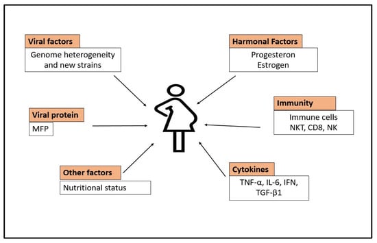
Hepatitis E virus (HEV) is a single-stranded, positive RNA virus. The HEV is the causing agent of hepatitis, with a high prevalence rate in low-income countries due to poor sanitary conditions. It can exhibit acute, continuous, or extrahepatic consequences in immunocompromised individuals such as those undergoing organ transplantation and having HIV infection. HEV infection is either self-limiting (silent), meaning the patient will possibly recover on his own, or symptomatic, causing acute liver injury or fulminant hepatitis, and may eventually cause death. It can also cause chronic hepatitis that can progress to cirrhosis or recovery. Pregnancy-related HEV infection has an incidence rate of 30%. HEV escape from innate immunity, hormonal imbalances, defective monocyte–macrophage function, downregulation of the T-cell-mediated immune system, high cytokine production, nutritional factors, and socioeconomic conditions may play fundamental roles in the prevalence of HEV infection. It is necessary to take particular measures to reduce the incidence burden of HEV infection in high endemic locations. 

hepatitis E virus (HEV) diagnosis treatments

1. Introduction

The hepatitis E virus (HEV) is one of the causal agents of hepatitis [1]. It typically results in 0.5% to 3% terminal illness in young adults, rising to 30% in expecting women [2]. HEV can exhibit acute hepatitis and continuous or extrahepatic consequences. HEV infects the liver and other organs and may get damaged indirectly. Chronic HEV infection is typically linked with immunocompromised individuals such as those undergoing organ transplantation or individuals with HIV infection or leukemia. Figure 1 shows various routes of HEV transmission.
Figure 1. Circulation of HEV infection. The diagram depicts various routes of HEV transmission.

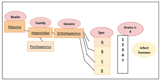
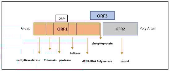
2. Classification and Molecular Characteristics of HEV

Hepatitis E virus (HEV) belongs to the Hepeviridae family of viruses, which consists of two genera. The genus Piscihepevirus comprises the cutthroat trout virus, whereas the genus Orthohepevirus consists of all avian and mammalian hepatitis E virus isolates. There are four distinct species within the genus Orthohepevirus, namely A, B, C, D [3]. Species A comprises HEV isolates from human, deer, wild and domestic pigs, rabbits, camels, and mongoose. Species B consists of all avian HEV isolates from birds. Species C consists of all HEV isolates from greater bandicoots, rats, Asian musk shrews, minks, and ferrets. Species D comprises HEV isolates from bats. All HEV isolates that are infectious to humans belong to species A, which comprises eight genotypes, as shown in Figure 2. Genotype 1 HEV (majorly Asian isolates) and genotype 2 HEV (Mexican and some African isolates) only infect humans. Genotype 3 (human, rabbit, pig, mongoose, deer) and 4 isolates infect humans and a few other animal species. Genotype 5 and 6 HEVs infect wild boars. Genotype 7 HEV infects camels and, reportedly, humans, while genotype 8 HEV infects Bactrian camels [4].
Figure 2. HEV classification. The family Hepeviridae has the genera ‘Orthohepevirus’. It has four species, of which species A contains eight viral genotypes. Genotypes 1, 2, 3, 4 and 7 mainly infect humans [4].
HEV is a nonenveloped virus that measures between 27 and 34 nanometers in diameter (nm). It comprises a single-stranded RNA genome (positive) and measures about 7.2 kb in length. Virological and hepatological characteristics of HEV are comparable to those of the hepatitis A virus (HAV). HEV is known to be present in blood with a coating of the lipid membrane, but it is not known to be encased in bile, and its primary source is fecal contamination. The HEV genome comprises a 5’ short non-coding region (NCR), ORF1, ORF2, ORF3, and ORF4 (specific to genotype 1) [4]. The non-coding regions of the viral genome are involved in non-structural proteins. The ORF3 region is responsible for encoding a small multipurpose protein, and ORF2 is related to the transcription of the viral coat protein. A poly adenosine tail of around 150–200 bases is present at the 3′ terminal (3′’ non-coding region of the genome). The ORF1 region of the HEV genome comprises the fourth open reading frame (ORF4) [5]. When endoplasmic reticulum stress occurs, ORF4 will take part and encodes a protein of 20 kDa. This protein regulates the viral RNA-dependent RNA polymerase activity via host–viral protein interactions. However, ORF4 is only expressed in HEV strains of genotype 1, even though it is present in all strains [6]. The functional domains of HEV are illustrated in Figure 3.
Figure 3. Functional domains of the HEV genome: methyltransferase domain, a Y domain, a PCP enzymatic domain, RNA helicase, phosphoprotein domain (ORF3), capsid-coding ORF2 region, and RNA-dependent RNA polymerase [5].

3. Manifestations of HEV Infection

After being exposed to HEV for two to nine weeks, patients begin to experience clinical symptoms such as:
  • Myalgia;
  • Arthralgia;
  • Anorexia;
  • Hepatomegaly;
  • Fever;
  • Weakness;
  • Vomiting;
  • Jaundice.
HEV can occasionally result in abrupt liver failure. Although chronic cases are rare, they are evident in immunocompromised people, and at the same time, acute hepatitis is more frequent in adults [7].
Even though the infection may not induce symptoms in some people, it may also cause clinical illness in another group. Notably, the clinical disease begins to manifest 15–60 days (on average, at 40 days) after HEV infection. At first, these symptoms are nonspecific and include restlessness, anorexia, nausea, fatigue, myalgia, and abdominal pain. Later, acute hepatitis symptoms include jaundice, dark urine, pale stools, and the appearance of hepatomegaly.
When it becomes symptomatic, IgM antibodies against HEV are elevated in the sera of infected persons and are detectable over 14 days to 12 weeks, with a sensitivity of 99.4% and specificity of 74.3%. IgG antibodies, which show a prior HEV infection in sick persons, develop in later stages and last for many years after the virus has dissipated. According to laboratory studies, HEV infection is responsible for high bilirubin levels in serum and a significant increase in enzymes in the liver [8]. The typical incubation time is 40 days (15–60 days).

4. Incidence Rate of HEV in Developing and Developed Nations

The rapidly spreading HEV causes acute viral hepatitis and is the leading cause of acute hepatitis infection in adults in Central Asia, the Indian subcontinent, and Southeast Asia. After hepatitis B, it is the second most common cause of acute hepatitis infection in the regions of the Middle East and North Africa. According to global estimates, over 20 million new cases of HEV infections occur yearly, of which 3.4 million are symptomatic [9]. The WHO reported 44,000 fatal HEV cases, roughly representing 3.3% of all deaths caused by viral hepatitis [10]. The frequency of HEV infection ranges from 7.2% to 35% in impoverished nations with generally poor health conditions. The prevalence rate, however, is approximately 3% in developed countries. The death rate for this virus, which is often self limiting, ranges from 1% to 3% and increases in cases of pregnant women [11].
Several variables, including varied degrees of virus exposure, different living standards in distinct regions, and various modes of viral transmission, can impact the distribution of HEV infection within a state. In nations with inadequate sanitation systems, gt1 and gt2, as human pathogens, can cause HEV endemics or epidemics [12].
In developed nations, food-borne zoonosis is thought to be the most typical method of HEV transmission. The discovery of HEV in pigs with high similarity to HEV strains reported in people was the first source of proof for HEV zoonosis. Pigs, deer, rabbits, mongooses, cattle, sheep, and horses are among the animals that can contract genotypes 3 and 4 [13]. In multiple case reports, food-borne strains of HEV are described. Undercooked or raw pork, pig liver, sausages, shellfish, green vegetables, and strawberries have recently been recognized as significant risk factors for human HEV infection [14].
Until now, only one incidence of genotype 7 infection has been reported in humans. The infected person is from the United Arab Emirates who frequently consumed camel milk and meat products and experienced a liver transplant. Therefore, paying more attention to the prevalence of camel-based zoonotic HEV in that region is essential [15]. The coliform tract is associated with HEV spread, like other viruses. HEV is self-limiting in young women indicating a low infectious load (0.1–4%).
Manifestations of gastrointestinal hepatitis are distributed differently worldwide, with genotype 1 being more widespread in Asia, Africa, and Latin America and genotype 2 being more widespread in sub-Saharan Africa and Mexico. Both vulnerable and healthy populations can contract genotypes 3 and 4, which are primarily found as sporadic cases in developed countries [9]. Viral hepatitis A (HAV) and HEV coinfection generally showed a higher prevalence in the summer, autumn, and winter (December to May).
Hepatitis E is an "emerging infection" in developed nations. It would be more accurate to call it locally acquired hepatitis E in these nations. The prevalence of IgG antibodies against HEV infection in developed countries was reported to be 5% in many early investigations, and therefore, hepatitis E is not a significant health concern in these areas.
Recently, HEV genotype 4 infection instances have been documented in Western nations like Belgium, Germany, and France. This genotype is highly prevalent in endemic in China, Japan, and Indonesia [16]. In 2011, Italy faced an epidemic of genotype 4 [17]. Since this endemic was not specific to travel-borne or food-borne illnesses from imported products, new strains are more likely to emerge. The most reported symptom in this outbreak was fatigue, which only affected male patients (mean age: 59 years). Children between 3 and 18 years old from low socioeconomic backgrounds have a 50% to 90% greater prevalence of HEV than their socioeconomically better-off peers. Young adults (15–39 years) have the highest infection rates, affecting both sexes equally.

5. Incidence Rate of HEV in Pregnant Females

According to studies, pregnant women demonstrate more significant signs of viral infection. Growing evidence shows that genotype 1 HEV infection, during the third trimester of pregnancy, is an important cause of maternal death in South Asia. A more severe infection that can occasionally lead to fulminant hepatitis is a hallmark of hepatitis E infection during pregnancy, especially in the third trimester, which raises morbidity and death rates for both mother and fetus [18].
Higher HEV rates have been associated with increased pregnancies in recently industrialized areas like Egypt, China, Pakistan, Nigeria, and Sudan. It is unclear which factor—viral pathogenicity, viral variations, immunological state, socioeconomic status, or a gap in prompt and effective care—is responsible for the catastrophic outcome of vaginal birth in each location. Pregnancy increases the risk of death for unknown reasons, especially during the second phase [19].
The prevalence of fulminant hepatitis is more common in pregnant women. A recent study demonstrated for the first time that pregnant women had greater hepatitis E incidence and fulminant rates than non-pregnant women and males. Lower progesterone receptor expression, greater interleukin, a higher viral load, and a poorer CD4/CD8 cell ratio are some of the mechanisms causing fulminant hepatitis during pregnancy [20]
The prevalence rate of HEV infection was higher in symptomatic women than in asymptomatic ones, as described in a multivariable meta-regression model. Maternal mortality, low birth weight, truncated gestational age, intrauterine deaths, and miscarriages were all linked to HEV infection. HEV during pregnancy was linked to several other outcomes, including a three-fold increase in intrauterine fetal immaturity, a two-to-three-fold increase in intrauterine fetal mortality, and additional effects.
The prevalence of HEV in the general population (women aged between 15 to 45 years) was between 5 and 22%, as predicted in earlier modelling research. Pregnant women are more susceptible to HEV infection than other hepatitis viruses such as A, B, and C [21]. The evidence suggests that hepatitis E contributes to pregnancy-related jaundice.
Another critical factor is the increased level of IgM in the serum of HEV-symptomatic women. In thirteen African countries, the ratio of immunoglobulin was higher in pregnant women in rural areas than in other demographics, with 0 to 84% HEV seroprevalence.
Regarding pregnancy outcomes, HEV infection in newborns delivered by HEV-positive mothers is generally harmful. However, children may typically survive without suffering a severe disease. Following a report from the Emirates, most children born to mothers who had HEV were either minimal, diagnosed with hepatitis, or passed away within a few hours of being born. However, the death of two newborns happened among HEV-infected mothers; one was due to an IUD and the other to an abortion (10%), which was 5% more than the rate among babies born to uninfected mothers. The incidence rate was significant in Ethiopia, with 14 IUDs and 10 preterm births in 40 pregnant women with chronic infection during the epidemic. 
Despite the colostrum’s anti-HEV antibodies and HEV genome presence, breastfeeding is safe in asymptomatic HEV-infected women. However, it is worrisome if the mother has high virus exposure or severe hepatitis. In these situations, formula feeding is suggested due to the danger of transfer from contaminated breast milk or from lesions on the nipple [22]

6. Relation of HEV with the Immune System during Pregnancy

6.1. T-Cell-Mediated Immune System

The adaptive immune system (T-cell mediated) is compromised during pregnancy to maintain the antigenic fetus inside the mother [23]. The ratio of T-helper type 1 and type 2 cells is changed, with a clear shift toward Th2 cells [24]. Most cytokine levels continue to lower until 20 weeks of gestation, a critical stage for the fetus’s survival. Through limiting cell-mediated immunity, cytokines, including TGF, IL-4, and IL-10, are released by the placenta, and immunological tolerance is aided by trophoblasts.

6.2. Defective Monocyte-Macrophage Function

Compared to HEV-induced acute liver injury, pregnant patients have impaired monocyte–macrophage function accompanied by decreased Toll-like receptor 3 and 7 expressions and reduced downstream signaling. Thus, an inadequate trigger for the innate immune response affects the severity and development of HEV-induced acute liver failure during pregnancy [25].

6.3. HEV-Induced Acute Liver Injury

In pregnant HEV-infected females with acute liver failure, it has been discovered that myeloid interactions and Toll-like receptor signaling are both compromised. As a result, the onset and intensity of hepatotoxicity brought on by HEV infection may be due to a deficiency of innate immunologic signals [25]. The DNA-binding activity of nuclear factor kappa B was considerably higher in pregnant patients with HIV-induced FHF compared to non-pregnant women and women with acute viral hepatitis (AVH) without FHF. These instances either had no p65 activation at all or had significantly less of it [26].

6.4. High Level of Cytokines

High levels of cytokines like TNF, IL-6, IFN, and TGF-1 may also be linked to worse pregnancy outcomes. HEV capsid protein decreases NF-B activity in the cell through blocking the UB-mediated proteasomal degradation of IκBα in human hepatic tissue, extending the viability of infected liver cells [27].

6.5. Hormonal Imbalance in HEV-Positive Pregnancy

Many steroid hormones are produced during pregnancy. These steroid hormones may inhibit CD4 cells while promoting virus replication. Women with acute liver failure infected with HEV have higher CD8 counts than CD4. Furthermore, compared to pregnancy without HEV or healthy pregnant women in the control group, expecting females with HEV-induced acute liver failure exhibited more significant estrogen, progesterone, and B-HCG levels [28]. In situations of pregnancy-related liver injury, fulminant hepatic failure and, ultimately, mortality are more likely due to the interaction between infectious agents and host immunological and hormonal variables.
Hormone levels in the control group were physiologically high but considerably higher in childbearing females with HEV infection, indicating a direct relationship between HEV and the immune system. While estrogen and ESR2 levels are biomarkers for maternal death during pregnancy with HEV infection, estrogen may also be a biomarker for preterm delivery [29]. Through the activation of the adaptor protein (ORF3 of HEV), the host factors induced by low immunity and hormonal imbalances are thought to harm cellular immunity and facilitate viral proliferation, leading to fulminant hepatitis during pregnancy.

6.6. Antibody-Mediated Hepatitis E Severity in Pregnancy

These results show a definite correlation between antibody levels and hepatitis E severity and imply antibody-mediated liver injury, as indicated in Figure 4. While IFNa levels declined in HEV-infected pregnant women in the later trimesters and were independent of healthy pregnancies, subclinical IFNa levels were higher, raising the question of whether these higher levels are responsible for the asymptomatic infection. The levels of four cytokines—CXCL10, IL10, sIL2RA, and IL6—and IgM-anti-HEV titers were correlated with ALT levels, and maternal females’ anti-HEV immunoglobulin was associated with how sick they were. Even if the relevant gene expression in the PBMCs is increased, the illness in pregnancy was linked to a considerable decrease in plasma cytokines [20].
Figure 4. Diagrammatic illustrations of HEV-induced liver injury during pregnancy. Various viral factors, hormonal factors, immunity, and nutritional status of a woman can play prominent roles during pregnancy. Several theories support that host factors, including immunological status, hormone levels, dietary imbalances, and viral factors, cause poor outcomes of HEV infection during pregnancy. Unfortunate pregnancy and birth outcomes are often linked to poor maternal nutrition. Micronutrient deficits, insufficient diet, and exposure to infectious disease results in immunologic compromise, diminished mucosal immunity, and altered cytokine expression that eventually increase the risk of HEV spread [20].
HEV recurrence in severe patients is linked to virus-related factors. HEV permanence is related to more significant quasispecies variability in the ORF1 and ORF2 domains during the acute stage of infection. Low quantities of the M domain protein of the virus are present in chronic HEV patients. The presence of T lymphocyte autoantigens in the M domains further emphasizes the significance of the cell-mediated immune response in eradicating HEV as well as HEV is inhibited via IRF-3 phosphorylation with help of X-domain [30]

7. Extrahepatic Manifestations of HEV

Over the past few years, our awareness of HEV-induced pathogenesis has greatly expanded. Hematological cancers, age, and prior liver condition are risk factors in the general population for developing fulminant hepatic failure (FHF) due to HEV gt1. Acute, non-travel-related hepatitis E is associated with HEV genotype 3,most prevalent in animals which can manifest as fulminant hepatitis with encephalopathy and coagulation issues [31].
Extrahepatic manifestations of HEV might be age dependent. HEV-highviral load in pregnant women may develop jaundice to neurological disease [32]. Moreover, a study from Pakistan said that neurological disorders were more prevalent among women (56.6%) with age of 30 to 50 years [33]. The majority of the patients were between the ages of 31 and 40. A related Indian study found that prevalence of neurological disorder is higher among women than men [34]. It is further reported that the most common symptom seen in twenty-eight (82%) patients is itching, which is followed by jaundice in twenty-seven (79%), nausea or vomiting in twenty-five (74%), neurological disease in eighteen (53%) cases, and damage to the gums or anterior naris in two cases. Eleven patients experienced sudden internal organ failure that caused sudden death.
There have been neurological symptoms associated with HEV genotypes 1 and 3. Patients who experienced neurological symptoms while infected with HEV had HEV RNA in their cerebrospinal fluid. Several studies have demonstrated a higher occurrence of HEV infection among Guillain–Barre syndrome (GBS) patients, and many of them have documented co-occurrence of acute hepatitis E with GBS.
Brachial neuritis or neuralgic amyotrophy (N.A.), also recognized as Parsonage–Turner syndrome, is a neurological ailment described by abrupt, severe pain in the shoulder, tailed by severe weakness. A European study compared 61 patients with HEV-NA to 61 patients without HEV-NA and found that HEV-NA more frequently causes bilateral irregular involvement (80.0% vs. 8.6%) and causes significant brachial plexus damage. 
HEV-associated membranoproliferative glomerulonephritis and membranous nephropathy are documented complications in kidney transplant cases. Kidney function and histology assessment in 51 SOT patients positively diagnosed with HEV genotype 3 infection showed a statistically noteworthy decline in glomerular filtration. Interestingly, cryoglobulinemia was cured, and renal function was amended along with improvement in proteinuria [35].
Hepatic viruses are also implicated in acute pancreatitis, often resulting from edema of the ampulla of Vater, which prevents the flow of pancreatic fluid. Acute pancreatitis was the only observable cause in single-center research reported from France, where 2.1% (16/790) of patients exhibited evidence of recent HEV infection through serological testing.

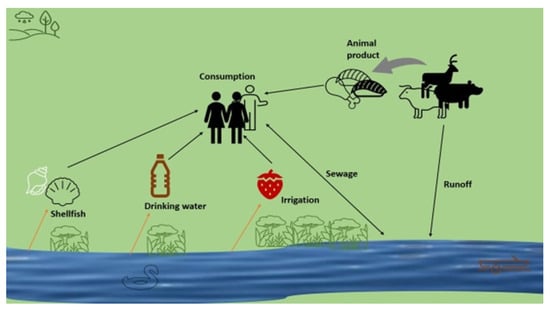
8. Transmission Route of HEV

Numerous investigations have shown the parenteral mode of infection to be via blood transfusion [36], along with the oro-fecal route being the primary route for hepatitis E infection. Studies regarding the transmission of HEV via dialysis demonstrate a significant seroprevalence of hepatitis E in the patients who received hemodialysis [37][38]; dialysis duration, however, does not correlate with HEV infection. Persons with uremia may be more vulnerable to HEV infection than people with normal renal function. Also, erythropoiesis is impaired in patients with final-stage renal failure and often needs blood transfusion. Although it has been shown that direct infection through blood transfusions can occur, blood products are not checked for HEV in most countries. The lack of a correlation between receiving blood products and the presence of HEV in serum, however, raises the possibility that HEV infection acquired via transfusion may not be the cause of the elevated anti-HEV IgG seroprevalence observed in the hemodialysis group. There may be brief or occasional outbreaks of HEV infection occurring within the dialysis unit that went unnoticed or were simply subclinical. This theory is supported by a study in which the patients receiving hemodialysis had anti-HEV IgM antibodies, even though none had a detectable HEV RNA genome [39].

9. Clinical Diagnosis of HEV

There is still no international alliance for lab HEV testing, and there are several different HEV tests with varying levels of specificity and sensitivity. Clinically, it is impossible to distinguish between severe acute hepatitis caused by HEV infection and other causes. A direct or indirect diagnosis of the disease can be made using a quantitative RT-PCR reaction to find HEV RNA in serum [40], plasma, or stool samples. Anti-HEV immunoglobulin M is detectable after acute HEV infection but is undetectable in the 16–24 weeks before clinical disease manifests.
Following the rapid development of anti-HEV immunoglobulin M, anti-HEV IgG persists several times, occasionally even for the remainder of the patient’s life [41]. Anti-HEV IgG is found in 95% of patients at their first clinical signs. Genomic DNA testing is essential to exclude active HEV infection due to the immunosuppressed group’s subpar antibody response. Additionally, immunochromatography and enzyme immunoassay [42] can be used for screening.
Standardizing diagnostic assays is crucial to identify as many cases of HEV infection as feasible. The World Health Organization is now working on developing reference materials for HEV serology that will eventually be accessible globally. 

10. Treatment Strategies for HEV Infection

10.1. Vaccination Therapy

Ribavirin and pegylated interferon are two primary options for HEV medical treatment. Pegylated interferon and ribavirin both prevent HEV replication in culture. Since then, pegylated interferon has been replaced with ribavirin as the primary medical therapy for acute and chronic HEV diseases. Focused investigations must clarify the ideal dosage and time length of ribavirin therapy [43]. Due to a high probability of acute repudiation, pegylated interferon is inappropriate in kidney transplant recipients. IFN-α is not advised for pregnant women [44].
It has been reported that some patients develop ribavirin tolerance and therapy inability, commonly attributed to a reduction in ribavirin dosage due to adverse effects, including anemia [45]. Ribavirin may cause viral clearance through applying mutagenesis pressure to the viral genome [46]; additionally, it changes the possibility of selecting resistant variations that do not respond. The viral polymerase variants Y1320H, K1383N, and G1634R are associated with resistance in addition to insertion in the hypervariable region. Because animal research showed that it has embryocidal and mutagenicity effects, therefore, pregnant women should not take ribavirin.
Childbearing females with HEV may be more inclined to use herbal remedies, which may also be a factor in the high mortality rates in some areas. High death rates among HEV-infected children under two have also been observed in Central Asia and Eastern Africa. HEV can sometimes be incorrectly diagnosed. Re-infection with HEV is also documented; it is recognized by a sharp rise in anti-HEV IgG levels and the appearance of HEV RNA [47].
One mTOR (mammalian target of rapamycin) inhibitor, everolimus, promotes HEV multiplication in vitro; it is well known that the calcineurin inhibitors tacrolimus and ciclosporin A have a pro-proliferative effect, in contrast to mycophenolate mofetil, which inhibits HEV replication in vitro [48].
Other drugs like rapamycin and everolimus promote HEV replication in vitro through blocking the mTOR, a molecular target of rapamycin, because the PI3K-PKB-mTOR pathway works as a cell limitation factor. As a result, when mTOR inhibitors are administered, increased HEV RNA amounts might be found in the blood [49].

10.2. Drug Development

Only 80% of patients treated with RBV experience viral clearance, and RBV is restricted in the significant-risk cohort of pregnant women, similar to pegIFN, highlighting the importance of novel therapy choices and clinical trials must be performed in the future. For the de novo discovery of drugs, screening libraries of compounds for their potential to interfere with viral lifespans can be used. It is necessary to have a molecular structure for the target to design antivirals for structure-guided development [50].

11. Conclusions

Due to unsanitary settings, HEV has a high prevalence rate in developing countries, which induces severe hepatitis infection. Humans are infected by genotypes 1 and 2, whereas genotypes 3 and 4 are animal-infection-causing agents. Pregnant women and others with impaired immune systems are seriously at risk. Because it is linked to undernourished populations and poor sanitation, HEV infection is understood to be a disease of poverty. Particular emphasis must be placed on reducing the burden of HEV infection in areas where HEV is highly endemic.

References

  1. Nishiyama, T.; Kobayashi, T.; Jirintai, S.; Kii, I.; Nagashima, S.; Primadharsini, P.P.; Nishizawa, T.; Okamoto, H. Screening of novel drugs for inhibiting hepatitis E virus replication. J. Virol. Methods 2019, 270, 1–11.
  2. Nimgaonkar, I.; Ding, Q.; Schwartz, R.E.; Ploss, A. Hepatitis E virus: Advances and challenges. Nat. Rev. Gastroenterol. Hepatol. 2018, 15, 96–110.
  3. Smith, D.B.; Purdy, M.A.; Simmonds, P. Genetic variability and the classification of hepatitis E virus. J. Virol. 2013, 87, 4161–4169.
  4. Sridhar, S.; Teng, J.L.L.; Chiu, T.-H.; Lau, S.K.P.; Woo, P.C.Y. Hepatitis E Virus Genotypes and Evolution: Emergence of Camel Hepatitis E Variants. Int. J. Mol. Sci. 2017, 18, 869.
  5. Kenney, S.P.; Meng, X.-J. Hepatitis E Virus Genome Structure and Replication Strategy. Cold Spring Harb. Perspect. Med. 2018, 9, a031724.
  6. Nair, V.P.; Anang, S.; Subramani, C.; Madhvi, A.; Bakshi, K.; Srivastava, A.; Shalimar; Nayak, B.; Ct, R.K.; Surjit, M. Endoplasmic Reticulum Stress Induced Synthesis of a Novel Viral Factor Mediates Efficient Replication of Genotype-1 Hepatitis E Virus. PLoS Pathog. 2016, 12, e1005521.
  7. Aslan, A.T.; Balaban, H.Y. Hepatitis E virus: Epidemiology, diagnosis, clinical manifestations, and treatment. World J. Gastroenterol. 2020, 26, 5543.
  8. Aggarwal, R.; Krawczynski, K. Hepatitis E: An overview and recent advances in clinical and laboratory research. J. Gastroenterol. Hepatol. 2000, 15, 9–20.
  9. Pérez-Gracia, M.T.; Suay, B.; Mateos-Lindemann, M.L. Hepatitis E: An emerging disease. Infect. Genet. Evol. 2014, 22, 40–59.
  10. World Health Organization (WHO). Hepatitis E. Fact Sheet. Geneva: WHO. 2018. Available online: http://www.who.int/news-room/fact-sheets/detail/hepatitis-e (accessed on 11 June 2023).
  11. Lin, S.; Zhang, Y.-J. Advances in Hepatitis E Virus Biology and Pathogenesis. Viruses 2021, 13, 267.
  12. Nelson, K.E.; Labrique, A.B.; Kmush, B.L. Epidemiology of Genotype 1 and 2 Hepatitis E Virus Infections. Cold Spring Harb. Perspect. Med. 2018, 9, a031732.
  13. Van der Poel, W.H. Food and environmental routes of Hepatitis E virus transmission. Curr. Opin. Virol. 2014, 4, 91–96.
  14. Yazaki, Y.; Mizuo, H.; Takahashi, M.; Nishizawa, T.; Sasaki, N.; Gotanda, Y.; Okamoto, H. Sporadic acute or fulminant hepatitis E in Hokkaido, Japan, may be food-borne, as suggested by the presence of hepatitis E virus in pig liver as food. J. Gen. Virol. 2003, 84, 2351–2357.
  15. Lee, G.-H.; Tan, B.-H.; Teo, E.C.-Y.; Lim, S.-G.; Dan, Y.-Y.; Wee, A.; Aw, P.P.K.; Zhu, Y.; Hibberd, M.L.; Tan, C.-K.; et al. Chronic Infection With Camelid Hepatitis E Virus in a Liver Transplant Recipient Who Regularly Consumes Camel Meat and Milk. Gastroenterology 2016, 150, 355–357.e3.
  16. der Honing, R.W.H.-V.; van Coillie, E.; Antonis, A.F.G.; van der Poel, W.H.M. First Isolation of Hepatitis E Virus Genotype 4 in Europe through Swine Surveillance in the Netherlands and Belgium. PLoS ONE 2011, 6, e22673.
  17. Garbuglia, A.R.; Scognamiglio, P.; Petrosillo, N.; Mastroianni, C.M.; Sordillo, P.; Gentile, D.; La Scala, P.; Girardi, E.; Capobianchi, M.R. Hepatitis E Virus Genotype 4 Outbreak, Italy, 2011. Emerg. Infect. Dis. 2013, 19, 110–114.
  18. Kar, P.; Sengupta, A. A guide to the management of hepatitis E infection during pregnancy. Expert Rev. Gastroenterol. Hepatol. 2019, 13, 205–211.
  19. Patra, S.; Kumar, A.; Trivedi, S.S.; Puri, M.; Sarin, S.K. Maternal and Fetal Outcomes in Pregnant Women with Acute Hepatitis E Virus Infection. Ann. Intern. Med. 2007, 147, 28–33.
  20. Wu, C.; Wu, X.; Xia, J. Hepatitis E virus infection during pregnancy. Virol. J. 2020, 17, 1–11.
  21. Fiore, S.; Savasi, V. Treatment of viral hepatitis in pregnancy. Expert Opin. Pharmacother. 2009, 10, 2801–2809.
  22. Chibber, R.M.; Usmani, M.A.; Al-Sibai, M.H. Should HEV infected mothers breast feed? Arch. Gynecol. Obstet. 2003, 270.
  23. Kraus, T.A.; Engel, S.M.; Sperling, R.S.; Kellerman, L.; Lo, Y.; Wallenstein, S.; Escribese, M.M.; Garrido, J.L.; Singh, T.; Loubeau, M.; et al. Characterizing the Pregnancy Immune Phenotype: Results of the Viral Immunity and Pregnancy (VIP) Study. J. Clin. Immunol. 2011, 32, 300–311.
  24. Zumla, A.; Bates, M.; Maeurer, M. Pregnancy and infection. New Engl. J. Med. 2014, 371, 1076.
  25. Sehgal, R.; Patra, S.; David, P.; Vyas, A.; Khanam, A.; Hissar, S.; Gupta, E.; Kumar, G.; Kottilil, S.; Maiwall, R.; et al. Impaired monocyte-macrophage functions and defective toll-like receptor signaling in hepatitis E virus-infected pregnant women with acute liver failure. Hepatology 2015, 62, 1683–1696.
  26. Prusty, B.K.; Hedau, S.; Singh, A.; Kar, P.; Das, B.C. Selective suppression of NF-kBp65 in hepatitis virus-infected pregnant women manifesting severe liver damage and high mortality. Mol. Med. 2007, 13, 518–526.
  27. Milan, S.; Varshney, B.; Lal, S.K. The ORF2 glycoprotein of hepatitis E virus inhibits cellular NF-κB activity by blocking ubiquitination mediated proteasomal degradation of IκBα in human hepatoma cells. BMC Biochem. 2012, 13, 7.
  28. Jilani, N.; Das, B.C.; Husain, S.A.; Baweja, U.K.; Chattopadhya, D.; Gupta, R.K.; Sardana, S.; Kar, P. Hepatitis E virus infection and fulminant hepatic failure during pregnancy. J. Gastroenterol. Hepatol. 2007, 22, 676–682.
  29. Swati, S.; Daga, M.K.; Kumar, A.; Husain, S.A.; Kar, P. Role of oestrogen and its receptors in HEV-associated feto-maternal outcomes. Liver Int. 2019, 39, 633–639.
  30. Nan, Y.; Yu, Y.; Ma, Z.; Khattar, S.K.; Fredericksen, B.; Zhang, Y.-J. Hepatitis E Virus Inhibits Type I Interferon Induction by ORF1 Products. J. Virol. 2014, 88, 11924–11932.
  31. Lhomme, S.; Marion, O.; Abravanel, F.; Chapuy-Regaud, S.; Kamar, N.; Izopet, J. Hepatitis E Pathogenesis. Viruses 2016, 8, 212.
  32. Dalton, H.R.; Kamar, N.; van Eijk, J.J.J.; Mclean, B.N.; Cintas, P.; Bendall, R.P.; Jacobs, B.C. Hepatitis E virus and neurological injury. Nat. Rev. Neurol. 2015, 12, 77–85.
  33. Awan, S.; Siddiqi, A.I.; Asif, A.; Ahmed, N.; Brohi, H.; Jalbani, S.; Wasay, M. Spectrum of neurological disorders in neurology outpatients clinics in urban and rural Sindh, Pakistan: A cross sectional study. BMC Neurol. 2019, 19, 1–6.
  34. Gourie-Devi, M.; Gururaj, G.; Satishchandra, P.; Subbakrishna, D. Prevalence of Neurological Disorders in Bangalore, India: A Community-Based Study with a Comparison between Urban and Rural Areas. Neuroepidemiology 2004, 23, 261–268.
  35. Kamar, N.; Weclawiak, H.; Guilbeau-Frugier, C.; Legrand-Abravanel, F.; Cointault, O.; Ribes, D.; Esposito, L.; Cardeau-Desangles, I.; Guitard, J.; Sallusto, F.; et al. Hepatitis E Virus and the Kidney in Solid-Organ Transplant Patients. Transplantation 2012, 93, 617–623.
  36. Bi, H.; Yang, R.; Wu, C.; Xia, J. Hepatitis E virus and blood transfusion safety. Epidemiology Infect. 2020, 148, 1–16.
  37. Ayoola, E.; Want, M.; Gadour, M.; Al-Hazmi, M.; Hamza, M. Hepatitis E virus infection in haemodialysis patients: A case-control study in Saudi Arabia. J. Med. Virol. 2002, 66, 329–334.
  38. Kamar, N.; Izopet, J.; Dalton, H.R. Chronic Hepatitis E Virus Infection and Treatment. J. Clin. Exp. Hepatol. 2013, 3, 134–140.
  39. Ouji, M.; Taherkhani, R.; Farshadpour, F. High prevalence of hepatitis E among regular hemodialysis patients in South of Iran. Int. J. Artif. Organs 2021, 44, 658–663.
  40. Al-Sadeq, D.W.; Majdalawieh, A.F.; Mesleh, A.G.; Abdalla, O.M.; Nasrallah, G.K. Laboratory challenges in the diagnosis of hepatitis E virus. J. Med. Microbiol. 2018, 67, 466–480.
  41. Aggarwal, R. Diagnosis of hepatitis E. Nat. Rev. Gastroenterol. Hepatol. 2013, 10, 24–33.
  42. Shrestha, A.C.; Flower, R.L.P.; Seed, C.R.; Stramer, S.L.; Faddy, H.M. A Comparative Study of Assay Performance of Commercial Hepatitis E Virus Enzyme-Linked Immunosorbent Assay Kits in Australian Blood Donor Samples. J. Blood Transfus. 2016, 2016, 9647675.
  43. Todt, D.; Meister, T.L.; Steinmann, E. Hepatitis E virus treatment and ribavirin therapy: Viral mechanisms of nonresponse. Curr. Opin. Virol. 2018, 32, 80–87.
  44. Kinast, V.; Burkard, T.L.; Todt, D.; Steinmann, E. Hepatitis E Virus Drug Development. Viruses 2019, 11, 485.
  45. Kamar, N.; Izopet, J.; Tripon, S.; Bismuth, M.; Hillaire, S.; Dumortier, J.; Radenne, S.; Coilly, A.; Garrigue, V.; D’Alteroche, L.; et al. Ribavirin for chronic hepatitis E virus infection in transplant recipients. New Engl. J. Med. 2014, 370, 1111–1120.
  46. Cameron, C.E.; Castro, C. The mechanism of action of ribavirin: Lethal mutagenesis of RNA virus genomes mediated by the viral RNA-dependent RNA polymerase. Curr. Opin. Infect. Dis. 2001, 14, 757–764.
  47. Choi, Y.; Zhang, X.; Skinner, B. Analysis of IgG Anti-HEV Antibody Protective Levels During Hepatitis E Virus Reinfection in Experimentally Infected Rhesus Macaques. J. Infect. Dis. 2019, 219, 916–924.
  48. Wang, Y.; Zhou, X.; Debing, Y.; Chen, K.; Van Der Laan, L.J.; Neyts, J.; Janssen, H.L.; Metselaar, H.J.; Peppelenbosch, M.P.; Pan, Q. Calcineurin inhibitors stimulate and mycophenolic acid inhibits replication of hepatitis E virus. Gastroenterology 2014, 146, 1775–1783.
  49. Zhou, X.; Wang, Y.; Metselaar, H.J.; Janssen, H.L.; Peppelenbosch, M.P.; Pan, Q. Rapamycin and everolimus facilitate hepatitis E virus replication: Revealing a basal defense mechanism of PI3K-PKB-mTOR pathway. J. Hepatol. 2014, 61, 746–754.
  50. Foster, T.L.; Thompson, G.S.; Kalverda, A.P.; Kankanala, J.; Bentham, M.; Wetherill, L.F.; Thompson, J.; Barker, A.M.; Clarke, D.; Noerenberg, M.; et al. Structure-guided design affirms inhibitors of hepatitis C virus p7 as a viable class of antivirals targeting virion release. Hepatology 2014, 59, 408–422.
More
Information
Contributors MDPI registered users' name will be linked to their SciProfiles pages. To register with us, please refer to https://encyclopedia.pub/register : , , , , , , , , ,
View Times: 224
Revisions: 2 times (View History)
Update Date: 28 Sep 2023
1000/1000
ScholarVision Creations