Submitted Successfully!
To reward your contribution, here is a gift for you: A free trial for our video production service.
Thank you for your contribution! You can also upload a video entry or images related to this topic.
Version Summary Created by Modification Content Size Created at Operation
1 -- 4604 2023-09-16 00:10:08 |
2 format change Meta information modification 4604 2023-09-18 04:51:38 |

Video Upload Options

Do you have a full video?

Confirm

Are you sure to Delete?
Cite
If you have any further questions, please contact Encyclopedia Editorial Office.
Iqbal, I.; Wilairatana, P.; Saqib, F.; Nasir, B.; Wahid, M.; Latif, M.F.; Iqbal, A.; Naz, R.; Mubarak, M.S. Plant Polyphenols and Their Benefits on Cardiovascular Health. Encyclopedia. Available online: https://encyclopedia.pub/entry/49256 (accessed on 04 August 2024).
Iqbal I, Wilairatana P, Saqib F, Nasir B, Wahid M, Latif MF, et al. Plant Polyphenols and Their Benefits on Cardiovascular Health. Encyclopedia. Available at: https://encyclopedia.pub/entry/49256. Accessed August 04, 2024.
Iqbal, Iram, Polrat Wilairatana, Fatima Saqib, Bushra Nasir, Muqeet Wahid, Muhammad Farhaj Latif, Ahmar Iqbal, Rabia Naz, Mohammad S. Mubarak. "Plant Polyphenols and Their Benefits on Cardiovascular Health" Encyclopedia, https://encyclopedia.pub/entry/49256 (accessed August 04, 2024).
Iqbal, I., Wilairatana, P., Saqib, F., Nasir, B., Wahid, M., Latif, M.F., Iqbal, A., Naz, R., & Mubarak, M.S. (2023, September 16). Plant Polyphenols and Their Benefits on Cardiovascular Health. In Encyclopedia. https://encyclopedia.pub/entry/49256
Iqbal, Iram, et al. "Plant Polyphenols and Their Benefits on Cardiovascular Health." Encyclopedia. Web. 16 September, 2023.
Plant Polyphenols and Their Benefits on Cardiovascular Health
Edit

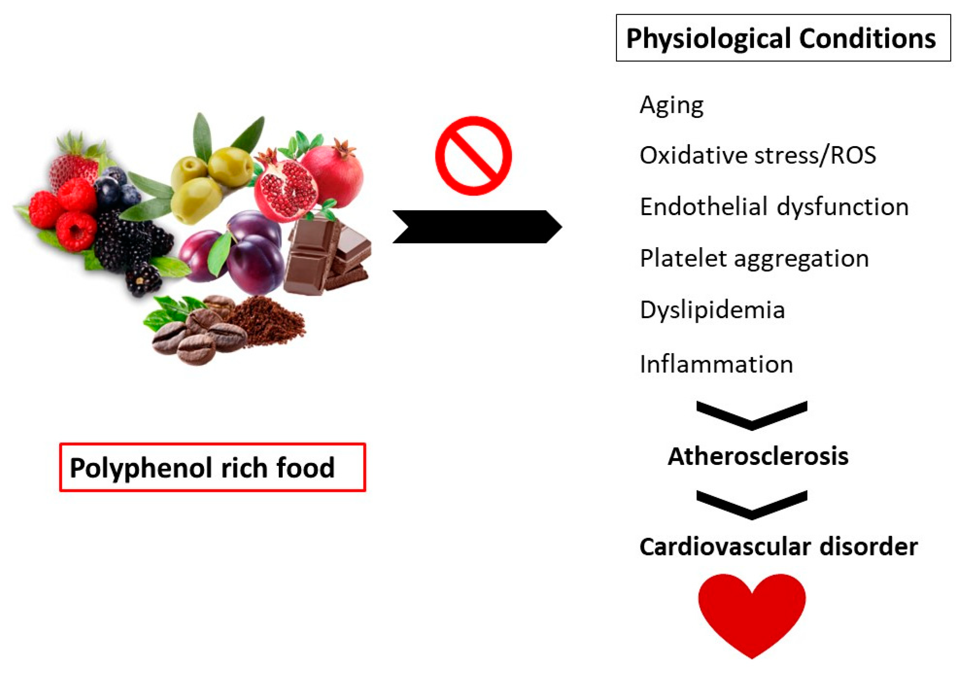
Polyphenols are secondary metabolites found in vegetables, fruits, and grains. These compounds exhibit several health benefits such as immune modulators, vasodilators, and antioxidants.

polyphenols cardiovascular atherosclerosis oxidative stress

1. Introduction

Cardiovascular disease (CVD), encompassing conditions such as atherosclerosis, hypertension, myocardial infarction, cardiomyopathy, arrhythmia, and heart failure (HF), is a major contributor to global mortality. The incidence of CVD has experienced a notable increase [1][2][3][4]. Despite the wide range of pharmaceuticals currently utilized for the management of CVD, such as statins, angiotensin-converting enzyme inhibitors (ACEIs), angiotensin receptor blockers (ARBs), calcium channel blockers (CCBs), fibrates, and β-blockers, it is important to acknowledge that a significant number of these medications are associated with adverse effects in the human population [4]. Hence, there exists a significant clinical requirement to discover and cultivate innovative therapeutic strategies for CVD [2]. The transport of oxygen and nutrients in the human body is carried out via blood circulation along with the removal of metabolic by-products and carbon dioxide through the cardiovascular system (CVS). Coronary artery disease (CAD), cerebrovascular disease (CVD), peripheral artery disease (PAD), congenital heart disease (CHD), hypertension, heart failure, and stroke are all disorders that affect the heart and blood arteries [1][2]. Within this context, cardiovascular diseases (CVDs) are among the leading causes of mortality throughout the world, claiming 17.9 million individual lives worldwide in 2019, which is approximately 32% of total fatalities. Approximately, 85% of these mortality rates were due to heart attacks and strokes (WHO site). Strokes kill 6.7 million people each year, and coronary heart disease claims 7.4 million lives [3][4].
Several pathologies can affect the cardiovascular system. Some of these pathologies include primary heart ailments, including cardiomyopathy and cardiac malignancies. Infectious and infectious-allergic damage to heart tissue, metabolic and systemic disorders, and diseases of other organs are also covered in this category [5][6]. CHD starts with inflammation of the blood artery walls, which narrows and causes angina pectoris [7]. In this respect, blood clots restrict arteries later in the disease’s progression, resulting in severe myocardial ischemia and myocardial infarction (heart attack). Heart failure can occur in severe cases of CHD when the heart muscle’s ability to pump blood around the body deteriorates [2]. Because these disorders are generally caused by arterial injury, symptoms and treatments vary depending on which arteries are afflicted [2].
Age and gender are among the most reported non-modifiable cardiovascular risk factors. Cardiovascular disorders become more common as people become older due to a rise in plasma cholesterol on one hand and the augmentation of arterial rigidity and peripheral vascular resistance on the other hand [8]. Although the risk of cardiovascular disorder varies with gender and age, the incidence is three to five times greater in men < 50 years of age compared to women. On the other hand, a considerable increase in the occurrence of CVD has been observed in women over the age of 50 years. Genetic factors, inactivity, hypertension, obesity, diabetes, smoking, and dyslipidemia are the prominent risk factors for cardiovascular disorders, as described in published reports [9][10][11][12].
Polyphenols have also been found useful in enhancing endothelial function, preventing aberrant platelet aggregation, decreasing inflammation, and improving plasma lipid profile, all of which benefit cardiovascular health. Because the processes by which these chemicals exert cardioprotective activities are not entirely known, although not conclusively shown, there may be a connection between the cardiovascular advantages of some diets and their polyphenol levels [13]. Depicted in Figure 1 is a sketch that shows how nutrition can help in preventing atherosclerosis, which contributes to CVD.
Figure 1. Nutrition can help prevent atherosclerosis, which is a pathophysiological process that contributes to the development of cardiovascular disease (CVD).

2. Polyphenols

Polyphenols are phytochemicals or secondary plant compounds that are considered non-essential nutrients in plants [14]. They are a rich collection of chemicals present in plants and algae, where their natural role is to defend the organism against UV radiation, infection, and herbivore ingestion. Polyphenols come in a variety of structural forms, from basic monomers to complex polymerized structures. Seaweed polyphenols may help lower hyperglycemia, hyperlipidemia, oxidative stress, chronic inflammation, metabolic abnormalities linked to CVDs, and diabetes sequelae. On the other hand, polyphenols from plants have been related to improved health in terms of obesity, diabetes, and CVD. A recent study has focused on marine macroalgae, presumably because of epidemiological evidence from Asian nations that suggests a diet high in seaweed lowers the occurrence of CVD, cancer, and other chronic disorders [1][15].
Polyphenols are also outstanding plant-derived secondary metabolites that exhibit anticancer, anti-cardiovascular, antidiabetic, and anti-neurodegenerative properties. Compounds like phenicic acid, stilbenes, flavonoids, coumarins, tannins, and lignins are present in numerous plants, including tonka bean (Dipteryx odorata), sweet grass (Hierochloe odorata), sweet woodruff (Galium odoratum), deer-tongue grass (Dichanthelium clandestinum), sweet clover (Verbascum spp.), and vanilla grass (Anthoxanthum odoratum). In addition to its antioxidant characteristics, resveratrol also exerts ameliorating effects against inflammation, cancer, aging, obesity, and diabetes, along with cardioprotective and neurological benefits [16].
Phenolic compounds are the most copious non-energetic components in plant-based meals. The aptitude of polyphenols to alter enzymatic activity and, consequently, the signal-transmitting mechanisms of several processes occurring in cells may be attributed to their physicochemical properties, which allow polyphenols to participate in numerous metabolic cellular redox processes. Thus, the antioxidant scavenging properties of polyphenols make them advantageous. These are the most predominant antioxidants in the diet; they are 20 times more prevalent than vitamin E and carotenoids and 10 times more prevalent than vitamin C. Nicotinamide-adenine-dinucleotide phosphate (NADP) oxidase and xanthine oxidase are two ROS-producing enzymes that polyphenols can inhibit [17]. Listed in Table 1 are polyphenol-rich plant foods.
Table 1. Polyphenol-rich plant foods.

Abbreviations: EC = epicatechin; EGCG = epigallocatechin gallate. x = In juices, wine, and other beverages: mg/100 mL. y = Concentration in mg/cm2. Note that the polyphenol content in purple potatoes is approximately five times higher than that in other varieties.

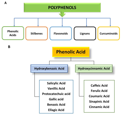
3. Classification of Polyphenols

Polyphenols are divided into several categories, including phenolic acids (hydroxybenzoic and hydroxycinnamic acids), flavonoids (flavones, flavonols, isoflavones, flavanones, and anthocyanins), stilbenes (resveratrol, piceatannol), lignans (sesamol, pinoresinol, sinol, enterodiol), and others, including tannins (hydrolyzable, non-hydrolyzable, and condensed tannins), lignins, xanthones, chromones, and anthraquinones, as shown in Figure 2A [23][24][25].
Figure 2. (A) Classification of polyphenols. (B) Classification and food sources of phenolic acids.

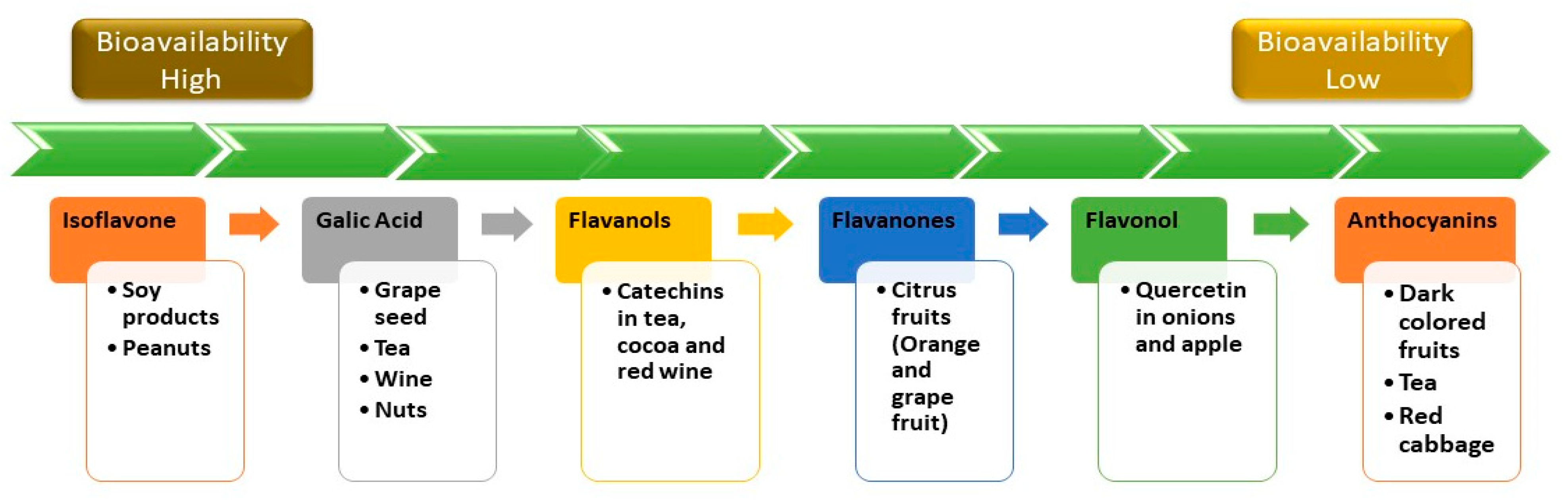
4. Bioavailability of Polyphenols

The bioavailability of phenolic compounds in our food is critical because only the most bioavailable phenolic compounds in our diet will have the most beneficial effects on the human body [26]. These will be different for each person depending on their relationship with food, how cell walls are made, and where glycosides are found. Many epidemiological studies have shown that phenolic compounds have a lot of health benefits, such as protecting against the buildup of fat, preventing microorganisms from decaying, lowering cardiovascular diseases, preventing diabetes, stroke, and cancer, and exerting anti-inflammatory effects [25].
Recent research has placed a strong emphasis on identifying the processes governing polyphenol metabolism and bioavailability in humans [27]. A wide array of fruits and vegetables contain compounds known as phenolics. Some plants contain as much as 750 mg/100 g of fruit, which is a significant amount [14][28]. The highest dietary sources of polyphenols are dark-colored fruits (especially small berries), chocolate, cereals made entirely of whole grains, coffee, and red wine, with the latter three accounting for the lion’s share of overall dietary polyphenol consumption [29]. Among the food groups consumed, polyphenols are primarily associated with carbohydrates, organic acids, and other food groups. They combine with arabinose to generate ester linkages in hemicellulose or core lignin, which allows them to form covalent connections with polysaccharides in the cell wall of the plant. While flavonoids can be found in the cytosol and endoplasmic reticulum, where they are formed, they are mostly found in free form in the cytosol and endoplasmic reticulum. Cell barriers and intracellular compartments must be damaged for the drug to be bioavailable [28]. Flavonoids found in nature are housed within plants as glycoside and non-glycosylated conjugate compounds, and as a result, the moiety’s type might affect their subsequent human bioavailability [30][31]. A summary of the comparative bioavailability of different polyphenols is shown in Figure 3 [32].
Figure 3. Comparative bioavailability of some common dietary polyphenols.

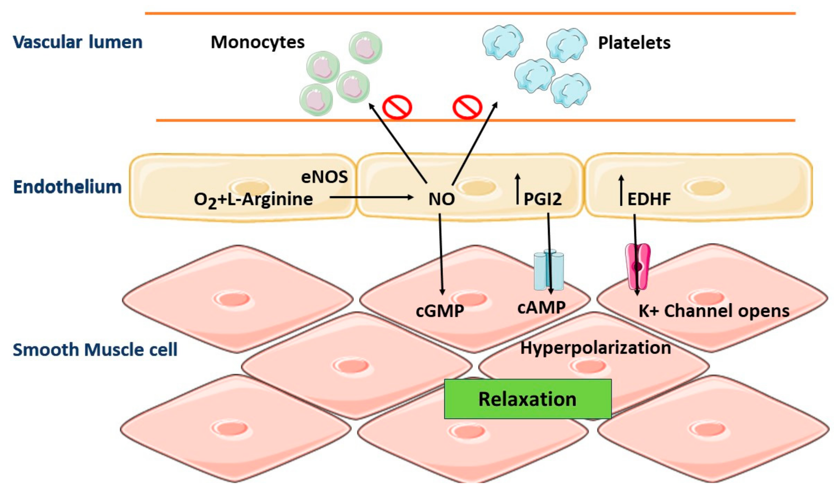
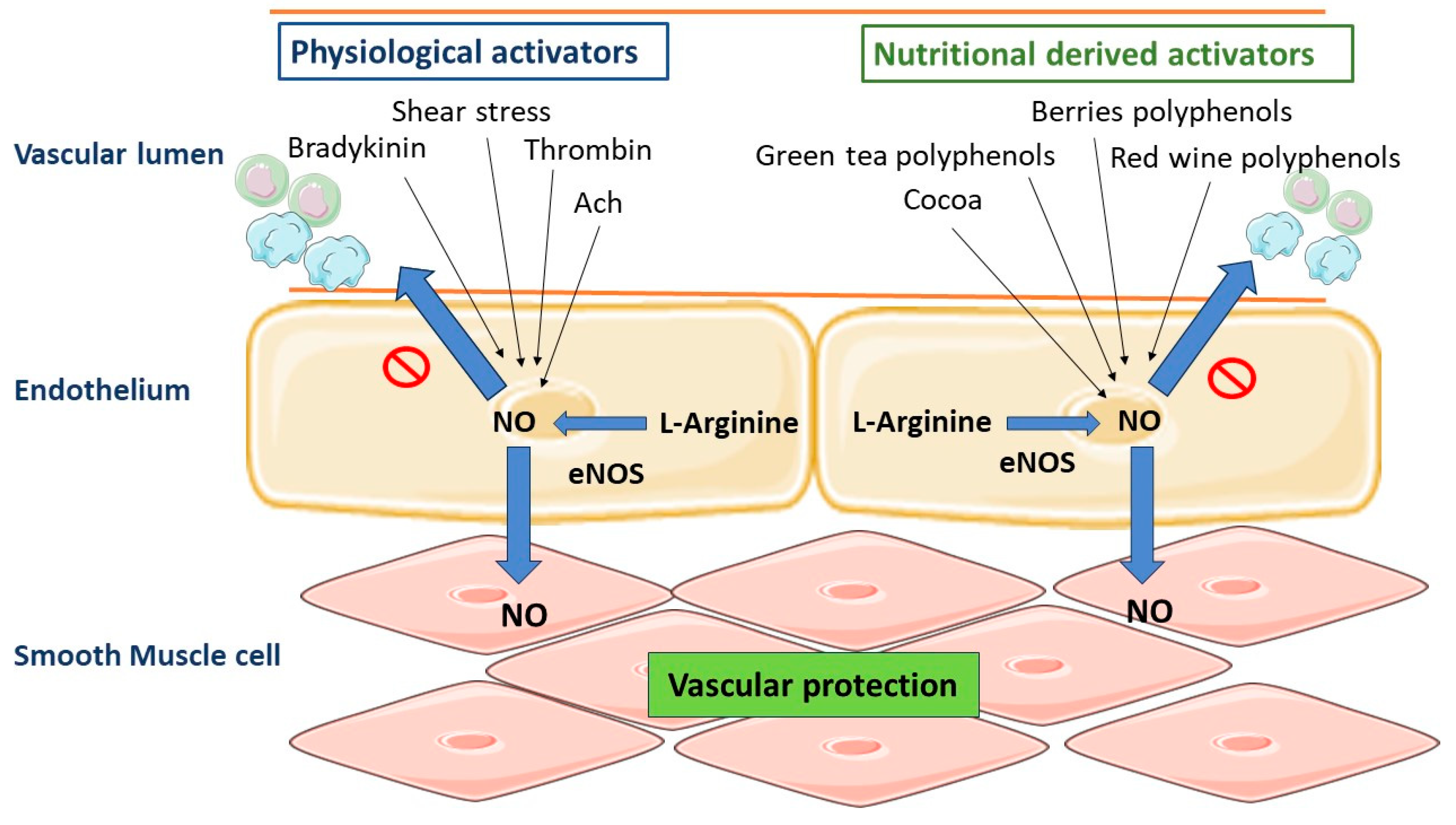
5. Role of Vascular Endothelium in the Regulation of Vascular Homeostasis

A monolayer of cells produces the endothelium, which makes up the interior of blood vessels. Vascular endothelium regulates the tone and homeostasis of the vasculature, as well as the morphological changes that occur in pathological circumstances. The endothelium regulates the balance of opposing processes such as vasodilation and constriction, pro-coagulant and antithrombotic actions, and cell proliferation and apoptosis [33][34].
Endothelial cells minimize the interaction of the bloodstream with the basal prothrombotic arterial wall due to their selective position [35]. The primary function of Endothelial cells is to regulate vascular tone by producing vasodilator and vasoconstrictor chemicals. The endothelial NO synthase (eNOS) enzyme produces NO from L-arginine, exerting a vasodilatory effect. NO can easily diffuse into the cells of vascular smooth muscle, where it triggers guanyl cyclase, thus accumulating cyclic guanosine monophosphate (cGMP), which ultimately activates the protein kinase G and causes endothelial vasorelaxation (Figure 4). The endothelium-derived hyperpolarizing factor (EDHF) plays its role in vasodilation by targeting the K+ channels in the blood vessels. Furthermore, prostacyclin (PGI2) produced during the cyclooxygenase (COX) pathway has vasodilatory effects. Some other factors can be produced by the endothelium having vasoconstrictive effects on blood vessels such as angiotensin II (Ang II), endothelin-1 (ET-1), and thromboxane A2 (TXA2) [36].
Figure 4. Normal regulation of vascular homeostasis. Vascular homeostasis is regulated in part by endothelium-derived NO. The endothelial NO synthase (eNOS) enzyme produces NO from L-arginine, exerting a vasodilatory effect. NO can easily diffuse into the cells of vascular smooth muscle, where it triggers guanyl cyclase, thus accumulating cyclic guanosine monophosphate (cGMP), which ultimately activates the protein kinase G and causes vasorelaxation in endothelial.
Regarding blood–tissue contact, the endothelium does play a crucial role, interacting directly with a variety of circulating substances, including antioxidants, oxidized LDLs, and pro-inflammatory cytokines such as tumor necrosis factor (TNF) and interleukins (IL) [33][34]. These variables can cause vasomotricity or the manufacturing of endothelial agents like nitric oxide (NO). Their role in a variety of physiological processes has been reported to influence biological processes such as the apoptosis, proliferation, and migration of endothelial cells [36][37][38]. Thus, endothelial dysfunction can have several detrimental effects on vascular cells and surrounding tissue, resulting in the development of cardiovascular disorders such as atherosclerosis and hypertension [39].
Diets like the Mediterranean diet have been linked to better cardiovascular health [40], which might be due to the high consumption of polyphenol-rich drinks and foods, as well as fruits and vegetables. Polyphenol-rich foods, including red wine, chocolate, green tea, and berries, also help to promote cardiovascular health [41][42]. Polyphenols have been linked to improving cardiovascular health in various ways. Their advantages include an improvement in lipid profiles. They also have direct effects on endothelial cells and have anti-atherosclerotic, anti-hypertensive, and anti-inflammatory properties (Figure 5).
Figure 5. Numerous physiological activators on the endothelium surface can boost endothelial NO production to produce vascular protection. Also, polyphenol-rich dietary items, including chocolate, berries, red wine, and green tea, can enhance endothelial NO production. The endothelial NO synthase (eNOS) enzyme produces NO from L-arginine, exerting a vasodilatory effect. NO can easily diffuse into the cells of vascular smooth muscle, where it triggers guanyl cyclase, thus accumulating cyclic guanosine monophosphate (cGMP), which ultimately activates the protein kinase G and causes vasorelaxation in endothelial.

6. Pathophysiology: Oxidative Stress and CVD

In healthy cells, antioxidant defense systems such as superoxide dismutase (SOD), catalase (CAT), and glutathione reductase (GSR) limit the generation of radicals during various physiological activities such as metabolism and cellular respiration [1][43][44]. Long-term exposure to stress [45], pollution [46], smoking, and excessive drinking [47][48], as well as aging [49], can cause an imbalance of oxidative species (also known as reactive oxygen species; ROS) in comparison to endogenous defenses, resulting in oxidative stress [1][43]. ROS can bind to proteins, lipids, and DNA, thus oxidizing them and changing a healthy state into a diseased one. An increase in the level of ROS results in oxidative stress, and the cell’s antioxidant system may become overburdened, endangering the health and integrity of the cell [50].
A similar pathological mechanism, atherosclerosis, underpins cardiovascular illnesses such as coronary artery disease, ischemic stroke, and peripheral artery disease [51]. Atherosclerosis is a multifactorial, degenerative ailment of the medium and great conduit arteries that is fueled by lipid buildup in the artery wall [52][53]. The risk factors of this disease include old age, chronic smoking, hyperlipidemia, hypertension, and a history of diabetes. The atherogenic process is tightly linked to inflammation and endothelial dysfunction [51][54]. Endothelial damage due to ROS leads to the development of atherosclerosis, which may result in myocardial infarction and ischemic reperfusion [55][56][57]. Oxidative stress and ROS target all body cells, especially smooth muscle cells and endothelial cells, with the help of neutrophils, macrophages, and platelets [58].
ROS has an impact on a variety of endothelium-related processes [2]. The most well-known is endothelium-dependent vasorelaxation, which has long been marked as a key component in the prognosis of cardiovascular health, which is harmed by a decrease in NO bioactivity and/or bioavailability [59][60]. There are some mechanisms responsible for reduced NO bioavailability. This can either be caused by a decreased expression of the enzyme responsible for the NO production in endothelial cells, i.e., endothelial NOS (eNOS), or a decrease in the existing NO owing to ROS destruction, among other things [60]. NO is a powerful vasodilator that also inhibits the activation and adherence of inflammatory cells [61].
In the vasculature, there are several sources of ROS, including mitochondrial enzymes such as NADH/NADPH oxidase and xanthine oxidase [60][62]. Endothelium-derived NO reacts quickly with the superoxide radical (O2) to create peroxynitrite (ONOO), a potent oxidant that is equally damaging to endothelial cells [63]. In this respect, studies have shown that the continued contact of endothelial cells with oxygen and different oxidants like hydrogen peroxide, ONOO, and/or oxidized LDL (ox-LDL) causes epithelial damage by promoting apoptosis, leading to cell damage and endothelial cell dysfunction, which has been reported as a critical early step in atherogenesis. Atherosclerotic lesions can arise from leaky and dysfunctional endothelium [51][52].
LDL normally diffuses easily in both directions across the compromised endothelium. Oxidative stress converts LDL to ox-LDL by peroxidation, which has cytotoxic effects and can cause inflammation [64]. The first step in the formation of atherosclerotic plaques is the oxidation of LDL and its subsequent passage through the endothelial barrier. Furthermore, the interaction of hypercholesterolemia, oxidative stress radicals, and inflammatory molecules creates an environment conducive to severe endothelial damage, which is a characteristic of atherosclerosis development [43][65].
A particular adhesion molecule called VCAM-1, which is crucial for binding monocytes and T cells before they transmigrate into the arterial wall, is produced by wounded or activated endothelial cells [64]. Reportedly VCAM-1, ICAM-1, and E-selectin enhance the adherence of leukocytes to the vascular endothelium at the sites of atherosclerotic lesions, consequently boosting signal transduction cascades [65]. These monocytes develop into macrophages after activation, which then become puffy with the uptake of ox LDL by scavenger receptor-mediated phagocytosis, resulting in fatty bands in the artery wall [51][59][66]. Furthermore, lipid-engorged macrophages (foam cells) eventually die in situ because of necrotic cell death, resulting in the creation of a tender and unstable core inside the atherosclerotic plaques, which has a high consistency of lipids [67]. This plaque is stabilized by a protective cap secreted by smooth muscle cells. It consists of a collagen-rich matrix comprising fibroblasts, which can stop the disease from progressing. Prolonged inflammation, on the other hand, might result in plaques that are unstable and prone to rupture [1][67]. Such ruptured plaques cause a fast thrombotic reaction, resulting in arterial blockage and, depending on the location of the atherosclerotic lesion, potentially causing heart attacks, ischemic strokes, or peripheral ischemia [52]. Research findings have linked the plaques in the walls of coronary arteries to chronic atherosclerotic lesions, which can limit the channel lumen. The current preferred hypothesis is that acute coronary syndrome (ACS) is caused by a rupture of the fibrous cap of an atherosclerotic plaque, which facilitates blood contact with extracellular matrix collagen and tissue factors previously deposited in the plaque, resulting in the formation of a thrombus [68][69][70][71]. On the other hand, increased platelet activation, including the adhesion, secretion, and aggregation at the site of arterial injury or in atherosclerotic arteries, plays a key part in the etiology of CVD [72][73]. When an atherosclerotic plaque ruptures, activated platelets can bind to the endothelium, causing fastening, aggregation, and thrombus development, which leads to embolism and the constriction of vessels, two features of myocardial infarction [74].
Physiological hemostasis is a natural defense against excessive blood loss that is based on the creation of a regulated thrombus at the site of the blood vessel injury. Platelets, the smallest (2–4 m) blood corpuscles, are formed at a rate of 40 × 103/mL/day in the bone marrow from megakaryocytes and play a key role in hemostasis. Platelets play a function in hemostasis that extends beyond forming the platelet plug, which also serves as the site of fibrin production (at the site of the vessel wall injury), to include a beneficial influence on vessel wall contraction and participation in clotting responses [8]. Collagen and tissue factor (TF) located in the sub-endothelial matrix come into contact with the lowing blood when the endothelium is injured, causing a clot to develop [75]. Denuded collagen directly promotes platelet pooling and activation, and the denuded tissue factor starts the synthesis of thrombin, which not only transforms fibrinogen to fibrin but also activates platelets [69]. The presence of collagen receptors (including integrin a2b1 and glycoprotein complex GPIb/IX/V) on the platelet surface allows platelets to connect with the subendothelial layer. Platelet attachment leads them to change shape from discoid to spherical, resulting in the creation of pseudopodia and the release of chemicals held in granules (e.g., ADP, P-selectin, von Willebrand factor [vWF], thrombospondin) and, as a result, platelet aggregation [68][76].

7. Beneficial Effects of Polyphenols on Cardiovascular Disorders

7.1. Polyphenols as Antioxidant Therapy

Antioxidant therapy is becoming better recognized as a strategy for reducing ROS in the vasculature and, as a result, reducing their harmful effects [77]. Blockers of the angiotensin-converting enzyme (ACE) that lower circulatory Ang II detoxify in addition to exhibiting their antihypertensive attributes. In this regard, statins (cholesterol-lowering drugs) are used for the same intent in addition to their cholesterol-lowering properties by regulating HMG CoA reductase. Similarly, vitamins E and C are widely used as dietary supplements in combination with other drugs to reduce oxidative stress [77]. Polyphenols, on the other hand, are gaining attention as possible therapeutic agents for reducing oxidative stress and thereby protecting people from heart diseases [78][79]. In the diet, polyphenols are the most prevalent antioxidants, and their consumption is ten times that of water-soluble vitamin C and one hundred times that of lipid-soluble vitamin E and carotenoids [36].

7.2. Polyphenols and Vascular Tone

The significance of endothelium-produced nitric oxide (NO) in controlling vascular tone and blood pressure is well understood. The central mechanism of NO action is the activation of the cGMP-protein kinase G cascade in artery smooth muscle cells. The potassium channels are triggered when the cascade is activated, resulting in membrane hyperpolarization and preventing intracellular calcium influx, which induces vasodilation. On the other hand, protein kinase G reduces smooth muscle vasoconstriction in arteries by phosphorylating myosin light chains [80][81]. NO generation is primarily responsible for the polyphenols’ effect on the endothelium [82][83][84].
After ingesting red wine or polyphenols (1 g/kg body weight) circulating NO concentrations reach 30 and 40 nM after 30 min in adults. A decrease in blood pressure (11 mmHg) and an increase in heart rate have also been observed [85]. Research findings have shown that olive oil can help hypertensive people lower their blood pressure [86], whereas red wine polyphenolic compounds (RWPC) can produce the endothelium-dependent relaxation of isolated arteries such as the rat’s mesenteric artery or aorta [82]. In addition, red wine polyphenols, polyphenols from grape skin, and quercetin exhibit antihypertensive effects. In this respect, short-term oral treatment with RWPC lowers blood pressure in normotensive rats. This hemodynamic effect was correlated with enhanced endothelium-dependent relaxation and the induction of the genes responsible for inducible NO synthase and COX-2 inside the artery wall, thus contributing to the maintenance of agonist-induced contractility [87]. The higher synthesis of NO in consequence to the impact of polyphenols found in wine extract is linked to the calcium ion-dependent pathway, among several other things [88]. Resveratrol and quercetin cause an increase in the intracellular ion concentration of (Ca2+) ions through the opening of potassium channels or the inhibition of Ca2+ ATP-ase within the endoplasmic reticulum of endothelial cells [89][90]. Similarly, delphinidin, an anthocyanin present in natural foods like red wine, can activate endothelial cells. This anthocyanin raises intracellular protein-Ca2+ and tyrosin phosphorylation, which controls eNOS. Tyrosine kinases and phospholipase C are both involved in Ca2+ signaling [91]. Furthermore, RWPC might even enhance endothelial NO production via the redox-responsive PI3/Akt channel, according to another report [92].
In addition, the effect of polyphenolic compounds on endothelial cells in preventing cardiovascular diseases is not limited to the stimulation of NO production. Because of the increased production of PGI2, the vasodilating effect is also boosted. In vitro studies on human endothelial cells exposed to the action of cocoa extract rich in procyanidins at a concentration of 2 mg/L and in vivo studies on procyanidins contained in chocolate administered to healthy volunteers showed that the ratio of cysteinyl leukotrienes (LTC4, LTD4, LTE4) to PGI2 can be reduced by 58 and 52%, respectively [93]. In contrast, isolavonoids, particularly genistein, limit the procoagulant action of vascular endothelium by, for example, lowering ET-1 expression [94]. Finally, polyphenols can affect endothelial cells’ NO levels by affecting PDE-2 and PDE-4, two phosphodiesterases [95][96]. Taken together, plant polyphenols may have complex effects on the circulatory system’s NO balance, which could account for their antihypertensive effects [97].

7.3. Polyphenols and Atherosclerosis

Atherosclerosis is the hardening and narrowing of the arteries, which is triggered by the buildup of lipids, cholesterol, and other substances in and on the artery walls over time. This then progresses into the endothelium, where they are oxidized by endothelial smooth muscle cells and activated macrophages [98][99]. ROS and reactive nitrogen species (RNS) production can enhance LDL oxidation. This causes a buildup of macrophages in this area, which clear oxidized LDL and transform them into foam cells. Endothelial dysfunction, as well as the concentration of monocytes/macrophages in the vascular intima under the influence of chemokines and adhesion molecules, foam cell development, and vascular smooth muscle proliferation, are all linked to the inflammatory backdrop of atherosclerotic lesions [100]. There is also a rise in extracellular matrix buildup surrounding the spot of inflammation, leading to plaque development which blocks the vessel, resulting in the loss of the blood artery’s natural capacity to relax [99][100].

7.4. Polyphenols and Anti-Platelet Action

The excessive activation of platelets is linked to several long-term vascular diseases. This is due to the many adhesion proteins in the granules that, when highly activated, can lead to different types of thrombotic diseases [101][102]. In this respect, numerous important things happen in the process of platelet activation. One of them is the conversion of arachidonic acid to thromboxane A2, an arachidonate metabolite, through the cyclooxygenase pathway [103]. In this context, polyphenols are valuable from the perspective of platelet activation, which includes the adhesion and aggregation of platelets, due to the antioxidant effect of polyphenols. The first step in platelet activation involves platelets sticking to the collagen in the body; as a result, the platelets become activated. In this respect, proteins like fibrinogen and thrombospondin act as adhesion proteins, and platelet receptors work together to help platelets stick together, leading to the start of a signaling process inside cells and the activation of platelets [104]. However, it has not been fully explained how polyphenols make platelets less likely to stick together. It turns out that extracts rich in polyphenolic compounds, like grape seed and Yucca schidigera extracts, can help stop platelets from sticking to collagen. These extracts contain resveratrol and its derivatives, which make platelets less likely to stick together when they are stimulated by thrombin [72].
Thromboxane A2 (TXA2) is the key compound that is formed from the breakdown of arachidonic acid (ARA). It has some surface receptors that make platelets clump together. Evidence from the literature has indicated that the anti-aggregative effect of polyphenols is linked to numerous complicated molecular processes [105]. The capacity of polyphenols to hinder the enzymes involved in the formation of TXA2, COX, and LOX is the primary method by which they exert their anti-platelet aggregate effects on platelets [105][106]. However, they are also antagonists of the thromboxane A2 receptor, which suggests that flavonoids, through their indirectly suppressive effect on COX1, can lower TXA2 levels in the blood [107]. In an in vivo dog model, researchers investigated the effects of grape juice and red and white wine on platelet aggregation activity. The results revealed the antiplatelet effects of red wine and grape juice, while white wine does not yet have this impact [108].
Flavonoids have been shown to lower platelet aggregation because collagen metabolism is altered by these compounds, in addition to their interference in arachidonic acid metabolism. This is expressed as the antiplatelet action of collagen in the early stages of the aggregation of platelets. Moreover, the oxidative stress results in the aggregation of platelet in response to collagen via the activation of the inositol pathway, boosting intracellular calcium levels in the process. Flavonoids such as quercetin, catechin, and kaempferol, among others, have been shown to decrease oxidative stress by impeding the enzyme NADPH-oxidase [101].

7.5. Polyphenols as Anti-Inflammatory Agents

Inflammatory response to injury is a complicated biotic process that happens in response to a damaging stimulus. Different enzymes, including cyclooxygenase (COX), lipoxygenase (LOX), tyrosine kinase (TK), phospholipase A2 (PLA2s), and protein kinase C), are responsible for the proper function of an inflammatory response. Certain flavonoids have been demonstrated to act directly on several such enzymes, blocking them and therefore directly affecting inflammation [109][110]. One of the most important elements in preventing and treating chronic inflammation, according to epidemiological research, is nutrition. Through ex vivo and in vivo models, researchers have discovered that some flavonoids exert anti-inflammatory effects. One of the key bodily functions that flavonoids have an impact on is the synthesis of prostaglandins. Hesperidin and diosmin can reduce the generation of prostaglandins, according to several in vivo studies [111].
The mobilization of leukocytes is known as a critical stage in the progression of inflammation that occurs in cardiovascular illnesses and other conditions. The production of arachidonic acid ultimately results in the generation of cytokines (IL-1) and chemokines (IL-8) by neutrophils, which is mediated by both COX and LOX. In this regard, quercetin, a polyphenol, is especially effective in suppressing the formation of prostaglandins (PGs), leukotrienes (LT), and thromboxanes (TXA) by preventing the enzymes COX and LOX, respectively [112][113][114]. Evidence from numerous ex vivo experiments shows that some flavonoids, for example, bilobetine, morelloflavone, amentoflavone, and those found in Sophora flavescens, exert their effect by inhibiting the production of arachidonic acid [115]. Furthermore, resveratrol is regarded as a molecule with anti-inflammatory properties, as it inhibits the production of PGs [116].

References

  1. Goszcz, K.; Duthie, G.G.; Stewart, D.; Leslie, S.J.; Megson, I.L. Bioactive polyphenols and cardiovascular disease: Chemical antagonists, pharmacological agents or xenobiotics that drive an adaptive response? Br. J. Pharmacol. 2017, 174, 1209–1225.
  2. Alam, M.A. Anti-hypertensive effect of cereal antioxidant ferulic acid and its mechanism of action. Front. Nutr. 2019, 6, 121.
  3. Jamee Shahwan, A.; Abed, Y.; Desormais, I.; Magne, J.; Preux, P.M.; Aboyans, V.; Lacroix, P. Epidemiology of coronary artery disease and stroke and associated risk factors in Gaza community-Palestine. PLoS ONE 2019, 14, e0211131.
  4. Li, J.; Liao, R.; Zhang, S.; Weng, H.; Liu, Y.; Tao, T.; Yu, F.; Li, G.; Wu, J. Promising remedies for cardiovascular disease: Natural polyphenol ellagic acid and its metabolite urolithins. Phytomedicine 2023, 18, 154867.
  5. Sharifi-Rad, J.; Rodrigues, C.F.; Sharopov, F.; Docea, A.O.; Can Karaca, A.; Sharifi-Rad, M.; Kahveci Karıncaoglu, D.; Gülseren, G.; Şenol, E.; Demircan, E.; et al. Diet, lifestyle and cardiovascular diseases: Linking pathophysiology to cardioprotective effects of natural bioactive compounds. Int. J. Environ. Res. Public Health 2020, 17, 2326.
  6. Blauwet, L.A.; Cooper, L.T. Myocarditis. Prog. Cardiovasc. Dis. 2010, 52, 274–288.
  7. Wirtz, P.H.; von Känel, R. Psychological stress, inflammation, and coronary heart disease. Curr. Cardiol. Rep. 2017, 19, 111.
  8. Khan, J.; Deb, P.K.; Priya, S.; Medina, K.D.; Devi, R.; Walode, S.G.; Rudrapal, M. Dietary flavonoids: Cardioprotective potential with antioxidant effects and their pharmacokinetic, toxicological and therapeutic concerns. Molecules 2021, 26, 4021.
  9. Curry, S.J.; Krist, A.H.; Owens, D.K.; Barry, M.J.; Caughey, A.B.; Davidson, K.W.; Doubeni, C.A.; Epling, J.W., Jr.; Kemper, A.R.; Kubik, M.; et al. Risk assessment for cardiovascular disease with nontraditional risk factors: US preventive services task force recommendation statement. JAMA 2018, 320, 272–280.
  10. Holvoet, P. Stress in obesity and associated metabolic and cardiovascular disorders. Scientifica 2012, 2012, 205027.
  11. Matsuda, M.; Shimomura, I. Increased oxidative stress in obesity: Implications for metabolic syndrome, diabetes, hypertension, dyslipidemia, atherosclerosis, and cancer. Obes. Res. Clin. Pract. 2013, 7, e330–e341.
  12. Knowles, J.W.; Ashley, E.A. Cardiovascular disease: The rise of the genetic risk score. PLoS Med. 2018, 15, e1002546.
  13. Murray, M.; Dordevic, A.L.; Ryan, L.; Bonham, M.P. An emerging trend in functional foods for the prevention of cardiovascular disease and diabetes: Marine algal polyphenols. Crit. Rev. Food Sci. Nutr. 2018, 58, 1342–1358.
  14. Manach, C.; Scalbert, A.; Morand, C.; Rémésy, C.; Jiménez, L. Polyphenols: Food sources and bioavailability. Am. J. Clin. Nutr. 2004, 79, 727–747.
  15. Golovinskaia, O.; Wang, C.K. The hypoglycemic potential of phenolics from functional foods and their mechanisms. Food Sci. Hum. Wellness 2023, 12, 986–1007.
  16. Leuci, R.; Brunetti, L.; Poliseno, V.; Laghezza, A.; Loiodice, F.; Tortorella, P.; Piemontese, L. Natural compounds for the prevention and treatment of cardiovascular and neurodegenerative diseases. Foods 2021, 10, 29.
  17. Quiñones, M.; Miguel, M.; Aleixandre, A. Beneficial effects of polyphenols on cardiovascular disease. Pharmacol. Res. 2013, 68, 125–131.
  18. Wahid, M.; Saqib, F.; Chicea, L.; Ahmedah, H.T.; Sajer, B.H.; Marc, R.A.; Pop, O.L.; Moga, M.; Gavris, C. Metabolomics analysis delineates the therapeutic effects of hydroethanolic extract of Cucumis sativus L. seeds on hypertension and isoproterenol-induced myocardial infarction. Biomed. Pharmacother. 2022, 148, 112704.
  19. Bouayed, J.; Deußer, H.; Hoffmann, L.; Bohn, T. Bioaccessible and dialysable polyphenols in selected apple varieties following in vitro digestion vs. their native patterns. Food Chem. 2012, 131, 1466–1472.
  20. Rothwell, J.A.; Perez-Jimenez, J.; Neveu, V.; Medina-Remon, A.; M’hiri, N.; García-Lobato, P.; Manach, C.; Knox, C.; Eisner, R.; Wishart, D.S. Phenol-Explorer 3.0: A major update of the Phenol-Explorer database to incorporate data on the effects of food processing on polyphenol content. Database 2013, 2013, bat070.
  21. Deußer, H.; Guignard, C.; Hoffmann, L.; Evers, D. Polyphenol and glycoalkaloid contents in potato cultivars grown in Luxembourg. Food Chem. 2012, 135, 2814–2824.
  22. Rojanathammanee, L.; Puig, K.L.; Combs, C.K. Pomegranate polyphenols and extract inhibit nuclear factor of activated T-cell activity and microglial activation in vitro and in a transgenic mouse model of Alzheimer disease. J. Nutr. 2013, 143, 597–605.
  23. Rasouli, H.; Farzaei, M.H.; Khodarahmi, R. Polyphenols and their benefits: A review. Int. J. Food Prop. 2017, 20, 1700–1741.
  24. Singla, R.K.; Dubey, A.K.; Garg, A.; Sharma, R.K.; Fiorino, M.; Ameen, S.M.; Haddad, M.A.; Al-Hiary, M. Natural Polyphenols: Chemical Classification, Definition of Classes, Subcategories, and Structures; Oxford University Press: Oxford, UK, 2019; Volume 102, pp. 1397–1400.
  25. Prabhu, S.; Molath, A.; Choksi, H.; Kumar, S.; Mehra, R. Classifications of polyphenols and their potential application in human health and diseases. Int. J. Physiol, Nutr. Phys. Educ. 2021, 6, 293–301.
  26. Karas, D.; Ulrichová, J.; Valentová, K. Galloylation of polyphenols alters their biological activity. Food Chem. Toxicol. 2017, 105, 223–240.
  27. Bohn, T. Dietary factors affecting polyphenol bioavailability. Nutr. Rev. 2014, 72, 429–452.
  28. Gonzalez, S.; Fernandez, M.; Cuervo, A.; Lasheras, C. Dietary intake of polyphenols and major food sources in an institutionalised elderly population. J. Hum. Nutr. Diet. 2014, 27, 176–183.
  29. Manach, C.; Williamson, G.; Morand, C.; Scalbert, A.; Rémésy, C. Bioavailability and bioefficacy of polyphenols in humans. I. Review of 97 bioavailability studies. Am. J. Clin. Nutr. 2005, 81 (Suppl. S1), 230s–242s.
  30. Crozier, A.; Del Rio, D.; Clifford, M.N. Bioavailability of dietary flavonoids and phenolic compounds. Mol. Aspects Med. 2010, 31, 446–467.
  31. Behl, T.; Bungau, S.; Kumar, K.; Zengin, G.; Khan, F.; Kumar, A.; Kaur, R.; Venkatachalam, T.; Tit, D.M.; Vesa, C.M.; et al. Pleotropic effects of polyphenols in cardiovascular system. Biomed. Pharmacother. 2020, 130, 110714.
  32. Laurent, C.; Besançon, P.; Caporiccio, B. Flavonoids from a grape seed extract interact with digestive secretions and intestinal cells as assessed in an in vitro digestion/Caco-2 cell culture model. Food Chem. 2007, 100, 1704–1712.
  33. Cines, D.B.; Pollak, E.S.; Buck, C.A.; Loscalzo, J.; Zimmerman, G.A.; McEver, R.P.; Pober, J.S.; Wick, T.M.; Konkle, B.A.; Schwartz, B.S.; et al. Endothelial cells in physiology and in the pathophysiology of vascular disorders. Blood 1998, 91, 3527–3561.
  34. Saqib, F.; Wahid, M.; AL-Huqail, A.A.; Ahmedah, H.T.; Bigiu, N.; Irimie, M.; Moga, M.; Marc, R.A.; Pop, O.L.; Chicea, L.M. Metabolomics based mechanistic insights to vasorelaxant and cardioprotective effect of ethanolic extract of Citrullus lanatus (Thunb.) Matsum. & Nakai. seeds in isoproterenol-induced myocardial infarction. Phytomedicine 2022, 100, 154069.
  35. Akimoto, S.; Mitsumata, M.; Sasaguri, T.; Yoshida, Y. Laminar shear stress inhibits vascular endothelial cell proliferation by inducing cyclin-dependent kinase inhibitor p21(Sdi1/Cip1/Waf1). Circ. Res. 2000, 86, 185–190.
  36. Sanches-Silva, A.; Testai, L.; Nabavi, S.F.; Battino, M.; Devi, K.P.; Tejada, S.; Sureda, A.; Xu, S.; Yousefi, B.; Majidinia, M.; et al. Therapeutic potential of polyphenols in cardiovascular diseases: Regulation of mTOR signaling pathway. Pharmacol. Res. 2020, 152, 104626.
  37. Ziche, M.; Morbidelli, L.; Masini, E.; Amerini, S.; Granger, H.J.; Maggi, C.A.; Geppetti, P.; Ledda, F. Nitric oxide mediates angiogenesis in vivo and endothelial cell growth and migration in vitro promoted by substance P. J. Clin. Investig. 1994, 94, 2036–2044.
  38. Matz, R.L.; Andriantsitohaina, R. Age-related endothelial dysfunction: Potential implications for pharmacotherapy. Drugs Aging 2003, 20, 527–550.
  39. Sofi, F.; Cesari, F.; Abbate, R.; Gensini, G.F.; Casini, A. Adherence to Mediterranean diet and health status: Meta-analysis. BMJ 2008, 9, 337.
  40. Rana, A.; Samtiya, M.; Dhewa, T.; Mishra, V.; Aluko, R.E. Health benefits of polyphenols: A concise review. J. Food Biochem. 2022, 46, e14264.
  41. Habauzit, V.; Morand, C. Evidence for a protective effect of polyphenols-containing foods on cardiovascular health: An update for clinicians. Ther. Adv. Chronic Dis. 2012, 3, 87–106.
  42. Khurana, S.; Venkataraman, K.; Hollingsworth, A.; Piche, M.; Tai, T.C. Polyphenols: Benefits to the cardiovascular system in health and in aging. Nutrients 2013, 5, 3779–3827.
  43. Brieger, K.; Schiavone, S.; Miller, F.J., Jr.; Krause, K.H. Reactive oxygen species: From health to disease. Swiss Med. Wkly. 2012, 142, w13659.
  44. Bouayed, J.; Rammal, H.; Soulimani, R. Oxidative stress and anxiety: Relationship and cellular pathways. Oxid. Med. Cell. Longevity 2009, 2, 63–67.
  45. Lodovici, M.; Bigagli, E. Oxidative stress and air pollution exposure. J. Toxicol. 2011, 2011, 487074.
  46. Li, S.; Tan, H.Y.; Wang, N.; Zhang, Z.J.; Lao, L.; Wong, C.W.; Feng, Y. The role of oxidative stress and antioxidants in liver diseases. Int. J. Mol. Sci. 2015, 16, 26087–26124.
  47. Chan, S.M.; Cerni, C.; Passey, S.; Seow, H.J.; Bernardo, I.; van der Poel, C.; Dobric, A.; Brassington, K.; Selemidis, S.; Bozinovski, S.; et al. Cigarette smoking exacerbates skeletal muscle injury without compromising its regenerative capacity. Am. J. Respir. Cell Mol. Biol. 2020, 62, 217–230.
  48. Finkel, T.; Holbrook, N.J. Oxidants, oxidative stress and the biology of ageing. Nature 2000, 408, 239–247.
  49. Selvaraju, V.; Joshi, M.; Suresh, S.; Sanchez, J.A.; Maulik, N.; Maulik, G. Diabetes, oxidative stress, molecular mechanism, and cardiovascular disease--an overview. Toxicol. Mech. Methods 2012, 22, 330–335.
  50. Le Brocq, M.; Leslie, S.J.; Milliken, P.; Megson, I.L. Endothelial dysfunction: From molecular mechanisms to measurement, clinical implications, and therapeutic opportunities. Antioxid. Redox Signal. 2008, 10, 1631–1674.
  51. Falk, E. Pathogenesis of atherosclerosis. J. Am. Coll. Cardiol. 2006, 47 (Suppl. S8), C7–C12.
  52. Megson, I.L.; Whitfield, P.D.; Zabetakis, I. Lipids and cardiovascular disease: Where does dietary intervention sit alongside statin therapy? Food Funct. 2016, 7, 2603–2614.
  53. Loke, W.M.; Proudfoot, J.M.; Hodgson, J.M.; McKinley, A.J.; Hime, N.; Magat, M.; Stocker, R.; Croft, K.D. Specific dietary polyphenols attenuate atherosclerosis in apolipoprotein E-knockout mice by alleviating inflammation and endothelial dysfunction. Arterioscler.; Thromb. Vasc. Biol. 2010, 30, 749–757.
  54. Dhalla, N.S.; Temsah, R.M.; Netticadan, T. Role of oxidative stress in cardiovascular diseases. J. Hypertens. 2000, 18, 655–673.
  55. Sugamura, K.; Keaney, J.F., Jr. Reactive oxygen species in cardiovascular disease. Free Radical Biol. Med. 2011, 51, 978–992.
  56. Raedschelders, K.; Ansley, D.M.; Chen, D.D. The cellular and molecular origin of reactive oxygen species generation during myocardial ischemia and reperfusion. Pharmacol. Ther. 2012, 133, 230–255.
  57. Park, W.H.; Kim, S.H. Involvement of reactive oxygen species and glutathione in gallic acid-induced human umbilical vein endothelial cell death. Oncol. Rep. 2012, 28, 695–700.
  58. Hopkins, P.N. Molecular biology of atherosclerosis. Physiol. Rev. 2013, 93, 1317–1542.
  59. Cai, H.; Harrison, D.G. Endothelial dysfunction in cardiovascular diseases: The role of oxidant stress. Circ. Res. 2000, 87, 840–844.
  60. Taniyama, Y.; Griendling, K.K. Reactive oxygen species in the vasculature: Molecular and cellular mechanisms. Hypertension 2003, 42, 1075–1081.
  61. Paravicini, T.M.; Touyz, R.M. NADPH oxidases, reactive oxygen species, and hypertension: Clinical implications and therapeutic possibilities. Diabetes Care 2008, 31 (Suppl. S2), S170–S180.
  62. Curtin, J.F.; Donovan, M.; Cotter, T.G. Regulation and measurement of oxidative stress in apoptosis. J. Immunol. Methods 2002, 265, 49–72.
  63. Gerhardt, T.; Ley, K. Monocyte trafficking across the vessel wall. Cardiovas. Res. 2015, 107, 321–330.
  64. Hansson, G.K.; Libby, P. The immune response in atherosclerosis: A double-edged sword. Nat. Rev. Immunol. 2006, 6, 508–519.
  65. Martínez-Cayuela, M. Oxygen free radicals and human disease. Biochimie 1995, 77, 147–161.
  66. Singh, R.B.; Mengi, S.A.; Xu, Y.J.; Arneja, A.S.; Dhalla, N.S. Pathogenesis of atherosclerosis: A multifactorial process. Exp. Clin. Cardiol. 2002, 7, 40–53.
  67. Jennings, L.K. Mechanisms of platelet activation: Need for new strategies to protect against platelet-mediated atherothrombosis. Thromb. Haemost. 2009, 102, 248–257.
  68. Furie, B.; Furie, B.C. Mechanisms of thrombus formation. N. Engl. J. Med. 2008, 359, 938–949.
  69. Falk, E.; Shah, P.K.; Fuster, V. Coronary plaque disruption. Circulation 1995, 92, 657–671.
  70. Fasolo, F.; Di Gregoli, K.; Maegdefessel, L.; Johnson, J.L. Non-coding RNAs in cardiovascular cell biology and atherosclerosis. Cardiovasc. Res. 2019, 115, 1732–1756.
  71. Olas, B.; Wachowicz, B.; Tomczak, A.; Erler, J.; Stochmal, A.; Oleszek, W. Comparative anti-platelet and antioxidant properties of polyphenol-rich extracts from: Berries of Aronia melanocarpa, seeds of grape and bark of Yucca schidigera in vitro. Platelets 2008, 19, 70–77.
  72. Jagroop, I.A.; Kakafika, A.I.; Mikhailidis, D.P. Platelets and vascular risk: An option for treatment. Curr. Pharm. Des. 2007, 13, 1669–1683.
  73. Gawaz, M. Role of platelets in coronary thrombosis and reperfusion of ischemic myocardium. Cardiovasc. Res. 2004, 61, 498–511.
  74. Davì, G.; Patrono, C. Platelet activation and atherothrombosis. N. Engl. J. Med. 2007, 357, 2482–2494.
  75. Jagroop, I.A.; Clatworthy, I.; Lewin, J.; Mikhailidis, D.P. Shape change in human platelets: Measurement with a channelyzer and visualisation by electron microscopy. Platelets 2000, 11, 28–32.
  76. van der Pol, A.; van Gilst, W.H.; Voors, A.A.; van der Meer, P. Treating oxidative stress in heart failure: Past, present and future. Eur. J. Heart Fail. 2019, 21, 425–435.
  77. Rudrapal, M.; Khairnar, S.J.; Khan, J.; Dukhyil, A.B.; Ansari, M.A.; Alomary, M.N.; Alshabrmi, F.M.; Palai, S.; Deb, P.K.; Devi, R. Dietary polyphenols and their role in oxidative stress-induced human diseases: Insights into protective effects, antioxidant potentials and mechanism (s) of action. Front. Pharmacol. 2022, 13, 283.
  78. Sies, H. Polyphenols and health: Update and perspectives. Arch. Biochem. Biophys. 2010, 501, 2–5.
  79. Hanif, S.; Shamim, U.; Ullah, M.F.; Azmi, A.S.; Bhat, S.H.; Hadi, S.M. The anthocyanidin delphinidin mobilizes endogenous copper ions from human lymphocytes leading to oxidative degradation of cellular DNA. Toxicology 2008, 249, 19–25.
  80. Puzserova, A.; Bernatova, I. Blood pressure regulation in stress: Focus on nitric oxide-dependent mechanisms. Physiol. Res. 2016, 65 (Suppl. S3), S309–S342.
  81. Andriambeloson, E.; Kleschyov, A.L.; Muller, B.; Beretz, A.; Stoclet, J.C.; Andriantsitohaina, R. Nitric oxide production and endothelium-dependent vasorelaxation induced by wine polyphenols in rat aorta. Br. J. Pharmacol. 1997, 120, 1053–1058.
  82. Duarte, J.; Andriambeloson, E.; Diebolt, M.; Andriantsitohaina, R. Wine polyphenols stimulate superoxide anion production to promote calcium signaling and endothelial-dependent vasodilatation. Physiol. Res. 2004, 53, 595–602.
  83. Zenebe, W.; Pechánová, O.; Andriantsitohaina, R. Red wine polyphenols induce vasorelaxation by increased nitric oxide bioactivity. Physiol. Res. 2003, 52, 425–432.
  84. Matsuo, S.; Nakamura, Y.; Takahashi, M.; Ouchi, Y.; Hosoda, K.; Nozawa, M.; Kinoshita, M. Effect of red wine and ethanol on production of nitric oxide in healthy subjects. Am. J. Cardiol. 2001, 87, 1029–1031.
  85. Ferrara, L.A.; Raimondi, A.S.; d’Episcopo, L.; Guida, L.; Dello Russo, A.; Marotta, T. Olive oil and reduced need for antihypertensive medications. Arch. Intern. Med. 2000, 160, 837–842.
  86. Diebolt, M.; Bucher, B.; Andriantsitohaina, R. Wine polyphenols decrease blood pressure, improve NO vasodilatation, and induce gene expression. Hypertension 2001, 38, 159–165.
  87. Andriambeloson, E.; Stoclet, J.C.; Andriantsitohaina, R. Mechanism of endothelial nitric oxide-dependent vasorelaxation induced by wine polyphenols in rat thoracic aorta. J. Cardiovasc. Pharmacol. 1999, 33, 248–254.
  88. Li, H.F.; Chen, S.A.; Wu, S.N. Evidence for the stimulatory effect of resveratrol on Ca2+-activated K+ current in vascular endothelial cells. Cardiovasc. Res. 2000, 45, 1035–1045.
  89. McKenna, E.; Smith, J.S.; Coll, K.E.; Mazack, E.K.; Mayer, E.J.; Antanavage, J.; Wiedmann, R.T.; Johnson, R.G., Jr. Dissociation of phospholamban regulation of cardiac sarcoplasmic reticulum Ca2+ ATPase by quercetin. J. Biol. Chem. 1996, 271, 24517–24525.
  90. Martin, S.; Andriambeloson, E.; Takeda, K.; Andriantsitohaina, R. Red wine polyphenols increase calcium in bovine aortic endothelial cells: A basis to elucidate signalling pathways leading to nitric oxide production. Br. J. Pharmacol. 2002, 135, 1579–1587.
  91. Ndiaye, M.; Chataigneau, M.; Lobysheva, I.; Chataigneau, T.; Schini-Kerth, V.B. Red wine polyphenol-induced, endothelium-dependent NO-mediated relaxation is due to the redox-sensitive PI3-kinase/Akt-dependent phosphorylation of endothelial NO-synthase in the isolated porcine coronary artery. FASEB J. 2005, 19, 455–457.
  92. Schramm, D.D.; Wang, J.F.; Holt, R.R.; Ensunsa, J.L.; Gonsalves, J.L.; Lazarus, S.A.; Schmitz, H.H.; German, J.B.; Keen, C.L. Chocolate procyanidins decrease the leukotriene-prostacyclin ratio in humans and human aortic endothelial cells. Am. J. Clin. Nutr. 2001, 73, 36–40.
  93. Fu, W.; Conklin, B.S.; Lin, P.H.; Lumsden, A.B.; Yao, Q.; Chen, C. Red wine prevents homocysteine-induced endothelial dysfunction in porcine coronary arteries. J. Surg. Res. 2003, 115, 82–91.
  94. Beretz, A.; Anton, R.; Cazenave, J.P. The effects of flavonoids on cyclic nucleotide phosphodiesterases. Prog. Clin. Biol. Res. 1986, 213, 281–296.
  95. Lugnier, C.; Schini, V.B. Characterization of cyclic nucleotide phosphodiesterases from cultured bovine aortic endothelial cells. Biochem. Pharmacol. 1990, 39, 75–84.
  96. Pechánová, O.; Bernátová, I.; Babál, P.; Martínez, M.C.; Kyselá, S.; Stvrtina, S.; Andriantsitohaina, R. Red wine polyphenols prevent cardiovascular alterations in L-NAME-induced hypertension. J. Hypertens. 2004, 22, 1551–1559.
  97. Aviram, M.; Rosenblat, M. Macrophage-mediated oxidation of extracellular low density lipoprotein requires an initial binding of the lipoprotein to its receptor. J. Lipid Res. 1994, 35, 385–398.
  98. Fuhrman, B.; Aviram, M. Flavonoids protect LDL from oxidation and attenuate atherosclerosis. Curr. Opin. Lipidol. 2001, 12, 41–48.
  99. Banach, M.; Markuszewski, L.; Zasłonka, J.; Grzegorczyk, J.; Okoński, P.; Jegier, B. The role of inflammation in the pathogenesis of atherosclerosis. Przegl. Epidemiol. 2004, 58, 663–670.
  100. Cordova, A.C.; Jackson, L.S.; Berke-Schlessel, D.W.; Sumpio, B.E. The cardiovascular protective effect of red wine. J. Am. Coll. Surg. 2005, 200, 428–439.
  101. El Haouari, M.; Rosado, J.A. Platelet signalling abnormalities in patients with type 2 diabetes mellitus: A review. Blood Cells Mol. Dis. 2008, 41, 119–123.
  102. Faggio, C.; Sureda, A.; Morabito, S.; Sanches-Silva, A.; Mocan, A.; Nabavi, S.F.; Nabavi, S.M. Flavonoids and platelet aggregation: A brief review. Eur. J. Pharmacol. 2017, 807, 91–101.
  103. Hubbard, G.P.; Stevens, J.M.; Cicmil, M.; Sage, T.; Jordan, P.A.; Williams, C.M.; Lovegrove, J.A.; Gibbins, J.M. Quercetin inhibits collagen-stimulated platelet activation through inhibition of multiple components of the glycoprotein VI signaling pathway. J. Thromb. Haemost. 2003, 1, 1079–1088.
  104. Vanhoutte, P.M. Endothelial dysfunction: The first step toward coronary arteriosclerosis. Circ. J. 2009, 73, 595–601.
  105. de Gaetano, G.; De Curtis, A.; di Castelnuovo, A.; Donati, M.B.; Iacoviello, L.; Rotondo, S. Antithrombotic effect of polyphenols in experimental models: A mechanism of reduced vascular risk by moderate wine consumption. Ann. N. Y. Acad. Sci. 2002, 957, 174–188.
  106. Mladěnka, P.; Zatloukalová, L.; Filipský, T.; Hrdina, R. Cardiovascular effects of flavonoids are not caused only by direct antioxidant activity. Free Radic. Biol. Med. 2010, 49, 963–975.
  107. Demrow, H.S.; Slane, P.R.; Folts, J.D. Administration of wine and grape juice inhibits in vivo platelet activity and thrombosis in stenosed canine coronary arteries. Circulation 1995, 91, 1182–1188.
  108. Hamid, A.A.; Aminuddin, A.; Yunus, M.H.M.; Murthy, J.K.; Hui, C.K.; Ugusman, A. Antioxidative and anti-inflammatory activities of Polygonum minus: A review of literature. Rev. Cardiovasc. Med. 2020, 21, 275–287.
  109. Choy, K.W.; Murugan, D.; Leong, X.F.; Abas, R.; Alias, A.; Mustafa, M.R. Flavonoids as natural anti-inflammatory agents targeting nuclear factor-kappa B (NFκB) signaling in cardiovascular diseases: A mini-review. Front. Pharmacol. 2019, 10, 1295.
  110. Dias, M.C.; Pinto, D.C.; Silva, A.M. Plant flavonoids: Chemical characteristics and biological activity. Molecules 2021, 26, 5377.
  111. Liao, H.; Ye, J.; Gao, L.; Liu, Y. The main bioactive compounds of Scutellaria baicalensis Georgi. for alleviation of inflammatory cytokines: A comprehensive review. Biomed. Pharmacother. 2021, 133, 110917.
  112. Al-Khayri, J.M.; Sahana, G.R.; Nagella, P.; Joseph, B.V.; Alessa, F.M.; Al-Mssallem, M.Q. Flavonoids as potential anti-inflammatory molecules: A review. Molecules 2022, 27, 2901.
  113. Krauth, V.; Bruno, F.; Pace, S.; Jordan, P.M.; Temml, V.; Romano, M.P.; Khan, H.; Schuster, D.; Rossi, A.; Filosa, R.; et al. Highly potent and selective 5-lipoxygenase inhibition by new, simple heteroaryl-substituted catechols for treatment of inflammation. Biochem. Pharmacol. 2023, 208, 115385.
  114. Sychrová, A.; Škovranová, G.; Čulenová, M.; Bittner Fialová, S. Prenylated flavonoids in topical infections and wound healing. Molecules 2022, 27, 4491.
  115. Martinez, J.; Moreno, J.J. Effect of resveratrol, a natural polyphenolic compound, on reactive oxygen species and prostaglandin production. Biochem. Pharmacol. 2000, 59, 865–870.
  116. Pey, A.L.; Megarity, C.F.; Timson, D.J. NAD(P)H quinone oxidoreductase (NQO1): An enzyme which needs just enough mobility, in just the right places. Biosci. Rep. 2019, 39, BSR20180459.
More
Information
Contributors MDPI registered users' name will be linked to their SciProfiles pages. To register with us, please refer to https://encyclopedia.pub/register : , , , , , , , ,
View Times: 241
Revisions: 2 times (View History)
Update Date: 18 Sep 2023
1000/1000
Video Production Service