Your browser does not fully support modern features. Please upgrade for a smoother experience.
Submitted Successfully!
 +1 credit
Thank you for your contribution! You can also upload a video entry or images related to this topic.
For video creation, please contact our Academic Video Service.
| Version | Summary | Created by | Modification | Content Size | Created at | Operation |
|---|---|---|---|---|---|---|
| 1 | R. Gayathri | -- | 2015 | 2023-09-11 09:28:00 | | | |
| 2 | Wendy Huang | Meta information modification | 2015 | 2023-09-11 12:35:37 | | |
Video Upload Options
We provide professional Academic Video Service to translate complex research into visually appealing presentations. Would you like to try it?
Cite
If you have any further questions, please contact Encyclopedia Editorial Office.
Gayathri, R.; Suchand Sandeep, C.S.; Vijayan, C.; Murukeshan, V.M. Biomedical Applications of Random Lasing. Encyclopedia. Available online: https://encyclopedia.pub/entry/49009 (accessed on 24 June 2026).
Gayathri R, Suchand Sandeep CS, Vijayan C, Murukeshan VM. Biomedical Applications of Random Lasing. Encyclopedia. Available at: https://encyclopedia.pub/entry/49009. Accessed June 24, 2026.
Gayathri, R., C. S. Suchand Sandeep, C. Vijayan, V. M. Murukeshan. "Biomedical Applications of Random Lasing" Encyclopedia, https://encyclopedia.pub/entry/49009 (accessed June 24, 2026).
Gayathri, R., Suchand Sandeep, C.S., Vijayan, C., & Murukeshan, V.M. (2023, September 11). Biomedical Applications of Random Lasing. In Encyclopedia. https://encyclopedia.pub/entry/49009
Gayathri, R., et al. "Biomedical Applications of Random Lasing." Encyclopedia. Web. 11 September, 2023.
Copy Citation
A disordered photonic medium is one in which scatterers are distributed randomly. Light entering such media experiences multiple scattering events, resulting in a “random walk”-like propagation. Micro- and nano-scale structured disordered photonic media offer platforms for enhanced light–matter interaction, and in the presence of an appropriate gain medium, coherence-tunable, quasi-monochromatic lasing emission known as random lasing can be obtained.
random lasing
media
light
biomedical applications
imaging applications
sensing applications
1. Introduction
The ramifications of light–matter interactions, as well as the quest for a control over these interactions, have opened new pathways to achieve a deeper understanding as well as novel applications in photonics. The presence of imperfections in the medium, specifically, scattering caused by micro- and nano-scale disorders are often treated as detrimental. A deep understanding of these disorders and their interactions with light is crucial for overcoming the limitations in such media and, thus, rendering them useful for applications in optics. Recent studies have revealed a wealth of interesting physics in systems with micro and nano-scale disorders [1]. These findings offer new paradigms of the physical process with potential for yet-unexplored applications in a variety of fields, given that most of the micro- and nano-scale structures around us, from beetle scales to human skin, for example, are disordered in nature.
Scattering centers are distributed randomly in a disordered medium, and light undergoes multiple scattering events in such media. The consequent “random walk”-like light propagation prolongs its path, thereby increasing the number of interactions with the medium that can potentially modify the optical processes involved. Properly harnessing light transport in micro- and nano-scale disordered materials opens up possibilities for light harvesting, sensing, limiting, and other applications [2]. The incorporation of appropriate optical gain mechanisms into such disordered structures gave rise to another exciting field of research called random lasing. The longer interaction time of light in disordered structures in the presence of a gain medium facilitates the amplification of light [3]. Thus, scattering-induced light localization acts as a cavity and provides feedback for lasing under appropriate conditions.
Unlike conventional lasers, random lasers are quasi-monochromatic, coherence-tunable, and multidirectional [4][5][6]. The degree of disorder in the medium primarily determines the emission characteristics of a random laser. This has several implications, one of which is the use of random lasing emission to probe phenomena involving disorder changes in the medium, making it an effective tool for sensing applications [7][8][9]. It also facilitates fundamental research on Anderson localization and other transport phenomena in disordered media [10][11]. Furthermore, the possibility of “mirrorless” cavity lasing leads to miniature laser sources. The complexity of random laser physics, as well as the diversity of materials that are capable of random lasing, motivates more fundamental research in this area. It also provides a scope for engineering random lasers tailored to the desired application. Random lasers have become increasingly popular for biomedical applications in recent years.
Random lasers piqued the attention of various scientific disciplines soon after their first demonstration. Some early notions of the applications of random lasers were to use them as stable optical frequency standards and laser paint [3][12]. Also, they were suggested for studying laser action in substances that cannot be manufactured in the form of homogenous large crystals, which led to the emergence of powder random lasers [13][14]. Furthermore, the mirror-less cavity model has enabled lasing emission at frequency ranges where obtaining high reflecting mirrors is difficult or expensive. Currently, random lasing frequencies range from deep UV to infrared and terahertz [15][16][17][18]. They are also suitable for display applications due to their wide angular distribution over the entire solid angle of 4π. Most importantly, the tunable spatial coherence, multimode behavior, and mode sensitivity with external disturbances make random lasers particularly interesting in biomedical applications such as imaging and bio-sensing [5]. The significant advancements which have been achieved in this direction are discussed in the following sections.
2. Imaging Applications
Wide-field imaging is one of the most commonly used microscopic imaging technique due to its simple configuration and low cost. It is a prominent technique for real-time, in vivo bio-imaging because of its potential to provide a larger field of view and higher temporal resolution, unlike other imaging techniques where the speed of acquisition is limited by the scanning optics, intricate illumination, and post-processing requirements. In wide-field imaging, the entire field of view is illuminated and imaged, and the image quality is largely dependent on the nature of the illumination source. Lasers are highly desirable sources for imaging applications due to their intense narrowband emission and spectral control. Nevertheless, because of the high spatial coherence, conventional lasers are often incompatible with wide-field imaging. Upon illumination with a highly coherent laser, light scattered from dust particles, the optical surfaces, inherent imperfections in the system, and the sample surface interfere, creating speckles and interference patterns [19]. Such interference patterns and speckles created by the coherent lasers are often known as coherent artefacts. They deteriorate the image quality in wide-field microscopy, making it difficult to interpret the information contained in the images. Several optical and computational techniques have been developed to suppress these coherent artefacts so that lasers can be used for wide-field imaging [19][20]. The most commonly used techniques involve vibrating multimode fibers, scanning micromirrors, and phase randomization techniques [21][22][23][24][25][26][27]. However, all of these methods are sequential decorrelation techniques producing time-varying independent speckle patterns that are to be averaged over many images. For instance, the use of rotating diffusers produces uncorrelated speckles in the images, and the averaging of N such images with independent speckle patterns helps to reduce the speckle contrast by a factor of N1/2 [22][24]. A simple estimation revealed that nearly a thousand images need to be captured and processed in order to reduce the speckle contrast to below the human perception level (speckle contrast ratio, C~3%) using this technique [28][29]. Hence, these techniques cannot be used for dynamic imaging or for real-time in vivo wide-field bioimaging applications because of the lengthy acquisition times, vibration noise caused by mechanical movements, and post-processing requirements [20].
Commercial wide-field imaging systems employ spatially incoherent sources such as LEDs, mercury vapor lamps, and xenon arc lamps in combination with excitation filters to avoid coherent artifacts. However, these are broadband sources relying on spontaneous emission and have low photon degeneracy, which implies that even if the source is bright, the number of photons available per mode is sparse. It was in this context that Redding et al. proposed and demonstrated the use of a random laser as an illumination source in wide-field imaging for the first time in 2012 [30]. Low spatial coherence and high photon degeneracy are the two key characteristics that make random lasers appealing for imaging applications. This unique combination is absent in other light sources (e.g., conventional lasers, thermal light sources, or LEDs); see Figure 1 for a comparison of different illumination sources. Previous works have also shown that random lasers perform as well as LEDs, even in scattering environments [30]. The findings suggest that random lasers could be used to image through turbid media. Ma et al. developed a multi-mode random fiber laser in 2019, and to demonstrate its potential for bio imaging, they used a cuvette filled with milk before and after the imaging sample in order to create a bio-scattering environment [31]. In the same year, Lee et al. used a curvature-tunable random laser to exhibit low-noise speckle contrast imaging of dynamic phenomena, such as the blood flow patterns in the ear skin of mice [32]. Random fiber laser has also been used to obtain high-contrast in vitro dental imaging in the backscattering configuration [33]. Recently, in 2021, Pramanik et al. developed a portable and low-cost continuous wave random laser for imaging applications [34]. Further, the tunable coherence of random lasers presents unique possibilities for tailoring light sources to specific imaging applications.
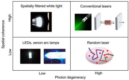
Figure 1. Comparison of light sources based on their spatial coherence and photon degeneracy.
Recently, random lasers have been investigated for wide-field fluorescence bio-imaging applications [35][36]. It has been observed that in both trans-illumination and epi-illumination configurations, random lasers outperform LEDs and conventional laser sources and provides high-contrast images with all the finer structural details. The photon degeneracy and the spectral characteristics of the illumination source have a significant impact on the emission intensity in fluorescence imaging. LEDs have broad emission spectra and low photon degeneracy by nature. Despite the fact that the LED’s bandwidth was limited to 25 nm using bandpass filters (similar to the filters commonly used in fluorescence microscopes), they were still inefficient in comparison to laser sources for the excitation of fluorescent molecules. Conversely, despite having a narrow band width and high photon degeneracy, the images recorded using the conventional laser had lower contrast than the images recorded using the random laser. This has been explained based on the non-uniform excitation caused by the inherent coherent artefacts in the case of the conventional laser, which led to output distortion, a decrease in overall intensity, and degradation of the image quality. The non-uniform excitation also causes the loss of information that may lead to misinterpretations and errors in quantitative fluorescence imaging. Random laser illumination was also demonstrated to provide better contrast and a higher signal-to-noise ratio in multi-layered diffusive tissue samples [35].
3. Sensing Applications
The spectral features of random lasers exhibit a strong dependence on the scattering environment. As a result, they display sensitivity to various external factors, including temperature, refractive index, pH, humidity, etc. [7][8][37][38]. This inherent sensitivity allows for a wide range of sensing applications using random lasing [39]. Random lasers that can sense multiple parameters simultaneously have also been developed [40]. A random-laser-based sensor using gold nanoparticles has demonstrated the ability to detect even nanomolar levels of dopamine (a neurotransmitter found in brain tissues) [41]. This has significant implications for the detection and management of Parkinson’s and Huntington’s diseases. Random lasing signals have been instrumental in identifying the morphological and structural alterations triggered by mutations in the Huntingtin gene [42]. In another recent report, a fiber-based plasmonic random laser was utilized to detect human immunoglobulin (IgG) and quantitatively monitor its concentration through specific binding interactions with protein A [8].
Chemical sensors based on random lasers, when operated near or above the threshold, exhibit a sensitivity more than 20 times higher than those relying on spontaneous emission or fluorescence [43]. Further, the use of biocompatible polymers has facilitated random-laser-based wearable sensors that may be transferred or implanted on the skin for the purpose of monitoring physical activities, detecting sweat, etc. [44][45]. Furthermore, recent research has successfully demonstrated self-healing random lasers which can self-recover their lasing action after being chopped into tens of pieces in only a few minutes [46]. Wearable systems like soft bioimaging implants, portable laser gadgets, and photonic skins could benefit greatly from such random lasers with stretchability, high deformability, and self-healing properties.
Random lasing has been conducted in various biological tissues and is being utilized for biosensing and biodiagnostic applications. Polson et al. used random lasing emission from tissues to identify cancer regions [9]. They observed that the cancerous tissues generated more laser lines than the healthy tissues did. This indicates the presence of more laser resonators as a result of increased tissue disorder associated with cancer progression. The random lasing thresholds were found to be related to the tumor malignancy grade, and could, thus, be utilized to classify tissues for diagnostic purposes [47]. Further, the cavity length of the laser resonators can be estimated by the power Fourier transform of random lasing spectra, which aids in the mapping of cancerous regions [48]. The random-laser-based biosensor developed by Song et al. could detect nanoscale structural and mechanical deformation in the bones [49]. Such mechanical behavior testing with random lasing emission has been extended to soft tissues as well [50]. In these techniques, the tissue acts as the scatterer, and the gain medium is a suitable fluorophore impregnated to the tissues. Recent research has also shown novel strategies for random lasing without the need to infiltrate the biological samples with dyes. Instead, the gain medium is encapsulated in a transparent spherical cell and attached at the ends of an optical fiber, allowing for non-invasive diagnostics of biological samples [51]. Proceeding one step further, a recent study demonstrated that random laser emission can be used to differentiate tissues, for example, fat, nerve, muscle, and skin tissues, even under room light conditions [52].
References
- Wiersma, D.S. Disordered Photonics. Nat. Photonics 2013, 7, 188–196.
- Burresi, M.; Pratesi, F.; Riboli, F.; Wiersma, D.S. Complex Photonic Structures for Light Harvesting. Adv. Opt. Mater. 2015, 3, 722–743.
- Letokhov, V.S. Generation of Light by a Scattering Medium with Negative Resonance Absorption. J. Exp. Theor. Phys. 1968, 26, 835–840.
- Polson, R.C.; Raikh, M.E.; Vardeny, Z.V. Universal Properties of Random Lasers. IEEE J. Sel. Top. Quantum Electron. 2003, 9, 120–123.
- Wiersma, D.S. The Physics and Applications of Random Lasers. Nat. Phys. 2008, 4, 359–367.
- Cao, H.; Xu, J.Y.; Ling, Y.; Burin, A.L.; Seeling, E.W.; Liu, X.; Chang, R.P.H. Random Lasers with Coherent Feedback. IEEE J. Sel. Top. Quantum Electron. 2003, 9, 111–119.
- Wiersma, D.S.; Cavalieri, S. Light Emission: A Temperature-Tunable Random Laser. Nature 2001, 414, 708–709.
- Shi, X.; Ge, K.; Tong, J.-H.; Zhai, T. Low-Cost Biosensors Based on a Plasmonic Random Laser on Fiber Facet. Opt. Express 2020, 28, 12233.
- Polson, R.C.; Vardeny, Z.V. Random Lasing in Human Tissues. Appl. Phys. Lett. 2004, 85, 1289–1291.
- Abaie, B.; Mobini, E.; Karbasi, S.; Hawkins, T.; Ballato, J.; Mafi, A. Random Lasing in an Anderson Localizing Optical Fiber. Light. Sci. Appl. 2017, 6, e17041.
- Choi, S.H.; Kim, S.-W.; Ku, Z.; Visbal-Onufrak, M.A.; Kim, S.-R.; Choi, K.-H.; Ko, H.; Choi, W.; Urbas, A.M.; Goo, T.-W.; et al. Anderson Light Localization in Biological Nanostructures of Native Silk. Nat. Commun. 2018, 9, 452.
- Balachandran, R.M.; Lawandy, N.M. Interface Reflection Effects in Photonic Paint. Opt. Lett. 1995, 20, 1271.
- Letokhov, V.S. Stimulated Emission of an Ensemble of Scattering Particles with Negative Absorption. JETP Lett. 1967, 5, 212–215.
- Noginov, M.A. Solid-State Random Lasers; Springer: Berlin/Heidelberg, Germany, 2005; ISBN 0387239138.
- Xu, X.; Lu, W.; Wang, T.; Gao, W.; Yu, X.; Qiu, J.; Yu, S.F. Deep UV Random Lasing from NaGdF 4:Yb 3+,Tm 3+ Upconversion Nanocrystals in Amorphous Borosilicate Glass. Opt. Lett. 2020, 45, 3095.
- Liang, H.K.; Meng, B.; Liang, G.; Tao, J.; Chong, Y.; Wang, Q.J.; Zhang, Y. Electrically Pumped Mid-Infrared Random Lasers. Adv. Mater. 2013, 25, 6859–6863.
- Schönhuber, S.; Brandstetter, M.; Hisch, T.; Deutsch, C.; Krall, M.; Detz, H.; Andrews, A.M.; Strasser, G.; Rotter, S.; Unterrainer, K. Random Lasers for Broadband Directional Emission. Optica 2016, 3, 1035.
- Zeng, Y.; Liang, G.; Qiang, B.; Wu, K.; Tao, J.; Hu, X.; Li, L.; Davies, A.G.; Linfield, E.H.; Liang, H.K.; et al. Two-Dimensional Multimode Terahertz Random Lasing with Metal Pillars. ACS Photonics 2018, 5, 2928–2935.
- Goodman, J.W. Speckle Phenomena in Optics: Theory and Applications, 2nd ed.; SPIE Press: Bellingham, WA, USA, 2020; ISBN 9781510631496.
- Silverstein, S.D.; O’Donnell, M. Theory of Frequency and Temporal Compounding in Coherent Imaging: Speckle Suppression and Image Resolution. J. Opt. Soc. Am. A 1988, 5, 104.
- Hard, R.; Zeh, R.; Allen, R.D. Phase-Randomized Laser Illumination for Microscopy. J. Cell Sci. 1977, 23, 335–343.
- Lowenthal, S.; Joyeux, D. Speckle Removal by a Slowly Moving Diffuser Associated with a Motionless Diffuser. J. Opt. Soc. Am. 1971, 61, 847.
- Ambar, H.; Aoki, Y.; Takai, N.; Asakura, T. Mechanism of Speckle Reduction in Laser-Microscope Images Using a Rotating Optical Fiber. Appl. Phys. B Photo. 1985, 38, 71–78.
- Stangner, T.; Zhang, H.; Dahlberg, T.; Wiklund, K.; Andersson, M. Step-by-Step Guide to Reduce Spatial Coherence of Laser Light Using a Rotating Ground Glass Diffuser. Appl. Opt. 2017, 56, 5427.
- Akram, M.N.; Tong, Z.; Ouyang, G.; Chen, X.; Kartashov, V. Laser Speckle Reduction Due to Spatial and Angular Diversity Introduced by Fast Scanning Micromirror. Appl. Opt. 2010, 49, 3297.
- Hansford, D.J.; Fells, J.A.J.; Elston, S.J.; Morris, S.M. Speckle Contrast Reduction of Laser Light Using a Chiral Nematic Liquid Crystal Diffuser. Appl. Phys. Lett. 2016, 109, 261104.
- Farrokhi, H.; Rohith, T.M.; Boonruangkan, J.; Han, S.; Kim, H.; Kim, S.-W.; Kim, Y.-J. High-Brightness Laser Imaging with Tunable Speckle Reduction Enabled by Electroactive Micro-Optic Diffusers. Sci. Rep. 2017, 7, 15318.
- Lee, Y.M.; Lee, D.U.; Park, J.M.; Park, S.Y.; Lee, S.G. P-45: A Study on the Relationships between Human Perception and the Physical Phenomenon of Speckle. SID Symp. Dig. Tech. Pap. 2008, 39, 1347.
- Roelandt, S.; Meuret, Y.; Jacobs, A.; Willaert, K.; Janssens, P.; Thienpont, H.; Verschaffelt, G. Human Speckle Perception Threshold for Still Images from a Laser Projection System. Opt. Express 2014, 22, 23965.
- Redding, B.; Choma, M.A.; Cao, H. Speckle-Free Laser Imaging Using Random Laser Illumination. Nat. Photonics 2012, 6, 355–359.
- Ma, R.; Rao, Y.J.; Zhang, W.L.; Hu, B. Multimode Random Fiber Laser for Speckle-Free Imaging. IEEE J. Sel. Top. Quantum. Electron. 2019, 25, 0900106.
- Lee, Y.J.; Yeh, T.W.; Yang, Z.P.; Yao, Y.C.; Chang, C.Y.; Tsai, M.T.; Sheu, J.K. A Curvature-Tunable Random Laser. Nanoscale 2019, 11, 3534–3545.
- Guo, J.Y.; Zhang, W.L.; Rao, Y.J.; Zhang, H.H.; Ma, R.; Lopes, D.S.; Lins, I.C.X.; Gomes, A.S.L. High Contrast Dental Imaging Using a Random Fiber Laser in Backscattering Configuration. OSA Contin. 2020, 3, 759.
- Pramanik, A.; Biswas, S.; Kumbhakar, P.; Kumbhakar, P. External Feedback Assisted Reduction of the Lasing Threshold of a Continuous Wave Random Laser in a Dye Doped Polymer Film and Demonstration of Speckle Free Imaging. J. Lumin. 2021, 230, 117720.
- Gayathri, R.; Suchand Sandeep, C.S.; Gummaluri, V.S.; Mohammed Asik, R.; Padmanabhan, P.; Gulyás, B.Z.; Vijayan, C.; Vadakke Matham, M. Plasmonic Random Laser Enabled Artefact-Free Wide-Field Fluorescence Bioimaging: Uncovering Finer Cellular Features. Nanoscale Adv. 2022, 4, 2278–2287.
- Carvalho, M.T.; Lotay, A.S.; Kenny, F.M.; Girkin, J.M.; Gomes, A.S.L. Random Laser Illumination: An Ideal Source for Biomedical Polarization Imaging? Proc. SPIE 2016, 9701, 97010Q.
- Gaio, M.; Caixeiro, S.; Marelli, B.; Omenetto, F.G.; Sapienza, R. Gain-Based Mechanism for PH Sensing Based on Random Lasing. Phys. Rev. Appl. 2017, 7, 034005.
- Tong, J.; Shi, X.; Wang, Y.; Han, L.; Zhai, T. Flexible Plasmonic Random Laser for Wearable Humidity Sensing. Sci. China Inf. Sci. 2021, 64, 222401.
- Ni, D.; Späth, M.; Klämpfl, F.; Hohmann, M. Properties and Applications of Random Lasers as Emerging Light Sources and Optical Sensors: A Review. Sensors 2022, 23, 247.
- Xu, Y.; Zhang, M.; Lu, P.; Mihailov, S.; Bao, X. Multi-Parameter Sensor Based on Random Fiber Lasers. AIP Adv. 2016, 6, 095009.
- Wan Ismail, W.Z.; Liu, G.; Zhang, K.; Goldys, E.M.; Dawes, J.M. Dopamine Sensing and Measurement Using Threshold and Spectral Measurements in Random Lasers. Opt. Express 2016, 24, A85.
- De Armas-Rillo, S.; Fumagallo-Reading, F.; Luis-Ravelo, D.; Abdul-Jalbar, B.; González-Hernández, T.; Lahoz, F. Random Lasing Detection of Mutant Huntingtin Expression in Cells. Sensors 2021, 21, 3825.
- Deng, C.; He, Q.; He, C.; Shi, L.; Cheng, J.; Lin, T. Conjugated Polymer-Titania Nanoparticle Hybrid Films: Random Lasing Action and Ultrasensitive Detection of Explosive Vapors. J. Phys. Chem. B 2010, 114, 4725–4730.
- Ge, K.; Guo, D.; Ma, X.; Xu, Z.; Hayat, A.; Li, S.; Zhai, T. Large-Area Biocompatible Random Laser for Wearable Applications. Nanomaterials 2021, 11, 1809.
- Ta, V.D.; Nguyen, T.T.; Nghiem, T.H.L.; Tran, H.N.; Le, A.T.; Dao, N.T.; Duong, P.D.; Mai, H.H. Silica Based Biocompatible Random Lasers Implantable in the Skin. Opt. Commun. 2020, 475, 126207.
- Hsu, Y.-T.; Tai, C.-T.; Wu, H.-M.; Hou, C.-F.; Liao, Y.-M.; Liao, W.-C.; Haider, G.; Hsiao, Y.-C.; Lee, C.-W.; Chang, S.-W.; et al. Self-Healing Nanophotonics: Robust and Soft Random Lasers. ACS Nano 2019, 13, 8977–8985.
- Wang, Y.; Duan, Z.; Qiu, Z.; Zhang, P.; Wu, J.; Zhang, D.; Xiang, T. Random Lasing in Human Tissues Embedded with Organic Dyes for Cancer Diagnosis. Sci. Rep. 2017, 7, 8385.
- Polson, R.C.; Vardeny, Z.V. Cancerous Tissue Mapping from Random Lasing Emission Spectra. J. Opt. 2010, 12, 024010.
- Song, Q.; Xu, Z.; Choi, S.H.; Sun, X.; Xiao, S.; Akkus, O.; Kim, Y.L. Detection of Nanoscale Structural Changes in Bone Using Random Lasers. Biomed. Opt. Express 2010, 1, 1401.
- Briones-Herrera, J.C.; Cuando-Espitia, N.; Sánchez-Arévalo, F.M.; Hernández-Cordero, J. Evaluation of Mechanical Behavior of Soft Tissue by Means of Random Laser Emission. Rev. Sci. Instrum. 2013, 84, 104301.
- Ignesti, E.; Tommasi, F.; Fini, L.; Martelli, F.; Azzali, N.; Cavalieri, S. A New Class of Optical Sensors: A Random Laser Based Device. Sci. Rep. 2016, 6, 35225.
- Hohmann, M.; Dörner, D.; Mehari, F.; Chen, C.; Späth, M.; Müller, S.; Albrecht, H.; Klämpfl, F.; Schmidt, M. Investigation of Random Lasing as a Feedback Mechanism for Tissue Differentiation during Laser Surgery. Biomed. Opt. Express 2019, 10, 807.
More
Information
Subjects:
Physics, Applied
Contributors
MDPI registered users' name will be linked to their SciProfiles pages. To register with us, please refer to https://encyclopedia.pub/register
:
View Times:
886
Revisions:
2 times
(View History)
Update Date:
11 Sep 2023
Table of Contents


Notice
You are not a member of the advisory board for this topic. If you want to update advisory board member profile, please contact office@encyclopedia.pub.
OK
Confirm
Only members of the Encyclopedia advisory board for this topic are allowed to note entries. Would you like to become an advisory board member of the Encyclopedia?
Yes
No
${ textCharacter }/${ maxCharacter }
Submit
Cancel
Back
Comments
${ item }
|
${ item.createdUser.fullName }
${ item.createdAt }
${ item.vote }
${ item.reply }
Delete
${ reply.createdUser.fullName }
${ reply.createdAt }
${ reply.vote }
Delete
There is no reply to this comment~
${ item.replyTextCharacter }/${ item.replyMaxCharacter }
Submit
Cancel
More
No more~
There is no comment~
${ textCharacter }/${ maxCharacter }
Submit
Cancel
${ selectedItem.replyTextCharacter }/${ selectedItem.replyMaxCharacter }
Submit
Cancel
Confirm
Are you sure to Delete?
Yes
No