Submitted Successfully!
To reward your contribution, here is a gift for you: A free trial for our video production service.
Thank you for your contribution! You can also upload a video entry or images related to this topic.
Version Summary Created by Modification Content Size Created at Operation
1 -- 5664 2023-09-08 20:21:34 |
2 Reference format revised. -36 word(s) 5628 2023-09-11 03:20:34 |

Video Upload Options

Do you have a full video?

Confirm

Are you sure to Delete?
Cite
If you have any further questions, please contact Encyclopedia Editorial Office.
Triantaphyllopoulos, K.A. Long Non-Coding RNAs in IBD and Johne’s Disease. Encyclopedia. Available online: https://encyclopedia.pub/entry/48984 (accessed on 07 September 2024).
Triantaphyllopoulos KA. Long Non-Coding RNAs in IBD and Johne’s Disease. Encyclopedia. Available at: https://encyclopedia.pub/entry/48984. Accessed September 07, 2024.
Triantaphyllopoulos, Kostas A.. "Long Non-Coding RNAs in IBD and Johne’s Disease" Encyclopedia, https://encyclopedia.pub/entry/48984 (accessed September 07, 2024).
Triantaphyllopoulos, K.A. (2023, September 08). Long Non-Coding RNAs in IBD and Johne’s Disease. In Encyclopedia. https://encyclopedia.pub/entry/48984
Triantaphyllopoulos, Kostas A.. "Long Non-Coding RNAs in IBD and Johne’s Disease." Encyclopedia. Web. 08 September, 2023.
Long Non-Coding RNAs in IBD and Johne’s Disease
Edit

Non-coding RNAs (ncRNA) have paved the way to new perspectives on the regulation of gene expression, not only in biology and medicine, but also in associated fields and technologies, ensuring advances in diagnostic means and therapeutic modalities. Critical in this multistep approach are the associations of long non-coding RNA (lncRNA) with diseases and their causal genes in their networks of interactions, gene enrichment and expression analysis, associated pathways, the monitoring of the involved genes and their functional roles during disease progression from one stage to another. Studies have shown that Johne’s Disease (JD), caused by Mycobacterium avium subspecies partuberculosis (MAP), shares common lncRNAs, clinical findings, and other molecular entities with Crohn’s Disease (CD). This has been a subject of vigorous investigation owing to the zoonotic nature of this condition, although results are still inconclusive.

long non-coding RNAs biomarker epigenetic regulation mycobacter Johne’s disease

1. Introduction

In recent years, incessant microbiological research has attracted the most powerful available resources to combat life-threatening infectious diseases, rendering this exploration significantly necessary and of high priority to assess the “quiver” that the pathogens carry and the susceptibility profile of the host. More importantly, new discoveries in the genetic and non-genetic components that have been identified in pathologies associated with bacterial invasion in human or animal cells have paved the way to advanced and in depth learning of the interactions between the molecular entities and environmental factors that are likely to lead to inflammatory bowel disease (IBD)-related pathogenicities. The ongoing research is significant and has strengthened the exploration and discovery of potential disease biomarkers that could improve diagnostic tools and the examination of new therapeutic modalities of high priority [1].
As mentioned earlier, accurate diagnosis and detection at the subclinical stage is a very important step for disease control. This is currently performed through the quantitative detection of Mycobacterium avium subspecies partuberculosis (MAP) DNA by real-time PCR or sequencing methods in animals with the disease; the latter gives the potential to also detect regions in the coding and non-coding parts of the genome, as has been reported for microRNAs (miRNAs), as well as providing the ability to study infectious disease biomarkers [2].
In recent years, non-coding RNAs are a novel and very promising diagnostic approach to infectious and non-infectious diseases that has become a major focus of interest in research, and a large number of ncRNAs have been identified through large-scale genomic analyses [3][4][5]. Conventionally, ncRNAs can be broadly classified into small ncRNAs (maximum length of ≈200 nucleotides, including microRNAs, siRNAs, and piRNAs) and longer transcripts, including as lncRNAs (>200 nucleotides in length) and small RNAs (50 to 300 nucleotides long). Of the above categories, miRNAs have been widely studied in various diseases [6]. These functionally act by blocking at the post-transcriptional level or translationally repressing the target mRNAs. On the contrary, only a small number of long non-coding (lncRNAs) have been functionally characterized so far.
LncRNAs are newly discovered potential biomarkers, which have introduced us to novel perspectives on the regulation of gene expression, not only in biology and medicine but also in associated scientific fields and technologies. Importantly, the complex interactions and dynamic equilibrium between the genetic code and epigenetic signatures during disease progression is modulated by environmental signals over the course of time under a traceable path. LncRNAs and long intervening noncoding RNAs (lincRNAs) are known to act as decoys, scaffolds, sponges, and guides to proteins and RNA molecules in cells, fulfilling essential functions associated with gene expression regulation. Like miRNAs, this class of long ncRNAs has emerged as important regulators of both normal and pathological states, while compelling evidence has been accumulated recently, indicating that lncRNAs are involved in a wide range of biological functions [7]. Importantly, lncRNAs can function as tumor suppressors [8], oncogenes [9] during the development of multiple cancers, or mediators of infectious diseases by controlling the basal and TLR2-inducible expression of TNFα in human monocytes [10].

2. Morphology and Biological Characteristics of Mycobacterium avium

Mycobacterium avium is a species of the phylum Actinobacteria, belonging to the genus Mycobacterium. Mycobacterium avium subspecies paratuberculosis is a thin, straight or slightly bent, acid-resistant, and immobile bacterium. It is mainly aerobic, but under anaerobic conditions, it has shown signs of survival [11]. Its dimensions are normally 0.5 × 1.5 μm. MAP ribosomes are no different from typical bacterial ribosomes.
Mycobacterium avium is a rod prokaryote, zoonotic microorganism that causes avian tuberculosis and Mycobacterium avium complex (MAC) in humans. A secondary infection to AIDS, MAC, also called Mycobacterium avium-intracellulare complex, is a microbial complex of two Mycobacterium species, Mycobacterium avium and Mycobacterium intracellulare, which are saprotrophic organisms that are present in soil and water. Interestingly, disease-isolated mycobacteria from CD patients are identical to Mycobacterium paratuberculosis, as determined by DNA probes that can distinguish between mycobacterial species [12]. This advocates for MAP being the causal pathogen of CD in humans.
The genus Mycobacterium represents the only entity within the Mycobacteriaceae family, which belongs to the order Mycobacteriales and the phylum Actinomycetota [13]. Whereas the great majority of about 130 described species in the genus are harmless environmental saprophytes, some mycobacteria have evolved to be major pathogens. The pathogenic species mainly belong to the slowly growing mycobacteria category and comprise well-known human pathogens, such as Mycobacterium tuberculosis, Mycobacterium leprae, and Mycobacterium ulcerans, while the confirmed animal pathogens are Mycobacterium bovis, Mycobacterium marinum, and MAP [14]. The MAP pathogen has been implicated in JD and also has the potential to infect humans and be the causal pathogen of CD. Also, MAP has the characteristics of an acid-resistant pathogen mycobacteria, including the human pathogen M. tuberculosis, which is not a typical Gram-positive mycobacterium. Its lipid-rich cell wall makes it resistant to Gram staining; thus, it can only be stained with carbolfuchsin, acid–alcohol, and methylene blue [15].

3. MAP, Crohn’s Disease, and Relative Pathologies

Currently, there are divided opinions on the possible link between MAP and Crohn’s disease, albeit that MAP is involved in a constantly increasing number of pathologic conditions of unknown etiology in humans and is considered a potent threat to animals and human health [16][17][18]. The available evidence covers a wide range of hosts including non-ruminant species such as pigs [19], rabbits [20], foxes and ferrets [21], and macaques [22], demonstrating MAP’s wide host infectivity and worldwide prevalence associated with the reduced productivity of infected animals [23].
More importantly, besides the threat to animal welfare, MAP is a controversial causative pathogen, since clinicians and scientists’ have divided opinions on the evidence of MAP isolation from human patients with Crohn’s disease, which was reformed recently by the positive MAP findings from patients [24][25][26][27]; thus, this pathogen poses a serious infectious threat to the public with its transmission occurring mainly through dairy products in livestock. The latter provides controversial evidence that MAP is zoonotic with a latent stage of infection similar to that of Mtb, the causative agent of TB in humans, where infection leads to a persistent immune response that controls but does not completely eliminate the pathogen. The latter makes JD difficult to document as a zoonotic disease that is associated with the pathogenesis of Crohn’s and related pathologies [24][25]; thus, the debate is still ongoing. The link of MAP zoonosis with CD has been a medical controversy for over one hundred years [26][28]; however, validation of the effort of Professor John Hermon-Taylor, an early elucidator of the zoonotic capacity of MAP [28], has come from numerous recent studies showing CD resolution with antimycobacterial therapies targeted against MAP [29][30][31][32][33].
Experimental evidence for MAP as the causative pathogen of IBD-related pathologies and CD has been reported in several studies in which the organism has been cultured from various tissues such as intestinal tissue, feces, breast milk, and/or blood from IBD patients and, recently, MAP DNA/RNA has been detected in patient samples versus healthy controls [26][27][34][35]. More importantly, exposure to MAP has not been accurately determined and the pathogen is easily spread by various means of contamination, posing a threat to the public, as the organism has been found in water, commercial milk, dairy products, etc., and has been shown to survive after pasteurization procedures that are sufficient to kill common contaminants [36]. This level of infection definitely leads to contamination of the environment via a contaminated water supply, dust bio-aerosols, milk, and the food supply. This contaminated environment not only affects the spread of MAP among animals, it may also be associated with the human intestinal disorders, i.e., IBD and CD [34][35][36][37]

4. Long Non-Coding RNAs (LncRNAs) and Their Footprint in Gene Regulation

4.1. LncRNAs in the Disease State

The human genome is pervasively transcribed, while a small fraction only of all RNAs processed inside the cell are protein-coding sequences. In fact, only ca. 2% of the transcripts of the human genome can encode proteins, and less than 3% compose protein-coding gene exons [38]. Consequently, the majority of the transcripts seem to be non-protein-coding sequences, which account for 98% of the human genome (ncRNAs), although budding yeast has been proven to be a powerful model organism for understanding the mechanisms ruling pervasive transcription [39][40].
Known types of non-coding RNAs are transfer RNAs (tRNAs); ribosomal RNAs(rRNAs); small RNAs, such as microRNAs, siRNAs, piRNAs, snoRNAs, snRNAs, and exRNAs; long ncRNAs (lncRNAs); long intergenic non-coding RNA (lincRNA) lincRNAs; circular RNAs (circRNAs); and examples of ncRNAs, such as the well-known Xist and HOTAIR. circRNAs are stable, evolutionarily conserved, and single-stranded RNA molecules. Unlike linear RNAs, circRNAs are closed-loop type RNAs with joined 3′ and 5′ ends [41]. Four types of circRNAs have been discovered, namely exonic circRNAs (ecircRNAs), circular intronic RNAs (ciRNAs), exon–intron circRNAs (EIciRNAs), and intergenic circRNAs [42][43]. circRNAs function as miRNA sponges and can regulate RNA expression by consuming miRNA targets [44].
It was shown that bacteria interfere with the expression of mammalian regulatory RNAs to modify immune signaling, autophagy, or the apoptotic machinery, and lncRNAs were reported to play a crucial role in the regulation of eukaryotic gene expression. This is in contrast to the known role of miRNAs in bacterial infections, which has been extensively studied and reviewed over the years [45][46][47]; thus, the emerging potential of RNA entities, collectively known as lncRNAs, has already begun to be manifested through their regulatory competence in complex machinery, illuminating the “usefulness” of read through pervasive transcription, i.e., a transcriptional read through of transcripts with non-established functions, leading to the accumulation of many opportunistic transcripts, whose synthesis is controlled by the cell at the co-transcriptional RNA processing level; this phenomenon can lead to chimeric transcripts and retained introns [48].
LncRNAs, among their many “charismatic” roles in gene activity, also play a critical role in organizing the 3D genome architecture and regulating in cis- or in trans- gene expression through numerous mechanisms that have been reported elsewhere [49][50][51][52]. They are DNA elements that can be encoded almost anywhere in the genome, e.g., within intergenic regions (lincRNAs), within protein-coding genes (in the antisense), and within introns [53]. Processed transcripts are a recent addition to this list. 
The first non-coding gene was discovered in humans by using differential hybridization screens of cDNA libraries, a suitable approach to cloning and studying genes with tissue-specific and temporal patterns of gene expression. The aforementioned non-coding gene was the imprinted and maternally expressed transcript H19, which was initially classified as mRNA, but the absence of a long and conserved open reading frame (ORF) within H19 and a lack of ribosomal interaction led to the conclusion that it is a non-coding transcript.
Strictly speaking, for immune cells and the pertinently activated mechanisms upon invasion by bacteria or other microorganisms, the competing endogenous lncRNAs are associated with dynamic changes in gene expression, the products of which combat infectious pathogens, initiate repair mechanisms, and resolve inflammatory responses in cells and tissues. The primary structure of lncRNAs shows that are at least 200 nucleotides, although there is a diversity that distinguishes lncRNAs from smaller non-coding RNAs such as tRNA, miRNA, piRNA (Piwi-interacting RNAs), snRNA, snoRNA, etc. 
LncRNAs function as protein scaffolds, activators or inhibitors of transcription, and antisense RNA or miRNA sponges [54] that exhibit lower cellular concentrations than protein-coding genes but with a higher degree of tissue specificity [55], despite their low evolutionary conservation. Importantly, lncRNAs have a key role in the regulation of gene expression and, as mentioned earlier, remain poorly identified and annotated and are characterized less emphatically in domesticated animals compared to in other species such as humans and mice [56]; however, progress has been made in this decade, starting with bovine and pig genomes and including non-coding transcripts [57][58].

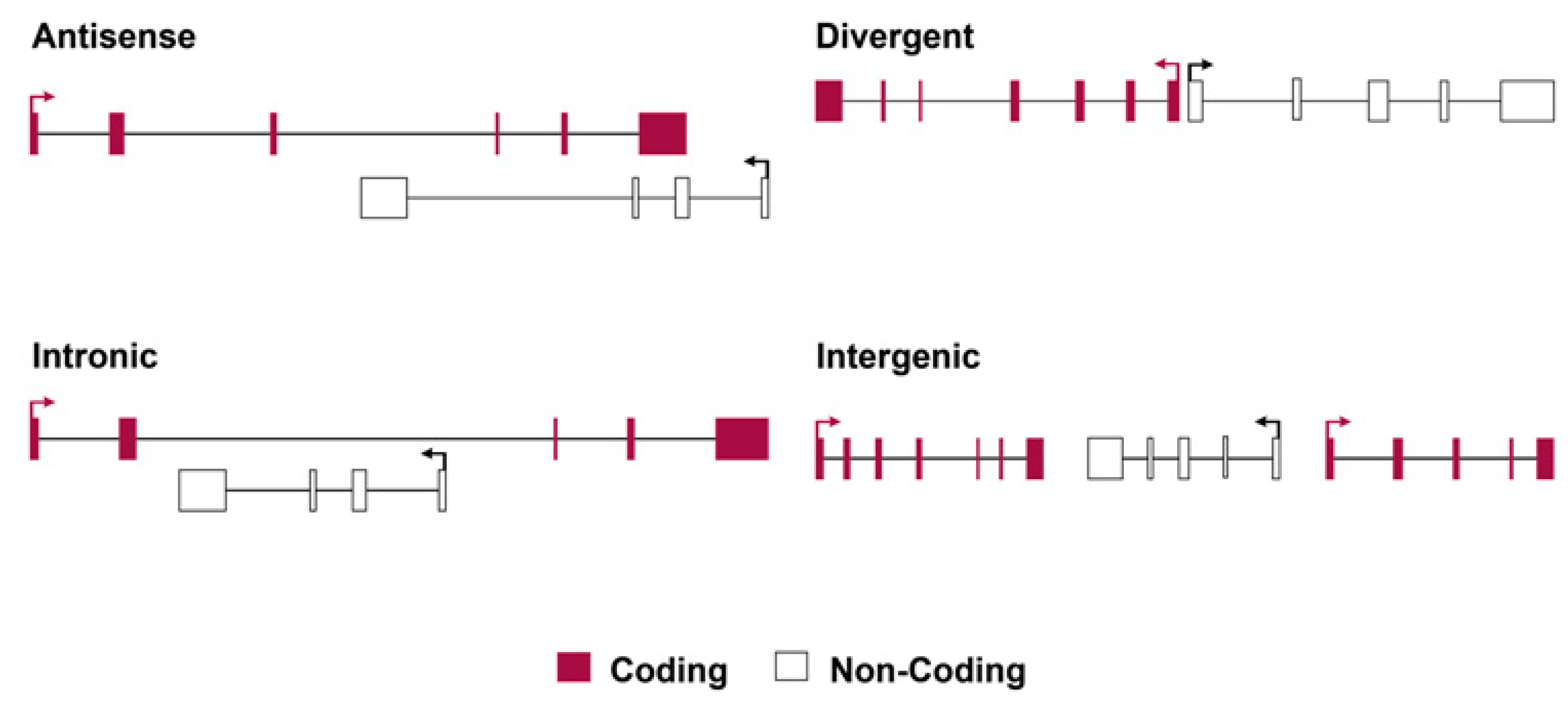
4.2. Principles of Classification for Long Non-Coding RNAs

Strictly speaking, for lncRNA classes, lncRNAs can be arbitrarily categorized into four types, as shown in Figure 1: (1) antisense lncRNAs are transcribed in the opposite direction to protein-coding genes, and they often overlap by at least one exon, while they initiate transcription within or at the 3′ end of coding genes. The antisense lncRNAs initiate transcription from the aligned antiparallel protein-coding gene sequence and transcribe in the opposite direction to the overlapping coding exons, e.g., antisense Tsix, which negatively regulates Xist in cis via chromatin modifications at the onset of X-inactivation. (2) Intronic lncRNAs, initiate bidirectional expression inside an intron of a protein-coding gene and terminate expression without overlapping exons. (3) Bidirectional lncRNAs initiate transcription in a divergent fashion from a promoter of a protein-coding gene; the distance of lncRNA that constitutes the bidirectionality in transcription is not defined and is associated with imprecise termination, but is generally within a few hundred (<1000) base pairs. (4) Intergenic lncRNAs (which are often referred to as large intervening/intergenic non-coding RNAs or lincRNAs) are sequences of lncRNA that do not overlap with protein-coding genes as they have separate transcriptional units from protein-coding genes.
Figure 1. The organization of different types of lncRNAs in the genome depends on the location of the lncRNA sequence relative to nearby protein-coding genes and the direction of its transcription. Categories of lncRNAs are often defined by their locations, as shown above. Antisense: these are lncRNAs that initiate inside a protein-coding gene and transcribe in the opposite direction to overlapping coding exons. Intronic: these are the lncRNAs that initiate inside an intron of a protein-coding gene in either direction and terminate without overlapping exons. Bidirectional: these are lncRNA transcripts that initiate in a divergent fashion from a promoter of a protein-coding gene; the precise distance cut-off that constitutes bidirectionality is not defined but is generally within a few hundred base pairs. Intergenic: these are lncRNAs (also termed large intervening non-coding RNAs or lincRNAs) with separate transcriptional units from protein-coding genes.

5. LncRNAs’ Involvement in Immune Dysregulation in IBD and JD

5.1. LncRNA Evidence in IBD-Related Pathologies

Recent evidence indicates that lncRNAs play important roles in the immune response, such as orchestrating the development of disparate types of immune cells, organizing chromatin and regulating the transcriptional programmes, and distinguished roles that feature immune homeostasis and gene activation. These are particularly important in infectious and inflammatory diseases, as perturbations in the non-coding component of the genome, such as RNA 3′ end processing and regulators of cleavage and polyadenylation, contribute to several human diseases [59]. LncRNAs also play important roles in directing the development of diverse immune cells and controlling the dynamic transcriptional programmes supported by alternative splicing and polyadenylation events that are hallmarks of immune cell activation.
Landmarks in the study of lncRNAs since their discovery 20 years ago include the significant progress in laboratory methods, computational biology, and algorithms invented to map new lncRNAs; however, the difficulty with accelerating progress resides in the low pace of characterization and the elucidation of their functional roles, which are mainly unexplored. 
One way to initiate the study of lncRNA characterization and functional analysis is by Gene Ontology (GO) term examination and enrichment analyses of the regional protein-coding genes [60][61]. The potential functions of the lncRNAs can be predicted by examining the Gene Ontology (GO) term enrichment of the nearest protein-coding gene. Additionally, for many tissues, the GO terms with highly significant enrichment are associated with relative functions of lncRNAs, lincRNAs, etc. that are essential for those tissues and have been examined and analyzed in previous studies of mammalian lncRNAs [62][63]. NFκB proteins are a family of five structurally related transcription factors that control the expression of inflammatory molecules, thereby counteracting bacterial infections. Many lncRNAs have been reported to interfere with NFκB signaling [64][65]. Specific examples of the latter interference account for (a) the induction of the lincRNA–Tnfaip3 complex, which is required for the transactivation of NFκB-regulated inflammatory genes in response to bacterial LPS stimulation [64] and (b) the reduction of HULC expression in a highly upregulated lncRNA (HULC) in liver cancer through treatment by TNF-a-induced apoptosis caused a reduction in HULC expression by the modulation of miRNA (miR-9) expression in association with DNA methyltransferases [66].
Despite the fact that a plethora of studies on IBD have focused on protein-coding genes, new advances in the field of lncRNAs, have shown their implications and altered expression in IBD patients compared to healthy controls. Although lncRNAs are novel gene regulators that have not been explored as much as miRNAs, an increasing number of lncRNAs are being implicated in IBD pathogeneses.

5.2. LncRNA Evidence from JD-Related Pathologies

Naturally infected cattle by MAP, studies have shown to contain lncRNAs with fewer exons than mRNAs (some of them incorporate less than three exons and some just have a single exon), while their length is also shorter than the length of mRNAs [56][67][68]. In more detail, Marete at al. published a report on the gene expression of highly differentially expressed gene targets associated with their affiliated lncRNA sequences and showed a pervasive lncRNA distribution on bovine autosomes (1–29) and on the X chromosome, which were experimentally localized in bovine chromosomes from macrophage cells of cows positive for JD vs. cows negative for JD (control) [68].
The lncRNAs identified in the Gupta et al. study were mostly intergenic (45.05%) with an average length of 600 bp long and ranged between 200 and 1000 bp [67], supporting the evidence that lncRNAs are mainly intergenic with a smaller overlap within genic regions [69]. The top 10 most significant differentially expressed (DEG) genes proximal to their lncRNA and a large amount of the above lncRNAs were found to be transcribed on chromosomes 18 and 19. These chromosomes are associated with signaling pathways for innate immune response, such as TNF signaling pathways (CCL5, CCL2, MAP2K3, MAP2K4, MAP3K14, MLKL, PIK3R5 and SOCS3), chemokine signaling pathways (CCL5, NFKBIB, ARRB2, CCL2, CCL4, CCL8, PIK3R5), NOD-like receptor signaling pathways (CCL5, NFKBIB, NLRP1, CCL2) and cytokine–cytokine receptor interactions (CCL5, CCL2, CCL4, CCL8, CSF3) [67]
RNA-seq studies in bovine macrophages infected with MA revealed a significant log2 fold-change for the DEG. The frizzled class receptor 1 gene (FZD1) was DGE, and its fold change was estimated to be 15.56 times more highly expressed in macrophages for positive vs. negative for JD macrophages in cattle [68]. In addition, a second significant enriched pathway was the RNA polymerase I promoter escape, which involves the TWISTNB gene and encodes RNA polymerase I subunit F. It is upregulated in macrophages from JD-diseased cows. 

5.3. Differences in LncRNA Profiles of CD and JD

A notable example is a variation in the genetic locus of protein tyrosine phosphatase 2 (PTPN2) in UC and IBD, which regulates cytokine signaling by acting on multiple phosphorylated proteins [70]. A study of patients with CD demonstrated a link between the SNP rs2542151 and lower levels of the PTPN2 protein in colonic fibroblasts, as well as the formation of aberrant autophagosomes in intestinal epithelial cells (IECs) [71]. The PTPN2 locus SNP rs2542151 is related to the variation in lncRNA LINC01882, which is primarily expressed in T cells and is also involved in IL-2 expression, affecting important events such as differentiation, immune responses, and the homeostasis of lymphocytes, including Tregs mechanisms. Specifically, the transcript LINC01882 has been reported to play significant roles in autoimmune diseases, including IBD, and in peripheral blood mononuclear cells (PBMCs) of UC patients [72]. Additionally, the lncRNA ROCKI negatively regulates its cognate encoding gene, myristoylated alanine-rich protein kinase C (MARCKS), which promotes inflammatory cytokine and chemokine production. Thus, the above consecutive events show that MARCKS’ gene expression, mediated by ROCKI, contributes to the IBD pathology [73]

Consistent with the above and the contribution of lncRNAs in IBD, which has been also shown in several studies, is their expression profile which can successfully distinguish IBD patients from healthy controls [74]. Furthermore, the transcription characteristics and clinically relevant parameters of lncRNAs indicate that they have strong potential to be used as prognostic biomarkers in IBD [75]

5.4. Expression Profiling in Crohn’s Disease vs. Healthy Controls

An analysis of datasets assigned with GEO accession number GSE75459 under platform GPL16956 from the Gene Expression Omnibus (GEO)/NCBI was performed. In more detail, the Platform GPL16956 Agilent-045997 Arraystar human lncRNA microarray V3 (Agilent Technologies Inc., Santa Clara, CA, USA) contains several series of samples from various human disease conditions, including (the GSE75459 dataset) CD samples under the title name “Plasma Long Non-coding RNA and mRNA Expression Profile of Crohn’s Disease identified by Microarray”. The latter dataset analysis yielded novel lncRNAs and mRNAs for gene expression profiling with new targets in CD through a genome-wide analysis. The limma package was applied for this analysis, which is the core of the underlying computational engine of the GEO2R (http://www.ncbi.nlm.nih.gov/geo/geo2r). This generated up to the 250 most statistically significant differentially expressed genes by calculating an adjusted p-value using the Benjamini–Hochberg method to control the false discovery rate (FDR) [76].
The LncRNAs GUSBP2, GAS5-AS1, LINC01272, DDX11-AS1, IFNG-AS1, KIF9-AS1, and BC012900 are specifically upregulated in IBD-related pathologies [77], while others, such as ALOX12P2, DPP10-AS1, DIO3OS, lnc-PTPN2-2, and TRIM52 are novel transcripts. Furthermore, in the GSE75459 dataset analysis, the lncRNAs TALAM1 and MALAT1 were shown to be downregulated in plasma samples from patients with CD [72][77][78][79][80]
Apart from the common transcripts/genes (the intersecting gene subset in both disease networks, i.e., IBD vs. JD), there are also unique molecules that are not found in databases or reported elsewhere, such as uncharacterized loci from the GSE75459 dataset analysis, e.g., XLOC and TCONS and the newly reported RNA genes, such as FIGNL2 Divergent Transcript), ALOX12P2, TALAM1, and others, as described below.

6. The Epigenetic Role of the LncRNAs Involved in Human IBD-Related Pathologies and Mycobacterial Infections of the Host

Epigenetic processes generate the epigenome, and these involve a variety of modulations and chemical modifications that do not affect the genetic code as such but instead include, and are not limited to, DNA methylation, histone modifications, chromatin remodeling, the regulation of gene expression by non-coding RNAs, genome instability, and any other force that contributes to the animal phenotype [81][82][83].
The International Human Epigenome Consortium and the Human Epigenome Projects have been initiated to understand and pass on knowledge on the overall epigenetic mechanisms involved in human health and disease [84]. The changes in non-coding RNA are believed to cause obesity, diabetes, and neurodegenerative diseases, affecting the lungs, liver, or other organs. Thus, the interactive roles of the microbiome and epigenetic regulation in human health are very important in intestinal dysfunction and IBD-related pathologies. 
Genome-wide association studies (GWAS) and exome sequencing have identified that only ~7% of disease-associated genetic variations (e.g., single nucleotide polymorphism, SNPs) identified-to-date are localized to protein-coding genes [85]. The vast majority of genetic disorders caused by sequence alterations (e.g., disease-associated SNPs) are localized to the non-coding regions of the genome, including genomic loci expressing lncRNAs [86].
The known DNA demethylase, (ten–eleven translocation protein that acts as an eraser protein in humans) (TET2) binds to the promoter region of the lncRNA CDKN2B-AS1 (or ANRIL) and regulates its expression and downstream genes. Interestingly, overexpression of the TET2 protein inhibits the abundance of CDKN2B-AS1, resulting in a decreased risk of gastric cancer [87].
In MAP-infected cattle, Mycobacterium sp. is capable of reprogramming the host cellular machinery, leading to the evasion of both the innate and adaptive immune responses, followed by the establishment of infection and dissemination. Lessons can be learnt from Mycobacterium tuberculosis infection in humans as well as infections in other mycobacterioses, e.g., M. leprae, as these pathogens reprogram the epigenetic mechanisms of the host epigenome by inducing changes in histone modification, DNA methylation, and non-coding RNA molecule expression or even reprogramming the cell potency [88].

7. The Triarchy of Infection, lncRNA Intervention, and Regulation (IIR)

7.1. The Infection Stage

Considering, on one hand, the infectious route of MAP pathogen invasion and the “immune disturbance” of the host and, on the other hand, the “breach” of intestinal mucosal homeostasis and barrier dysfunction caused by IBD in humans, the following events represent a common ground for these pathologies: either (a) starting with the steps of an immune challenge, triggered by MAP, and followed by antigen presentation from the host macrophages or (b) a gradual rising chronic intestinal barrier dysfunction of an unknown trigger that is lurking and deteriorating and is manifested by the aforementioned associated signaling pathways in the previous sections.
Evidence has accumulated through research over the years to show the interplay of genetic susceptibility, including non-coding regulatory transcripts, the environmental impact on the microbiome, and the immune condition of the host leading to or triggered by dysbiosis, which contributes to intestinal barrier dysfunction and inappropriate intestinal immune activation, thus resulting in disturbed intestinal mucosal homeostasis, which has become the hallmark of IBD [89][90]. The pathogen (MAP) starts its transmission from the oral mucosa and spreads to the ileum, which has been implicated as the primary point of MAP invasion upon the entrance of MAP into the microfold (M) cells of Peyer’s patches in the intestinal mucosa (Figure 2).
Figure 2. The MAP pathogen is the infectious trigger for the chronic development of JD and is also likely to be associated with IBD syndrome. Infection starts by MAP invasion and internalization in the M cells of Peyer’s patches within the intestinal mucosa, which play the role of antigen carriers. Mycobacteria (MAP) are bound to fibronectin and integrin receptors on M cells in the intestinal mucosa and to intraepithelial macrophages through complement receptors where they are phagocytosed. The later events produce “regulatory imbalance” in non-coding RNAs (ncRNAs) which play a critical role in modulating host–microbe interactions; thus, lncRNAs have been specifically proposed as potential modulators of the host response to microbiome-linked pathologies, such as cancers and obesity. Thus, one of the roles of lncRNAs during infection is to regulate the polarization of M1 and M2 macrophages. The corresponding lncRNAs that are produced to polarize either M1 or M2 macrophages in humans are shown inside the colored boxes (pink and green, respectively). (Detailed descriptions of the picture and terms are found inside the text). MΦ, macrophages, DC, dendritic cells; P, Paneth cells; IEC, intestinal epithelial cells. Arrows next to the genes or cells types: upwards or downwards arrows show increases or decreases in the number of cells or gene expression levels, respectively. All the other arrows show direction or end product.

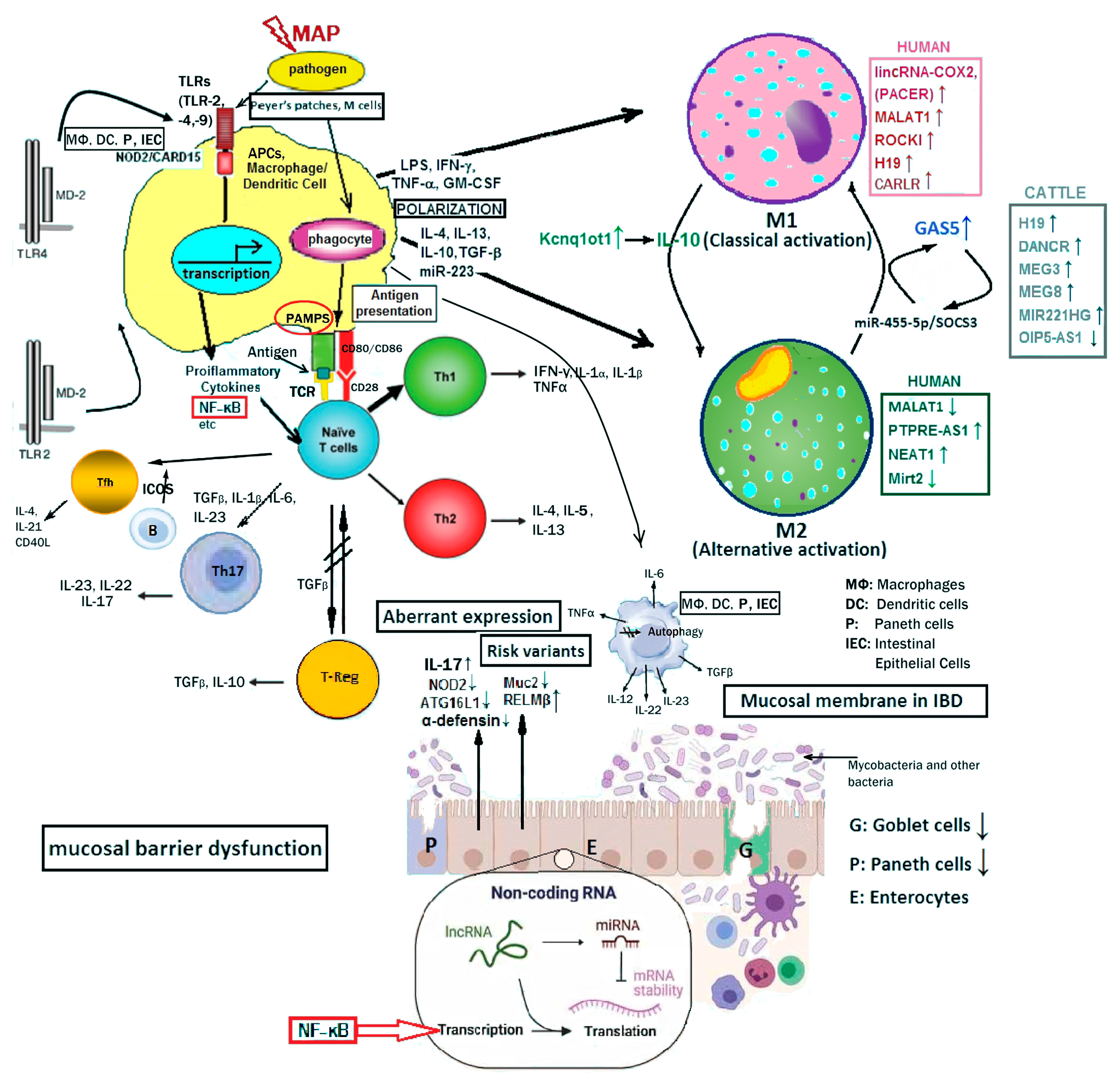
7.2. Signaling Event Pathways and lncRNA Intervention

The molecular basis of antigen uptake by M cells has been progressively identified in the last decade in humans, although unresolved points are being investigated in ongoing research. The follicle-associated epithelium (FAE) covering mucosa-associated lymphoid tissue is important and is distinct from the villous epithelium in terms of its cellular composition and functions [91]. The excretion of interleukin-22 binding protein (IL-22BP) by dendritic cells at the subepithelial region results in the inhibition of the IL-22-mediated secretion of antimicrobial peptides by the FAE, while notch signaling from stromal cells underneath the FAE reduces goblet cell differentiation [91]. The latter events diminish the mucosal barrier functions, allowing luminal microorganisms to promptly gain access to the luminal surface of the FAE.
An important aspect of inflammatory response production is the participating receptors that internalize the pathogen. However, the intestinal microbiota is composed of a considerable population of microorganisms, which is maintained under balanced dynamic host–microbiota interactions, beyond which a dynamic equilibrium imbalance may occur, leading to dysbiosis of the enteric microbiota, as has been demonstrated in IBD and CD, but is not evident in UC patients [89][92].

7.3. Coding Genes and LncRNAs Regulate the Pathological Phenotype Variables

The genetic and epigenetic variables influenced by environmental factors are the phenotype variables. Their regulated values are coordinated by the following factors: (a) genetic variations and polymorphisms, e.g., the genetic variability includes the associated susceptibility genes, such as NOD2, SLC11A1, and ATG16L1 [93] (Gao et al., 2022), including the important autophagy-gene-associated functions, thus rendering the host with an increased susceptibility to infection and thereby a propensity to manifest the disease. IBD patients with the ATG16L1 and NOD2 mutations, as well as studies on murine models have demonstrated the disturbed secretory apparatus of P cells, resulting in defects of antibacterial autophagy [94]; (b) non-coding RNA regulators and their polymorphisms in combination with epigenetic factors alter gene regulation; and (c) specific ncRNA actions, such as the miR-31 binding potential on cytokine receptors, are crucial to inflammation control, as found in DSS-induced colitis [95].
In more detail, NOD2 and the autophagy gene ATG16L1 are expressed by an important type of cell, the Paneth (P) cells, in the intestinal mucosa upon bacterial challenge and inflammation [96]. P cell granule secretion into the lumen of crypts is governed by cholinergic and bacterial factors, such as the gene associated with IBD, NOD2 [97], including the degranulation of P cells via the toll-like receptor 9 gene (TLR9). CD is characterized by a defective intestinal barrier towards intestinal microbes, while the cellular and molecular basis of this defect is likely to include P cells as major participants. P cells predominantly reside in the small intestine; however, P cells may also be induced by inflammation as metaplastic cells, owing to various stressors (e.g., cigarette smoking, stomach acid, excessive hormones, etc.) in other parts of the intestine, such as the colon.
Important risk variants in murine models as well as IBD patients impair P cell function, leading to colitis; notably, the autophagy gene ATG16L1 and NOD2 mutations, with disturbed secretory P cells, result in antibacterial autophagy defects. In IBD, there is an abnormal goblet cell function, including the MUC2 and RELMβ proteins, while mucosal barrier dysfunction is represented by goblet (G) and P cell (P) functional disruption inside the intact intestinal epithelium.
LncRNA regulators and genetic variations in combination with epigenetic factors play significant role in the gene regulation of proinflammatory genes. One of the roles of lncRNAs during infection is to regulate the polarization of M1 and M2 macrophages. 

As shown in Figure 2, LncRNAs in humans, such as PACER, MALAT1, HOTAIR and CARLR, directly or indirectly, interact with NF-κB pathway components to regulate target gene transcription. Likewise bovine lncRNAs such as, H19, DANCR, MEG3, MEG8, MIR221HG and OIP5-AS1 are differentially expressed upon MAP challenge to regulate biological functions and disease state.

8. The Prospect of LncRNAs in Contemporary Therapies of GI

IBD-related pathogenesis is a challenging subject for medicine to deal with in terms of the initial trigger of the disease, the diagnosis, the involved mechanism(s), and potentially, an effective therapy. Likewise, concerning JD, there is an ongoing and severe problem with animals at their productive age, whereby almost all animals develop critical pathological signs including weight loss, diarrhea, and secondary infections [98]. Reports are incessant concerning MAP infection in a wide range of hosts, including subjects not only from cattle but also from other important ruminants such as sheep, goat, deer, pigeons, etc., which are significant dairy product resources, or even non-ruminant species [98], which can easily transmit the MAP-resistant pathogen to other animals and the human population.
As mentioned earlier, another important parameter is the environmental basis of the disease, including pathogenic infection as one external trigger that precipitates intestinal inflammation [99][100], while recurrent pathogen infections in mice can develop as a consequential IBD-like syndrome [100]. Therefore, the first preventive strategy of the immune system is to minimize the risks of both inflammatory damage to the tissue and the development of chronic inflammatory diseases (e.g., CD), and the second is that pathogens need to be managed in ways that maintain the benefits of symbiosis in the intestinal flora in order to sustain the “harmonious” balance in the intestine [99].
There are a few examples presented below that support the rationale behind the idea of using lncRNA applications, particularly for disease diagnosis and therapy. Ge et al., illustrated that the level of lncRNA ANRIL (CDKN2B-AS1) can be used to distinguish patients with CD from healthy controls [101] and that ANRIL can serve as a biomarker under multiple conditions. In fact, changes in ANRIL expression are associated with the infliximab treatment response in patients with CD, as ANRIL expression in responders of infliximab treatment was increased, whereas that from non-responders remained unchanged.
It is well-known that corticosteroids are commonly prescribed drugs for IBD, while glucocorticoids (GCs), in particular, show anti-inflammatory and immunosuppressive effects which are used to induce remission in UC patients, and also, they are of benefit in CD [102].

9. Conclusions

Recent advances in information technologies, in association with genetic and epigenetic discoveries, have revealed unknown folds in the regulatory networks of molecular pathogenesis for the intestine that promise to harness diseases of the gut and control therapeutic outcomes. A significant profile of molecules in the array of molecular networks, including the non-coding RNAs of long transcripts (>200 ntds), has recently been implicated in various diseases through significant regulatory roles.
In recent years, a limited number of proteins or peptides encoded by ncRNAs have been demonstrated to exhibit significant biological and pathological functions associated with the triggering and progression of intestinal barrier dysfunction and the importance of the intestinal microbiota. Numerous studies have shown a link between intestinal dysbiosis and IBD. Although few studies have focused on ncRNAs in the modulation of dynamic host–microbiota interactions, the molecular regulators of ncRNAs in the intestinal microbiota are still not fully understood; likewise, the roles of intestinal microorganisms in initiating and facilitating the IBD-related pathologies are not known. Notably, many of the lncRNAs involved in JD and mycobacterioses are also major contributors to dysfunctional gene expression in humans and are mostly related to various types of cancer, including CRCs.

References

  1. Cullen, C.M.; Aneja, K.K.; Beyhan, S.; Cho, C.E.; Woloszynek, S.; Convertino, M.; McCoy, S.J.; Zhang, Y.; Anderson, M.Z.; Alvarez-Ponce, D.; et al. Emerging Priorities for Microbiome Research. Front. Microbiol. 2020, 11, 136.
  2. Tribolet, L.; Kerr, E.; Cowled, C.; Bean, A.G.D.; Stewart, C.R.; Dearnley, M.; Farr, R.J. MicroRNA Biomarkers for Infectious Diseases: From Basic Research to Biosensing. Front. Microbiol. 2020, 11, 1197.
  3. Huang, Z.; Liu, J.; Li, L.; Guo, Y.; Luo, Q.; Li, J. Long non-coding RNA expression profiling of macrophage line RAW264.7 infected by Mycobacterium tuberculosis. Biotech. Histochem. 2020, 95, 403–410.
  4. Wei, L.; Liu, K.; Jia, Q.; Zhang, H.; Bie, Q.; Zhang, B. The Roles of Host Noncoding RNAs in Mycobacterium tuberculosis Infection. Front. Immunol. 2021, 12, 664787.
  5. Yang, X.; Yang, J.; Wang, J.; Wen, Q.; Wang, H.; He, J.; Hu, S.; He, W.; Du, X.; Liu, S.; et al. Microarray analysis of long noncoding RNA and mRNA expression profiles in human macrophages infected with Mycobacterium tuberculosis. Sci. Rep. 2016, 6, 38963.
  6. Pekarek, L.; Torres-Carranza, D.; Fraile-Martinez, O.; Garcia-Montero, C.; Pekarek, T.; Saez, M.A.; Rueda-Correa, F.; Pimentel-Martinez, C.; Guijarro, L.G.; Diaz-Pedrero, R.; et al. An Overview of the Role of MicroRNAs on Carcinogenesis: A Focus on Cell Cycle, Angiogenesis and Metastasis. Int. J. Mol. Sci. 2023, 24, 7268.
  7. Chen, Y.; Li, Z.; Chen, X.; Zhang, S. Long non-coding RNAs: From disease code to drug role. Acta Pharm. Sin. B 2021, 11, 340–354.
  8. Zhang, Z.; Shi, S.; Li, J.; Costa, M. Long Non-Coding RNA MEG3 in Metal Carcinogenesis. Toxics 2023, 11, 157.
  9. Wang, B.; Wang, M.; Jia, S.; Li, T.; Yang, M.; Ge, F. Systematic Survey of the Regulatory Networks of the Long Noncoding RNA BANCR in Cervical Cancer Cells. J. Proteome Res. 2022, 21, 1137–1152.
  10. Song, X.; Gao, F.; Li, H.; Qin, W.; Chai, C.; Shi, G.; Yang, H. Long noncoding RNA THRIL promotes foam cell formation and inflammation in macrophages. Cell Biol. Int. 2023, 47, 156–166.
  11. Bates, A.; O’Brien, R.; Liggett, S.; Griffin, F. The effect of sub-clinical infection with Mycobacterium avium subsp. paratuberculosis on milk production in a New Zealand dairy herd. BMC Vet. Res. 2018, 14, 93.
  12. Zarei-Kordshouli, F.; Geramizadeh, B.; Khodakaram-Tafti, A. Prevalence of Mycobacterium avium subspecies paratuberculosis IS 900 DNA in biopsy tissues from patients with Crohn’s disease: Histopathological and molecular comparison with Johne’s disease in Fars province of Iran. BMC Infect. Dis. 2019, 19, 23.
  13. King, H.C.; Khera-Butler, T.; James, P.; Oakley, B.B.; Erenso, G.; Aseffa, A.; Knight, R.; Wellington, E.M.; Courtenay, O. Environmental reservoirs of pathogenic mycobacteria across the Ethiopian biogeographical landscape. PLoS ONE 2017, 12, e0173811.
  14. Riojas, M.A.; McGough, K.J.; Rider-Riojas, C.J.; Rastogi, N.; Hazbon, M.H. Phylogenomic analysis of the species of the Mycobacterium tuberculosis complex demonstrates that Mycobacterium africanum, Mycobacterium bovis, Mycobacterium caprae, Mycobacterium microti and Mycobacterium pinnipedii are later heterotypic synonyms of Mycobacterium tuberculosis. Int. J. Syst. Evol. Microbiol. 2018, 68, 324–332.
  15. Rossini, N.O.; Dias, M.V.B. Mutations and insights into the molecular mechanisms of resistance of Mycobacterium tuberculosis to first-line. Genet. Mol. Biol. 2023, 46, e20220261.
  16. Mataragka, A.; Leousi, E.; Liandris, E.; Ntafis, V.; Leontides, L.; Aggelidou, E.; Bossis, I.; Triantaphyllopoulos, K.A.; Theodoropoulou, I.; Ikonomopoulos, J. Faecal shedding of Mycobacterium avium subspecies paratuberculosis reduces before parturition in sheep? Small Rumin. Res. 2017, 147, 32–36.
  17. Mataragka, A.; Sotirakoglou, K.; Gazouli, M.; Triantaphyllopoulos, K.A.; Ikonomopoulos, J. Parturition affects test-positivity in sheep with subclinical paratuberculosis; investigation following a preliminary analysis. J. King Saud Univ. Sci. 2019, 31, 1399–1403.
  18. Garcia, A.B.; Shalloo, L. Invited review: The economic impact and control of paratuberculosis in cattle. J. Dairy Sci. 2015, 98, 5019–5039.
  19. Thoen, C.; Jarnagin, J.; Richards, W. Isolation and identification of mycobacteria from porcine tissues: A three-year summary. Am. J. Vet. Res. 1975, 36, 1383–1386.
  20. Greig, A.; Stevenson, K.; Perez, V.; Pirie, A.; Grant, J.; Sharp, J. Paratuberculosis in wild rabbits (Oryctolagus cuniculus). Vet. Rec. 1997, 140, 141–143.
  21. Beard, P.; Daniels, M.; Henderson, D.; Pirie, A.; Rudge, K.; Buxton, D.; Rhind, S.; Greig, A.; Hutchings, M.; McKendrick, I.; et al. Paratuberculosis infection of nonruminant wildlife in Scotland. J. Clin. Microbiol. 2001, 39, 1517–1521.
  22. Fechner, K.; Matz-Rensing, K.; Lampe, K.; Kaup, F.J.; Czerny, C.P.; Schafer, J. Detection of Mycobacterium avium subsp. paratuberculosis in non-human primates. J. Med. Primatol. 2017, 46, 211–217.
  23. Byrne, A.; Ollier, S.; Tahlan, K.; Biet, F.; Bissonnette, N. Genomic epidemiology of Mycobacterium avium subsp. paratuberculosis isolates from Canadian dairy herds provides evidence for multiple infection events. Front. Genet. 2023, 14, 1043598.
  24. Davis, W.C. On deaf ears, Mycobacterium avium paratuberculosis in pathogenesis Crohn’s and other diseases. World J. Gastroenterol. 2015, 21, 13411–13417.
  25. McNees, A.L.; Markesich, D.; Zayyani, N.R.; Graham, D.Y. Mycobacterium paratuberculosis as a cause of Crohn’s disease. Expert Rev. Gastroenterol. Hepatol. 2015, 9, 1523–1534.
  26. Sechi, L.A.; Dow, C.T. Mycobacterium avium ss. paratuberculosis Zoonosis—The Hundred Year War—Beyond Crohn’s Disease. Front. Immunol. 2015, 6, 96.
  27. Elmagzoub, W.A.; Idris, S.M.; Isameldin, M.; Arabi, N.; Abdo, A.; Ibrahim, M.; Khan, M.A.A.; Tanneberger, F.; Bakhiet, S.M.; Okuni, J.B.; et al. Mycobacterium avium subsp. paratuberculosis and microbiome profile of patients in a referral gastrointestinal diseases centre in the Sudan. PLoS ONE 2022, 17, e0266533.
  28. Dow, C.T. Hermon-Taylor: M. paratuberculosis and Crohn’s Disease-The Book of Revelation According to John. Pathogens 2021, 10, 1469.
  29. Agrawal, G.; Clancy, A.; Huynh, R.; Borody, T. Profound remission in Crohn’s disease requiring no further treatment for 3-23 years: A case series. Gut Pathog. 2020, 12, 16.
  30. Agrawal, G.; Hamblin, H.; Clancy, A.; Borody, T. Anti-Mycobacterial Antibiotic Therapy Induces Remission in Active Paediatric Crohn’s Disease. Microorganisms 2020, 8, 1112.
  31. Ekundayo, T.C.; Okoh, A.I. Systematic Assessment of Mycobacterium avium Subspecies Paratuberculosis Infections from 1911-2019: A Growth Analysis of Association with Human Autoimmune Diseases. Microorganisms 2020, 8, 1212.
  32. Gupta, S.; Chaubey, K.K.; Agarwal, P.; Kuenstner, J.T.; Parashar, D.; Singh, S.V. Therapeutic management of Mycobacterium avium subspecies paratuberculosis infection with complete resolution of symptoms and disease in a patient with advanced inflammatory bowel syndrome. Mol. Biol. Rep. 2021, 48, 7013–7020.
  33. Savarino, E.; Bertani, L.; Ceccarelli, L.; Bodini, G.; Zingone, F.; Buda, A.; Facchin, S.; Lorenzon, G.; Marchi, S.; Marabotto, E.; et al. Antimicrobial treatment with the fixed-dose antibiotic combination RHB-104 for Mycobacterium avium subspecies paratuberculosis in Crohn’s disease: Pharmacological and clinical implications. Expert Opin. Biol. Ther. 2019, 19, 79–88.
  34. Eslami, M.; Shafiei, M.; Ghasemian, A.; Valizadeh, S.; Al-Marzoqi, A.H.; Shokouhi Mostafavi, S.K.; Nojoomi, F.; Mirforughi, S.A. Mycobacterium avium paratuberculosis and Mycobacterium avium complex and related subspecies as causative agents of zoonotic and occupational diseases. J. Cell. Physiol. 2019, 234, 12415–12421.
  35. Timms, V.J.; Daskalopoulos, G.; Mitchell, H.M.; Neilan, B.A. The Association of Mycobacterium avium subsp. paratuberculosis with Inflammatory Bowel Disease. PLoS ONE 2016, 11, e0148731.
  36. Pierce, E.S. Could Mycobacterium avium subspecies paratuberculosis cause Crohn’s disease, ulcerative colitis… and colorectal cancer? Infect. Agents Cancer 2018, 13, 1.
  37. Okuni, J.B.; Hansen, S.; Eltom, K.H.; Eltayeb, E.; Amanzada, A.; Omega, J.A.; Czerny, C.P.; Abd El Wahed, A.; Ojok, L. Paratuberculosis: A Potential Zoonosis and a Neglected Disease in Africa. Microorganisms 2020, 8, 1007.
  38. Zhang, H.; Xue, C.; Wang, Y.; Shi, J.; Zhang, X.; Li, W.; Nunez, S.; Foulkes, A.S.; Lin, J.; Hinkle, C.C.; et al. Deep RNA Sequencing Uncovers a Repertoire of Human Macrophage Long Intergenic Noncoding RNAs Modulated by Macrophage Activation and Associated with Cardiometabolic Diseases. J. Am. Heart Assoc. 2017, 6, e007431.
  39. Palazzo, A.F.; Koonin, E.V. Functional Long Non-coding RNAs Evolve from Junk Transcripts. Cell 2020, 183, 1151–1161.
  40. Villa, T.; Porrua, O. Pervasive transcription: A controlled risk. FEBS J. 2022, 290, 3723–3736.
  41. Panda, A.C. Circular RNAs Act as miRNA Sponges. Adv. Exp. Med. Biol. 2018, 1087, 67–79.
  42. Li, J.; Xu, Q.; Huang, Z.J.; Mao, N.; Lin, Z.T.; Cheng, L.; Sun, B.; Wang, G. CircRNAs: A new target for the diagnosis and treatment of digestive system neoplasms. Cell Death Dis. 2021, 12, 205.
  43. Wang, F.; Nazarali, A.J.; Ji, S. Circular RNAs as potential biomarkers for cancer diagnosis and therapy. Am. J. Cancer Res. 2016, 6, 1167–1176.
  44. Zhou, Z.; Wang, X.; Hu, Q.; Yang, Z. CircZfp609 contributes to cerebral infarction via sponging miR-145a-5p to regulate BACH1. Metab. Brain Dis. 2023, 38, 1971–1981.
  45. Das, K.; Garnica, O.; Dhandayuthapani, S. Modulation of Host miRNAs by Intracellular Bacterial Pathogens. Front. Cell. Infect. Microbiol. 2016, 6, 79.
  46. Aguilar, C.; Mano, M.; Eulalio, A. Multifaceted Roles of microRNAs in Host-Bacterial Pathogen Interaction. Microbiol. Spectr. 2019, 7, 10–1128.
  47. Kim, J.K.; Kim, T.S.; Basu, J.; Jo, E.K. MicroRNA in innate immunity and autophagy during mycobacterial infection. Cell. Microbiol. 2017, 19, e12687.
  48. Statello, L.; Guo, C.J.; Chen, L.L.; Huarte, M. Gene regulation by long non-coding RNAs and its biological functions. Nat. Rev. Mol. Cell Biol. 2021, 22, 96–118.
  49. Chedin, F. Nascent Connections: R-Loops and Chromatin Patterning. Trends Genet. TIG 2016, 32, 828–838.
  50. Lan, Y.; Xiao, X.; He, Z.; Luo, Y.; Wu, C.; Li, L.; Song, X. Long noncoding RNA OCC-1 suppresses cell growth through destabilizing HuR protein in colorectal cancer. Nucleic Acids Res. 2018, 46, 5809–5821.
  51. Qin, W.; Li, X.; Xie, L.; Li, S.; Liu, J.; Jia, L.; Dong, X.; Ren, X.; Xiao, J.; Yang, C.; et al. A long non-coding RNA, APOA4-AS, regulates APOA4 expression depending on HuR in mice. Nucleic Acids Res. 2016, 44, 6423–6433.
  52. Sridhar, B.; Rivas-Astroza, M.; Nguyen, T.C.; Chen, W.; Yan, Z.; Cao, X.; Hebert, L.; Zhong, S. Systematic Mapping of RNA-Chromatin Interactions In Vivo. Curr. Biol. 2017, 27, 610–612.
  53. Asim, M.N.; Ibrahim, M.A.; Imran Malik, M.; Dengel, A.; Ahmed, S. Advances in Computational Methodologies for Classification and Sub-Cellular Locality Prediction of Non-Coding RNAs. Int. J. Mol. Sci. 2021, 22, 8719.
  54. Laham-Karam, N.; Laitinen, P.; Turunen, T.A.; Yla-Herttuala, S. Activating the Chromatin by Noncoding RNAs. Antioxid. Redox Signal. 2018, 29, 813–831.
  55. Yang, F.; Yang, Y.; Chen, L.; Zhang, Z.; Liu, L.; Zhang, C.; Mai, Q.; Chen, Y.; Chen, Z.; Lin, T.; et al. The gut microbiota mediates protective immunity against tuberculosis via modulation of lncRNA. Gut Microbes 2022, 14, 2029997.
  56. Weikard, R.; Demasius, W.; Kuehn, C. Mining long noncoding RNA in livestock. Anim. Genet. 2017, 48, 3–18.
  57. Koufariotis, L.T.; Chen, Y.P.; Chamberlain, A.; Vander Jagt, C.; Hayes, B.J. A catalogue of novel bovine long noncoding RNA across 18 tissues. PLoS ONE 2015, 10, e0141225.
  58. Zhou, Z.Y.; Li, A.M.; Adeola, A.C.; Liu, Y.H.; Irwin, D.M.; Xie, H.B.; Zhang, Y.P. Genome-wide identification of long intergenic noncoding RNA genes and their potential association with domestication in pigs. Genome Biol. Evol. 2014, 6, 1387–1392.
  59. Gruber, A.J.; Zavolan, M. Alternative cleavage and polyadenylation in health and disease. Nat. Rev. Genet. 2019, 20, 599–614.
  60. Kern, C.; Wang, Y.; Chitwood, J.; Korf, I.; Delany, M.; Cheng, H.; Medrano, J.F.; Van Eenennaam, A.L.; Ernst, C.; Ross, P.; et al. Genome-wide identification of tissue-specific long non-coding RNA in three farm animal species. BMC Genom. 2018, 19, 684.
  61. Kern, C.; Wang, Y.; Xu, X.; Pan, Z.; Halstead, M.; Chanthavixay, G.; Saelao, P.; Waters, S.; Xiang, R.; Chamberlain, A.; et al. Functional annotations of three domestic animal genomes provide vital resources for comparative and agricultural research. Nat. Commun. 2021, 12, 1821.
  62. Mele, M.; Mattioli, K.; Mallard, W.; Shechner, D.M.; Gerhardinger, C.; Rinn, J.L. Chromatin environment, transcriptional regulation, and splicing distinguish lincRNAs and mRNAs. Genome Res. 2017, 27, 27–37.
  63. Washietl, S.; Kellis, M.; Garber, M. Evolutionary dynamics and tissue specificity of human long noncoding RNAs in six mammals. Genome Res. 2014, 24, 616–628.
  64. Ma, S.; Ming, Z.; Gong, A.Y.; Wang, Y.; Chen, X.; Hu, G.; Zhou, R.; Shibata, A.; Swanson, P.C.; Chen, X.M. A long noncoding RNA, lincRNA-Tnfaip3, acts as a coregulator of NF-kappaB to modulate inflammatory gene transcription in mouse macrophages. FASEB J. 2017, 31, 1215–1225.
  65. Zhao, K.; Tan, J.Y.; Mao, Q.D.; Ren, K.Y.; He, B.G.; Zhang, C.P.; Wei, L.Z. Overexpression of long non-coding RNA TUG1 alleviates TNF-alpha-induced inflammatory injury in interstitial cells of Cajal. Eur. Rev. Med. Pharmacol. Sci. 2019, 23, 312–320.
  66. Yu, X.; Zheng, H.; Chan, M.T.; Wu, W.K. HULC: An oncogenic long non-coding RNA in human cancer. J. Cell. Mol. Med. 2017, 21, 410–417.
  67. Gupta, P.; Peter, S.; Jung, M.; Lewin, A.; Hemmrich-Stanisak, G.; Franke, A.; von Kleist, M.; Schutte, C.; Einspanier, R.; Sharbati, S.; et al. Analysis of long non-coding RNA and mRNA expression in bovine macrophages brings up novel aspects of Mycobacterium avium subspecies paratuberculosis infections. Sci. Rep. 2019, 9, 1571.
  68. Marete, A.; Ariel, O.; Ibeagha-Awemu, E.; Bissonnette, N. Identification of Long Non-coding RNA Isolated From Naturally Infected Macrophages and Associated with Bovine Johne’s Disease in Canadian Holstein Using a Combination of Neural Networks and Logistic Regression. Front. Vet. Sci. 2021, 8, 639053.
  69. Talyan, S.; Andrade-Navarro, M.A.; Muro, E.M. Identification of transcribed protein coding sequence remnants within lincRNAs. Nucleic Acids Res. 2018, 46, 8720–8729.
  70. Bussieres-Marmen, S.; Vinette, V.; Gungabeesoon, J.; Aubry, I.; Perez-Quintero, L.A.; Tremblay, M.L. Loss of T-cell protein tyrosine phosphatase in the intestinal epithelium promotes local inflammation by increasing colonic stem cell proliferation. Cell. Mol. Immunol. 2018, 15, 367–376.
  71. Lin, L.; Zhou, G.; Chen, P.; Wang, Y.; Han, J.; Chen, M.; He, Y.; Zhang, S. Which long noncoding RNAs and circular RNAs contribute to inflammatory bowel disease? Cell Death Dis. 2020, 11, 456.
  72. Houtman, M.; Shchetynsky, K.; Chemin, K.; Hensvold, A.H.; Ramskold, D.; Tandre, K.; Eloranta, M.L.; Ronnblom, L.; Uebe, S.; Catrina, A.I.; et al. T cells are influenced by a long non-coding RNA in the autoimmune associated PTPN2 locus. J. Autoimmun. 2018, 90, 28–38.
  73. Zhang, Q.; Chao, T.C.; Patil, V.S.; Qin, Y.; Tiwari, S.K.; Chiou, J.; Dobin, A.; Tsai, C.M.; Li, Z.; Dang, J.; et al. The long noncoding RNA ROCKI regulates inflammatory gene expression. EMBO J. 2019, 38, e100041.
  74. Xu, J.; Xu, H.M.; Yang, M.F.; Liang, Y.J.; Peng, Q.Z.; Zhang, Y.; Tian, C.M.; Wang, L.S.; Yao, J.; Nie, Y.Q.; et al. New Insights Into the Epigenetic Regulation of Inflammatory Bowel Disease. Front. Pharmacol. 2022, 13, 813659.
  75. Yarani, R.; Mirza, A.H.; Kaur, S.; Pociot, F. The emerging role of lncRNAs in inflammatory bowel disease. Exp. Mol. Med. 2018, 50, 1–14.
  76. Ritchie, M.E.; Phipson, B.; Wu, D.; Hu, Y.; Law, C.W.; Shi, W.; Smyth, G.K. limma powers differential expression analyses for RNA-sequencing and microarray studies. Nucleic Acids Res. 2015, 43, e47.
  77. Qiao, C.; Yang, L.; Wan, J.; Liu, X.; Pang, C.; You, W.; Zhao, G. Long noncoding RNA ANRIL contributes to the development of ulcerative colitis by miR-323b-5p/TLR4/MyD88/NF-kappaB pathway. Biochem. Biophys. Res. Commun. 2019, 508, 217–224.
  78. Wang, S.; Hou, Y.; Chen, W.; Wang, J.; Xie, W.; Zhang, X.; Zeng, L. KIF9-AS1, LINC01272 and DIO3OS lncRNAs as novel biomarkers for inflammatory bowel disease. Mol. Med. Rep. 2018, 17, 2195–2202.
  79. Gomes, C.P.; Nobrega-Pereira, S.; Domingues-Silva, B.; Rebelo, K.; Alves-Vale, C.; Marinho, S.P.; Carvalho, T.; Dias, S.; Bernardes de Jesus, B. An antisense transcript mediates MALAT1 response in human breast cancer. BMC Cancer 2019, 19, 771.
  80. Li, Y.; Zhu, L.; Chen, P.; Wang, Y.; Yang, G.; Zhou, G.; Li, L.; Feng, R.; Qiu, Y.; Han, J.; et al. MALAT1 Maintains the Intestinal Mucosal Homeostasis in Crohn’s Disease via the miR-146b-5p-CLDN11/NUMB Pathway. J. Crohn’s Colitis 2021, 15, 1542–1557.
  81. Bure, I.V.; Nemtsova, M.V.; Kuznetsova, E.B. Histone Modifications and Non-Coding RNAs: Mutual Epigenetic Regulation and Role in Pathogenesis. Int. J. Mol. Sci. 2022, 23, 5801.
  82. Triantaphyllopoulos, K.A.; Ikonomopoulos, I.; Bannister, A.J. Epigenetics and inheritance of phenotype variation in livestock. Epigenetics Chromatin 2016, 9, 31.
  83. Wei, J.W.; Huang, K.; Yang, C.; Kang, C.S. Non-coding RNAs as regulators in epigenetics (Review). Oncol. Rep. 2017, 37, 3–9.
  84. Bujold, D.; Morais, D.A.L.; Gauthier, C.; Cote, C.; Caron, M.; Kwan, T.; Chen, K.C.; Laperle, J.; Markovits, A.N.; Pastinen, T.; et al. The International Human Epigenome Consortium Data Portal. Cell Syst. 2016, 3, 496–499 e492.
  85. Consortium, E.P.; Bernstein, B.E.; Birney, E.; Dunham, I.; Green, E.D.; Gunter, C.; Snyder, M. An integrated encyclopedia of DNA elements in the human genome. Nature 2012, 489, 57–74.
  86. Gonzalez-Serna, D.; Villanueva-Martin, G.; Acosta-Herrera, M.; Marquez, A.; Martin, J. Approaching Shared Pathophysiology in Immune-Mediated Diseases through Functional Genomics. Genes 2020, 11, 1482.
  87. Deng, W.; Zhang, Y.; Cai, J.; Zhang, J.; Liu, X.; Yin, J.; Bai, Z.; Yao, H.; Zhang, Z. LncRNA-ANRIL promotes gastric cancer progression by enhancing NF-kB signaling. Exp. Biol. Med. 2019, 244, 953–959.
  88. Rajeev, R.; Dwivedi, A.P.; Sinha, A.; Agarwaal, V.; Dev, R.R.; Kar, A.; Khosla, S. Epigenetic interaction of microbes with their mammalian hosts. J. Biosci. 2021, 46, 94.
  89. Ramos, G.P.; Papadakis, K.A. Mechanisms of Disease: Inflammatory Bowel Diseases. Mayo Clin. Proc. 2019, 94, 155–165.
  90. Schoultz, I.; Keita, A.V. Cellular and Molecular Therapeutic Targets in Inflammatory Bowel Disease-Focusing on Intestinal Barrier Function. Cells 2019, 8, 193.
  91. Nakamura, Y.; Kimura, S.; Hase, K. M cell-dependent antigen uptake on follicle-associated epithelium for mucosal immune surveillance. Inflamm. Regen. 2018, 38, 15.
  92. Nishino, K.; Nishida, A.; Inoue, R.; Kawada, Y.; Ohno, M.; Sakai, S.; Inatomi, O.; Bamba, S.; Sugimoto, M.; Kawahara, M.; et al. Analysis of endoscopic brush samples identified mucosa-associated dysbiosis in inflammatory bowel disease. J. Gastroenterol. 2018, 53, 95–106.
  93. Gao, P.; Liu, H.; Huang, H.; Sun, Y.; Jia, B.; Hou, B.; Zhou, X.; Strober, W.; Zhang, F. The Crohn Disease-associated ATG16L1(T300A) polymorphism regulates inflammatory responses by modulating TLR- and NLR-mediated signaling. Autophagy 2022, 18, 2561–2575.
  94. Larabi, A.; Barnich, N.; Nguyen, H.T.T. New insights into the interplay between autophagy, gut microbiota and inflammatory responses in IBD. Autophagy 2020, 16, 38–51.
  95. Qu, X.; Zheng, C.; Wang, B.; Wang, F.; Sun, X.; Gao, Y.; Xia, Q.; Kong, X. Comprehensive analysis of circular RNAs from steatotic livers after ischemia and reperfusion injury by next-generation RNA sequencing. FEBS Lett. 2021, 595, 99–109.
  96. Wehkamp, J.; Stange, E.F. An Update Review on the Paneth Cell as Key to Ileal Crohn’s Disease. Front. Immunol. 2020, 11, 646.
  97. Ntunzwenimana, J.C.; Boucher, G.; Paquette, J.; Gosselin, H.; Alikashani, A.; Morin, N.; Beauchamp, C.; Thauvette, L.; Rivard, M.E.; Dupuis, F.; et al. Functional screen of inflammatory bowel disease genes reveals key epithelial functions. Genome Med. 2021, 13, 181.
  98. Seva, J.; Sanes, J.M.; Mas, A.; Ramis, G.; Sanchez, J.; Parraga-Ros, E. Prevalence of Mycobacterium avium Subsp. paratuberculosis in Feral Pigeons (Columba livia) Associated with Difficulties Controlling Paratuberculosis in a Bovine Herd (Fighting Bull Breed). Animals 2022, 12, 3314.
  99. Johnson, A.M.F.; DePaolo, R.W. Infectious Scarring: Setting the Trigger for Intestinal Inflammation. Cell Host Microbe 2018, 23, 154–156.
  100. Yang, W.H.; Heithoff, D.M.; Aziz, P.V.; Sperandio, M.; Nizet, V.; Mahan, M.J.; Marth, J.D. Recurrent infection progressively disables host protection against intestinal inflammation. Science 2017, 358, eaao5610.
  101. Ge, Q.; Dong, Y.; Lin, G.; Cao, Y. Long Noncoding RNA Antisense Noncoding RNA in the INK4 Locus Correlates With Risk, Severity, Inflammation and Infliximab Efficacy in Crohn’s Disease. Am. J. Med. Sci. 2019, 357, 134–142.
  102. Aslam, N.; Lo, S.W.; Sikafi, R.; Barnes, T.; Segal, J.; Smith, P.J.; Limdi, J.K. A review of the therapeutic management of ulcerative colitis. Ther. Adv. Gastroenterol. 2022, 15, 17562848221138160.
More
Information
Contributor MDPI registered users' name will be linked to their SciProfiles pages. To register with us, please refer to https://encyclopedia.pub/register :
View Times: 239
Revisions: 2 times (View History)
Update Date: 11 Sep 2023
1000/1000
ScholarVision Creations