Submitted Successfully!
To reward your contribution, here is a gift for you: A free trial for our video production service.
Thank you for your contribution! You can also upload a video entry or images related to this topic.
Version Summary Created by Modification Content Size Created at Operation
1 -- 4452 2023-08-30 12:32:40 |
2 references update -1 word(s) 4451 2023-09-01 10:15:33 |

Video Upload Options

Do you have a full video?

Confirm

Are you sure to Delete?
Cite
If you have any further questions, please contact Encyclopedia Editorial Office.
Ouologuem, D.T. Plasmodium falciparum Development from Gametocyte to Oocyst. Encyclopedia. Available online: https://encyclopedia.pub/entry/48643 (accessed on 21 July 2024).
Ouologuem DT. Plasmodium falciparum Development from Gametocyte to Oocyst. Encyclopedia. Available at: https://encyclopedia.pub/entry/48643. Accessed July 21, 2024.
Ouologuem, Dinkorma T.. "Plasmodium falciparum Development from Gametocyte to Oocyst" Encyclopedia, https://encyclopedia.pub/entry/48643 (accessed July 21, 2024).
Ouologuem, D.T. (2023, August 30). Plasmodium falciparum Development from Gametocyte to Oocyst. In Encyclopedia. https://encyclopedia.pub/entry/48643
Ouologuem, Dinkorma T.. "Plasmodium falciparum Development from Gametocyte to Oocyst." Encyclopedia. Web. 30 August, 2023.
Plasmodium falciparum Development from Gametocyte to Oocyst
Edit

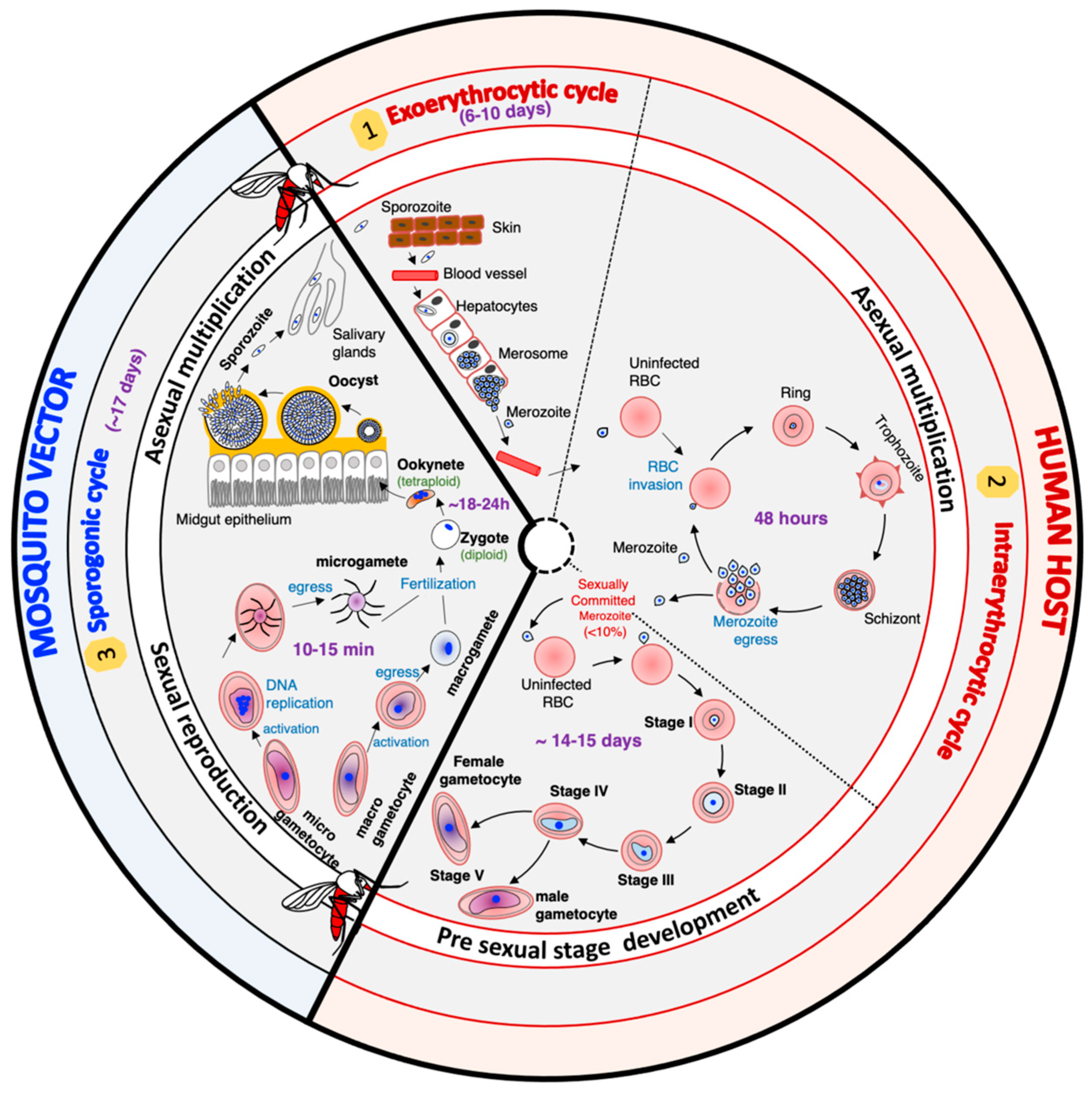
Malaria elimination never succeed without the implementation of transmission-blocking strategies. The transmission of Plasmodium spp. parasites from the human host to the mosquito vector depends on circulating gametocytes in the peripheral blood of the vertebrate host. Once ingested by the mosquito during blood meals, these sexual forms undergo a series of radical morphological and metabolic changes to survive and progress from the gut to the salivary glands, where they will be waiting to be injected into the vertebrate host. The design of effective transmission-blocking strategies requires a thorough understanding of all the mechanisms that drive the development of gametocytes, gametes, sexual reproduction, and subsequent differentiation within the mosquito. The drastic changes in Plasmodium falciparum shape and function throughout its life cycle rely on the tight regulation of stage-specific gene expression.

Plasmodium Gametocytes Gametes Differentiation

1. Introduction

Malaria is a mosquito-borne infection caused by an apicomplexan parasite of the genus Plasmodium and is transmitted through the bite of a mosquito vector of the genus Anopheles. In humans, malaria is caused by six Plasmodium species, including Plasmodium falciparum, Plasmodium vivax, Plasmodium malariae, Plasmodium ovale curtisi, Plasmodium ovale wallikeri, and the zoonotic parasite Plasmodium knowlesi. Of the human malaria parasites, P. falciparum causes the most significant morbidity and mortality. In 2021, clinical malaria affected 247,000 million individuals worldwide, leading to 619,000 deaths, with the highest burden observed in sub-Saharan Africa [1]. Over the last two decades, artemisinin-based combination therapies (ACTs), intermittent preventive treatments (IPTs), and vector control approaches have significantly reduced the burden of malaria in some areas [1]. However, despite tremendous efforts to control malaria, the emergence of resistant parasites to current antimalarials and the resistance of mosquitos to insecticides are significant obstacles to the global malaria eradication/elimination program [1].
With the renewed interest in malaria elimination and eradication, it has been recognized that intervention strategies should target all stages of the parasite life cycle with a particular interest in preventing transmission [2]. Plasmodium transmission from the human host to the mosquito vector and the parasite stages within the mosquito are promising targets for successfully interrupting the parasite life cycle and reducing the malaria burden in endemic areas [3].
The life cycle of Plasmodium falciparum is very complex. It includes different hosts, different cell types within the same host, asexual multiplication phases, and a sexual replication phase (Figure 1). In humans, the cycle begins with the inoculation of the sporozoite forms of the parasite by an infected mosquito during its blood meal. These sporozoites migrate through the bloodstream, reach the liver, and enter the hepatocytes, where they multiply asexually. After six to ten days, the rupture of infected hepatocytes releases thousands of merozoites, a new form of the parasite, into the bloodstream. The merozoites invade the red blood cells (RBCs) to initiate the intraerythrocytic cycle. After two days, new merozoites are released, and these offspring enter new RBCs to perpetuate the intraerythrocytic cycle responsible for the clinical manifestations of malaria in humans. A small percentage of merozoites differentiate into sexual forms called gametocytes, which mediate transmission. Gametocytes are morphologically (shape, size, and flexibility) [4][5][6] and functionally different from asexual parasites [6][7][8][9]. Mosquitos ingest male and female gametocytes during a blood meal, which results in a wave of differentiation processes and developmental stages within the mosquito [6][10][11][12]. In the mosquito’s gut lumen, gametocytes transform into male and female gametes. These gametes then fuse to produce a zygote that differentiates into a highly motile and invasive ookinete [6][11][13]. The ookinete invades the mosquito epithelial cell wall to form an oocyst, which in turn will ultimately produce thousands of sporozoites through a process known as sporogony [6][12][14]. The sporozoites will migrate and reside in the mosquito salivary gland [12], waiting to be transmitted to the vertebrate host during a blood meal and perpetuating the parasite’s life cycle.
Figure 1. The life cycle of Plasmodium falciparum parasites.

2. Gametocyte Development

The development of male and female gametocytes within the vertebrate host and the sexual recombination in the mosquito vector are critical steps in the Plasmodium falciparum life cycle. These biological stages ensure parasite virulence and transmission. During the erythrocytic cycle, 1–5% of asexual parasites switch from the asexual multiplication pathway to a sexual differentiation pathway. The differentiation of sexually committed parasites into mature gametocytes is divided into five developmental stages (stage I–V) over 14 days. The differentiation process is characterized by dramatic alterations in parasite size, shape, and flexibility. Only early stages (stage I) and mature stage V are detectable in the peripheral blood, as the other stages are sequestered in the bone marrow and possibly other tissues [15]. The differentiation process underlying asexual to stage V gametocyte formation is governed by specific expression mechanisms extensively described in other reviews [16][17][18]. The following paragraphs summarize gene expression regulation mechanisms involved in asexual to early gametocyte formation.
Several environmental and metabolic factors that trigger sexual commitment, including antimalarial drugs [19][20], lysophosphatidylcholine [21][22], host immune response [23][24], and parasite factors [25][26], have been reported. ApiAP2-G was the first transcription factor shown to govern the transcription of gametocyte-associated genes [27][28], and its regulation requires interaction with a second transcription factor, PfAP2-I [29]. The cell surface receptors involved in signal transduction and the signaling cascade leading to the activation of ApiAp2-G transcription factors still need to be characterized.
While the P. falciparum genome is predominantly acetylated during the asexual developmental cycle, ApiAP2-G appears to be repressed, and its locus was shown to be associated with histone heterochromatin marks, including H3K4ac, H3K9me1, H3K36me2, PfHP1, and the gametocyte essential factor PfAP2-G5 [30]. PfHP1-dependent gene silencing is antagonized by a sexual commitment activator protein called PfGDV1 (Plasmodium falciparum gametocyte development 1) [31][32]. PfGDV1 targets heterochromatin and triggers the eviction of PfHP1. Interestingly, PfGDV1 activation is, in turn, controlled by a multi-exon long non-coding gdv1 antisense RNA (Pfgdv1asRNA) that initiates downstream of the gdv1 locus [31].
The gametocyte essential factor AP2-G5 further prevents sexual commitment [32]. Indeed, PfAP2-G5 binds upstream of the Pfap2-g locus and other exogenic regions, hence suppressing ap2-g expression [32]. Although findings strongly suggest that ApiAP2 genes are under epigenetic control, histone post-translational modification linked to parasite conversion remains to be characterized. In addition, the signaling cascade events leading to the removal of the repressive state by PfGDV1 and the activation of PfApiAP2-G remain unknown.
Sexual conversion can occur by two possible routes following specific histone modification marks that promote changes in gene expression and sexual differentiation [29][33]. One route suggests that the decision to undergo gametocyte development occurs during the asexual cycle preceding the erythrocyte reinvasion event that leads to gametocyte formation. In this model, the decision to undergo gametocyte development is linked to PfAP2-G expression in the schizont of the preceding cycle [33]. The second route implies that the decision occurs within the same cycle, during the initial PfAP2-G expression in ring stages [33]. Recently, Li et al. identified an ApiAP2 transcription factor in P. yoelii, AP2-O3, implicated in a gender-specific transcription program [34]. AP2-O3, expressed predominantly in female gametocytes, represses the expression of male-specific genes. Genetic studies have shown that the depletion of either HP1 or the histone deacetylase enzyme 2 (Hda2) resulted in the activation of ap2-g transcription and many heterochromatic genes [34].
Gametocyte development from stage I to stage V is characterized by euchromatic post-translational modifications and an abundance of repressive methylation marks on histone 3. Ealy gametocyte stages (I to III) are characterized by the H3K9me3, H3K27me2, H3K27me3, H3K36me2, H3K37me1, H3R17me1, and H3R17me2 modifications [30][35]. The role of arginine methylation as a key feature for the epigenetic regulation of gametocyte development and maturation was suggested by Von Grüning et al. [30].
Three-dimensional structure analyses of the P. falciparum genome revealed that the localization and interaction of sexual differentiation genes in a repressive center are critical for regulating sexual conversion [36]. Further studies are needed to unravel the machinery regulating genome localization and the signaling pathway governing sexual commitment. The repertoire of environmental triggers for sexual conversion is far from being exhaustive. Characterizing proteins interacting with the histone-associated protein PfHP1 and PfGDV1 will undoubtedly unravel the molecular player involved in parasite conversion.
Despite their similitude with human enzymes, histone-modifying enzymes could be potential targets for developing antimalarial drugs with transmission-blocking activities. Stenzel et al. [37] designed, synthesized, and tested the biological activity of thirteen terephthalic-acid-based HDAC inhibitors. A subset of these compounds had moderate activity against P. falciparum gametocytes but showed sub-micromolar transmission-blocking activity against the rodent malaria parasite P. berghei. Hence, histone-modifying enzymes are potential targets to prevent malaria infection in the mosquito.

3. Gamete Development

Establishing a successful malaria infection in the Anopheles mosquito and the subsequent spread of competent parasites depend on the coordinated development of the Plasmodium parasite in the mosquito midgut. The differentiation process in the mosquito midgut, lasting almost 20 h, is defined by the rapid conversion of mature stage V female and male gametocytes into female macrogametes and male microgametes, respectively [6][10][11]. This step is followed by the sexual reproduction between macro- and microgametes and, finally, the conversion of newly formed zygotes into ookinetes [6][11]. The transition in the mosquito midgut constitutes a significant bottleneck in the Plasmodium life cycle as there is a 316-fold loss in parasite abundance from the gametocyte to ookinete stage and a 100-fold loss from the ookinete to oocyst stage [38]. Due to this considerable reduction in the parasite population, the plasmodial midgut stages are attractive targets for developing transmission-blocking interventions. Hence, malaria parasite developmental stages in the mosquito midgut have been extensively studied, and several key mechanisms and proteins orchestrating the differentiation process have been described, providing potential targets for vaccines and drug development [39].
Gamete formation is morphologically characterized by a rounding-up of the cell, followed by the parasitophorous vacuole membrane (PVM) rupture and parasite egress from the erythrocyte [10][40]. The differentiation of male gametocytes into male gametes involves three successive mitotic DNA replications that will produce eight motile microgametes through a process called exflagellation. Upon completion of DNA replication, the microgametes egress from the host cell [10]. In contrast, the differentiation of female gametocytes results in a rounding-up of the parasite with no DNA replication and the emergence of the forming gamete from the infected red blood cell [6].

3.1. Gene Expression Regulation Controlling Gametogenesis

Transcriptomic analysis of P. falciparum gametocytes and gametes revealed that small transcriptome changes characterize gametogenesis compared to other life-stage differentiation processes [41]. Most studies investigating gamete to sporozoite formation rely on Plasmodium berghei, causing rodent malaria. The rodent parasites are not of direct practical concern to humans. However, the easy access to Plasmodium life stages in the mosquito and the practicability for the experimental study of human malaria made them a good model for human malaria.
In Plasmodium berghei, the change in gene expression necessary for female gametogenesis is determined by the translation of a large number of mRNAs maintained in a repressive translational state [42][43]. Indeed, in female gametocytes, many transcripts are synthesized but translationally repressed until needed for macrogamete formation and zygote-to-ookinete transformation. Using the Plasmodium berghei model system, Guerreiro et al. revealed that approximately 50% of the transcriptome is maintained in this translationally repressed state by a messenger ribonucleoprotein (mRNP) composed of 16 major factors, including the RNA helicase DOZI (development of zygote inhibited) and the Sm-like factor CITH (homolog of worm CAR-I and fly Trailer Hitch) [44]. Using an in vitro translation assay, Tarique et al. characterized P. falciparum DOZI (PfDZ50) and demonstrated that the protein inhibits translation [45], suggesting a function similar to that reported for P. berghei female gametogenesis.
The expression of stage-specific genes that are necessary for microgamete formation is regulated by transient and reversible protein phosphorylation followed by the de novo synthesis of genes involved in DNA replication, axoneme assembly and motility, chromatin condensation, cytokinesis, and exflagellation [46][47]. Proteomic studies in P. berghei gametes revealed that proteins implicated in RNA translation, protein biosynthesis, glycolysis, environmental stress response, and tubulin-associated cytoskeleton dynamics are predominantly regulated during gamete formation [48]. Gene expression and protein synthesis regulation during both male and female gametogenesis depend highly on the signaling cascade orchestrated by stage-specific kinases and phosphatases [46][49][50].

3.2. Signaling Cascade Controlling Gametogenesis

Gametocyte activation is stimulated by environmental stimuli, including a temperature drop by approximately 5 °C, the presence of mosquito-derived xanthurenic acid (XA), and an increase in extracellular pH from 7.2 to about 8 [51][52][53]. Plasmodium receptors responsible for sensing temperature drops and binding to XA have not been identified yet. The exposure of P. falciparum gametocytes to XA has been shown to increase cyclic GMP (cGMP) levels in the gametocyte, suggesting parasite guanylyl cyclase (GC) activation [54]. In the rodent malaria parasites, the increase in cGMP eventually leads to an increase in the cytoplasmic calcium level necessary to trigger a calcium-dependent stage-specific gene expression and regulation pathway [55][56][57].
The Plasmodium falciparum genome encodes two guanylyl cyclase proteins (GCα and GCβ) that are only expressed in sexual-stage parasites. Functional studies in P. falciparum revealed that these proteins are active guanylyl cyclases and likely important in gametocyte activation [58]. Pharmacological and genetic studies investigating the signaling cascade of P. falciparum and P. berghei gamete formation have shown that GC activation leads to the formation of a secondary messenger cyclic GMP (cGMP), which in turn activates the protein kinase G (PKG), the unique effector identified in Plasmodium [54][59]. Taylor et al. revealed that the tight regulation of cGMP concentration is critical for P. falciparum gametocyte conversion and that premature high cGMP levels are deleterious for gamete formation [60]. Bennink et al. suggested that activation of PKG leads to the hydrolysis of phosphatidylinositol-(4,5)-bisphosphate (PIP2) by the phosphoinositide-specific phospholipase C (PI-PLC) and the production of two secondary messengers, diacylglycerol (DAG) and inositol triphosphate (IP3) [61][62]. Although IP3 induces the opening of calcium channels on the Plasmodium endoplasmic reticulum membrane, no ortholog of an IP3 receptor channel has yet been identified. A recent biochemical approach of an IP3 affinity chromatography column combined with bioinformatics has revealed a potential transporter associated with multidrug resistance in P. falciparum [63]. Additional work will elucidate the function of this novel protein.
The rapid release of calcium in mature gametocytes mediates a stage-specific calcium-dependent effector pathway in all gametogenesis steps, including gamete formation, gamete egress, microgamete mitotic maturation, and exflagellation [54][59][61]. In Plasmodium, the intracellular calcium level is sensed by calcium-dependent protein kinases (CDPKs) [64]. Macro- and microgamete-specific CDPKs that regulate the expression and activation of genes necessary for converting gametocytes into gametes have been identified [47][65][66].
In mature female gametocytes, the increase in cytoplasmic Ca2+ is sensed by CDPK1 [65]. CDPK1 has been identified as the key protein that regulates the translation of mRNAs in a temporal and stage-specific manner during macrogamete formation [65].
The differentiation of male gametocytes is regulated by a male-specific calcium-dependent protein kinase CDPK4, which initiates DNA replication, axoneme assembly, and cell motility [47][67]. In P. berghei, the lysis of the host cell membrane(s) surrounding the microgametocyte is mediated by CDPK1 [65]. In addition, CDPK1 translationally activates mRNA species in the developing zygote that remain repressed in macrogametes [65]. The cell-division cycle protein 20 (CDC20) appears to regulate male gametocyte mitotic division but not the one during schizogony [68]. Whether CDC20 works with CDPK1 and anaphase-promoting complex 3 (APC3) to modulate chromosome condensation and cytokinesis for microgamete formation is not yet determined [69]. In P. berghei, a histone chaperone protein termed FACT-L (facilitates chromatin transcription), which facilitates chromatin transcription, was shown to be involved in male gametocyte DNA replication and the production of fertile microgametes [70]. The formation of the flagella requires the formation of basal bodies and the assembly of axonemes.
Upon completion of DNA replication, axonemes become motile, facilitating the egress of the microgametes from the host cell. Genetic studies in P. berghei have revealed that axoneme assembly and motility require several proteins, including the stage-specific Actin II [71], the armadillo-repeat motif protein Pf16 [72], the spindle-assembly-related protein SAS6 [73], the SR protein kinase (SRPK) [74], and a gametocyte-specific mitogen-activated protein kinase 2 (MAP2) [75][76]. The Plasmodium falciparum kinome includes four NIMA-related kinases (PfNEK 1 to 4). While PfNEK-1 is expressed in asexual and sexual stages, the mRNA transcripts of PfNEK-2, PfNEk-3, and PfNEK-4 are exclusively expressed in gametocytes. NEK1 and NEK3 have been shown to activate the atypical MAP2 protein through phosphorylation [77][78]. PfNEK-2 and PfNEK-4 are required for meiosis completion in the ookinete [79][80]. A metallo-dependent protein phosphatase, PPM1, also plays an important role in P. berghei male gametocyte exflagellation [50].
Kinases and phosphatases involved in gamete formation can be considered promising targets for drug development. A high-throughput screening of P. falciparum-cGMP-dependent protein kinase identified a thiazole scaffold that kills erythrocytic and sexual-stage parasites [81]. Since the malarian PKG differs from the mammalian PKGs, this scaffold represents a good starting point for developing a new class of antimalarial drugs. Recently, Xitong et al. [82] assessed the activity of 25 phosphatase inhibitors against Plasmodium berghei sexual development and transmissibility to the mosquito. Two compounds from the panel effectively inhibited different development stages, from gametogenesis to ookinete maturation. These examples highlight that in silico modeling and screening combined with in vivo and ex vivo approaches using mouse models and human malaria could help identify parasite kinase and phosphatase inhibitors. The kinases regulating male gamete exflagellation (CDPK4 and atypical MAP-2) and DNA replication (Nek-2, Nek-4) should be of great interest in the screening.
Although rodent malaria parasites are sound model systems to understand P. falciprum gametogenesis, more studies relying on human malaria parasites are necessary to grasp the complexity of the early stages of malaria transmission. Those studies should consider including field studies to better understand the biology of transmission during natural infection.

4. Zygote to Ookinete Development

4.1. Gamete Fusion and Zygote Formation

The zygote results from the fusion between a fertile male microgamete and a female macrogamete. Fertilization of a macrogamete by a microgamete is mediated by stage- and sex-specific proteins synthesized in the respective gamete before the fusion event. The proteins involved in gamete fusion also mediate the prerequisite recognition and attachment steps. Genetic studies of P. falciparum and P. berghei genes have revealed that P48/45 and P230, two members of a protein family defined by a disulfide bonding pattern of six conserved cysteine residues, are essential for male gamete fertility and fusion with macrogametes [83][84][85]. In P. falciparum, P48/45 and P230 are localized on the gamete surface and have been shown to form a complex necessary for the fusion of the microgamete to the macrogamete. In vitro and in vivo studies in the mouse model have demonstrated the critical implication of P48/45 and P230 in fertilization. These findings led to their use as targets for transmission-blocking vaccines [39][86]. In macrogamete Pfs47, a paralog of P48/45 was identified [87]. Functional studies demonstrated the expression of Pfs47 exclusively on the surface of female gametes, but the protein did not appear crucial for fertilization [87].
The evolutionarily conserved class II gamete fusogen HAP2/GCS1 (hapless 2/Generative-cell-specific 1) has also been implicated in the fusion of the plasma membranes of two haploid gametes [88]. Gene disruption studies of p48/45 or hap2/gcs1 resulted in sterility due to the inability of the male gamete to either attach or fuse to fertile female gametes [85][88]. Studies investigating gamete fusion in Plasmodium revealed that a histone chaperone protein named FACT (facilitates chromatin transcription) plays an essential role in fertilization. However, the mechanism of action of this nuclear protein is still unclear [70].
The fusion of the two gametes requires the prior disassembly of a peculiar organelle located underneath the parasite plasma membrane and is named the inner membrane complex (IMC) [5]. The IMC will be reassembled later to ensure the pellicle’s integrity and parasite polarity [5][89]. The IMC is a membranous scaffold in the Alveolata, a group of diverse unicellular eukaryotes, including Plasmodium spp., Toxoplasma gondii, the ciliates, and dinoflagellates. This membranous patchwork is anchored to the subpellicular microtubule network. It also interacts with various parasite-specific proteins to coordinate parasite morphological changes, the segmentation of daughter cells during asexual replication, and parasite motility. Interestingly, the localization and interaction of many IMC proteins are regulated by post-translational S-palmitoylation mediated by the palmitoyl-acyl-transferase DHHC2 (the ortholog of PfDHHC1) [90]. Inhibitor studies revealed that DHHC2 palmitoylation is critical to zygote differentiation during the initial mosquito infection with P. berghei [91]. Hence, stage-specific palmitoylation enzymes could be novel targets for disrupting IMC assembly and zygote formation and differentiation into ookinetes.
The concept of transmission-blocking immunity is mostly antibody-mediated. Therefore, the development of transmission-blocking vaccines (TBVs) focuses on inducing potent antibodies sustained at adequate levels. Extensive efforts toward the clinical development of P. falciparum TBVs are undertaken, but the functional activities associated with most antibodies remain modest. Further study must be conducted to determine the players involved in gamete fusion and their association with each other to orchestrate gamete fusion. The disassembly of the IMC is a pre-requisite for gamete fusion, and targeting the enzymes modulating this biological process could be an approach to interfere with the establishment of the infection in the mosquito gut. Plasmodium falciparum is predicted to encode 12 putative palmitoyl acyl-transferases thought to ensure lipid-based palmitoylation of parasite proteins and act as a biological rheostat for protein–protein interactions and subcellular trafficking [92]. Inhibitors of palmitoylation enzymes could constitute a new class of antimalarial drugs targeting multiple parasite life stages and other apicomplexan parasites of medical importance [92][93][94].

4.2. Molecular and Genetic Mechanisms of Fertilization

Gene expression studies in P. berghei revealed that zygote development and differentiation into ookinetes require the transcriptional activation of several maternal silenced mRNAs in the zygote. A study investigating the parental contribution of transcripts implicated in zygote differentiation revealed that while inherited maternal mRNAs are activated to drive the early stage of zygote differentiation, the paternal alleles are initially silenced and then reactivated [95][96].
The derepression of a maternal transcript post fertilization will drive significant morphological changes defined by the formation of the IMC and the secretory organelles, as well as the elongation and apical polarization of the differentiating zygote [5][97][98]. The dynamic organization of the IMC and the parasite cytoskeleton is essentially coordinated post-translationally by reversible phosphorylation [65][74][99] and palmitoylation [91][100]. A systematic analysis of the Plasmodium kinome, combined with genetic studies in P. berghei, revealed that protein kinase 7 (PK7) and cyclin-G-associated protein (GAK) are essential in ookinete formation [74][99]. GAK is predicted to regulate clathrin-mediated vesicle trafficking and membrane fusion, suggesting its involvement in the formation of the secretory organelles or the assembly of the IMC [74]. PK7 mutants were blocked in ookinete formation, but the mechanism by which this kinase regulates ookinete development remains undetermined. Reverse genetics studies revealed that the Plasmodium phosphatase PPKL (protein phosphatase with kelch-like domains) is essential during ookinete differentiation and is involved in defining ookinete polarity, pellicle morphology and integrity, and ookinete motility [101]. Plasmodium falciparum DHHC1, a palmitoyl-S-acyl-transferase (PAT) containing a conserved DH(H/Y)C motif, was shown to be exclusively localized to the IMC [91][100]. DHHC1 is apicomplexan-specific and was implicated in ookinete formation and morphogenesis [91][100].
Subsequent regulation of stage-specific gene expression requires a de novo synthesis of ookinete-specific genes by the transcription factor ApiAp2-O [102]. ApiAp2-O appears to be associated with more than 500 genes involved in ookinete development, motility, midgut invasion, and parasite escape from mosquito immunity [103]. The ookinete-specific genes mediate nuclear fusion in the diploid zygote, DNA replication, and meiosis that will produce a motile tetraploid ookinete [104]. The tetraploid state was shown to persist throughout the ookinete stage until the formation of sporozoites in the oocyst [104][105]. Like in eukaryotic model systems, meiosis and cell cycle progression in the Plasmodium parasite are regulated by NIMA-related kinases (Neks). Gene disruption studies in P. berghei revealed that Nek-4 and Nek-2 are abundantly expressed during the gametocyte stages. Nek-4 and Nek-2 are essential for zygote differentiation into the ookinete stage but not for gamete formation and fertilization [79][80]. Given the success in developing drugs targeting human kinases, Plasmodium kinases are attractive targets for the next generation of antimalarials. Indeed, ongoing efforts attempt to characterize Plasmodium kinases while evaluating them as antimalarial drug targets [106][107].

4.3. Formation and Maturation of Ookinete

Ookinete maturation is completed 19 to 36 h post-gametocyte ingestion in the blood meal. The ookinete will quickly exit the midgut lumen via an intracellular or intercellular route [108]. Ookinete motility is regulated by kinases. The cGMP-dependent protein kinase (PKG) pathway activates the gliding motility apparatus on the IMC, and the calcium-dependent protein kinase 3 (CDPK3) mobilizes the intracellularly stored calcium necessary for signaling [109][110]. The traversal of the midgut wall is mediated by an impressive set of microneme proteins discharged by the mature ookinete [111]. Proteomic [111] and genetics studies identified key proteins for midgut traversal, including the circumsporozoite-TRAP-related protein CTRP [112], the membrane-attack ookinete protein (MAOP) [113], the secreted ookinete adhesive protein SOAP [114], the von-Willebrand-Factor-A-domain-related protein WARP [115], the cell-traversal protein for ookinetes and sporozoites CelTOS [116], and the chitinase 1 (CHT1) [117][118].
The ookinete stage is an attractive target for transmission-blocking strategies as the mosquito immune system naturally kills many ookinetes. Ookinete proteins involved in the attachment and invasion of the midgut epithelial cells are potential vaccine targets. Two ookinetes surface proteins, P25 and P28, that play a role in ookinete adhesion to the midgut and differentiation into oocyst are candidates for transmission-blocking vaccines [39]. Transmission-blocking strategies comprise gametocytocidal drugs, transmission-blocking vaccines (TBV), and the engineering of genetically modified mosquitos refractory to Plasmodium infection [119]. The rationale for transmission-blocking drugs is to promote gametocyte clearance in the human host and to block onward parasite transmission by targeting blood stage parasites [120]. On the contrary, transmission-blocking vaccines aim to induce, in the human host, the production of antibodies against specific proteins accessible in the mosquito gut to prevent mosquitoes from carrying and spreading the parasites. Targeted proteins include surface proteins of Plasmodium stages found in the mosquito [39] or mosquito-specific effectors implicated in the infection [121]. Antibodies will be ingested by the mosquitoes during their blood meal and are expected to interact with the target in the mosquito gut before the ookinete transversal of the midgut wall. Therefore, essential ookinete surface proteins and potentially micronemal proteins secreted by the parasite at the time of gut lumen exit could be targeted by TBV approaches.
As mentioned above, parasite-specific kinases, whose functions are required for parasite motility, are also good targets for Plasmodium multi-stage drugs.

5. Ookinete to Oocyst Development

Following the traversal of the midgut epithelium, ookinetes settle in the basal lamina surrounding the gut and differentiate into oocysts. Ookinetes can be found in the basal lamina 18–24 h after an infective blood meal [12]. The mechanisms triggering the differentiation process are still unclear, but the extracellular matrices composed of collagen, fibronectin, laminin, and chondroitin sulfate are thought to play a role [122]. With the current proteomic and genomic tools, future studies should further investigate mosquito factors involved in ookinete differentiation.
Compared with other developmental stages, relatively few transcriptomics and proteomics studies assess changes associated with the early stage of oocyst development. Sequencing strand-specific cDNA libraries of seven Plasmodium stages has revealed an abundance of antisense transcripts in gametocytes and ookinetes [123]. These findings suggest that antisense RNA plays a role in gene expression regulation in sexual stages. However, the roles of these antisense transcripts still have to be investigated [123]. Studies investigating the ookinete to oocyst differentiation are required to understand these processes better.

References

  1. World Health Organization. World Malaria Report 2022; 9789240064898; World Health Organization: Geneva, Switzerland, 2022.
  2. Alonso, P.L.; Brown, G.; Arevalo-Herrera, M.; Binka, F.; Chitnis, C.; Collins, F.; Doumbo, O.K.; Greenwood, B.; Hall, B.F.; Levine, M.M.; et al. A research agenda to underpin malaria eradication. PLoS Med. 2011, 8, e1000406.
  3. Kamiya, T.; Paton, D.G.; Catteruccia, F.; Reece, S.E. Targeting malaria parasites inside mosquitoes: Ecoevolutionary consequences. Trends Parasitol. 2022, 38, 1031–1040.
  4. Aingaran, M.; Zhang, R.; Law, S.K.; Peng, Z.; Undisz, A.; Meyer, E.; Diez-Silva, M.; Burke, T.A.; Spielmann, T.; Lim, C.T.; et al. Host cell deformability is linked to transmission in the human malaria parasite Plasmodium falciparum. Cell. Microbiol. 2012, 14, 983–993.
  5. Kono, M.; Herrmann, S.; Loughran, N.B.; Cabrera, A.; Engelberg, K.; Lehmann, C.; Sinha, D.; Prinz, B.; Ruch, U.; Heussler, V.; et al. Evolution and architecture of the inner membrane complex in asexual and sexual stages of the malaria parasite. Mol. Biol. Evol. 2012, 29, 2113–2132.
  6. Aikawa, M. Plasmodium: The fine structure of malarial parasites. Exp. Parasitol. 1971, 30, 284–320.
  7. Beri, D.; Balan, B.; Tatu, U. Commit, hide and escape: The story of Plasmodium gametocytes. Parasitology 2018, 145, 1772–1782.
  8. Lasonder, E.; Rijpma, S.R.; van Schaijk, B.C.; Hoeijmakers, W.A.; Kensche, P.R.; Gresnigt, M.S.; Italiaander, A.; Vos, M.W.; Woestenenk, R.; Bousema, T.; et al. Integrated transcriptomic and proteomic analyses of P. falciparum gametocytes: Molecular insight into sex-specific processes and translational repression. Nucleic Acids Res. 2016, 44, 6087–6101.
  9. Silvestrini, F.; Bozdech, Z.; Lanfrancotti, A.; Di Giulio, E.; Bultrini, E.; Picci, L.; Derisi, J.L.; Pizzi, E.; Alano, P. Genome-wide identification of genes upregulated at the onset of gametocytogenesis in Plasmodium falciparum. Mol. Biochem. Parasitol. 2005, 143, 100–110.
  10. Yahiya, S.; Jordan, S.; Smith, H.X.; Gaboriau, D.C.A.; Famodimu, M.T.; Dahalan, F.A.; Churchyard, A.; Ashdown, G.W.; Baum, J. Live-cell fluorescence imaging of microgametogenesis in the human malaria parasite Plasmodium falciparum. PLoS Pathog. 2022, 18, e1010276.
  11. Bannister, L.H.; Sinden, R.E. New knowledge of parasite morphology. Br. Med. Bull. 1982, 38, 141–145.
  12. Sinden, R.E.; Strong, K. An ultrastructural study of the sporogonic development of Plasmodium falciparum in Anopheles gambiae. Trans. R. Soc. Trop. Med. Hyg. 1978, 72, 477–491.
  13. Vlachou, D.; Zimmermann, T.; Cantera, R.; Janse, C.J.; Waters, A.P.; Kafatos, F.C. Real-time, in vivo analysis of malaria ookinete locomotion and mosquito midgut invasion. Cell. Microbiol. 2004, 6, 671–685.
  14. Yoshikawa, Y.; Kimura, S.; Soga, A.; Sugiyama, M.; Ueno, A.; Kondo, H.; Zhu, Z.; Ochiai, K.; Nakayama, K.; Hakozaki, J.; et al. Plasmodium berghei Brca2 is required for normal development and differentiation in mice and mosquitoes. Parasites Vectors 2022, 15, 244.
  15. Joice, R.; Nilsson, S.K.; Montgomery, J.; Dankwa, S.; Egan, E.; Morahan, B.; Seydel, K.B.; Bertuccini, L.; Alano, P.; Williamson, K.C.; et al. Plasmodium falciparum transmission stages accumulate in the human bone marrow. Sci. Transl. Med. 2014, 6, 244re245.
  16. Josling, G.A.; Llinas, M. Sexual development in Plasmodium parasites: Knowing when it’s time to commit. Nat. Rev. Microbiol. 2015, 13, 573–587.
  17. Nilsson, S.K.; Childs, L.M.; Buckee, C.; Marti, M. Targeting Human Transmission Biology for Malaria Elimination. PLoS Pathog. 2015, 11, e1004871.
  18. Meibalan, E.; Marti, M. Biology of Malaria Transmission. Cold Spring Harb. Perspect. Med. 2017, 7, a025452.
  19. Buckling, A.; Ranford-Cartwright, L.C.; Miles, A.; Read, A.F. Chloroquine increases Plasmodium falciparum gametocytogenesis in vitro. Parasitology 1999, 118, 339–346.
  20. Barkakaty, B.N.; Sharma, G.K.; Chakravorty, N.K. Studies on efficacy of treatment with sulfamethoxazole + trimethoprim and sulfalene + pyrimethamine combinations in Plasmodium falciparum malaria of known and unknown resistant status. J. Commun. Dis. 1988, 20, 165–174.
  21. Brancucci, N.M.B.; Gerdt, J.P.; Wang, C.; De Niz, M.; Philip, N.; Adapa, S.R.; Zhang, M.; Hitz, E.; Niederwieser, I.; Boltryk, S.D.; et al. Lysophosphatidylcholine Regulates Sexual Stage Differentiation in the Human Malaria Parasite Plasmodium falciparum. Cell 2017, 171, 1532–1544.e15.
  22. Abdi, A.I.; Achcar, F.; Sollelis, L.; Silva-Filho, J.L.; Mwikali, K.; Muthui, M.; Mwangi, S.; Kimingi, H.W.; Orindi, B.; Andisi Kivisi, C.; et al. Plasmodium falciparum adapts its investment into replication versus transmission according to the host environment. Elife 2023, 12, e85140.
  23. Bruce, M.C.; Alano, P.; Duthie, S.; Carter, R. Commitment of the malaria parasite Plasmodium falciparum to sexual and asexual development. Parasitology 1990, 100 Pt 2, 191–200.
  24. Nixon, C.P.; Nixon, C.E.; Michelow, I.C.; Silva-Viera, R.A.; Colantuono, B.; Obeidallah, A.S.; Jha, A.; Dockery, D.; Raj, D.; Park, S.; et al. Antibodies to PfsEGXP, an Early Gametocyte-Enriched Phosphoprotein, Predict Decreased Plasmodium falciparum Gametocyte Density in Humans. J. Infect. Dis. 2018, 218, 1792–1801.
  25. Ayanful-Torgby, R.; Oppong, A.; Abankwa, J.; Acquah, F.; Williamson, K.C.; Amoah, L.E. Plasmodium falciparum genotype and gametocyte prevalence in children with uncomplicated malaria in coastal Ghana. Malar. J. 2016, 15, 592.
  26. Chawla, J.; Goldowitz, I.; Oberstaller, J.; Zhang, M.; Pires, C.V.; Navarro, F.; Sollelis, L.; Wang, C.C.Q.; Seyfang, A.; Dvorin, J.; et al. Phenotypic Screens Identify Genetic Factors Associated with Gametocyte Development in the Human Malaria Parasite Plasmodium falciparum. Microbiol. Spectr. 2023, 11, e0416422.
  27. Kafsack, B.F.; Rovira-Graells, N.; Clark, T.G.; Bancells, C.; Crowley, V.M.; Campino, S.G.; Williams, A.E.; Drought, L.G.; Kwiatkowski, D.P.; Baker, D.A.; et al. A transcriptional switch underlies commitment to sexual development in malaria parasites. Nature 2014, 507, 248–252.
  28. Sinha, A.; Hughes, K.R.; Modrzynska, K.K.; Otto, T.D.; Pfander, C.; Dickens, N.J.; Religa, A.A.; Bushell, E.; Graham, A.L.; Cameron, R.; et al. A cascade of DNA-binding proteins for sexual commitment and development in Plasmodium. Nature 2014, 507, 253–257.
  29. Josling, G.A.; Russell, T.J.; Venezia, J.; Orchard, L.; van Biljon, R.; Painter, H.J.; Llinas, M. Dissecting the role of PfAP2-G in malaria gametocytogenesis. Nat. Commun. 2020, 11, 1503.
  30. von Gruning, H.; Coradin, M.; Mendoza, M.R.; Reader, J.; Sidoli, S.; Garcia, B.A.; Birkholtz, L.M. A Dynamic and Combinatorial Histone Code Drives Malaria Parasite Asexual and Sexual Development. Mol. Cell. Proteom. 2022, 21, 100199.
  31. Filarsky, M.; Fraschka, S.A.; Niederwieser, I.; Brancucci, N.M.B.; Carrington, E.; Carrio, E.; Moes, S.; Jenoe, P.; Bartfai, R.; Voss, T.S. GDV1 induces sexual commitment of malaria parasites by antagonizing HP1-dependent gene silencing. Science 2018, 359, 1259–1263.
  32. Shang, X.; Shen, S.; Tang, J.; He, X.; Zhao, Y.; Wang, C.; He, X.; Guo, G.; Liu, M.; Wang, L.; et al. A cascade of transcriptional repression determines sexual commitment and development in Plasmodium falciparum. Nucleic Acids Res. 2021, 49, 9264–9279.
  33. Bancells, C.; Llora-Batlle, O.; Poran, A.; Notzel, C.; Rovira-Graells, N.; Elemento, O.; Kafsack, B.F.C.; Cortes, A. Revisiting the initial steps of sexual development in the malaria parasite Plasmodium falciparum. Nat. Microbiol. 2019, 4, 144–154.
  34. Li, Z.; Cui, H.; Guan, J.; Liu, C.; Yang, Z.; Yuan, J. Plasmodium transcription repressor AP2-O3 regulates sex-specific identity of gene expression in female gametocytes. EMBO Rep. 2021, 22, e51660.
  35. Coetzee, N.; Sidoli, S.; van Biljon, R.; Painter, H.; Llinas, M.; Garcia, B.A.; Birkholtz, L.M. Quantitative chromatin proteomics reveals a dynamic histone post-translational modification landscape that defines asexual and sexual Plasmodium falciparum parasites. Sci. Rep. 2017, 7, 607.
  36. Bunnik, E.M.; Cook, K.B.; Varoquaux, N.; Batugedara, G.; Prudhomme, J.; Cort, A.; Shi, L.; Andolina, C.; Ross, L.S.; Brady, D.; et al. Changes in genome organization of parasite-specific gene families during the Plasmodium transmission stages. Nat. Commun. 2018, 9, 1910.
  37. Stenzel, K.; Chua, M.J.; Duffy, S.; Antonova-Koch, Y.; Meister, S.; Hamacher, A.; Kassack, M.U.; Winzeler, E.; Avery, V.M.; Kurz, T.; et al. Design and synthesis of terephthalic acid-based histone deacetylase inhibitors with dual-stage anti-Plasmodium activity. Chem. Med. Chem. 2017, 12, 1627–1636.
  38. Vaughan, J.A.; Noden, B.H.; Beier, J.C. Sporogonic development of cultured Plasmodium falciparum in six species of laboratory-reared Anopheles mosquitoes. Am. J. Trop. Med. Hyg. 1994, 51, 233–243.
  39. Sauerwein, R.W.; Bousema, T. Transmission blocking malaria vaccines: Assays and candidates in clinical development. Vaccine 2015, 33, 7476–7482.
  40. Wirth, C.C.; Pradel, G. Molecular mechanisms of host cell egress by malaria parasites. Int. J. Med. Microbiol. 2012, 302, 172–178.
  41. Ngwa, C.J.; Scheuermayer, M.; Mair, G.R.; Kern, S.; Brügl, T.; Wirth, C.C.; Aminake, M.N.; Wiesner, J.; Fischer, R.; Vilcinskas, A.; et al. Changes in the transcriptome of the malaria parasite Plasmodium falciparum during the initial phase of transmission from the human to the mosquito. BMC Genom. 2013, 14, 256.
  42. Mair, G.R.; Braks, J.A.M.; Garver, L.S.; Wiegant, J.C.A.G.; Hall, N.; Dirks, R.W.; Khan, S.M.; Dimopoulos, G.; Janse, C.J.; Waters, A.P. Regulation of sexual development of Plasmodium by translational repression. Science 2006, 313, 667–669.
  43. Mair, G.R.; Lasonder, E.; Garver, L.S.; Franke-Fayard, B.M.; Carret, C.K.; Wiegant, J.C.; Dirks, R.W.; Dimopoulos, G.; Janse, C.J.; Waters, A.P. Universal features of post-transcriptional gene regulation are critical for Plasmodium zygote development. PLoS Pathog. 2010, 6, e1000767.
  44. Guerreiro, A.; Deligianni, E.; Santos, J.M.; Silva, P.A.G.C.; Louis, C.; Pain, A.; Janse, C.J.; Franke-Fayard, B.; Carret, C.K.; Siden-Kiamos, I.; et al. Genome-wide RIP-Chip analysis of translational repressor-bound mRNAs in the Plasmodium gametocyte. Genome Biol. 2014, 15, 493.
  45. Tarique, M.; Ahmad, M.; Ansari, A.; Tuteja, R. Plasmodium falciparum DOZI, an RNA helicase interacts with eIF4E. Gene 2013, 522, 46–59.
  46. Invergo, B.M.; Brochet, M.; Yu, L.; Choudhary, J.; Beltrao, P.; Billker, O. sub-minute phosphoregulation of cell cycle systems during Plasmodium gamete formation. Cell. Rep. 2017, 21, 2017–2029.
  47. Billker, O.; Dechamps, S.; Tewari, R.; Wenig, G.; Franke-Fayard, B.; Brinkmann, V. Calcium and a calcium-dependent protein kinase regulate gamete formation and mosquito transmission in a malaria parasite. Cell 2004, 117, 503–514.
  48. Garcia, C.H.S.; Depoix, D.; Queiroz, R.M.L.; Souza, J.M.F.; Fontes, W.; de Sousa, M.V.; Santos, M.D.M.; Carvalho, P.C.; Grellier, P.; Charneau, S. Dynamic molecular events associated to Plasmodium berghei gametogenesis through proteomic approach. J. Proteom. 2018, 180, 88–98.
  49. Alonso-Morales, A.; Gonzalez-Lopez, L.; Cazares-Raga, F.E.; Cortes-Martinez, L.; Torres-Monzon, J.A.; Gallegos-Perez, J.L.; Rodriguez, M.H.; James, A.A.; Hernandez-Hernandez Fde, L. Protein phosphorylation during Plasmodium berghei gametogenesis. Exp. Parasitol. 2015, 156, 49–60.
  50. Guttery, D.S.; Poulin, B.; Ramaprasad, A.; Wall, R.J.; Ferguson, D.J.; Brady, D.; Patzewitz, E.M.; Whipple, S.; Straschil, U.; Wright, M.H.; et al. Genome-wide functional analysis of Plasmodium protein phosphatases reveals key regulators of parasite development and differentiation. Cell Host Microbe 2014, 16, 128–140.
  51. Billker, O.; Shaw, M.K.; Margos, G.; Sinden, R.E. The roles of temperature, pH and mosquito factors as triggers of male and female gametogenesis of Plasmodium berghei in vitro. Parasitology 1997, 115 Pt 1, 1–7.
  52. Garcia, G.E.; Wirtz, R.A.; Barr, J.R.; Woolfitt, A.; Rosenberg, R. Xanthurenic acid induces gametogenesis in Plasmodium, the malaria parasite. J. Biol. Chem. 1998, 273, 12003–12005.
  53. Kawamoto, F.; Alejo-Blanco, R.; Fleck, S.L.; Sinden, R.E. Plasmodium berghei: Ionic regulation and the induction of gametogenesis. Exp. Parasitol. 1991, 72, 33–42.
  54. McRobert, L.; Taylor, C.J.; Deng, W.; Fivelman, Q.L.; Cummings, R.M.; Polley, S.D.; Billker, O.; Baker, D.A. Gametogenesis in malaria parasites is mediated by the cGMP-dependent protein kinase. PLoS Biol. 2008, 6, e139.
  55. Brochet, M.; Balestra, A.C.; Brusini, L. cGMP homeostasis in malaria parasites-The key to perceiving and integrating environmental changes during transmission to the mosquito. Mol. Microbiol. 2021, 115, 829–838.
  56. Muhia, D.K.; Swales, C.A.; Deng, W.; Kelly, J.M.; Baker, D.A. The gametocyte-activating factor xanthurenic acid stimulates an increase in membrane-associated guanylyl cyclase activity in the human malaria parasite Plasmodium falciparum. Mol. Microbiol. 2001, 42, 553–560.
  57. Brochet, M.; Collins, M.O.; Smith, T.K.; Thompson, E.; Sebastian, S.; Volkmann, K.; Schwach, F.; Chappell, L.; Gomes, A.R.; Berriman, M.; et al. Phosphoinositide metabolism links cGMP-dependent protein kinase G to essential Ca2+ signals at key decision points in the life cycle of malaria parasites. PLoS Biol. 2014, 12, e1001806.
  58. Carucci, D.J.; Witney, A.A.; Muhia, D.K.; Warhurst, D.C.; Schaap, P.; Meima, M.; Li, J.L.; Taylor, M.C.; Kelly, J.M.; Baker, D.A. Guanylyl cyclase activity associated with putative bifunctional integral membrane proteins in Plasmodium falciparum. J. Biol. Chem. 2000, 275, 22147–22156.
  59. Wang, P.P.; Jiang, X.; Zhu, L.; Zhou, D.; Hong, M.; He, L.; Chen, L.; Yao, S.; Zhao, Y.; Chen, G.; et al. A G-Protein-coupled receptor modulates gametogenesis via PKG-mediated signaling cascade in Plasmodium berghei. Microbiol. Spectr. 2022, 10, e0015022.
  60. Taylor, C.J.; McRobert, L.; Baker, D.A. Disruption of a Plasmodium falciparum cyclic nucleotide phosphodiesterase gene causes aberrant gametogenesis. Mol. Microbiol. 2008, 69, 110–118.
  61. Bennink, S.; Kiesow, M.J.; Pradel, G. The development of malaria parasites in the mosquito midgut. Cell. Microbiol. 2016, 18, 905–918.
  62. Martin, S.K.; Jett, M.; Schneider, I. Correlation of phosphoinositide hydrolysis with exflagellation in the malaria microgametocyte. J. Parasitol. 1994, 80, 371–378.
  63. Alves, E.; Nakaya, H.; Guimarães, E.; Garcia, C.R.S. Combining IP3 affinity chromatography and bioinformatics reveals a novel protein-IP3 binding site on Plasmodium falciparum MDR1 transporter. Curr. Res. Microb. Sci. 2022, 4, 100179.
  64. Holder, A.A.; Mohd Ridzuan, M.A.; Green, J.L. Calcium dependent protein kinase 1 and calcium fluxes in the malaria parasite. Microbes Infect. 2012, 14, 825–830.
  65. Sebastian, S.; Brochet, M.; Collins, M.O.; Schwach, F.; Jones, M.L.; Goulding, D.; Rayner, J.C.; Choudhary, J.S.; Billker, O. A Plasmodium calcium-dependent protein kinase controls zygote development and transmission by translationally activating repressed mRNAs. Cell Host Microbe 2012, 12, 9–19.
  66. Bansal, A.; Molina-Cruz, A.; Brzostowski, J.; Liu, P.; Luo, Y.; Gunalan, K.; Li, Y.; Ribeiro, J.M.C.; Miller, L.H. PfCDPK1 is critical for malaria parasite gametogenesis and mosquito infection. Proc. Natl. Acad. Sci. USA 2018, 115, 774–779.
  67. Kumar, S.; Haile, M.T.; Hoopmann, M.R.; Tran, L.T.; Michaels, S.A.; Morrone, S.R.; Ojo, K.K.; Reynolds, L.M.; Kusebauch, U.; Vaughan, A.M.; et al. Plasmodium falciparum Calcium-Dependent Protein Kinase 4 is Critical for Male Gametogenesis and Transmission to the Mosquito Vector. mBio 2021, 12, e0257521.
  68. Guttery, D.S.; Ferguson, D.J.; Poulin, B.; Xu, Z.; Straschil, U.; Klop, O.; Solyakov, L.; Sandrini, S.M.; Brady, D.; Nieduszynski, C.A.; et al. A putative homologue of CDC20/CDH1 in the malaria parasite is essential for male gamete development. PLoS Pathog. 2012, 8, e1002554.
  69. Wall, R.J.; Ferguson, D.J.P.; Freville, A.; Franke-Fayard, B.; Brady, D.; Zeeshan, M.; Bottrill, A.R.; Wheatley, S.; Fry, A.M.; Janse, C.J.; et al. Plasmodium APC3 mediates chromosome condensation and cytokinesis during atypical mitosis in male gametogenesis. Sci. Rep. 2018, 8, 5610.
  70. Laurentino, E.C.; Taylor, S.; Mair, G.R.; Lasonder, E.; Bartfai, R.; Stunnenberg, H.G.; Kroeze, H.; Ramesar, J.; Franke-Fayard, B.; Khan, S.M.; et al. Experimentally controlled downregulation of the histone chaperone FACT in Plasmodium berghei reveals that it is critical to male gamete fertility. Cell Microbiol. 2011, 13, 1956–1974.
  71. Deligianni, E.; Morgan, R.N.; Bertuccini, L.; Kooij, T.W.; Laforge, A.; Nahar, C.; Poulakakis, N.; Schuler, H.; Louis, C.; Matuschewski, K.; et al. Critical role for a stage-specific actin in male exflagellation of the malaria parasite. Cell. Microbiol. 2011, 13, 1714–1730.
  72. Straschil, U.; Talman, A.M.; Ferguson, D.J.; Bunting, K.A.; Xu, Z.; Bailes, E.; Sinden, R.E.; Holder, A.A.; Smith, E.F.; Coates, J.C.; et al. The Armadillo repeat protein PF16 is essential for flagellar structure and function in Plasmodium male gametes. PLoS ONE 2010, 5, e12901.
  73. Marques, S.R.; Ramakrishnan, C.; Carzaniga, R.; Blagborough, A.M.; Delves, M.J.; Talman, A.M.; Sinden, R.E. An essential role of the basal body protein SAS-6 in Plasmodium male gamete development and malaria transmission. Cell Microbiol. 2015, 17, 191–206.
  74. Tewari, R.; Straschil, U.; Bateman, A.; Bohme, U.; Cherevach, I.; Gong, P.; Pain, A.; Billker, O. The systematic functional analysis of Plasmodium protein kinases identifies essential regulators of mosquito transmission. Cell Host Microbe 2010, 8, 377–387.
  75. Rangarajan, R.; Bei, A.K.; Jethwaney, D.; Maldonado, P.; Dorin, D.; Sultan, A.A.; Doerig, C. A mitogen-activated protein kinase regulates male gametogenesis and transmission of the malaria parasite Plasmodium berghei. EMBO Rep. 2005, 6, 464–469.
  76. Tewari, R.; Dorin, D.; Moon, R.; Doerig, C.; Billker, O. An atypical mitogen-activated protein kinase controls cytokinesis and flagellar motility during male gamete formation in a malaria parasite. Mol. Microbiol. 2005, 58, 1253–1263.
  77. Dorin, D.; Le Roch, K.; Sallicandro, P.; Alano, P.; Parzy, D.; Poullet, P.; Meijer, L.; Doerig, C. Pfnek-1, a NIMA-related kinase from the human malaria parasite Plasmodium falciparum. Eur. J. Biochem. 2001, 268, 2600–2608.
  78. Lye, Y.M.; Chan, M.; Sim, T.S. Pfnek3: An atypical activator of a MAP kinase in Plasmodium falciparum. FEBS Lett. 2006, 580, 6083–6092.
  79. Reininger, L.; Tewari, R.; Fennell, C.; Holland, Z.; Goldring, D.; Ranford-Cartwright, L.; Billker, O.; Doerig, C. An essential role for the Plasmodium Nek-2 Nima-related protein kinase in the sexual development of malaria parasites. J. Biol. Chem. 2009, 284, 20858–20868.
  80. Reininger, L.; Billker, O.; Tewari, R.; Mukhopadhyay, A.; Fennell, C.; Dorin-Semblat, D.; Doerig, C.; Goldring, D.; Harmse, L.; Ranford-Cartwright, L.; et al. A NIMA-related protein kinase is essential for completion of the sexual cycle of malaria parasites. J. Biol. Chem. 2005, 280, 31957–31964.
  81. Penzo, M.; de Las Heras-Duena, L.; Mata-Cantero, L.; Diaz-Hernandez, B.; Vazquez-Muniz, M.J.; Ghidelli-Disse, S.; Drewes, G.; Fernandez-Alvaro, E.; Baker, D.A. High-throughput screening of the Plasmodium falciparum cGMP-dependent protein kinase identified a thiazole scaffold which kills erythrocytic and sexual stage parasites. Sci. Rep. 2019, 9, 7005.
  82. Jia, X.; Liu, F.; Bai, J.; Zhang, Y.; Cui, L.; Cao, Y.; Luo, E. Phosphatase inhibitors BVT-948 and alexidine dihydrochloride inhibit sexual development of the malaria parasite Plasmodium berghei. Int. J. Parasitol. Drugs Drug Resist. 2022, 19, 81–88.
  83. Williamson, K.C. Pfs230: From malaria transmission-blocking vaccine candidate toward function. Parasite Immunol. 2003, 25, 351–359.
  84. Rener, J.; Graves, P.M.; Carter, R.; Williams, J.L.; Burkot, T.R. Target antigens of transmission-blocking immunity on gametes of Plasmodium falciparum. J. Exp. Med. 1983, 158, 976–981.
  85. van Dijk, M.R.; Janse, C.J.; Thompson, J.; Waters, A.P.; Braks, J.A.; Dodemont, H.J.; Stunnenberg, H.G.; van Gemert, G.J.; Sauerwein, R.W.; Eling, W. A central role for P48/45 in malaria parasite male gamete fertility. Cell 2001, 104, 153–164.
  86. Ko, K.T.; Lennartz, F.; Mekhaiel, D.; Guloglu, B.; Marini, A.; Deuker, D.J.; Long, C.A.; Jore, M.M.; Miura, K.; Biswas, S.; et al. Structure of the malaria vaccine candidate Pfs48/45 and its recognition by transmission blocking antibodies. Nat. Commun. 2022, 13, 5603.
  87. van Schaijk, B.C.; van Dijk, M.R.; van de Vegte-Bolmer, M.; van Gemert, G.J.; van Dooren, M.W.; Eksi, S.; Roeffen, W.F.; Janse, C.J.; Waters, A.P.; Sauerwein, R.W. Pfs47, paralog of the male fertility factor Pfs48/45, is a female specific surface protein in Plasmodium falciparum. Mol. Biochem. Parasitol. 2006, 149, 216–222.
  88. Liu, Y.; Tewari, R.; Ning, J.; Blagborough, A.M.; Garbom, S.; Pei, J.; Grishin, N.V.; Steele, R.E.; Sinden, R.E.; Snell, W.J.; et al. The conserved plant sterility gene HAP2 functions after attachment of fusogenic membranes in Chlamydomonas and Plasmodium gametes. Genes Dev. 2008, 22, 1051–1068.
  89. Patil, H.; Hughes, K.R.; Lemgruber, L.; Philip, N.; Dickens, N.; Starnes, G.L.; Waters, A.P. Zygote morphogenesis but not the establishment of cell polarity in Plasmodium berghei is controlled by the small GTPase, RAB11A. PLoS Pathog. 2020, 16, e1008091.
  90. Tremp, A.Z.; Al-Khattaf, F.S.; Dessens, J.T. Palmitoylation of Plasmodium alveolins promotes cytoskeletal function. Mol. Biochem. Parasitol. 2017, 213, 16–21.
  91. Santos, J.M.; Kehrer, J.; Franke-Fayard, B.; Frischknecht, F.; Janse, C.J.; Mair, G.R. The Plasmodium palmitoyl-S-acyl-transferase DHHC2 is essential for ookinete morphogenesis and malaria transmission. Sci. Rep. 2015, 5, 16034.
  92. Frenal, K.; Tay, C.L.; Mueller, C.; Bushell, E.S.; Jia, Y.; Graindorge, A.; Billker, O.; Rayner, J.C.; Soldati-Favre, D. Global analysis of apicomplexan protein S-acyl transferases reveals an enzyme essential for invasion. Traffic 2013, 14, 895–911.
  93. Tay, C.L.; Jones, M.L.; Hodson, N.; Theron, M.; Choudhary, J.S.; Rayner, J.C. Study of Plasmodium falciparum DHHC palmitoyl transferases identifies a role for PfDHHC9 in gametocytogenesis. Cell. Microbiol. 2016, 18, 1596–1610.
  94. Yadav, P.; Ayana, R.; Garg, S.; Jain, R.; Sah, R.; Joshi, N.; Pati, S.; Singh, S. Plasmodium palmitoylation machinery engineered in E. coli for high-throughput screening of palmitoyl acyl-transferase inhibitors. FEBS Open Bio 2019, 9, 248–264.
  95. Akinosoglou, K.A.; Bushell, E.S.; Ukegbu, C.V.; Schlegelmilch, T.; Cho, J.S.; Redmond, S.; Sala, K.; Christophides, G.K.; Vlachou, D. Characterization of Plasmodium developmental transcriptomes in Anopheles gambiae midgut reveals novel regulators of malaria transmission. Cell. Microbiol. 2015, 17, 254–268.
  96. Ukegbu, C.V.; Cho, J.-S.; Christophides, G.K.; Vlachou, D. Transcriptional silencing and activation of paternal DNA during Plasmodium berghei zygotic development and transformation to oocyst. Cell. Microbiol. 2015, 17, 1230–1240.
  97. Volkmann, K.; Pfander, C.; Burstroem, C.; Ahras, M.; Goulding, D.; Rayner, J.C.; Frischknecht, F.; Billker, O.; Brochet, M. The alveolin IMC1h is required for normal ookinete and sporozoite motility behaviour and host colonisation in Plasmodium berghei. PLoS ONE 2012, 7, e41409.
  98. Poulin, B.; Patzewitz, E.M.; Brady, D.; Silvie, O.; Wright, M.H.; Ferguson, D.J.; Wall, R.J.; Whipple, S.; Guttery, D.S.; Tate, E.W.; et al. Unique apicomplexan IMC sub-compartment proteins are early markers for apical polarity in the malaria parasite. Biol. Open 2013, 2, 1160–1170.
  99. Dorin-Semblat, D.; Sicard, A.; Doerig, C.; Ranford-Cartwright, L.; Doerig, C. Disruption of the PfPK7 gene impairs schizogony and sporogony in the human malaria parasite Plasmodium falciparum. Eukaryot. Cell 2008, 7, 279–285.
  100. Wetzel, J.; Herrmann, S.; Swapna, L.S.; Prusty, D.; John Peter, A.T.; Kono, M.; Saini, S.; Nellimarla, S.; Wong, T.W.; Wilcke, L.; et al. The role of palmitoylation for protein recruitment to the inner membrane complex of the malaria parasite. J. Biol. Chem. 2015, 290, 1712–1728.
  101. Guttery, D.S.; Poulin, B.; Ferguson, D.J.; Szoor, B.; Wickstead, B.; Carroll, P.L.; Ramakrishnan, C.; Brady, D.; Patzewitz, E.M.; Straschil, U.; et al. A unique protein phosphatase with kelch-like domains (PPKL) in Plasmodium modulates ookinete differentiation, motility and invasion. PLoS Pathog. 2012, 8, e1002948.
  102. Yuda, M.; Iwanaga, S.; Shigenobu, S.; Mair, G.R.; Janse, C.J.; Waters, A.P.; Kato, T.; Kaneko, I. Identification of a transcription factor in the mosquito-invasive stage of malaria parasites. Mol. Microbiol. 2009, 71, 1402–1414.
  103. Kaneko, I.; Iwanaga, S.; Kato, T.; Kobayashi, I.; Yuda, M. Genome-Wide Identification of the Target Genes of AP2-O, a Plasmodium AP2-Family Transcription Factor. PLoS Pathog. 2015, 11, e1004905.
  104. Janse, C.J.; Van der Klooster, P.F.; Van der Kaay, H.J.; Van der Ploeg, M.; Overdulve, J.P. Rapid repeated DNA replication during microgametogenesis and DNA synthesis in young zygotes of Plasmodium berghei. Trans. R. Soc. Trop. Med. Hyg. 1986, 80, 154–157.
  105. Janse, C.J.; van der Klooster, P.F.; van der Kaay, H.J.; van der Ploeg, M.; Overdulve, J.P. DNA synthesis in Plasmodium berghei during asexual and sexual development. Mol. Biochem. Parasitol. 1986, 20, 173–182.
  106. Zhang, V.M.; Chavchich, M.; Waters, N.C. Targeting protein kinases in the malaria parasite: Update of an antimalarial drug target. Curr. Top. Med. Chem. 2012, 12, 456–472.
  107. Moolman, C.; Sluis, R.V.; Beteck, R.M.; Legoabe, L.J. An Update on Development of Small-Molecule Plasmodial Kinase Inhibitors. Molecules 2020, 25, 5182.
  108. Baton, L.A.; Ranford-Cartwright, L.C. How do malaria ookinetes cross the mosquito midgut wall? Trends Parasitol. 2005, 21, 22–28.
  109. Moon, R.W.; Taylor, C.J.; Bex, C.; Schepers, R.; Goulding, D.; Janse, C.J.; Waters, A.P.; Baker, D.A.; Billker, O. A cyclic GMP signalling module that regulates gliding motility in a malaria parasite. PLoS Pathog. 2009, 5, e1000599.
  110. Ishino, T.; Orito, Y.; Chinzei, Y.; Yuda, M. A calcium-dependent protein kinase regulates Plasmodium ookinete access to the midgut epithelial cell. Mol. Microbiol. 2006, 59, 1175–1184.
  111. Lal, K.; Prieto, J.H.; Bromley, E.; Sanderson, S.J.; Yates, J.R., 3rd; Wastling, J.M.; Tomley, F.M.; Sinden, R.E. Characterisation of Plasmodium invasive organelles; an ookinete microneme proteome. Proteomics 2009, 9, 1142–1151.
  112. Dessens, J.T.; Beetsma, A.L.; Dimopoulos, G.; Wengelnik, K.; Crisanti, A.; Kafatos, F.C.; SINDEN, R.E. CTRP is essential for mosquito infection by malaria ookinetes. EMBO J. 1999, 18, 6221–6227.
  113. Kadota, K.; Ishino, T.; Matsuyama, T.; Chinzei, Y.; Yuda, M. Essential role of membrane-attack protein in malarial transmission to mosquito host. Proc. Natl. Acad. Sci. USA 2004, 101, 16310–16315.
  114. Dessens, J.T.; Siden-Kiamos, I.; Mendoza, J.; Mahairaki, V.; Khater, E.; Vlachou, D.; Xu, X.J.; Kafatos, F.C.; Louis, C.; Dimopoulos, G.; et al. SOAP, a novel malaria ookinete protein involved in mosquito midgut invasion and oocyst development. Mol. Microbiol. 2003, 49, 319–329.
  115. Yuda, M.; Yano, K.; Tsuboi, T.; Torii, M.; Chinzei, Y. von Willebrand Factor A domain-related protein, a novel microneme protein of the malaria ookinete highly conserved throughout Plasmodium parasites. Mol. Biochem. Parasitol. 2001, 116, 65–72.
  116. Kariu, T.; Ishino, T.; Yano, K.; Chinzei, Y.; Yuda, M. CelTOS, a novel malarial protein that mediates transmission to mosquito and vertebrate hosts. Mol. Microbiol. 2006, 59, 1369–1379.
  117. Vinetz, J.M.; Valenzuela, J.G.; Specht, C.A.; Aravind, L.; Langer, R.C.; Ribeiro, J.M.; Kaslow, D.C. Chitinases of the avian malaria parasite Plasmodium gallinaceum, a class of enzymes necessary for parasite invasion of the mosquito midgut. J. Biol. Chem. 2000, 275, 10331–10341.
  118. Viswanath, V.K.; Gore, S.T.; Valiyaparambil, A.; Mukherjee, S.; Lakshminarasimhan, A. Plasmodium chitinases: Revisiting a target of transmission-blockade against malaria. Protein. Sci. 2021, 30, 1493–1501.
  119. Hoermann, A.; Habtewold, T.; Selvaraj, P.; Del Corsano, G.; Capriotti, P.; Inghilterra, M.G.; Kebede, T.M.; Christophides, G.K.; Windbichler, N. Gene drive mosquitoes can aid malaria elimination by retarding Plasmodium sporogonic development. Sci. Adv. 2022, 8, eabo1733.
  120. Burrows, J.N.; Duparc, S.; Gutteridge, W.E.; Hooft van Huijsduijnen, R.; Kaszubska, W.; Macintyre, F.; Mazzuri, S.; Mohrle, J.J.; Wells, T.N.C. New developments in anti-malarial target candidate and product profiles. Malar. J. 2017, 16, 26.
  121. Dinglasan, R.R.; Kalume, D.E.; Kanzok, S.M.; Ghosh, A.K.; Muratova, O.; Pandey, A.; Jacobs-Lorena, M. Disruption of Plasmodium falciparum development by antibodies against a conserved mosquito midgut antigen. Proc. Natl. Acad. Sci. USA 2007, 104, 13461–13466.
  122. Kaslow, D.C.; Nussenzweig, V.; Miller, L. Meeting on Parasites and the invertebrate vector. John D and Catherine T MacArthur Foundation, 18–21 November, 1993. Mem. Inst. Oswaldo. Cruz. 1994, 89, 279–295.
  123. López-Barragán, M.J.; Lemieux, J.; Quiñones, M.; Williamson, K.C.; Molina-Cruz, A.; Cui, K.; Barillas-Mury, C.; Zhao, K.; Su, X.-Z. Directional gene expression and antisense transcripts in sexual and asexual stages of Plasmodium falciparum. BMC Genom. 2011, 12, 587.
More
Information
Subjects: Cell Biology
Contributor MDPI registered users' name will be linked to their SciProfiles pages. To register with us, please refer to https://encyclopedia.pub/register :
View Times: 343
Revisions: 2 times (View History)
Update Date: 01 Sep 2023
1000/1000
Video Production Service