Submitted Successfully!
To reward your contribution, here is a gift for you: A free trial for our video production service.
Thank you for your contribution! You can also upload a video entry or images related to this topic.
Version Summary Created by Modification Content Size Created at Operation
1 -- 2666 2023-08-01 08:13:06 |
2 format change Meta information modification 2666 2023-08-01 08:24:05 |

Video Upload Options

Do you have a full video?

Confirm

Are you sure to Delete?
Cite
If you have any further questions, please contact Encyclopedia Editorial Office.
Murdaca, G.; Greco, M.; Tonacci, A.; Negrini, S.; Borro, M.; Puppo, F.; Gangemi, S. IL-33/IL-31 Axis in Autoimmune Disorders. Encyclopedia. Available online: https://encyclopedia.pub/entry/47470 (accessed on 01 July 2024).
Murdaca G, Greco M, Tonacci A, Negrini S, Borro M, Puppo F, et al. IL-33/IL-31 Axis in Autoimmune Disorders. Encyclopedia. Available at: https://encyclopedia.pub/entry/47470. Accessed July 01, 2024.
Murdaca, Giuseppe, Monica Greco, Alessandro Tonacci, Simone Negrini, Matteo Borro, Francesco Puppo, Sebastiano Gangemi. "IL-33/IL-31 Axis in Autoimmune Disorders" Encyclopedia, https://encyclopedia.pub/entry/47470 (accessed July 01, 2024).
Murdaca, G., Greco, M., Tonacci, A., Negrini, S., Borro, M., Puppo, F., & Gangemi, S. (2023, August 01). IL-33/IL-31 Axis in Autoimmune Disorders. In Encyclopedia. https://encyclopedia.pub/entry/47470
Murdaca, Giuseppe, et al. "IL-33/IL-31 Axis in Autoimmune Disorders." Encyclopedia. Web. 01 August, 2023.
IL-33/IL-31 Axis in Autoimmune Disorders
Edit

Several allergic and immunologic diseases including asthma, food allergy (FA), chronic spontaneous urticaria (CSU), atopic dermatitis (AD), systemic lupus erythematosus (SLE), systemic sclerosis (SSc), rheumatoid arthritis (RA), and Behçet’s disease (BD) are characterized by the involvement of Th2 immunity. Several mediators lead to immunoglobulin (Ig)E production, thus including key cytokines such as interleukin (IL)-4, IL-5, and IL-13. Among them, IL-31 and IL-33 have been recently studied as novel biomarkers and future therapeutic targets for allergic and immunological disorders. IL-31 is a proinflammatory cytokine—it regulates cell proliferation and is involved in tissue remodeling. IL-33, acting through its receptor suppression of tumorigenity (ST2L), is an alarmin cytokine from the IL-1 family, whose expression is mediated by tissue damage. The latter has a pleiotropic effect, as it may modulate specific and innate immune cells functions.

cytokine IL-33 IL-31 inflammation autoimmune disease

1. Introduction

Allergic and autoimmune diseases are multifactorial conditions, in which both genetic and environmental factors play a crucial role. Although they are characterized by different phenotypes, these disorders often share a common and complex milieu of cytokines that are involved in their pathogenesis. Among them, interleukin (IL)-31 and IL-33 have been extensively studied. Indeed, since 2003, when IL-33 was firstly identified, many researchers investigated its functioning and its complex relations with other immune-regulatory pathways, suggesting that these inflammatory patterns were connected. Thus, this led to the newest theory of an “IL-31/IL-33 axis” that could be involved in several conditions such as allergies, autoimmune-diseases, and cancer [1][2][3][4][5].

2. From IL-33 and IL-31 “Single Molecules” to the Idea of an IL-31/IL-33 Axis

IL-33 is a member of the “alarmins” family. The family encompasses several endogenous peptides and proteins that are released in response to cellular damage, apoptosis, or immune activation.
Alarmins act as intercellular signals by interacting with chemotactic and pattern recognition receptors (PRRs) to boost immune cells in host defense. Moreover, on the basis of their ability to activate dendritic cells (DC) to mature ones, alarmins cooperate with adaptive immunity and T cell-dependent long term immune memory [6].
IL-33 is a tissue-derived nuclear cytokine produced by endothelial cells, epithelial cells, fibroblast-like cells, and myofibroblasts. It was recently hypothesized that IL-33 is a two-faced molecule. It can work both intracellularly as a nuclear factor able to regulate gene expression and extracellularly as an IL-1 family cytokine [1]. The ability to serve as an extracellular receptor able to activate immune cells is mainly due to its structure. Indeed, IL-33 is made up of two evolutionary conserved domains, the N-terminal nuclear domain and the C-terminal IL-1-like cytokine domain, divided by a divergent central part. Thanks to its IL-1 cytokine domain, IL-33 binds to its membrane receptor named ST2, which is a member of the toll-like/IL-1-receptor superfamily, to interact with IL-1 receptor accessory protein (IL1RAcP), a co-receptor made by a central five-stranded sheet rounded by five helices placed on the cytosolic end of the protein that is shared with other IL-1 family members (IL1α, IL1β, IL-36). The IL-33/ST2/IL1RAcP complex then induces the dimerization of the toll-interleukin receptor (TIR) domain, which leads to the activation of intracellular signaling through myeloid differentiation primary response 88 (MyD88) adaptor, interleukin receptor- associated kinase (IRAK)1 and IRAK4 kinases, and tumor necrosis factor receptor- associated factors (TRAF)6, which culminates in the activation of mitogen-activated protein (MAP) kinases and nuclear factor κB (NFκB) transcription factors, thus promoting the pro-inflammatory cascade. Moreover, this complex activates Jun kinase and extracellular signal-regulated kinase (ERK) expression, which downregulates forkhead box p3 (Foxp3) and GATA3 expression. As ST2 is mainly expressed by mast cells, group 2 innate lymphoid cells (ILC2s), eosinophils, and regulatory T cells (Tregs), these cells represent the major target of IL-33 [2]. Thus, this leads to the concept that IL-33 plays a crucial role in modulating immune cells functioning in several conditions such as asthma and lung diseases. The second molecule of interest is IL-31. This a gp130/IL-6 family cytokine with a four-helix bundle structure. IL-31 is mainly produced by cluster of differentiation (CD) 4+ T helper (Th2 cells), although mast cells and dendritic cells can produce it too but to a lower extent. The main targets of IL-31 are fibroblasts and eosinophils, which are activated through IL-31 receptor (IL-31R). To date, several isoforms of IL-31 receptor have been identified. Among them, CRL and IL-31 receptor alpha (RA)v2 are the soluble forms showing no transmembrane region, whereas IL-31RAv1 and IL-31RAv4 display the classical features of type I cytokine receptors, which are made of a cytokine receptor homology domain with two pairs of conserved cysteine residues and a WSDWS signature motif, followed by three fibronectin type III-like domains and a single transmembrane region connected to an intracellular tail. Within the cytoplasmic tail, there is a box-1 motif typically involved in the association with cytoplasmic tyrosine kinases of the Jak family [7][8]. The final signaling is then mediated by the binding of IL-31 receptor alpha (IL-31RA) and oncostatin-M receptor beta (OSMR), which are expressed on IL-31-activated monocytes.
IL-31 action is achieved through three signaling pathways: JAK/STAT pathway (Janus-activated kinase/signal transducer and activator of transcription), PI3K/AKT (phosphatidylinositol 3′-kinase/protein kinase) pathway, and MAPK (mitogen-activated protein kinase) pathway. Thus, the IL-31 receptor is mainly expressed in nonhematopoietic tissue, skin, and endothelium, suggesting that IL-31 has several functions in regulating these tissue responses. Indeed, several pieces of research have demonstrated that IL-31 stimulates pro-inflammatory cytokines, regulates cell proliferation, and is involved also in tissue remodeling [1][7][9][10].
Shortly after the discovery of IL-31 and IL-33, researchers investigated the possible relationship between these two molecules. Di Salvo et al. [1] published interesting research that highlighted the IL-33/IL-31 axis as a potential inflammatory pathway in allergic and inflammatory diseases. Researchers assumed that the presence of one interleukin might stimulate the induction of the other, amplifying inflammation and the consequent detrimental processes. Moreover, two experimental studies by Maier et al. [11] and Stott et al. [12] demonstrated the presence of a complex interplay between these two cytokines. Indeed, they noticed that IL-31 genetic expression and release from TH2 cells is induced by IL-4. Afterward, IL-33 enhances IL-4-induced IL-31 release. They also reported that IL-31 protein induction is mediated by IL-4/STAT6 and IL-33/NF-κB signaling and is downregulated by suppressor of cytokine signaling (SOCS)3.
To conclude, over recent years researchers’ attention has moved from the single molecule approach to a more complex idea of a structured pro-inflammatory axis. These discoveries help us, not only because they shed new light on disease pathogenesis, but also in that they improve more targeted therapies.

3. Autoimmune Disorders

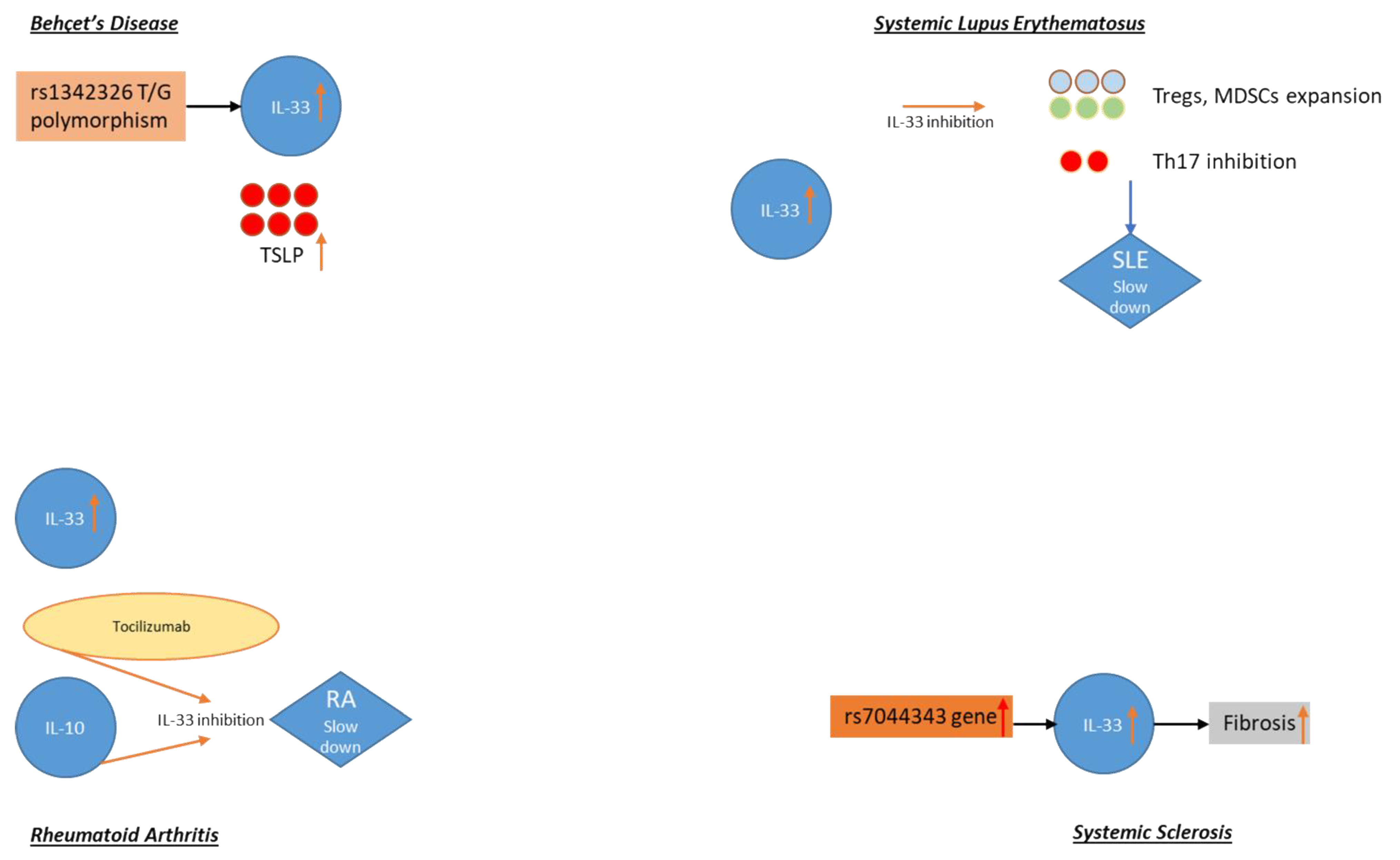
3.1. Behçet’s Disease

Behçet’s disease (BD) is a multi-systemic vasculitis with the highest prevalence among countries along the ancient Silk Road from the Mediterranean basin to East Asia.
The diagnosis is made on clinical criteria, and as to date there is no specific test, although an association with human leukocyte antigen (HLA)-B51 is a known genetic predisposition factor.
BD-typical features are recurrent oral and genital aphthous ulcers, ocular disease, and skin lesions. However, along with other systemic diseases, vascular, articular, gastrointestinal, neurologic, urogenital, pulmonary, and cardiac systems are often involved [13][14][15].
As is the case with other immune-related disorders, BD is characterized by multifactorial pathogenesis and several immunological abnormalities both regarding innate and humoral immunity have been detected. Among them, IL-1-related cytokines, thus including IL-33, have been recently studied. Indeed, Talei et al. [16] demonstrated that a specific polymorphism of the IL-33 gene, the rs1342326 T/G, may explain genetic susceptibility to BD, highlighting that this polymorphism may up-regulate IL-33 expression. Notably, Çerçi et al. [17] conducted a study to investigate the role of IL-33 among BD patients. They enrolled 54 BD patients, 31 had active BD and 23 had the inactive disease, and compared them with 18 healthy subjects to measure IL-33 levels using an enzyme-linked immunosorbent assay (ELISA). They found that serum IL-33 levels were significantly higher in patients with BD compared with the healthy controls (p < 0.01). Moreover, they noticed that among active BD patients with arthritis the mean serum IL-33 level was higher, but this finding was not statistically significant (p = 0.122). Another interesting study conducted by Kacem et al. [18] conducted on 40 BD patients demonstrated that messenger RNA (mRNA) expression of thymic stromal lymphopoïetin (TSLP) and IL-33 was increased in active BD with skin lesions. TSLP and IL-33 are both pro-inflammatory cytokines released from epithelial cells when facing stressing stimuli. Also, this represents the link between the environment and systemic immune responses.
High levels of IL-33 were also demonstrated in BD patients with neurologic involvement. Central nervous system (CNS) complications are rare but with high morbidity and mortality. Hamzaoui et al. [19] analyzed IL-33 levels in cerebrospinal fluid (CSF) of neuro BD (NBD), hypothesizing that this cytokine could be involved in oligodendrocyte and neuronal injury. They noticed that IL-33 levels were significantly higher in NBD patients compared to those who had the non-inflammatory neurological disease (NIND) and those with headache attributed to BD. Regarding the association between IL-31 and BD, data are lacking. However, as emerged from a study by Takeuchi et al. [20], IL-31 levels among BD patients with ocular involvement significantly reduced after infliximab (IFX) treatment. Thus, this suggests its role on disease course.

3.2. Systemic Lupus Erythematosus (SLE)

SLE is a multi-systemic disease characterized by the presence of several autoantibodies and immune dysregulations with a high prevalence in females [21][22]. Disease pathogenesis is still challenging as it is a multi-factorial condition in which several mechanisms are involved, including epigenetics [23]. Although great progress has been done on the development of new therapies, SLE patients still have great morbidity and mortality, which are mainly due to cardiovascular and renal involvement [24]. Among the plethora of immune-mediators that are currently under investigation, researchers recently focused on IL-33. Indeed, Yang et al. [25] conducted a study on 70 SLE patients, noticing that SLE patients had higher serum IL-33 levels compared to healthy controls. This study also highlighted that, although IL-33 may have a crucial role in the acute phase of the disease, specifically targeting erythrocytes and platelets, it was not associated with its course. Analogous results were obtained from a Guo et al. [26] study, as they noticed that IL-33 serum levels were higher in SLE patients. Moreover, they investigated the possible association between cytokine levels and clinical manifestations, noticing that there was a significant difference between IL-33 levels and C-reactive protein (CRP) levels and the erythrocyte sedimentation rate (ESR). Thus, this strengthened the idea that IL-33 may play a crucial role in the acute phase of the disease. Pre-clinical studies also hypothesized the role of IL-33 as an active player in SLE pathogenesis. Li et al. [27] conducted a study on lupus-prone mice, reporting that IL-33 inhibition may slow SLE through the expansion of T regulatory cells (T regs) and myeloid-derived suppressor cells (MDSCs) and inhibition of Th17 cells and proinflammatory responses. Thus, this indicated that the blockade of IL-33 has a protective effect on SLE. Genetic studies regarding IL-33 gene and its polymorphisms have also been conducted. Indeed, Zhu et al. [28] analyzed two IL-33 single nucleotide polymorphisms (SNPs), demonstrating that both were potential risk factors for developing SLE. On the other hand, at least two studies reported different results.

3.3. Rheumatoid Arthritis (RA)

Rheumatoid arthritis (RA) is an autoimmune disease characterized by systemic inflammation of diarthrodial joints, which may lead to articular irreversible damage. It affects almost 1% of the global population and systemic involvement may be present. As is the case with other inflammatory diseases, several mediators are involved in RA pathogenesis, including IL-1 family members. Thanks to the discoveries of this field, new target therapies have entered the market with encouraging results [29][30][31].
Indeed, over recent years, evidence has been collected regarding a relationship between IL-33 and RA. Chen et al. [32], focusing on the protective role of IL-10, demonstrated that in mice IL-33 levels were down-regulated by IL-10. Therefore, they demonstrated that IL-33 expression, rather than its receptor (ST2) is positively correlated with IL-10 level in active RA. More recently, Macedo et al. [33] confirmed the triggering role of IL-33 in collagen-induced arthritis in experimental models. They demonstrated that the administration of interleukin-33 intensifies the process. Moreover, they found a correlation between cytokine concentrations in serum and synovial fluid of patients with RA and disease activity. Other interesting studies have been conducted to determine if there was any association between IL-33 levels and RA therapy.
Firstly, Sellam et al. [34] investigated IL-33 and rituximab (RTX), a genetically engineered chimeric mouse/human monoclonal antibody representing a glycosylated immunoglobulin with human IgG1 constant regions and murine light-chain and heavy-chain variable region sequences, which binds specifically to the transmembrane antigen, CD20, a non-glycosylated phosphoprotein, located on pre-B and mature B lymphocytes. The authors found that serum IL-33 may predict clinical response to RTX independently of auto-antibodies. Therefore, they proposed IL-33 as a new biomarker in addition to auto-antibody status in predicting RTX response in RA patients. An interesting study published by Choi et al. [35] evaluated the effects of tocilizumab IL-33 in patients with RA. Tocilizumab is a humanized monoclonal antibody that acts as an IL-6 receptor antagonist, which can be administered both intravenously or subcutaneously [36]. This study was conducted on 83 RA patients, and serum cytokine levels were analyzed at baseline and after 24 weeks of tocilizumab therapy. Data confirmed that IL-33 levels were significantly higher in RA patients than in healthy controls (p < 0.001). Moreover, a significant correlation with rheumatoid factor titer and IL-33 was found. Aside from this, the authors demonstrated that serum IL-33 levels decreased significantly after 24 weeks of tocilizumab therapy (p < 0.001), thus strengthening the concept that IL-33 could be used as a marker to monitor therapy response in RA.
On the other hand, a study by Rivière et al. [37] found that there was no association between IL-33 and response to tumor necrosis factor-alpha inhibitors (TNFi), as well as to non-TNFi drugs overall or analyzed separately. Likewise, there was no difference when comparing the levels of serum IL-33 between responders and non-responders in TNFi and non-TNFi groups. To sum up, this study corroborates the association between serum IL-33 detection and seropositivity in RA patients. However, it did not reproduce the results obtained from the study by Sellam et al. [34].

3.4. Systemic Sclerosis (SSc)

Systemic sclerosis (SSc) is a complex disease characterized by fibrosis, vasculopathy, and immune dysregulation. Several systems may be involved, and disease triggers and pathogenesis are still under investigation. Indeed, actual therapies are organ-specific, but no curative therapies have emerged. New promising results came from autologous hematopoietic stem cell transplantation (AHSCT), however, it still stands as a major procedure with several complications, including infections, which leads to the concept that AHSCT could be proposed only to a small number of selected SSc patients [38][39][40]. As fibrosis is one of the cardinal characteristics of SSc, researchers have investigated its pathogenesis, noticing that IL-1 family cytokines are actively involved. Indeed, IL-33 may be considered as a biomarker of fibrosis involvement [41][42][43]. Zhang et al. [44] conducted a study on 56 Chinese SSc patients, reporting that IL-33 levels in SSc patients were significantly higher than in healthy controls. However, any significant correlation was found between cytokine levels and disease characteristics. On the other hand, Wagner et al. [45] found a significant correlation between IL-33 sST2 levels and skin involvement. They demonstrated that sST2 is elevated in late phase limited cutaneous SSc (lcSSc) as compared to patients with shorter disease duration or with the diffuse subtype of SSc. Moreover, they noticed that sST2 levels were decreased by prostanoid treatment. Analogous results were referred by Yanaba et al. [46]. Moreover, they reported that SSc patients with pulmonary fibrosis and decreased forced vital capacity presented higher IL-33 levels.
A brief, simple graphical overview of the IL-31 and IL-33 involvement in the autoimmune disorders above is shown in Figure 1, allowing the reader to understand at a glance their role in such conditions.
Figure 1. Interleukin (IL)-31 and IL-33 involvement in autoimmune disorders: Behçet’s disease (BD), systemic lupus erythematosus (SLE), rheumatoid arthritis (RA), systemic sclerosis (SSc). TSLP: thymic stromal lymphopoïetin, Tregs: regulatory T cells, MDSCs: myeloid-derived suppressor cells.

References

  1. Di Salvo, E.; Ventura-Spagnolo, E.; Casciaro, M.; Navarra, M.; Gangemi, S. IL-33/IL-31 Axis: A Potential Inflammatory Pathway. Mediat. Inflamm. 2018, 2018, 1–8.
  2. Cayrol, C.; Girard, J.P. Interleukin-33 (IL-33): A nuclear cytokine from the IL-1 family. Immunol. Rev. 2018, 281, 154–168.
  3. Ferretti, E.; Corcione, A.; Pistoia, V. The IL-31/IL-31 receptor axis: General features and role in tumor microenvironment. J. Leukoc. Biol. 2017, 102, 711–717.
  4. Ginaldi, L.; De Martinis, M.; Saitta, S.; Sirufo, M.M.; Mannucci, C.; Casciaro, M.; Ciccarelli, F.; Gangemi, S. Interleukin-33 serum levels in postmenopausal women with osteoporosis. Sci. Rep. 2019, 9, 3786.
  5. Ieni, A.; Casciaro, M.; Cardia, R.; Di Salvo, E.; Tuccari, G.; Ieni, A.; Gangemi, S. Interleukin-33 involvement in nonsmall cell lung carcinomas: An update. Biomolecules 2019, 9, 1–7.
  6. Yang, D.; Han, Z.; Oppenheim, J.J. Alarmins and immunity. Immunol. Rev. 2017, 280, 41–56.
  7. Zhang, Q.; Putheti, P.; Zhou, Q.; Liu, Q.; Gao, W. Structures and biological functions of IL-31 and IL-31 receptors. Cytokine Growth Factor Rev. 2008, 19, 347–356.
  8. Dillon, S.R.; Sprecher, C.; Hammond, A.; Bilsborough, J.; Rosenfeld-Franklin, M.; Presnell, S.R.; Haugen, H.S.; Maurer, M.; Harder, B.; Johnston, J.; et al. Interleukin 31, a cytokine produced by activated T cells, induces dermatitis in mice. Nat. Immunol. 2004, 5, 752–760.
  9. Furue, M.; Yamamura, K.; Kido-Nakahara, M.; Nakahara, T.; Fukui, Y. Emerging role of interleukin-31 and interleukin-31 receptor in pruritus in atopic dermatitis. Allergy Eur. J. Allergy Clin. Immunol. 2018, 73, 29–36.
  10. Gibbs, B.F.; Patsinakidis, N.; Raap, U. Role of the Pruritic Cytokine IL-31 in Autoimmune Skin Diseases. Front. Immunol. 2019, 10, 1–6.
  11. Maier, E.; Werner, D.; Duschl, A.; Bohle, B.; Horejs, J. Europe PMC Funders Group Human Th2 but not Th9 cells release IL-31 in a STAT6/NF-κB- dependent way. J. Immunol. 2016, 193, 645–654.
  12. Stott, B.; Lavender, P.; Lehmann, S.; Pennino, D.; Durham, S.; Schmidt-Weber, C.B. Human IL-31 is induced by IL-4 and promotes TH2-driven inflammation. J. Allergy Clin. Immunol. 2013, 132, 446–454.
  13. Skef, W.; Hamilton, M.J.; Arayssi, T. Gastrointestinal behçet’s disease: A. review. World, J. Gastroenterol. 2015, 21, 3801–3812.
  14. Alpsoy, E. Behçet’s disease: A comprehensive review with a focus on epidemiology, etiology and clinical features, and management of mucocutaneous lesions. J. Dermatol. 2016, 43, 620–632.
  15. Tong, B.; Liu, X.; Xiao, J.; Su, G. Immunopathogenesis of Behcet’s disease. Front. Immunol. 2019, 10, 665.
  16. Talei, M.; Abdi, A.; Shanebandi, D.; Jadidi-Niaragh, F.; Khabazi, A.; Babaie, F.; Alipour, S.; Afkari, B.; Sakhinia, E.; Babaloo, Z. Interleukin-33 gene expression and rs1342326 polymorphism in Behçet’s disease. Immunol. Lett. 2019, 212, 120–124.
  17. Çerçi, P.; Altıner, S.; İnal, A.; Köse, K.; Keskin, G.; Ölmez, Ü. Investigating the role of IL-33 in the pathogenesis of Behçet’s Disease. Acta Clin. Belg. 2017, 72, 434–438.
  18. Kacem, O.; Kaabachi, W.; Dhifallah, I.; Ben Hamzaoui, A.; Hamzaoui, K. Elevated expression of TSLP and IL-33 in Behçet’s disease skin lesions: IL-37 alleviate inflammatory effect of TSLP. Clin. Immunol. 2018, 192, 14–19.
  19. Hamzaoui, K.; Borhani-Haghighi, A.; Kaabachi, W.; Hamzaoui, A. Increased interleukin 33 in patients with neuro-Behcet’s disease: Correlation with MCP-1 and IP-10 chemokines. Cell. Mol. Immunol. 2014, 11, 613–616.
  20. Takeuchi, M.; Karasawa, Y.; Harimoto, K.; Tanaka, A.; Shibata, M.; Sato, T.; Caspi, R.R.; Ito, M. Analysis of Th Cell-related Cytokine Production in Behçet Disease Patients with Uveitis Before and After Infliximab Treatment. Ocul. Immunol. Inflamm. 2017, 25, 52–61.
  21. Aringer, M.; Costenbader, K.; Daikh, D.; Brinks, R.; Mosca, M.; Ramsey-Goldman, R.; Smolen, J.S.; Wofsy, D.; Boumpas, D.T.; Kamen, D.L.; et al. 2019 European League Against Rheumatism/American College of Rheumatology classification criteria for systemic lupus erythematosus. Arthritis Rheumatol 2019, 71, 1400–1412.
  22. D’Cruz, D.P.; Khamashta, M.A.; Hughes, G.R. Systemic lupus erythematosus. Lancet 2007, 369, 1208–1219.
  23. Ghodke-Puranik, Y.; Niewold, T.B. Immunogenetics of systemic lupus erythematosus: A comprehensive review. J. Autoimmun. 2015, 64, 125–136.
  24. Ippolito, A.; Petri, M. An update on mortality in systemic lupus erythematosus. Clin. Exp. Rheumatol. 2008, 26, S72–79.
  25. Yang, Z.; Liang, Y.; Xi, W.; Li, C.; Zhong, R. Association of increased serum IL-33 levels with clinical and laboratory characteristics of systemic lupus erythematosus in Chinese population. Clin. Exp. Med. 2011, 11, 75–80.
  26. Guo, J.; Xiang, Y.; Peng, Y.F.; Huang, H.T.; Lan, Y.; Wei, Y.S. The association of novel IL-33 polymorphisms with sIL-33 and risk of systemic lupus erythematosus. Mol. Immunol. 2016, 77, 1–7.
  27. Li, P.; Lin, W.; Zheng, X. IL-33 neutralization suppresses lupus disease in lupus-prone mice. Inflammation 2014, 37, 824–832.
  28. Zhu, X.; Xie, L.; Qin, H.; Liang, J.; Yang, Y.; Xu, J.; Zhang, T. Interaction between IL-33 gene polymorphisms and current smoking with susceptibility to systemic lupus erythematosus. J. Immunol. Res. 2019, 2019, 1547578.
  29. Burmester, G.R.; Pope, J.E. Novel treatment strategies in rheumatoid arthritis. Lancet 2017, 389, 2338–2348.
  30. Huizinga, T.; Knevel, R. Rheumatoid arthritis: 2014 treat-To-Target RA recommendations-Strategy is key. Nat. Rev. Rheumatol. 2015, 11, 509–511.
  31. Firestein, G.S. Evolving concepts of rheumatoid arthritis. Nature 2003, 423, 356–361.
  32. Chen, S.; Chen, B.; Wen, Z.; Huang, Z.; Ye, L. IL-33/ST2-mediated inflammation in macrophages is directly abrogated by IL-10 during rheumatoid arthritis. Oncotarget 2017, 8, 32407–32418.
  33. Macedo, R.B.V.; Kakehasi, A.M.; de Andrade, M.V.M. IL33 in rheumatoid arthritis: Potencial contribution to pathogenesis. Rev. Bras. Reumatol. 2016, 56, 451–457.
  34. Sellam, J.; Rivière, E.; Courties, A.; Rouzaire, P.O.; Tolusso, B.; Vital, E.V.; Emery, P.; Ferraccioli, G.; Soubrier, M.; Ly, B.; et al. Serum IL-33, a new marker predicting response to rituximab in rheumatoid arthritis. Arthritis Res. Ther. 2016, 18, 1–8.
  35. Choi, I.A.; Lee, S.J.; Park, W.; Park, S.H.; Shim, S.C.; Baek, H.J.; Yoo, D.H.; Kim, H.A.; Lee, S.K.; Lee, Y.J.; et al. Effects of tocilizumab therapy on serum interleukin-33 and interleukin-6 levels in patients with rheumatoid arthritis. Arch. Rheumatol. 2018, 33, 389–394.
  36. Scott, L.J. Tocilizumab: A Review in Rheumatoid Arthritis. Drugs 2017, 77, 1865–1879.
  37. Rivière, E.; Sellam, J.; Pascaud, J.; Ravaud, P.; Gottenberg, J.E.; Mariette, X. Serum IL-33 level is associated with auto-antibodies but not with clinical response to biologic agents in rheumatoid arthritis. Arthritis Res. Ther. 2018, 20, 18–20.
  38. Sullivan, K.M.; Goldmuntz, E.A.; Keyes-Elstein, L.; McSweeney, P.A.; Pinckney, A.; Welch, B.; Mayes, M.D.; Nash, R.A.; Crofford, L.J.; Eggleston, B.; et al. Myeloablative autologous stem-cell transplantation for severe scleroderma. N. Engl. J. Med. 2018, 378, 35–47.
  39. Denton, C.P.; Khanna, D. Systemic sclerosis. Lancet 2017, 390, 1685–1699.
  40. Nagaraja, V.; Denton, C.P.; Khanna, D. Old medications and new targeted therapies in systemic sclerosis. Rheumatology 2015, 54, 1944–1953.
  41. Manetti, M.; Guiducci, S.; Ceccarelli, C.; Romano, E.; Bellando-Randone, S.; Conforti, M.L.; Ibba-Manneschi, L.; Matucci-Cerinic, M. Increased circulating levels of interleukin 33 in systemic sclerosis correlate with early disease stage and microvascular involvement. Ann. Rheum. Dis. 2011, 70, 1876–1878.
  42. Vettori, S.; Cuomo, G.; Iudici, M.; D’Abrosca, V.; Giacco, V.; Barra, G.; De Palma, R.; Valentini, G. Early Systemic Sclerosis: Serum Profiling of Factors Involved in Endothelial, T-cell, and Fibroblast Interplay is Marked by Elevated Interleukin-33 Levels. J. Clin. Immunol. 2014, 34, 663–668.
  43. Terras, S.; Opitz, E.; Moritz, R.K.; Höxtermann, S.; Gambichler, T.; Kreuter, A. Increased serum IL-33 levels may indicate vascular involvement in systemic sclerosis. Ann. Rheum. Dis. 2013, 72, 144–145.
  44. Zhang, Y.J.; Zhang, Q.; Yang, G.J.; Tao, J.H.; Wu, G.C.; Huang, X.L.; Duan, Y.; Li, X.P.; Ye, D.Q.; Wang, J. Elevated serum levels of interleukin-1β and interleukin-33 in patients with systemic sclerosis in Chinese population. Z. Rheumatol. 2018, 77, 151–159.
  45. Wagner, A.; Köhm, M.; Nordin, A.; Svenungsson, E.; Pfeilschifter, J.M.; Radeke, H.H. Increased Serum Levels of the IL-33 Neutralizing sST2 in Limited Cutaneous Systemic Sclerosis. Scand. J. Immunol. 2015, 82, 269–274.
  46. Yanaba, K.; Yoshizaki, A.; Asano, Y.; Kadono, T.; Sato, S. Serum IL-33 levels are raised in patients with systemic sclerosis: Association with extent of skin sclerosis and severity of pulmonary fibrosis. Clin. Rheumatol. 2011, 30, 825–830.
More
Information
Contributors MDPI registered users' name will be linked to their SciProfiles pages. To register with us, please refer to https://encyclopedia.pub/register : , , , , , ,
View Times: 256
Revisions: 2 times (View History)
Update Date: 01 Aug 2023
1000/1000
Video Production Service