Submitted Successfully!
To reward your contribution, here is a gift for you: A free trial for our video production service.
Thank you for your contribution! You can also upload a video entry or images related to this topic.
Version Summary Created by Modification Content Size Created at Operation
1 -- 1883 2023-07-13 07:29:49 |
2 format correct Meta information modification 1883 2023-07-17 04:22:17 |

Video Upload Options

Do you have a full video?

Confirm

Are you sure to Delete?
Cite
If you have any further questions, please contact Encyclopedia Editorial Office.
Pagliaro, P.; Penna, C. Mechanisms Involved in MIRI and Interactions with NLRP3. Encyclopedia. Available online: https://encyclopedia.pub/entry/46728 (accessed on 27 July 2024).
Pagliaro P, Penna C. Mechanisms Involved in MIRI and Interactions with NLRP3. Encyclopedia. Available at: https://encyclopedia.pub/entry/46728. Accessed July 27, 2024.
Pagliaro, Pasquale, Claudia Penna. "Mechanisms Involved in MIRI and Interactions with NLRP3" Encyclopedia, https://encyclopedia.pub/entry/46728 (accessed July 27, 2024).
Pagliaro, P., & Penna, C. (2023, July 13). Mechanisms Involved in MIRI and Interactions with NLRP3. In Encyclopedia. https://encyclopedia.pub/entry/46728
Pagliaro, Pasquale and Claudia Penna. "Mechanisms Involved in MIRI and Interactions with NLRP3." Encyclopedia. Web. 13 July, 2023.
Mechanisms Involved in MIRI and Interactions with NLRP3
Edit

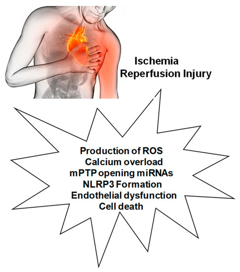
Myocardial ischemia-reperfusion injury (MIRI) is caused by several mechanisms, including the production of reactive oxygen species (ROS), altered cellular osmolarity, and inflammatory response. Calcium overload, altered oxygen levels, and mitochondrial ROS are also involved in these MIRI processes, resulting in the irreversible opening of the mitochondrial permeability transition pore (mPTP). These mechanisms and processes are associated with NLRP3 inflammasome priming and activation, which can also induce cell death by pyroptosis through the up-regulation of the caspase-1 pathway and IL-18 release. In addition, endothelial dysfunction, both in the presence and absence of MIRI, is also accompanied by altered oxygen levels, decreased nitric oxide production, and ROS overproduction, resulting in the expression of adhesion molecules and leukocyte infiltration in which the NLRP3 inflammasome plays a central role, thus contributing, through endothelial dysfunction, to the alteration of coronary flow, typical of ischemic heart disease. Given the intricate interrelationship between ROS and NLRP3, ROS inhibitors can reduce NLRP3 inflammasome activation, while NLRP3 inhibitors can reduce oxidative stress and inflammation. 

inflammation reactive oxygen species ischemia/reperfusion injury

1. Introduction

Myocardial ischemia-reperfusion injury (MIRI) is the aggravation of myocardial damage caused by the rapid reperfusion of ischemic myocardium. In fact, reperfusion causes further damage to the ischemic tissue in the experimental and clinical scenarios of ischemic heart diseases (IHD). The reperfusion damage and subsequent cell death occur primarily as a consequence of massive reactive oxygen production (ROS) production, altered cellular osmolarity, and subsequent inflammatory response, including neutrophil infiltration. The inflammatory response not only affects the local aspect but is generalized and, in severe cases, can lead to a syndrome of multiorgan dysfunction [1]. Coronary circulation is both the culprit and victim of MIRI, which is caused by complex mechanisms that include intracellular, extracellular, and mechanical processes, all intertwined with inflammatory processes [2][3]. Among the inflammatory processes that are triggered by ROS but which in turn generate and exacerbate redox-related pathologies, researchers are paying particular attention to the activation of Nod-like receptor (NLR) proteins, particularly the NLR pyrin domain containing 3 (NLRP3) [4], which is the main and most characterized member of the NLR family [5].
Actually, NLRs are a protein family of intracellular sensors, whose members share a conserved central nucleotide bond, an oligomerization domain (NOD), and a variable N-terminal effector domain and are rich in leucine repeat (LRR) [6][7][8].
Therefore, the innate immune system intervenes in IHD through the recognition of receptor patterns (PRR) capable of detecting the presence of both pathogenic microbes and other endogenous or exogenous pathogens, such as damage-associated molecular patterns (DAMPs) or pathogens (PAMPs) [9].
The activation and regulation of the NLRP3 inflammasome is a complex two-step process consisting of priming and activation. Upon recognition of PAMP or DAMP, the inflammasome is primed. The first phase, or priming, is carried out through the activation of the nuclear factor kappa B (NF-κB) pathway, which leads to the up-regulation of NLRP3 and pro-IL-1β proteins and changes in the post-translational modifications (PTMs) of NLRP3, such as ubiquitination and phosphorylation, which empower NLRP3 and promote inflammasome assembly. After priming, the second phase, or activation phase, leads to conformational changes in NLRP3 and PTMs that enable NLRP3 oligomerization and consequently inflammasome activation. This phase is characterized by an up-regulation of the protein expression of some components of inflammasomes (NLRP3, apoptosis-associated speck-like protein (ASC), caspase-1, and pro-interleukin (IL-1β). Upstream, inflammasome activation is also regulated by various ion signals (K+, Ca2+, Cl), mitochondrial dysfunction, and lysosome disruption [8][10].

MIRI Activates Various Cell Death Pathways

Numerous cell death pathways, including but not limited to pyroptosis, apoptosis, necrosis, and autophagy, are present in MIRI [6][7]. An important mechanism of MIRI is sterile inflammation with subsequent pyroptosis cell death [7]. Therefore, NLRP3 perceives cardiomyocyte injury and enrolls an ASC (apoptosis-associated speck-like protein carrying a CARD (C-terminal caspase recruitment domain) and procaspase-1 to shape inflammasome complexes, which initiate the pyroptosis pathway. This involves gasdermin D (GSDMD) activation, which represents the classical pathway of pyroptosis cell death [11]. NLRP3 activation is the major player in this type of controlled cardiomyocyte cell death, which is characterized by the development of cell membrane holes, the release of pro-inflammatory cytokines, and cell lysis [8]. Specifically, pyroptosis is a mode of cell death by which the immune system responds to endogenous damage and pathogens. Indeed, pyroptosis has been defined as a sort of programmed GSDMD/NLRP3-dependent necrosis and is mediated by GSDMD cleavage by caspase-1 [12]. Apoptosis and pyroptosis are similar in that both involve DNA damage, a positive terminal deoxynucleotidyl TUNEL (transferase-mediated UTP nick end-labeling) result, and the presence of annexin-5. However, in contrast to apoptosis, pyroptosis damages the cell membrane quickly and irreparably [13]. Both pyroptosis and apoptosis exhibit chromatin condensation, even though the nucleus remains intact in apoptosis. Since in pyroptosis, a pivotal role is played by GSDMD, the deletion of the GSDMD gene prevents both pyroptosis and IL-1β secretion by macrophage cells [14].
Of note, functional and physiological autophagy, another regulated cell death, would have the overall effect of minimizing NLRP3 activation and reducing cytokine secretion. However, abnormalities in autophagy pathways may result in ineffective or incomplete autophagy and mitophagy, thereby inducing the activation of NLRP3 and exacerbating myocardial damage [12][15]. Mitophagy is also among the mechanisms activated by MIRI. Lemasters introduced the term mitophagy in 2005 to highlight a very particular autophagic process that affected the mitochondria [16]. Indeed, mitophagy is a process aimed at maintaining mitochondrial homeostasis at the cellular level, which causes the degradation of dysfunctional mitochondria [17].
Recently, ferroptosis has been reported among the cellular death modalities activated by MIRI [18]. Ferroptosis is an iron-dependent form of controlled cell death characterized by intracellular iron overload and redox system dysfunction, inducing lipid peroxidation. It is closely correlated with the induction of inflammatory signaling pathways, such as JAK-STAT, NF-κB, inflammasome, cyclic GMP–AMP synthase (cGAS)–stimulator of interferon genes (STING) pathway (cGAS-STING), and MAPK signaling pathways, and vice versa. As a matter of fact, the inflammatory response is a significant factor in challenges with iron metabolism and redox system dysfunction [19]. Additionally, mTOR is involved because its deletion causes an excessive amount of cell death, while high levels of mTOR block the cell death brought on by the inducer of ferroptosis, erastin [18]. Actually, the mechanism underlying ferroptosis is not fully understood, but it is unquestionably related to mitochondrial dysfunction [20]. Indeed, a reduction in mitochondrial volume, an increase in the density of the bilayer membrane, and a rupture of the outer mitochondrial membrane have all been observed, suggesting that the morphology and size of the mitochondria can be affected [21].

2. Mechanisms Involved in MIRI and Interactions with NLRP3

The main mechanisms involved in MIRI (Figure 1) and intertwined with NLRP3 include ROS production, calcium overload, mPTP opening, MiRNAs, and endothelial dysfunction.
Figure 1. Factors involved in determining myocardial ischemia/reperfusion injury. Mitochondrial permeability transition pores, mPTP.

2.1. Production of ROS

Following MIRI, a large amount of ROS is produced during the early stages of reperfusion. During ischemia and reperfusion, ROS are produced by different mechanisms at the mitochondrial level, but they can also be produced by the intervention of cellular enzymes, such as NADPH oxidases (NOX) family, cyclooxygenase (COX) and lipoxygenase (LOX) [22]. These ROS are capable of causing cellular damage by inducing lipid peroxidation, disrupting cell signaling pathways, and activating pro-inflammatory factors. The high production of ROS results in the opening of mitochondrial permeability transition pores (mPTP), leading to mitochondrial damage [17] and the induction of ROS-induced ROS release (RIRR). ROS during MIRI promotes tissue inflammation and the activation of the NLRP3 complex in various organs, including the heart. In addition, it appears that NLRP3 itself directly or indirectly induces ROS production at the mitochondrial level [23]. ROS are able to induce the synthesis of cytokines, including IL-18, inducing the inflammatory state of tissues and promoting both apoptosis and calcium overload. Thus, the high amount of ROS results in cell death, including apoptosis and pyroptosis. ROS production also appears to be induced by its ability to induce the release of inflammatory factors. Thus, ROS are involved in a number of early and late self-ingravescent mechanisms. Despite the well-known deleterious effects, low levels of ROS have an essential, albeit double-edged, role in cardioprotection [24][25][26]. Similarly, a double-edged role has been reported for NLRP3 [27][28]. This double-edged role for NLRP3 is shown in animal model NLRP3 knockout (KO) where the cardioprotective effects of ischemic preconditioning are reduced and the levels of signal transducers and activators of transcription 3 (STAT3) proteins are decreased in this animal model [27][28]. ASC, another component associated with NLRP3, appears to have a protective role, although the number of studies is currently very small. In a mouse model of ASC−/−, no action on ischemic preconditioning-induced protection was demonstrated for this factor. Indeed, ASC deficiency had no effect on an ischemic preconditioning protocol [28]. However, in another work, it was reported that either the absence of NLRP3 or of ASC determined the loss of protection [29]. Obviously, further studies are necessary to clarify these discrepancies.

2.2. Calcium Overload

As a result of MIRI, a disordered calcium distribution is established, leading to dysfunctional calcium homeostasis, and an anomalous increase in intracellular calcium concentration, also referred to as calcium overload, develops. This overload also affects the mitochondrial level and is accompanied by both a decrease in mitochondrial membrane potential and ATP content and mPTP opening. A close correlation between calcium overload/NLRP3 expression/pyrotosis has been demonstrated in several tissues, including the heart [11][30]. Calcium overload in MIRI operates as an excitatory element of oxidative stress, which in turn induces inflammasome formation [11]. Notably, Mo et al., in a hypoxia/reperfusion (H/R) model of adult rat cardiomyocytes, demonstrated that calcium overload can induce pyroptosis by up-regulating the NLRP3/caspase-1 pathway [31].

2.3. Role of mPTP Opening

Mitochondria are the central nodes of the cell and are in charge of cellular energy production through oxidative phosphorylation (OXPHOS) [32]. These organelles are influenced negatively by both low oxygen levels following ischemia and, as seen previously, by ROS produced during the early stages of reperfusion. Both of these factors participate in cell death due to the irreversible opening of the mPTP [33]. The opening of the mPTP leads to a loss of mitochondrial membrane potential (ΔΨm), a loss of OXPHOS activity, and a subsequent drastic reduction of ATP [34]. This leads to mitochondrial osmotic shock and the rupture of the outer mitochondrial membrane and, consequently, to all forms of cardiomyocyte death, including pyroptosis [7][35].

2.4. Endothelial Dysfunction

Endothelial dysfunction involves the decreased production of nitric oxide (NO), expression of adhesion molecules, the adhesion of leukocytes to the endothelium, and the infiltration of leukocytes. All these phenomena are mechanisms involved in MIRI, leading to the so-called “no-reflow phenomenon”, which is especially due to limited NO availability, resulting in vasoconstriction and micro-thrombus formation in the lumen of small vessels [3][36][37]. In endothelial dysfunction, the activation of NF-κB and other transcription factors, with increased expression of cell adhesion molecules, play a pivotal role in inducing inflammatory processes [3][36]. Of note, NF-κB is central to NLRP3 priming and activation [4][38].
Another harmful aspect induced by early reperfusion is inflammatory damage mediated by the activation of mast cells and neutrophils, whose products act as chemoattractants for other leukocytes [39]. Recently cathepsins, proteases involved in multiple pathophysiological roles, have attracted the attention of researchers. In particular, cathepsin G, a modulator of neutrophil chemoattractant, induces morphological modifications that break focal adhesion and intracellular contacts with cardiomyocytes and is involved in MIRI [39][40]. Indeed, cathepsin G inhibition with DCCI (dual inhibitor of cathepsin G and chymase) limits MIRI and damages [41].

References

  1. Carden, D.L.; Granger, D.N. Pathophysiology of ischaemia-reperfusion injury. J. Pathol. 2000, 190, 255–266.
  2. Davidson, S.M.; Ferdinandy, P.; Andreadou, I.; Bøtker, H.E.; Heusch, G.; Ibáñez, B.; Ovize, M.; Schulz, R.; Yellon, D.M.; Hausenloy, D.J.; et al. Multitarget Strategies to Reduce Myocardial Ischemia/Reperfusion Injury: JACC Review Topic of the Week. J. Am. Coll. Cardiol. 2019, 73, 89–99.
  3. Penna, C.; Comità, S.; Tullio, F.; Alloatti, G.; Pagliaro, P. Challenges facing the clinical translation of cardioprotection: 35 years after the discovery of ischemic preconditioning. Vascul. Pharmacol. 2022, 144, 106995.
  4. Zuurbier, C.J.; Abbate, A.; Cabrera-Fuentes, H.A.; Cohen, M.V.; Collino, M.; De Kleijn, D.P.V.; Downey, J.M.; Pagliaro, P.; Preissner, K.T.; Takahashi, M.; et al. Innate immunity as a target for acute cardioprotection. Cardiovasc. Res. 2019, 115, 1131–1142.
  5. Pasqua, T.; Pagliaro, P.; Rocca, C.; Angelone, T.; Penna, C. Role of NLRP-3 Inflammasome in Hypertension: A Potential Therapeutic Target. Curr. Pharm. Biotechnol. 2018, 19, 708–714.
  6. Eltzschig, H.K.; Eckle, T. Ischemia and reperfusion--from mechanism to translation. Nat. Med. 2011, 17, 1391–1401.
  7. Davidson, S.M.; Adameová, A.; Barile, L.; Cabrera-Fuentes, H.A.; Lazou, A.; Pagliaro, P.; Stensløkken, K.O.; Garcia-Dorado, D.; EU-CARDIOPROTECTION COST Action (CA16225). Mitochondrial and mitochondrial-independent pathways of myocardial cell death during ischaemia and reperfusion injury. J. Cell. Mol. Med. 2020, 24, 3795–3806.
  8. Lamkanfi, M.; Dixit, V.M. Mechanisms and functions of inflammasomes. Cell 2014, 157, 1013–1022.
  9. Mastrocola, R.; Aragno, M.; Alloatti, G.; Collino, M.; Penna, C.; Pagliaro, P. Metaflammation: Tissue-Specific Alterations of the NLRP3 Inflammasome Platform in Metabolic Syndrome. Curr. Med. Chem. 2018, 25, 1294–1310.
  10. Zhao, J.; Li, J.; Li, G.; Chen, M. The role of mitochondria-associated membranes mediated ROS on NLRP3 inflammasome in cardiovascular diseases. Front. Cardiovasc. Med. 2022, 9, 1059576.
  11. Liu, Y.; Zhang, J.; Zhang, D.; Yu, P.; Zhang, J.; Yu, S. Research Progress on the Role of Pyroptosis in Myocardial Ischemia-Reperfusion Injury. Cells 2022, 11, 3271.
  12. Popov, S.V.; Maslov, L.N.; Naryzhnaya, N.V.; Mukhomezyanov, A.V.; Krylatov, A.V.; Tsibulnikov, S.Y.; Ryabov, V.V.; Cohen, M.V.; Downey, J.M. The Role of Pyroptosis in Ischemic and Reperfusion Injury of the Heart. J. Cardiovasc. Pharmacol. Ther. 2021, 26, 562–574.
  13. Jorgensen, I.; Miao, E.A. Pyroptotic cell death defends against intracellular pathogens. Immunol. Rev. 2015, 265, 130–142.
  14. Shi, J.; Zhao, Y.; Wang, K.; Shi, X.; Wang, Y.; Huang, H.; Zhuang, Y.; Cai, T.; Wang, F.; Shao, F. Cleavage of GSDMD by inflammatory caspases determines pyroptotic cell death. Nature 2015, 526, 660–665.
  15. Toldo, S.; Mauro, A.G.; Cutter, Z.; Abbate, A. Inflammasome, pyroptosis, and cytokines in myocardial ischemia-reperfusion injury. Am. J. Physiol. Heart Circ. Physiol. 2018, 315, H1553–H1568.
  16. Lemasters, J.J. Selective mitochondrial autophagy, or mitophagy, as a targeted defense against oxidative stress, mitochondrial dysfunction, and aging. Rejuvenation Res. 2005, 8, 3–5.
  17. Penna, C.; Perrelli, M.G.; Pagliaro, P. Mitochondrial pathways, permeability transition pore, and redox signaling in cardioprotection: Therapeutic implications. Antioxid. Redox Signal. 2013, 18, 556–599.
  18. Liu, C.; Li, Z.; Li, B.; Liu, W.; Zhang, S.; Qiu, K.; Zhu, W. Relationship be-tween ferroptosis and mitophagy in cardiac ischemia reperfusion injury: A mini-review. PeerJ 2023, 11, e14952.
  19. Decout, A.; Katz, J.D.; Venkatraman, S.; Ablasser, A. The cGAS–STING pathway as a therapeutic target in inflammatory diseases. Nat. Rev. Immunol. 2021, 21, 548–569.
  20. Gan, B. Mitochondrial regulation of ferroptosis. J. Cell Biol. 2021, 220, e202105043.
  21. Battaglia, A.M.; Chirillo, R.; Aversa, I.; Sacco, A.; Costanzo, F.; Biamonte, F. Ferroptosis and Cancer: Mitochondria Meet the “Iron Maiden”. Cell Death Cells 2020, 9, 1505.
  22. Pagliaro, P.; Moro, F.; Tullio, F.; Perrelli, M.G.; Penna, C. Cardioprotective pathways during reperfusion: Focus on redox signaling and other modalities of cell signaling. Antioxid. Redox Signal. 2011, 14, 833–850.
  23. Minutoli, L.; Puzzolo, D.; Rinaldi, M.; Irrera, N.; Marini, H.; Arcoraci, V.; Bitto, A.; Crea, G.; Pisani, A.; Squadrito, F.; et al. ROS-Mediated NLRP3 Inflammasome Activation in Brain, Heart, Kidney, and Testis Ischemia/Reperfusion Injury. Oxid. Med. Cell. Longev. 2016, 2016, 2183026.
  24. Penna, C.; Rastaldo, R.; Mancardi, D.; Raimondo, S.; Cappello, S.; Gattullo, D.; Losano, G.; Pagliaro, P. Post-conditioning induced cardioprotection requires signaling through a redox-sensitive mechanism, mitochondrial ATP-sensitive K+ channel and protein kinase C activation. Basic Res. Cardiol. 2006, 101, 180–189.
  25. Downey, J.M.; Cohen, M.V. A really radical observation: A comment on Penna et al. in Basic Res Cardiol (2006) 101:180-189. Basic Res. Cardiol. 2006, 101, 190–191.
  26. Tsutsumi, Y.M.; Yokoyama, T.; Horikawa, Y.; Roth, D.M.; Patel, H.H. Reactive oxygen species trigger ischemic and pharmacological postconditioning: In vivo and in vitro characterization. Life Sci. 2007, 81, 1223–1227.
  27. Zuurbier, C.J. NLRP3 Inflammasome in Cardioprotective Signaling. J. Cardiovasc. Pharmacol. 2019, 74, 271–275.
  28. Zuurbier, C.J.; Jong, W.M.; Eerbeek, O.; Koeman, A.; Pulskens, W.P.; Butter, L.M.; Leemans, J.C.; Hollmann, M.W. Deletion of the innate immune NLRP3 receptor abolishes cardiac ischemic preconditioning and is associated with decreased IL-6/STAT3 signaling. PLoS ONE 2012, 7, e40643.
  29. Sandanger, Ø.; Gao, E.; Ranheim, T.; Bliksøen, M.; Kaasbøll, O.J.; Alfsnes, K.; Nymo, S.H.; Rashidi, A.; Ohm, I.K.; Attramadal, H.; et al. NLRP3 inflammasome activation during myocardial ischemia reperfusion is cardioprotective. Biochem. Biophys. Res. Commun. 2016, 469, 1012–1020.
  30. Wu, X.; Ren, G.; Zhou, R.; Ge, J.; Chen, F.H. The role of Ca2+ in acid-sensing ion channel 1a-mediated chondrocyte pyroptosis in rat adjuvant arthritis. Lab. Investig. 2019, 99, 499–513.
  31. Mo, G.; Liu, X.; Zhong, Y.; Mo, J.; Li, Z.; Li, D.; Zhang, L.; Liu, Y. IP3R1 regulates Ca2+ transport and pyroptosis through the NLRP3/Caspase-1 pathway in myocardial ischemia/reperfusion injury. Cell Death Discov. 2021, 7, 31.
  32. García-Niño, W.R.; Zazueta, C.; Buelna-Chontal, M.; Silva-Palacios, A. Mitochondrial Quality Control in Cardiac-Conditioning Strategies against Ischemia-Reperfusion Injury. Life 2021, 11, 1123.
  33. Zhou, H.; Zhu, P.; Wang, J.; Zhu, H.; Ren, J.; Chen, Y. Pathogenesis of cardiac ischemia reperfusion injury is associated with CK2α-disturbed mitochondrial homeostasis via suppression of FUNDC1-related mitophagy. Cell Death Differ. 2018, 25, 1080–1093.
  34. Urbani, A.; Giorgio, V.; Carrer, A.; Franchin, C.; Arrigoni, G.; Jiko, C.; Abe, K.; Maeda, S.; Shinzawa-Itoh, K.; Bogers, J.F.M.; et al. Purified F-ATP synthase forms a Ca2+-dependent high-conductance channel matching the mitochondrial permeability transition pore. Nat. Commun. 2019, 10, 4341.
  35. Robichaux, D.J.; Harata, M.; Murphy, E.; Karch, J. Mitochondrial permeability transition pore-dependent necrosis. J. Mol. Cell. Cardiol. 2023, 174, 47–55.
  36. Heusch, G. Myocardial ischaemia-reperfusion injury and cardioprotection in perspective. Nat. Rev. Cardiol. 2020, 17, 773–789.
  37. Heusch, G. Coronary microvascular obstruction: The new frontier in cardioprotection. Basic Res. Cardiol. 2019, 114, 45.
  38. Bauernfeind, F.G.; Horvath, G.; Stutz, A.; Alnemri, E.S.; MacDonald, K.; Speert, D.; Fernandes-Alnemri, T.; Wu, J.; Monks, B.G.; Fitzgerald, K.A.; et al. Cutting edge: NF-κB activating pattern recognition and cytokine receptors license NLRP3 inflammasome activation by regulating NLRP3 expression. J. Immunol. 2009, 183, 787–791.
  39. Huertas, J.; Lee, H.T. Multi-faceted roles of cathepsins in ischemia reperfusion injury (Review). Mol. Med. Rep. 2022, 26, 368.
  40. Sabri, A.; Alcott, S.G.; Elouardighi, H.; Pak, E.; Derian, C.; Andrade-Gordon, P.; Kinnally, K.; Steinberg, S.F. Neutrophil cathepsin G promotes detachment-induced cardiomyocyte apoptosis via a protease-activated receptor-independent mechanism. J. Biol. Chem. 2003, 278, 23944–23954.
  41. Hooshdaran, B.; Kolpakov, M.A.; Guo, X.; Miller, S.A.; Wang, T.; Tilley, D.G.; Rafiq, K.; Sabri, A. Dual inhibition of cathepsin G and chymase reduces myocyte death and improves cardiac remodeling after myocardial ischemia reperfusion injury. Basic Res. Cardiol. 2017, 112, 62.
More
Information
Subjects: Physiology
Contributors MDPI registered users' name will be linked to their SciProfiles pages. To register with us, please refer to https://encyclopedia.pub/register : ,
View Times: 257
Revisions: 2 times (View History)
Update Date: 17 Jul 2023
1000/1000
Video Production Service