Submitted Successfully!
To reward your contribution, here is a gift for you: A free trial for our video production service.
Thank you for your contribution! You can also upload a video entry or images related to this topic.
Version Summary Created by Modification Content Size Created at Operation
1 -- 1341 2023-07-11 09:15:35 |
2 layout Meta information modification 1341 2023-07-11 09:42:00 |

Video Upload Options

Do you have a full video?

Confirm

Are you sure to Delete?
Cite
If you have any further questions, please contact Encyclopedia Editorial Office.
Vertyshev, A.Y.; Akberdin, I.R.; Kolpakov, F.A. Kinases/Protein Phosphatases in Signaling Pathways Activation. Encyclopedia. Available online: https://encyclopedia.pub/entry/46626 (accessed on 29 April 2024).
Vertyshev AY, Akberdin IR, Kolpakov FA. Kinases/Protein Phosphatases in Signaling Pathways Activation. Encyclopedia. Available at: https://encyclopedia.pub/entry/46626. Accessed April 29, 2024.
Vertyshev, Alexander Yu., Ilya R. Akberdin, Fedor A. Kolpakov. "Kinases/Protein Phosphatases in Signaling Pathways Activation" Encyclopedia, https://encyclopedia.pub/entry/46626 (accessed April 29, 2024).
Vertyshev, A.Y., Akberdin, I.R., & Kolpakov, F.A. (2023, July 11). Kinases/Protein Phosphatases in Signaling Pathways Activation. In Encyclopedia. https://encyclopedia.pub/entry/46626
Vertyshev, Alexander Yu., et al. "Kinases/Protein Phosphatases in Signaling Pathways Activation." Encyclopedia. Web. 11 July, 2023.
Kinases/Protein Phosphatases in Signaling Pathways Activation
Edit

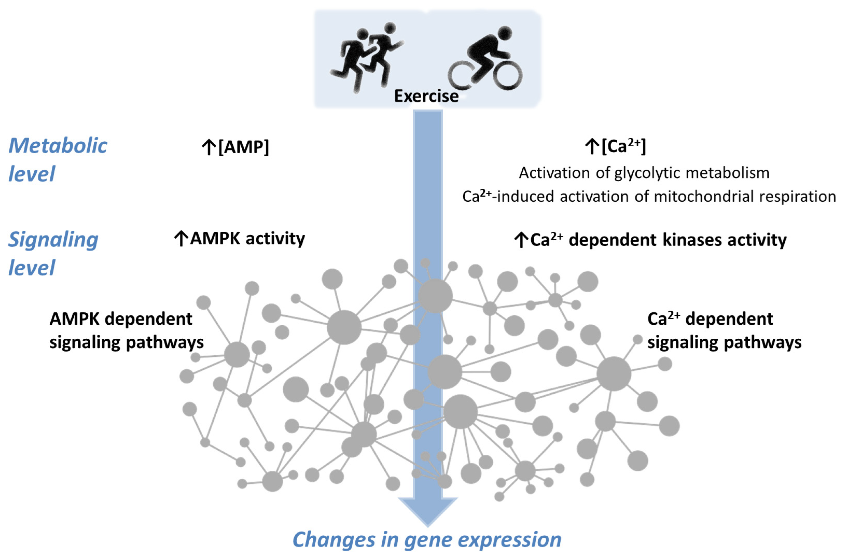
Optimizing physical training regimens to increase muscle aerobic capacity requires an understanding of the internal processes that occur during exercise that initiate subsequent adaptation. During exercise, muscle cells undergo a series of metabolic events that trigger downstream signaling pathways and induce the expression of many genes in working muscle fibers. There are a number of studies that show the dependence of changes in the activity of AMP-activated protein kinase (AMPK), one of the mediators of cellular signaling pathways, on the duration and intensity of single exercises. The activity of various AMPK isoforms can change in different directions, increasing for some isoforms and decreasing for others, depending on the intensity and duration of the load.

skeletal muscle physical exercise Ca2+-dependent signalling mathematical modeling toggle-switch AMPK

1. Introduction

Regular physical exercise initiates a number of adaptation processes in various systems of the human body. These exercises induce many metabolic and signaling events in skeletal muscle cells, which in turn activate downstream signaling pathways and induce the expression of many genes in skeletal muscle. One of the areas of research is mathematical modeling of metabolic and signaling processes (in order to test ideas about their mechanisms, simulate the response of signaling pathways under a wide range of loads, and then optimize training regimes). Based on models of metabolic processes [1][2], a modular mathematical model was developed in which exercise-induced metabolic processes are complemented by signal transduction and gene expression modules in human skeletal muscle [3]. The model includes Ca2+- and AMPK-dependent signaling pathways (Figure 1) and has been tested by modeling cyclic exercise on a bicycle ergometer and knee extension exercises of varying intensity.
Figure 1. A simplified scheme of Ca2+- and AMPK-dependent signaling. Exercise causes numerous changes at the metabolic level. An increase in [AMP] and [Ca2+] concentrations, indicated by an arrow (↑), activates coupled signaling pathways that form a complex network with numerous feedback loops and protein–protein interactions. This leads to changes in gene expression.

2. AMPK and Ca2+—Dependent Signaling Transients

2.1. Dynamics of AMPK and CaMKII Activity during Exercise

During the onset of exercises, AMPK isoforms demonstrate different activities. In general, changes in AMPK isoform activity depend on exercise intensity and duration. A muscle biopsy is used to determine AMPK activity in vivo. Typically, a biopsy is taken at a limited number of time points due to the complexity of this invasive procedure. For example, samples were taken six times per participant (before and during training) [4], but usually there are even fewer samples. Since the minimum time required for a biopsy procedure is about 15–30 s, this also imposes limitations on the exercise regimens that can be studied (including interval exercises). After the biopsy, a complex multistage procedure [5] is performed to measure the activity of the obtained AMPK and its isoforms using various substrates.
While α2 AMPK, especially α2β2γ3 isoform activity, commonly rises after exercise at intensities above 50% VO2peak and higher [4][6][7][8], the α1 AMPK activity changes are controversial. The controversies in the data from different studies may be partly due to the different training statuses of participants and different exercise intensities. In untrained subjects, α1 AMPK activity increases or tends to increase [9][10][11], while the activity of α1 AMPK during exercise does not change or tends to decrease in trained subjects [4][6][7][8][12][13][14][15]. One study even showed a decrease of α1 AMPK activity after high-intensity all-out sprint exercise [8].
Among the cited works [6][7][10][11][12] used the SAMS peptide, while [4][8][14][15] used the AMARA peptide. In various studies, the amount of lysate used in measurements varied from 50 to 300 μg.
If the time points at which AMPK activity was measured were far apart (10 min or more) then a progressive increase in the activity was observed as a rule.
If the biopsy was taken more often, then signs of transient processes would be observed. At the beginning of the exercise, a downward trend in the activity of α1β2γ1 and α2β2γ1 isoforms of AMPK was observed [4], as well as a slow rise in α2β2γ3 activity. Then, the AMPK activity increased during the progression of exercise.
Therefore, the decrease in AMPK activity after high-intensity exercise observed in a number of studies, including [8], may not be a fundamental dependence, but represent only part of a transient process.
An opposite transition but fairly similar trend was observed for CaMKII phosphorylation and autonomous activity during exercise on a bicycle ergometer at intensity corresponding to 67 ± 2% of VO2peak for 90 min [16]. In this study, biopsies were taken from the vastus lateralis muscle before and after 1, 10, 30, 60, and 90 min of exercise. At the 1 min time point, there was a significant increase in CaMKII phosphorylation and autonomous activity, which decreased significantly at the time point of 10 min and beyond. The trained subjects (VO2peak 55 mL/kg/min) participated in this study [16], just like in the [4] study (VO2peak 55 mL/kg/min).
The transients in CaMKII autonomous activity may be related to differences in Ca2+ uptake and release kinetics and thus Ca2+–CaM binding, or changes in the activity of protein phosphatases dephosphorylating CaMKII over time [16].
It should be noted that assumptions about transient processes were made using the data obtained in mixed fiber muscle extracts (homogenate and lysate). Individual muscle fibers of various types and motor units may have significant differences in the concentration of metabolic enzymes, kinases, and phosphatases. During exercise of different intensities, the concentrations of metabolites in different muscle fibers can vary greatly, both due to uneven recruitment and differences in fiber types.
Taken together, the data suggests that some transient processes occur in a short period of time (1–10 min from exercise onset), the underlying causes of which are not yet clear. Unfortunately, most of the studies discussed above were performed using continuous exercises with constant loads of different intensities. There is no data on what transients exist during variable intensity training and competitions. For example, during various interval training exercises or when moving along a course with uphills, downhills, and flat terrains.

2.2. Possible Effects of Repeated Exercise

It is known that during repeated bouts of physical exercise, different kinetics of oxygen consumption [17][18][19][20] as well as different rates of activation of a number of metabolic enzymes [21][22][23] and, respectively, different kinetics of metabolic processes (ON-kinetics) [17][24][25][26] are observed.
Unfortunately, the kinetics of the activity of metabolic enzymes and kinases in the pause between exercise bouts or after exercise cessation (OFF kinetics) have not been comprehensively studied. To researchers' knowledge, there are no in vivo investigations with data on OFF-kinetics with sufficient time resolution, although the data are very important since they can additionally characterize and support a hypothesis on the mechanism of transient processes. Such processes can play an essential role in actual training practice, determine the effectiveness of various training programs and exercise regimens, and provide insight into how to optimize training regimens.

2.3. Simulations Based on the Current Model and Their Limitations

The current model by [3] simulates a non-linear increase in AMPK phosphorylation as a surrogate for its activity (ON-kinetics) during moderate-intensity exercise, which is similar to the increase in AMPK activity that was proposed by [27] based on experimental data.
The simulated AMPK activity during recovery shows an almost exponential decline in the model [3]. At the same time, there is a study demonstrating that the decrease in AMPK activity occurs with some delay [28]. The decrease of AMPK activity in vitro takes a sigmoid-like shape after the addition of protein phosphatase with about a 10-minute delay. The reasons for such a delay are unexplained, though. A rather similar pattern of AMPK activity in vitro is shown by [29], but the sigmoid-like shape is almost indiscernible, probably due to different experimental conditions and a much higher rate of dephosphorylation.
The current set of model equations [3] cannot simulate changes in AMPK activity different from exponential-based rise and drop dynamics and particularly transient processes. To adequately simulate the transient processes, it is necessary to find out the underlying causes and incorporate them into the model.
One of the directions was the interrogation for data on the kinetics of protein phosphatase activity during exercise (ON and OFF phases). Since it is necessary to clarify the shape of the decrease in the activity of AMPK and other kinases after exercise and in pauses of 1–10 min during interval training.

References

  1. Li, Y.; Dash, R.K.; Kim, J.; Saidel, G.M.; Cabrera, M.E.; White, A.T.; Schenk, S.; Solomon, T.P.J.; Haus, J.M.; Kirwan, J.P. Role of NADH/NAD+ transport activity and glycogen store on skeletal muscle energy metabolism during exercise: In silico studies. Am. J. Physiol.-Cell Physiol. 2009, 296, C25–C46.
  2. Kiselev, I.; Akberdin, I.; Vertyshev, A.; Popov, D.; Kolpakov, F. A Modular Visual Model of Energy Metabolism in Human Skeletal Muscle. Math. Biol. Bioinform. 2019, 14, 373–392.
  3. Akberdin, I.R.; Kiselev, I.N.; Pintus, S.S.; Sharipov, R.N.; Vertyshev, A.Y.; Vinogradova, O.L.; Popov, D.V.; Kolpakov, F.A. A Modular Mathematical Model of Exercise-Induced Changes in Metabolism, Signaling, and Gene Expression in Human Skeletal Muscle. Int. J. Mol. Sci. 2021, 22, 10353.
  4. Treebak, J.T.; Birk, J.B.; Rose, A.J.; Kiens, B.; Richter, E.A.; Wojtaszewski, J.F.P. AS160 phosphorylation is associated with activation of α2β2γ1- but not α2β2γ3-AMPK trimeric complex in skeletal muscle during exercise in humans. Am. J. Physiol. Metab. 2007, 292, E715–E722.
  5. Birk, J.B.; Wojtaszewski, J.F.P. Kinase Activity Determination of Specific AMPK Complexes/Heterotrimers in the Skeletal Muscle. Methods Mol. Biol. 2018, 1732, 215–228.
  6. Wojtaszewski, J.F.P.; Nielsen, P.; Hansen, B.F.; Richter, E.A.; Kiens, B. Isoform-specific and exercise intensity-dependent activation of 5′-AMP-activated protein kinase in human skeletal muscle. J. Physiol. 2000, 528 Pt 1, 221–226.
  7. Wojtaszewski, J.F.; Mourtzakis, M.; Hillig, T.; Saltin, B.; Pilegaard, H. Dissociation of AMPK activity and ACCβ phosphorylation in human muscle during prolonged exercise. Biochem. Biophys. Res. Commun. 2002, 298, 309–316.
  8. Birk, J.B.; Wojtaszewski, J.F.P. Predominant α2/β2/γ3 AMPK activation during exercise in human skeletal muscle. J. Physiol. 2006, 577 Pt 3, 1021–1032.
  9. Chen, Z.-P.; McConell, G.K.; Michell, B.J.; Snow, R.J.; Canny, B.J.; Kemp, B.E. AMPK signaling in contracting human skeletal muscle: Acetyl-CoA carboxylase and NO synthase phosphorylation. Am. J. Physiol. Metab. 2000, 279, E1202–E1206.
  10. Nielsen, J.N.; Wojtaszewski, J.F.P.; Haller, R.G.; Hardie, D.G.; Kemp, B.E.; Richter, E.A.; Vissing, J. Role of 5′AMP-activated protein kinase in glycogen synthase activity and glucose utilization: Insights from patients with McArdle’s disease. J. Physiol. 2002, 541 Pt 3, 979–989.
  11. Nielsen, J.N.; Mustard, K.J.W.; Graham, D.A.; Yu, H.; MacDonald, C.S.; Pilegaard, H.; Goodyear, L.J.; Hardie, D.G.; Richter, E.A.; Wojtaszewski, J.F.P.; et al. 5′-AMP-activated protein kinase activity and subunit expression in exercise-trained human skeletal muscle. J. Appl. Physiol. 2003, 94, 631–641.
  12. Fujii, N.; Hayashi, T.; Hirshman, M.F.; Smith, J.T.; Habinowski, S.A.; Kaijser, L.; Mu, J.; Ljungqvist, O.; Birnbaum, M.J.; Witters, L.A.; et al. Exercise Induces Isoform-Specific Increase in 5′AMP-Activated Protein Kinase Activity in Human Skeletal Muscle. Biochem. Biophys. Res. Commun. 2000, 273, 1150–1155.
  13. Roepstorff, C.; Thiele, M.; Hillig, T.; Pilegaard, H.; Richter, E.A.; Wojtaszewski, J.F.P.; Kiens, B. Higher skeletal muscle α2AMPK activation and lower energy charge and fat oxidation in men than in women during submaximal exercise. J. Physiol. 2006, 574 Pt 1, 125–138.
  14. Kristensen, D.E.; Albers, P.H.; Prats, C.; Baba, O.; Birk, J.B.; Wojtaszewski, J.F.P. Human muscle fibre type-specific regulation of AMPK and downstream targets by exercise. J. Physiol. 2015, 593, 2053–2069.
  15. McConell, G.K.; Wadley, G.D.; Le Plastrier, K.; Linden, K.C. Skeletal muscle AMPK is not activated during 2 h of moderate intensity exercise at ∼65% VO2 peak in endurance trained men. J. Physiol. 2020, 598, 3859–3870.
  16. Rose, A.J.; Kiens, B.; Richter, E.A. Ca2+-calmodulin-dependent protein kinase expression and signalling in skeletal muscle during exercise. J. Physiol. 2006, 574 Pt 3, 889–903.
  17. Bangsbo, J.; Krustrup, P.; González-Alonso, J.; Saltin, B. ATP production and efficiency of human skeletal muscle during intense exercise: Effect of previous exercise. Am. J. Physiol. Metab. 2001, 280, E956–E964.
  18. Burnley, M.; Doust, J.H.; Ball, D.; Jones, A.M. Effects of prior heavy exercise on VO(2) kinetics during heavy exercise are related to changes in muscle activity. J. Appl. Physiol. 2002, 93, 167–174.
  19. Gurd, B.J.; Scheuermann, B.W.; Paterson, D.H.; Kowalchuk, J.M.; Niemeijer, V.M.; Spee, R.F.; Schoots, T.; Wijn, P.F.F.; Kemps, H.M.C.; Williams, A.M.; et al. Prior heavy-intensity exercise speeds VO2 kinetics during moderate-intensity exercise in young adults. J. Appl. Physiol. 2005, 98, 1371–1378.
  20. Niemeyer, M.; Leithäuser, R.; Beneke, R. Effect of intensive prior exercise on muscle fiber activation, oxygen uptake kinetics, and oxygen uptake plateau occurrence. Eur. J. Appl. Physiol. 2020, 120, 2019–2028.
  21. Parolin, M.L.; Chesley, A.; Matsos, M.P.; Spriet, L.L.; Jones, N.L.; Heigenhauser, G.J.F. Regulation of skeletal muscle glycogen phosphorylase and PDH during maximal intermittent exercise. Am. J. Physiol. Metab. 1999, 277, E890–E900.
  22. Tupling, A.R.; Green, H.J.; Roy, B.D.; Grant, S.; Ouyang, J. Paradoxical effects of prior activity on human sarcoplasmic reticulum Ca2+-ATPase response to exercise. J. Appl. Physiol. 2003, 95, 138–144.
  23. Gurd, B.J.; Peters, S.J.; Heigenhauser, G.J.F.; LeBlanc, P.J.; Doherty, T.J.; Paterson, D.H.; Kowalchuk, J.M. Prior heavy exercise elevates pyruvate dehydrogenase activity and speeds O2 uptake kinetics during subsequent moderate-intensity exercise in healthy young adults. J. Physiol. 2006, 577 Pt 3, 985–996.
  24. Rico-Sanz, J. Progressive decrease of intramyocellular accumulation of H+ and Pi in human skeletal muscle during repeated isotonic exercise. Am. J. Physiol.-Cell Physiol. 2003, 284, C1490–C1496.
  25. Jones, A.M.; Fulford, J.; Wilkerson, D.P. Influence of prior exercise on muscle and deoxygenation kinetics during high-intensity exercise in men. Exp. Physiol. 2008, 93, 468–478.
  26. Layec, G.; Bringard, A.; Le Fur, Y.; Vilmen, C.; Micallef, J.-P.; Perrey, S.; Cozzone, P.J.; Bendahan, D. Effects of a prior high-intensity knee-extension exercise on muscle recruitment and energy cost: A combined local and global investigation in humans. Exp. Physiol. 2009, 94, 704–719.
  27. Jensen, T.E.; Wojtaszewski, J.F.P.; Richter, E.A. AMP-activated protein kinase in contraction regulation of skeletal muscle metabolism: Necessary and/or sufficient? Acta Physiol. 2009, 196, 155–174.
  28. Ross, F.A.; Rafferty, J.N.; Dallas, M.L.; Ogunbayo, O.; Ikematsu, N.; McClafferty, H.; Tian, L.; Widmer, H.; Rowe, I.C.M.; Wyatt, C.N.; et al. Selective Expression in Carotid Body Type I Cells of a Single Splice Variant of the Large Conductance Calcium- and Voltage-activated Potassium Channel Confers Regulation by AMP-activated Protein Kinase. J. Biol. Chem. 2011, 286, 11929–11936.
  29. Xiao, B.; Sanders, M.J.; Underwood, E.; Heath, R.; Mayer, F.V.; Carmena, D.; Jing, C.; Walker, P.A.; Eccleston, J.F.; Haire, L.F.; et al. Structure of mammalian AMPK and its regulation by ADP. Nature 2011, 472, 230–233.
More
Information
Contributors MDPI registered users' name will be linked to their SciProfiles pages. To register with us, please refer to https://encyclopedia.pub/register : , ,
View Times: 247
Revisions: 2 times (View History)
Update Date: 11 Jul 2023
1000/1000