Submitted Successfully!
To reward your contribution, here is a gift for you: A free trial for our video production service.
Thank you for your contribution! You can also upload a video entry or images related to this topic.
Version Summary Created by Modification Content Size Created at Operation
1 -- 2742 2023-06-29 23:28:14 |
2 Format correct Meta information modification 2742 2023-06-30 06:12:52 |

Video Upload Options

Do you have a full video?

Confirm

Are you sure to Delete?
Cite
If you have any further questions, please contact Encyclopedia Editorial Office.
Shabbir, H.; Csapó, E.; Wojnicki, M. Functional Groups' Role in Metal Ion Sensing Mechanism. Encyclopedia. Available online: https://encyclopedia.pub/entry/46243 (accessed on 26 August 2024).
Shabbir H, Csapó E, Wojnicki M. Functional Groups' Role in Metal Ion Sensing Mechanism. Encyclopedia. Available at: https://encyclopedia.pub/entry/46243. Accessed August 26, 2024.
Shabbir, Hasan, Edit Csapó, Marek Wojnicki. "Functional Groups' Role in Metal Ion Sensing Mechanism" Encyclopedia, https://encyclopedia.pub/entry/46243 (accessed August 26, 2024).
Shabbir, H., Csapó, E., & Wojnicki, M. (2023, June 29). Functional Groups' Role in Metal Ion Sensing Mechanism. In Encyclopedia. https://encyclopedia.pub/entry/46243
Shabbir, Hasan, et al. "Functional Groups' Role in Metal Ion Sensing Mechanism." Encyclopedia. Web. 29 June, 2023.
Functional Groups' Role in Metal Ion Sensing Mechanism
Edit

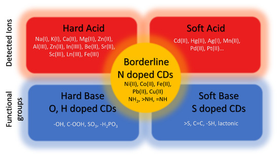
Carbon dots (CDs) are zero-dimensional nanomaterials composed of carbon and surface groups attached to their surface. CDs have a size smaller than 10 nm and have potential applications in different fields such as metal ion detection, photodegradation of pollutants, and bio-imaging, in this research, the capabilities of CDs in metal ion detection will be described. Quantum confinement is generally viewed as the key factor contributing to the uniqueness of CDs characteristics due to their small size and the lack of attention on the surface functional groups and their roles is given, however, in this research, the focus will be on the functional group and the composition of CDs. The surface functional groups depend on two parameters: (i) the oxidation of precursors and (ii) their composition.

carbon dots surface functional groups metal ion sensing mechanism oxidation

1. Introduction

Due to their small size, CDs properties are mostly conceptualized in terms of quantum confinement. However, in reality, almost all of these characteristics depend on the CDs’ original precursor and final chemical composition [1][2]. The origin of CDsphotoluminescent behavior is due to their small size, but their chemical characteristics can also influence them. The surface of CDs is covered by a variety of functional groups that are joined to the carbon core structure. Thus, it can be said that CDs’ properties depend on their size and functional groups present on the surface [3][4].
The functional groups, which contain sp2 and sp3 hybridized carbons atoms, are located on the surface of CDs. The CDs can also show dichroism (different colors from different angles) due to the complex nature of the functional group. Functional groups originate from oxidation during synthesis and afterward. When the specific absorption wavelength matches with one of the functional group’s absorption, it can absorb it, which also leads to multicolor CDs, and so different functional groups can emit light with different wavelengths [5][6]. If the amount of oxygen is increased, the number of functional groups also increases, which traps excitons and causes redshift. Hui Ding et al.[7]  reported the emission wavelength from 400 to 625 nm after increasing the oxidation of CDs due to more traps by the functional group. Yuan et al. [8] reported that the color from green to the red of emission changes when the functional group is changed by a solvent. The green emissive CDs have pyridinic N and pyrrolic N groups when measured by FTIR which are not present in red color CDs. Fluorescent molecules which are attached to the core or surface of the CDs can also exhibit fluorescence directly [9].
CDs that can emit light in the whole visible spectrum can open doors for practical applications. There are no such CDs reported until now that can emit full visible spectra. Meng Li Liu et al. [10] reported the synthesis of CDs by means of the Schiff base reaction by utilizing P-benzoquinone and triethylenetetramine (TETA) as a precursor. These CDs can be separated into yellow- and green-color CDs using a silica gel column.
The emergent absorption band of CDs can range from UV to visible range and rarely up to the NIR region [11]. This shift can also be observed during surface functionalization [11]  and doping [12]. The absorption involves π–π* aromatic (C=C bonds) transition, which corresponds to a peak from 300 to 400 nm and a peak above 400 nm to n-π* (C=O bond) transition. Arul et al. proposed  [13] that the n–π* and π–π* transition is linked with the carbonyl and hydroxyl functional group because the absorption peak changes after adding liquid ammonia to the CDs.

2. Origin of Optical Properties

Transmission electron microscopy (TEM) reveals the crystallized core structure and amorphous functional group on the surface of CDs. The π-electron system’s energy gap transitions possessed intrinsic photoluminescence properties [14]. The energy gap transitions of a π-electron system exhibit intrinsic PL emission, while the sp2 bonding in CDs also depends on its core size [15]. CDs functional groups can also capture some of the light excitons and contribute to photoluminescence phenomena. It is reported that the oxidation of CDs can enable them to emit different color light in the spectrum from blue to orange [16]. CDs multicolor emissions are associated with their surface defects. A CD’s surface has a lot of functional groups, surface defects (broken bonds), and sp2 and sp3 hybridized carbon [17].
Oxygen-abundant functional groups such as hydroxyl, carboxyl, carbonyl, sulfoxide, etc., depend on the oxidation of CDs. These functional groups are attached to CDs core atoms and are the origin of high solubility in polar solvents like water, while also a source of surface defects [18][19]. Rigu Su et al. [20] reported the synthesis of multi-color zinc-doped CDs. They confirmed, using the TEM method, that color change does not depend on CDs size because different emissive colors have a similar size. FTIR and XPS results reveal that zinc doping can decrease the graphic carbon percentage by increasing oxidation, which decreases the other functional group amount. The red emissive CDs have a low number of oxygen-related functional groups, while blue emissive CDs have more oxygen-related functional groups. They use a different ratio of p-phenylenediamine and ZnCl2 precursor to obtain multicolor CDs.
Hydroxyl functional groups (–OH) are mostly present on the surface of CDs due to the organic nature of precursors, and oxidation also contributes to the amount of -OH groups, which was confirmed by FTIR spectroscopy methods. FTIR spectra also confirmed the presence of stretching vibrations and in-plane bending vibrations of -OH [21]. These functional groups have a strong influence on the properties of CDs, while carboxyl groups (–COOH) are also produced when oxidation occurs on a CD’s surface. The FTIR spectrum of amino groups (–NH2) produced during the surface functionalization of CDs has been observed, and the these CDs are termed as nitrogen-doped CDs[22].
Nitrogen-based functional groups such as amino, pyridine, nitro, amide, etc., are the second most common functional groups present on the surface and core of CDs. Thanks to the similar atomic radius and electronic structure of carbon and nitrogen, this type of doping is effective [23][24].
Shanshan Wang et al.[25]  reported the synthesis of CDs via laser ablation and hydrothermal carbonization methods (HTC). Xylose was used as a precursor and was placed at 200 °C for six hours in an HTC reactor. The CDs obtained from this method are also named HTC to avoid confusion. The HTC CDs were further annealed for two hours at 850 °C with an argon flow rate of 50 cm3/min.
For the laser ablation method, the annealed carbon powder and 20 wt% Teflon powder were used as precursors. The repetition rate was 50 Hz, the pulse width was 1–2 ns, and the produced CDs were named LA-CDs. The hydrothermal carbonization process frequently produces green-emission CDs, while laser ablation produces blue-emission CDs under excitation at 360 nm; although both have similar sizes (4.72 ± 0.7 nm), they show different PL properties. The XPS result shows that oxygen comprising the functional group demonstrates π–π* transition, while nitrogen comprising the functional group demonstrates n–π* and π–π* transitions which cause changes in the PL properties of both. They observed that LA-CDs have an N-H bond in their FTIR spectra, while the other two do not have the presence of nitrogen in their FTIR spectra. The LA-CDs had a bright blue emission, whereas the HTC-CDs had a green emission, while annealed CDs did not show any emission when excited by UV. The annealing process probably affects the removal of functional groups. This confirms how vital functional groups are in the phenomenon of CD photoluminescence.
Sulfur-doped CDs are also frequently studied due to similar electron mobility. The sulfur atom has six valence electrons which can alter the optical properties of CDs. The sulfur can attach to the core and the surface of CDs [26], as the functionalization of CDs with thiol can produce an S–H functional group at the peak of 2532 cm−1 [27]. The phosphorus-doped CDs are also studied because phosphorus can produce substitutional defects in CDs surfaces, act as an n-type donor, and alter the optical and electronic properties of CDs [28]. Figure 1 shows the CDs functional group structure comparison of hard acid and soft acid detection using CDs.
Figure 1. Detection of metal ions having a hard or soft character using CDs with diverse surface functional groups.

3. Mercury Detection

Mercury is one of the heavy metals that can cause significant health problems. It possesses biological toxicity, is non-biodegradable, and has high mobility, which means it stays in the environment for a long time. The Hg–C bond-based organometallic compounds can be present in the air and water for a longer time and cause an imbalance in the biological system, and when ingested by a human, they can potentially damage the nervous system. Because mercury is present in significant quantities near areas where mercury-based minerals are extracted, it is vital to limit the amount of mercury in nature, and thus detecting it in nature is the first step. There are some well-known methods to detect mercury in the samples. Surface-enhanced Raman scattering (SERS), mass spectroscopy, and electrochemical methods are among them. All of these methods require large and complex equipment with an expert operator, limiting their utilization. As already discussed, CDs have good luminescence properties and can form a luminescence turn-on sensor for mercury. The CDs obtained from eggshell membranes are useful, acting as label-free methods to determine mercury, with a detection limit of 2.6 µM [29]. The CDs obtained from L-cysteine were also used as a detecting probe for mercury, and were used with several different metals ions such as Cu2+, Pb2+, Ag+, Cd2+, Cr3+, and Co2+, but these metal ions do not influence the emission spectra of CDs, even when the concentration is increased up to 10 times, while with mercury, the emission spectra are decreased sharply down to 60% of the total emission spectra [30].
Citric acid monohydrated and ammonia-based nitrogen-doped high luminescent CDs (quantum yield 40.5%) prepared by the one-step hydrothermal method employed as a sensor for Hg2+ with a limit of detection up to 0.087 µM, which is 30 times higher than eggshell-membrane-based CDs, and it is suitable in a natural water sample with recovery in the range of 96.6–105.5% [31].
Pineapple-peel-based CDs were prepared by simple hydrothermal treatment and acted as a label-free probe for Hg2+ ions. The CDs were utilized to detect Hg2+ in lake and tap water samples and found them useful. The logic gate sensor based on NOT operation utilized Hg2+ as a input. This shows that CDs can also be used commercially as sensors [32]. A large group of CDs also do not detect mercury ions. For example, hydrothermally produced CDs using Phyllanthus acidus do not detect Hg2+, but can detect Fe3+ up to the detection limit of 0.9 µM [33].

4. Lead Detection

Lead is a toxic transition metal, a non-biodegradable compound, and can react with blood [34]. It can be found in drinking water due to its presence in small amounts in water pipes, and the corrosion of pipes can cause lead to migrate with water. When consumed by humans, even in significantly low amounts of more than 5 mmol/L, it can cause memory loss, mental diseases, and other medical issues [35]. It has three oxidation states, but Pb(II) is mainly found in nature, which can cause mental disabilities, migraine [35], memory loss, and “dullness” [36] in humans, especially children. Different traditional and new techniques are used to detect Pb(II), such as inductively coupled plasma (ICP) mass spectrometry, electrochemical sensors [37], atomic absorption spectroscopy (AAS), DNAzyme [38], and some inorganic nanomaterials. However, all of these methods are expensive, and the demand for new low-cost and sensitive methods is high.

5. Silver Detection

Silver is an essential element that has many applications, such as antimicrobial agents in water [39], electrical devices [40], medicine, and electrical devices, and waste related to these applications in the environment is harmful to humans. The recycling of silver is expensive, so it is important to minimize the amount of silver converted into waste [41]. Ag+ ions can change and destroy the healthy nature of pure drinking water, so it is essential to control the number of Ag+ ions in nature. Ag+ ions can be detected using spectroscopic methods such as X-ray fluorescence spectroscopy [42], fluorescence spectrometry, and inductively coupled plasma–atomic emission spectrometry, but these methods require a complex operation. CDs can be used to overcome this situation, which act as probes for Ag+ detection.

6. Chromium Detection

Chromium is a highly toxic element found in industrial wastewater. It has two major oxidation states—the non-toxic (at low concentrations) trivalent chromium Cr(III), and hexavalent chromium Cr(VI), which is, even in low amounts, very toxic. Cr(VI) causes cancer and hormonal problems if consumed in sufficient amounts by humans, and its recommended quantity in drinking water is lower than 100 ppb, according to the U.S. Environmental Protection Agency. It can be measured through conventional methods, but fluorescence probes are proven helpful in detecting chromium(VI).
Additionally, CDs exhibit high selectivity and are accessible in use. Researchers are working on the detection of both oxidation states of chromium. MMF Chang et al. reported the synthesis protocol of CDs using a sucrose precursor at a low temperature of 85 °C. These CDs have a yellow color emission. These CDs show pH-dependent fluorescence quenching when treated with Cr(III), which depends on concentration. The limit of detection was found to be 24.58 ± 0.02 μM [43].

7. Iron(III) and Iron(II) Detection

Iron(III) is one the most commonly used metal ions, and is essential for humans up to a certain level; above that level, it can cause diseases such as type 2 diabetes, inflammation [44] and Alzheimer’s disease, etc. [45], while a deficiency of iron in the human body can cause anemia (IDA). Iron(III) in the environment also influences plant growth, so it is essential to monitor the quantity of Fe3+ in the environment, and more specifically patients with diseases caused by Iron(III) [46].
Fe3+ can also cause problems with the production of zinc. During the electrochemical process of zinc production, the efficiency of the process is significantly decreased by iron dissolved in the electrolyte. Ferrite and zinc oxide are involved in the hydrometallurgical production of zinc, and a high temperature is required to remove the ferrite, so it is also essential to control the amount of Fe3+ and Fe2+ [47].
The detection method for Fe3+ is similar to that for other heavy metals, including voltammetry and coupled plasma mass spectrometry (ICP-MS), so now the focus is on detecting iron with the help of a fluorescent probe. Examples of fluorescence probes are conjugated polymer, quantum dot, and CDs, which is also a potential contender for Fe3+. Fe2+ ions also exist in nature and are essential for humans and must be monitored, but they oxidize to Fe3+ in an open environment, making it difficult to detect them accurately. So, there is very little research on Fe2+ detection [48].

8. Copper(II) Detection

Copper (Cu2+) is essential for the healthy growth of biological activity because it strengthens [49] the bones and immune system, but an excessive amount of Cu2+ can cause vomiting, pain, and disturbance of biological activity [50]. So, it is necessary to develop an easy and inexpensive sensing method to detect Cu2+. Researchers are using CDs to detect Cu2+ [51][52]. Van Dien Dang et al. [53] synthesized the nitrogen-doped CDs by using citric acid as an oxygen source and ethylenediamine as a nitrogen source. The CDs had a quantum yield of about 84%, and their fluorescence activity decreased after adding different concentrations of Cu2+ ions. The limit of detection was observed as 0.076 nM. Xiaochun Zheng et al. [54] described the mechanism of detection of Cu2+ based on the functional group by using citric acid and polyethylenimine as precursors for synthesis. The CDs were synthesized by means of the hydrothermal method. The quantum yield was 25% when excited at the wavelength 365 nm. They linked the detection of Cu2+ to the presence of amino groups on the surface of CDs, which caused the splitting of Cu2+ d-orbital that produced the new path for CDs excited states. The -NHx group peak CDs in FTIR also disappeared after reaction with Cu2+.
CDs can detect metal ions by transferring the electron from the excited state in CDs to metals and then reverting back to the ground state of CDs. As the redox potential plays an important part in the study carried out by Xiaochun Zheng et al., the redox potential play an important factor in metal ion sensing [54]. Redox potential of CDs should also be investigated to further analyze the role of CDs because metal redox potential should be more negative than the holes on CDs and positive than the electrons on CDs. Therefore, measuring the redox potential of different metals ions and comparing it with CDs produced using various precursors can provide further insights.

References

  1. Zhen Tian, Xutao Zhang, Di Li, Ding Zhou, Pengtao Jing, Dezhen Shen, Songnan Qu, Radek Zboril, Andrey L. Rogach Full-Color Inorganic Carbon Dot Phosphors for White-Light-Emitting Diodes. Advanced Optical Materials 2017, 5, 1, https://doi.org/10.1002/adom.201700416.
  2. Xiang Miao, Dan Qu, Dongxue Yang, Bing Nie, Yikang Zhao, Hongyou Fan and Zaicheng Sun Synthesis of Carbon Dots with Multiple Color Emission by Controlled Graphitization and Surface Functionalization. Advanced Materials 2018, 30, 1, https://doi.org/10.1002/adma.201704740.
  3. Ting Yuan, Ting Meng, Ping He,YuXin Shi, Yunchao Li, Xiaohong Li, Louzhen Fan ORCID logo and Shihe Yang Carbon quantum dots: an emerging material for optoelectronic applications. Journal of Materials Chemistry C 2019, 7, 6820-6835, DOI https://doi.org/10.1039/C9TC01730E.
  4. Yuan Xiong , Julian Schneider , Elena V. Ushakova , Andrey L. Rogach Influence of molecular fluorophores on the research field of chemically synthesized carbon dots. Nano Today 2018, 23, 124-139, https://doi.org/10.1016/j.nantod.2018.10.010.
  5. Jingjing Yu ,Chang Liu ,Kang Yuan ,Zunming Lu ,Yahui Cheng ,Lanlan Li ,Xinghua Zhang ,,Peng Jin ,Fanbin Meng and Hui Liu Luminescence Mechanism of Carbon Dots by Tailoring Functional Groups for Sensing Fe3+ Ions. https://doi.org/10.3390/nano8040233 2018, 8, 4, Nanomaterials.
  6. Hui Ding; Xue-Hua Li; Xiao-Bo Chen; Ji-Shi Wei; Xiao-Bing Li and Huan-Ming Xiong Surface states of carbon dots and their influences on luminescence. Journal of Applied Physics 2020, 127, 23, https://doi.org/10.1063/1.5143819.
  7. Hui Ding, Shang-Bo Yu, Ji-Shi Wei, and Huan-Ming Xiong Full-Color Light-Emitting Carbon Dots with a Surface-State-Controlled Luminescence Mechanism. ACS Nano 2016, 1, 484–491, https://doi.org/10.1021/acsnano.5b05406.
  8. Biao Yuan, Shanyue Guan, Xingming Sun, Xiaoming Li, Haibo Zeng, Zheng Xie, Ping Chen, and Shuyun Zhou Highly Efficient Carbon Dots with Reversibly Switchable Green–Red Emissions for Trichromatic White Light-Emitting Diodes. ACS Appl. Mater. Interfaces 2018, 18, 16005–16014, https://doi.org/10.1021/acsami.8b02379.
  9. Venkatesh Gude, Ananya Das, Tanmay Chatterjeea and Prasun K. Mandal Molecular origin of photoluminescence of carbon dots: aggregation-induced orange-red emission. Physical Chemistry Chemical Physics 2016, 18, 28274-28280, DOI https://doi.org/10.1039/C6CP05321A.
  10. Meng Li Liu, Lin Yang, Rong Sheng Li, Bin Bin Chen, Hui Liu and Cheng Zhi Huang Large-scale simultaneous synthesis of highly photoluminescent green amorphous carbon nanodots and yellow crystalline graphene quantum dots at room temperature. Green Chemistry 2017, 19, 3611-3617, DOI https://doi.org/10.1039/C7GC01236E.
  11. Evgeny V. Kundelev, Nikita V. Tepliakov, Mikhail Yu. Leonov, Vladimir G. Maslov, Alexander V. Baranov, Anatoly V. Fedorov, Ivan D. Rukhlenko, and Andrey L. Rogach Amino Functionalization of Carbon Dots Leads to Red Emission Enhancement. The Journal of Physical Chemistry Letters 2019, 17, 5111–5116, https://doi.org/10.1021/acs.jpclett.9b01724.
  12. Ying Li, Hechun Lin, Chunhua Luo,Yunqiu Wang,Chunli Jiang,Ruijuan Qi, Rong Huang,Jadranka Travas-sejdic and Hui Peng Aggregation induced red shift emission of phosphorus doped carbon dots. RSC Advances 2017, 7, 32225-32228, DOI https://doi.org/10.1039/C7RA04781A.
  13. Velusamy Arul and Mathur Gopalakrishnan Sethuraman Facile green synthesis of fluorescent N-doped carbon dots from Actinidia deliciosa and their catalytic activity and cytotoxicity applications. Optical Materials 2017, 78, 181-190, https://doi.org/10.1016/j.optmat.2018.02.029.
  14. Siyu Lu, Laizhi Sui, Min Wu, Shoujun Zhu, Xue Yong and Bai Yang Graphitic Nitrogen and High-Crystalline Triggered Strong Photoluminescence and Room-Temperature Ferromagnetism in Carbonized Polymer Dots. Advanced Science 2018, 6, 2, https://doi.org/10.1002/advs.201801192.
  15. Zifei Wang, Fanglong Yuan, Xiaohong Li, Yunchao Li, Haizheng Zhong, Louzhen Fa and Shihe Yang 53% Efficient Red Emissive Carbon Quantum Dots for High Color Rendering and Stable Warm White-Light-Emitting Diodes. Advanced Materials 2017, 29, 37, https://doi.org/10.1002/adma.201702910.
  16. Lei Bao, Cui Liu, Zhi-Ling Zhang and Dai-Wen Pang Photoluminescence-Tunable Carbon Nanodots: Surface-State Energy-Gap Tuning. Advanced materials 2015, 27, 1663-1667, https://doi.org/10.1002/adma.201405070.
  17. Jin Gao,Mengmeng Zhu, Hui Huang, Yang Liu and Zhenhui Kang Advances, challenges and promises of carbon dots. Inorganic Chemistry Frontiers 2017, 4, 1963-1986, DOI https://doi.org/10.1039/C7QI00614D.
  18. Kai-Kai Liu, Shi-Yu Song, Lai-Zhi Sui, Si-Xuan Wu, Peng-Tao Jing, Ruo-Qiu Wang, Qing-Yi Li, Guo-Rong Wu, Zhen-Zhong Zhang, Kai-Jun Yuan and Chong-Xin Shan Efficient Red/Near-Infrared-Emissive Carbon Nanodots with Multiphoton Excited Upconversion Fluorescence. Advanced Science 2019, 6, 17, https://doi.org/10.1002/advs.201900766.
  19. Di Li, Chao Liang, Elena V. Ushakova, Minghong Sun, Xiaodan Huang, Xiaoyu Zhang, Pengtao Jing, Seung Jo Yoo, Jin-Gyu Kim, Enshan Liu, Wei Zhang, Lihong Jing, Guichuan Xing, Weitao Zheng, Zikang Tang, Songnan Qu and Andrey L. Rogach Thermally Activated Upconversion Near-Infrared Photoluminescence from Carbon Dots Synthesized via Microwave Assisted Exfoliation. Small 2019, 15, 1, https://doi.org/10.1002/smll.201905050.
  20. Rigu Su,Qingwen Guan, Wei Cai, Wenjing Yang, Quan Xu, Yongjian Guo, Lipeng Zhang, Ling Fei and Meng Xu*d Multi-color carbon dots for white light-emitting diodes. RSC Advances 2019, 9, 9700-9708, https://doi.org/10.1039/C8RA09868A.
  21. LiQin Liu, YuanFang Li, Lei Zhan, Yue Liu and ChengZhi Huang One-step synthesis of fluorescent hydroxyls-coated carbon dots with hydrothermal reaction and its application to optical sensing of metal ions. Science China Chemistry 2011, 54, 1342–1347 , https://doi.org/10.1007/s11426-011-4351-6.
  22. N. Tsubokawa and M. Hosoya Reactive carbon black having acyl imidazole or acid anhydride groups: Preparation and reaction with functional polymers having hydroxyl or amino groups. Reactive Polymers 1990, 14, 33-40, https://doi.org/10.1016/0923-1137(91)90245-J.
  23. Takashi Ogi, Kana Aishima,Fitri Aulia Permatasari, Ferry Iskandar, Eishi Tanabec and Kikuo Okuyamaa Kinetics of nitrogen-doped carbon dot formation via hydrothermal synthesis. New Journal of Chemistry 2016, 40, 5555-5561, DOI https://doi.org/10.1039/C6NJ00009F.
  24. Xiaojuan Gong, Qingyan Zhang, Yifang Gao, Shaomin Shuang, Martin M. F. Choi, and Chuan Dong Phosphorus and Nitrogen Dual-Doped Hollow Carbon Dot as a Nanocarrier for Doxorubicin Delivery and Biological Imaging. ACS Applied Materials & Interfaces 2016, 18, 11288–11297, https://doi.org/10.1021/acsami.6b01577.
  25. Shanshan Wang, Dong-Sheng Yang and Fuqian Yang Nitrogen-induced shift of photoluminescence from green to blue emission for xylose-derived carbon dots. Nano Express 2020, 1, 2, 10.1088/2632-959X/aba771.
  26. Dr. Feng Li, Prof. Dayong Yang, Prof and Huaping Xu Non-Metal-Heteroatom-Doped Carbon Dots: Synthesis and Properties. Chemistry: A European Journal 2018, 25, 1165-1176, https://doi.org/10.1002/chem.201802793.
  27. Yeji Kim and Jongsung Kim Bioinspired thiol functionalized carbon dots for rapid detection of lead (II) ions in human serum. Optical Materials 2020, 99, 1, https://doi.org/10.1016/j.optmat.2019.109514.
  28. Jin Zhou, Xiaoyue Shan,Juanjuan Ma, Yamin Gu, Zhaosheng Qian, Jianrong Chena and Hui Feng Facile synthesis of P-doped carbon quantum dots with highly efficient photoluminescence. RSC Advances 2013, 4, 5465-5468, https://doi.org/10.1039/C3RA45294H.
  29. Lei Wang, Shou-Jun Zhu, Hai-Yu Wang, Song-Nan Qu, Yong-Lai Zhang, Jun-Hu Zhang, Qi-Dai Chen, Huai-Liang Xu, Wei Han, Bai Yang, and Hong-Bo Sun Common Origin of Green Luminescence in Carbon Nanodots and Graphene Quantum Dots. ACS Nano 2014, 3, 2541–2547, https://doi.org/10.1021/nn500368m.
  30. Huifang Wu and Changlun Tong Nitrogen- and Sulfur-Codoped Carbon Dots for Highly Selective and Sensitive Fluorescent Detection of Hg2+ Ions and Sulfide in Environmental Water Samples. Journal of Agricultural and Food Chemistry 2019, 10, 2794–2800, https://doi.org/10.1021/acs.jafc.8b07176.
  31. Zhiguo Ye,Yanhui Zhang,Guixin Li and Baoxin Li Fluorescent Determination of Mercury(II) by Green Carbon Quantum Dots Synthesized from Eggshell Membrane. Analytical Letters 2020, 53, 18, https://doi.org/10.1080/00032719.2020.1759618.
  32. RETURN TO ISSUEPREVARTICLENEXT Pineapple Peel-Derived Carbon Dots: Applications as Sensor, Molecular Keypad Lock, and Memory Device Somasundaram Anbu Anjugam Vandarkuzhali, Sampathkumar Natarajan, Shanmugapriya Jeyabalan, Gandhi Sivaraman*, Subramanian Singaravadivel*, Shanmugam Muthusubramanian, and Balasubramanian Viswanathan Pineapple Peel-Derived Carbon Dots: Applications as Sensor, Molecular Keypad Lock, and Memory Device. ACS Omega 2018, 10, 12584–12592, https://doi.org/10.1021/acsomega.8b01146.
  33. Raji Atchudan , Thomas Nesakumar Jebakumar Immanuel Edison , Kanikkai Raja Aseer , Suguna Perumal , Namachivayam Karthik and Yong Rok Lee Highly fluorescent nitrogen-doped carbon dots derived from Phyllanthus acidus utilized as a fluorescent probe for label-free selective detection of Fe3+ ions, live cell imaging and fluorescent ink. Biosensors and Bioelectronics 2017, 99, 303-311, https://doi.org/10.1016/j.bios.2017.07.076.
  34. Wassana Yantasee , Yuehe Lin, Kitiya Hongsirikarn, Glen E Fryxell, Raymond Addleman and Charles Timchalk Electrochemical sensors for the detection of lead and other toxic heavy metals: the next generation of personal exposure biomonitors. Environ Health Perspect 2007, 12, 1683-90, DOI: 10.1289/ehp.10190.
  35. M S Bhatia and R Gupta, S Srivastava Migraine associated with water deprivation and progressive myopia. Cephalalgia . 2006, 6, 758-760, DOI: 10.1111/j.1468-2982.2006.01083.x.
  36. Papanikolaou NC , Hatzidaki EG , Belivanis S , Tzanakakis GN and Tsatsakis AM Lead toxicity update. A brief review. Med Sci Monit . 2005, 26, 1, doi: 10.2147/IMCRJ.S404885.
  37. Wassana Yantasee , Yuehe Lin, Kitiya Hongsirikarn, Glen E Fryxell, Raymond Addleman and Charles Timchalk Electrochemical sensors for the detection of lead and other toxic heavy metals: the next generation of personal exposure biomonitors. Environ Health Perspect . 2007, 12, 90, DOI: 10.1289/ehp.10190.
  38. Xiaofang Niu, Yanjun Liu, Fei Wang and D. Luo Highly sensitive and selective optical sensor for lead ion detection based on liquid crystal decorated with DNAzyme. Optics express 2019, 1, 1, DOI:10.1364/oe.27.030421.
  39. Federica Paladini and Mauro Pollini Antimicrobial Silver Nanoparticles for Wound Healing Application: Progress and Future Trends. Materials 2019, 16, 2540, doi: 10.3390/ma12162540.
  40. Dapeng Chen, X. Qiao, X. Qiu and Jianguo Chen less Synthesis and electrical properties of uniform silver nanoparticles for electronic applications. Journal of Materials Science 2009, 1, 1, DOI:10.1007/S10853-008-3204-Y.
  41. S. Syed Silver recovery aqueous techniques from diverse sources: Hydrometallurgy in recycling. Waste Management 2016, 50, 234-256, https://doi.org/10.1016/j.wasman.2016.02.006.
  42. Germarie Sánchez-Pomales , Thilak K Mudalige, Jin-Hee Lim and Sean W Linder Rapid determination of silver in nanobased liquid dietary supplements using a portable X-ray fluorescence analyzer. Journal of Agricultural and Food Chemistry 2013 , 30, 1, DOI: 10.1021/jf402018t.
  43. Melissa May Fung Chang, Irine Runnie Ginjom, Maria Ngu-Schwemlein and Sing Muk Ng Synthesis of yellow fluorescent carbon dots and their application to the determination of chromium(III) with selectivity improved by pH tuning. Microchimica Acta 2016, 183, 1899–1907 , DOI 10.1007/s00604-016-1819-2.
  44. Elizabeta Nemeth and Tomas Ganz The role of hepcidin in iron metabolism. Acta Haematol 2009, 2, 78-86, DOI: 10.1159/000243791.
  45. Judith A Simcox and Donald A McClain Iron and diabetes risk. Cell Metab 2013, 3, 329-41, DOI: 10.1016/j.cmet.2013.02.007.
  46. Guanxiong Liu , Baoqiang Li , Ying Liu , Yujie Feng , Dechang Jia and , Yu Zhou Rapid and high yield synthesis of carbon dots with chelating ability derived from acrylamide/chitosan for selective detection of ferrous ions. Applied Surface Science 2009, 487, 1167-1175, https://doi.org/10.1016/j.apsusc.2019.05.069.
  47. Swarnkar, S. R.; Gupta, B. L. and Sekharan, R. Dhana Iron control in zinc plant residue leach solution. Hydrometallurgy, 1996, 42, 21-26, 10.1016/0304-386X(95)00077-T .
  48. Jing Shi, G. Ni, Jinchun Tu, Xiaoyong Jin and Juan Peng less Green synthesis of fluorescent carbon dots for sensitive detection of Fe2+ and hydrogen peroxide. Journal of Nanoparticle Research 2017, 1, 1, DOI:10.1007/s11051-017-3888-5.
  49. Daniel López de Romaña , Manuel Olivares, Ricardo Uauy and Magdalena Araya Risks and benefits of copper in light of new insights of copper homeostasis. Journal of Trace Elements in Medicine and Biology 2011, 1, 3-13, doi: 10.1016/j.jtemb.2010.11.004..
  50. Peng Wang , Yonghui Yuan , Ke Xu , Hongshan Zhong , Yinghui Yang , Shiyu Jin , Ke Yang and Xun Qi Biological applications of copper-containing materials. Bioactive Materials 2021, 6, 916-927, https://doi.org/10.1016/j.bioactmat.2020.09.017.
  51. Alfonso Salinas-Castillo, Maria Ariza-Avidad, Christian Pritz, Maria Camprubí-Robles, Belen Fernández, Maria J. Ruedas-Rama, Alicia Megia-Fernández, Alejandro Lapresta-Fernández, Francisco Santoyo-Gonzalez, Annelies Schrott-Fischer and Luis F. Capitan-Vallvey Carbon dots for copper detection with down and upconversion fluorescent properties as excitation sources. Chemical Communications 2012, 49, 1103-1105, DOI https://doi.org/10.1039/C2CC36450F.
  52. Van Dien Dang, Akhilesh Babu Ganganboina, and Ruey-An Doong Bipyridine- and Copper-Functionalized N-doped Carbon Dots for Fluorescence Turn Off–On Detection of Ciprofloxacin. ACS Appl. Mater. Interfaces 2020, 29, 32247–32258, https://doi.org/10.1021/acsami.0c04645.
  53. Van Dien Dang, Akhilesh Babu Ganganboina, and Ruey-An Doong Bipyridine- and Copper-Functionalized N-doped Carbon Dots for Fluorescence Turn Off–On Detection of Ciprofloxacin. ACS Appl. Mater. Interfaces 2020, 29, 32247–32258, https://doi.org/10.1021/acsami.0c04645.
  54. Dr. Xiaochun Zheng, Prof. Wenjun Liu, Dr. Qixiao Gai, Prof. Zhaoshuo Tian and Prof. Shoutian Ren A Carbon-Dot-Based Fluorescent Probe for the Sensitive and Selective Detection of Copper(II) Ions. ChemistrySelect 2019, 4, 2392-2397, https://doi.org/10.1002/slct.201803584.
More
Information
Contributors MDPI registered users' name will be linked to their SciProfiles pages. To register with us, please refer to https://encyclopedia.pub/register : , ,
View Times: 303
Revisions: 2 times (View History)
Update Date: 30 Jun 2023
1000/1000
ScholarVision Creations